Showing posts with label Cardiology journals impact factor. Show all posts

Monday, 13 March 2023

Lupine Publishers| Thresholds and Upper Limits of Training-Induced Cardiovascular, Respiratory, Immune, and Musculoskeletal Adaptations in Able-Bodied People and Neurologically Impaired Patients

  Lupine Publishers| Journal of Cardiology Research & Reports


Abstract

The benefits of exercising are well-recognized–experts and governmental health agencies agree that regular endurance training generally reduces the risks of metabolic and cardiovascular diseases. In turn, the consequences of a sedentary lifestyle and physical inactivity caused by disease or trauma are also well-documented- e.g., walking or being moderately active physically less than 30 minutes per day generally increases the risks of developing obesity, infections, osteoporosis, metabolic disorders, cardiovascular problems, and dyslipidemia in able-bodied persons. However, the threshold levels of exercising for meaningful health benefits in people suffering from paralysis or the upper limits of endurance training beyond which, injuries, sequelae, and long-term secondary problems that may be induced in able-bodied and people with disabilities remain unclear. This said, it is becoming increasingly clear that significant health problems including severe stress injuries and sudden heart failures may be experienced during long-distance running - e.g., marathons, triathlons, and ultra-triathlons. This review summarizes data on injuries and fatal events associated with endurance exercise training. Impacts on the immune, cardiovascular, respiratory, and musculoskeletal systems in disabled and nondisabled individuals are also discussed.

Keywords: Spinal Cord Injury; Performances; Marathon; Iron Man; Tendinitis; Cardiac Arrest; Infections; Stressed Fractures; Overtraining

Introduction

The Marathon as we know it today exists since the late 19th century. It is essentially a long-distance race of 42 kilometers (42.195 kilometers) usually performed on the road. It can be completed by running or running/walking for able-bodied persons as well as by wheel chairing for disabled individuals. About 800 marathons are held each year worldwide. Most of the competitors are recreational athletes. Progressively, over the years, longdistance races have become even harder. In the late 80s and early 90s – e.g., ultra-marathons (e.g., 100 kilometers), triathlons and Ironman races (1.5 kilometer swim, 40 kilometer bike ride and 10 kilometer run), or ultra-triathlons (e.g., the Uber Man created in 2017 is composed of a 33 kilometer swim, a 643 kilometer bike ride, and a 217 kilometer run).
Until recently, it was generally believed that marathon runners were not particularly at risks for cardiovascular and respiratory problems since cardiac deaths were reported to be almost ‘nonsignificant’ Maron et al. [1]. However, as exciting and healthy as it may look like, marathons can alter the function of several biological systems and organs. It is not uncommon indeed for runners to experience acute musculoskeletal injuries, gastrointestinal problems, and life-threatening hyponatremia Sanchez et al. [2]. The latter is increasingly encountered due to overhydration with hypotonic fluids.
The recent enthusiasm for extreme events such as triathlons, ultra-marathons, and ultra-triathlons has enabled more data to be generated and new findings to be made. Among them, the number of injuries and fatal deaths has increased significantly.
For instance, sudden cardiac death (SCD) is described as an event that is non-traumatic, non-violent, unexpected and resulting from sudden cardiac arrest within six hours of previously witnessed normal health.
Prolonged exercise can trigger unheralded ventricular arrhythmias and SCD in individuals without no previous heart problems. In apparently healthy long-distance runners, the incidence of SCD ranges may reach 1:15,000 per year. SCD is more frequent in male and black athletes as well as in older individuals Kim et al., Ghio et al. [3, 4].
The heart of lifelong male endurance athletes generally contains more plaque or other signs of heart problems (e.g., myocardial fibrosis, late gadolinium enhancement), such as scarring and inflammation, than the hearts of less active men of the same age.
Physiological adaptations to triathlon and ultramarathon training include increased left ventricular cavity size and/or wall thickness. However, after a competition, cardiac problems associated with heart muscle fatigue may be found. Musculoskeletal adaptations to proper training include increased muscle respiratory capacity and substrate utilization modifications. The musculoskeletal system is the site of most injuries for those performing high endurance exercise training – i.e., non-traumatic overuse injuries are found in 80 to 85% of the musculoskeletal injuries. An imbalance caused by overly intensive training and inadequate recovery leads to a breakdown in tissue reparative mechanisms and eventually to overuse injuries Cosca et al. [5].
Overuse injuries associated with inflammatory mechanisms are also often found. They include patellofemoral pain syndrome, iliotibial band friction syndrome, medial tibial stress syndrome, Achilles tendinopathy, plantar fasciitis, and lower extremity stress fractures. Moreover, endurance athletes are more at risk for exercise-associated medical conditions, including exerciseinduced asthma, exercise-associated collapse, and overtraining syndrome. Exercise-induced immunodepression and exaggerated inflammatory response Castell et al. [6] affecting the upper respiratory tract and the gastrointestinal system may also be found – e.g., intensive triathlon training induces low peripheral CD34+ stem cells Phillip et al. [7]. Endurance athletes often quickly develop exercise-induced bronchoconstriction, two- or threefold greater (incidence) than the rate of asthma in the general population Knopfli et al. [8]. Overall, up to 70% of recreational and competitive runners sustain overuse injuries during any 12-month period (Australian Sports Commission’s 2006).
It was clearly demonstrated recently by a large-scale study with more than 9 million participants that SCD affected that is 135 people Harris et al. [9]. The victims were 47 years of age on average, and 85 percent were male. Overall, 90 deaths and cardiac arrests occurred during the swimming portion of races. The incidence of cardiovascular events was strikingly lower in female triathletes, 3.5-fold less than in men. Several experts believe that they may constitute an underestimation of the reality since only data from the finishers were considered. Yet, it shows that deaths and SCD during long-distance events are not rare Harris et al. [9].
In people living with disabilities such as with paralysis caused by a spinal cord injury (SCI), the consequences of physical inactivity are also well-known-within a few weeks to a few months post-injury, several organs and systems including bones, muscles, immune cells, guts, skin, brain, blood cells, and heart, generally undergo significant dysregulations that lead to the development of chronic diseases and polymorbidity problems. Many of those socalled ‘secondary complications’ are not associated with the injury per se but, instead, with the main consequence of physical inactivity mainly in completely paralyzed patients Wilson et al. [10].
Extensive work, mainly from Bauman, has characterized the extent after SCI of lean body mass loss, bone loss, fractures, adiposity increase, obesity, anabolic hormone decrease, insulin resistance, hypertension, cholesterol, incidence of type II diabetes, cardiovascular problems, anxiety and depression Bauman et al. ; Bauman [11, 12]; Battalio et al. [13]. Immunodeficiency and its role in frequent infections (e.g., UTIs, skin sores, septicaemia, pneumonia) and premature death has also been established Brommer et al. [14]. Overall, 19 classes of drugs and 300 different compounds are used against chronic comorbidities post-SCI Rouleau et al. [15].
Although it has clearly been shown in able-bodied persons with a sedentary lifestyle that significant exercise training such as active walking more than 30 minutes per day can prevent or reverse these problems, comparable effects for those with SCI are incompletely characterized Krassioukov et al. [16]. Yet, it was shown in paraplegic mice treated with Spinalon, an experimental tritherapy that activates during 30 minutes the spinal locomotor networks, that an increase of muscle mass (25%), femoral bone mineral density (10%), red blood cells/haematocrit levels (10%), and lymphocyte counts (25%) occurs after just a few weeks of metabolically challenging exercising on a treadmill induced pharmacologically Guertin et al. [17]; Ung et al. [18]. Preliminary evidence of efficacy in patients was recently reported also Radhakrishna et al. [19].
In patients with less severe injuries (incomplete SCIs), specialized rehabilitation approaches such as body-weightsupported treadmill training (BWSTT, Barbeau et al. [20]; Wernig et al. [21] and functional electrical stimulation (FES) biking have been shown to improve voluntary walking capabilities via longlasting effects on spinal plasticity as well as cardiovascular and cardiometabolic functions, to some extent Coupaud et al. [22]; Kapadia et al. [23]; Graham et al. [24].
However, for those with severe SCIs, BWSTT and FES used separately do not generally induce comparable health benefits although small effects can occasionally be found Frotzler et al. [25]. For instance, epidural stimulation (ES) + BWSTT was recently shown in 4 volunteers with AIS-B injuries to increase lean body mass, decrease body fat, and improve android/gynoid ratio, resting metabolic rate and VO2 max Terson de Paleville et al. [26]. BWSTT+exoskeletons (EXs) in 5 volunteers with SCI provided preliminary evidence of moderate benefits on VO2 max and peak heart rates Evans et al. [27].
All in all, it appears that medical devices used alone or with BWSTT can lead, to some extent, to detectable but moderate benefits essentially in individuals with incomplete SCIs. However, with complete SCIs, these approaches are promising, but not yet ideally suited for significant metabolic outcomes Ditors et al. [28]; Ter Woerds et al. [29]; van Duijnhoven et al. [30] suggesting that other combinatorial approaches such as drug + device need to be explored Gerasimenko et al. [31]; Freyvert et al. [32]; Bloch [33].

Concluding Remarks

The long-term consequences on health degradation and system/organ dysregulations of physical inactivity or of a sedentary lifestyle are undisputable. In able-bodied persons, walking daily more than 30 minutes is generally recognized as the threshold level for preventing those complications. However, for disabled and particularly for wheelchair-bound persons -e.g., with a severe SCI- the threshold for preventing polymorbidities, systemic dysregulations, and overmedication problems remains unclear and is currently being explored. New solutions using pharmacological, robotic, and electrical approaches as combinatorial therapies are promising. In turn, for able-bodied and wheelchair athletes capable of performing long-distance competitions such as marathons, ultra-marathons, and other comparable events, the risk of deaths is significant but relatively low. In turn, the incidence of myocardial fibrosis and other cardiac problems is high over time as well as the incidence of overuse problems that is by far the more important type of injuries.

Read More about Lupine Publishers Journal of Cardiology Research & Reports Please Click on Below Link: https://lupine-publishers-cardiovascular.blogspot.com/

Wednesday, 8 March 2023

Lupine Publishers| Acute Myocardial Infarction” (IMA)”

 Lupine Publishers| Journal of Cardiology Research & Reports


Introduction

Myocardial infarction is the most common presentation of ischemic heart disease. WHO estimated that in 2002 12.6% of deaths worldwide were due to ischemic heart disease, which is the leading cause of death in developed countries and the third leading cause of death in developing countries, post-AIDS and low respiratory infections. In developed countries such as the United States, deaths from heart disease are more numerous than cancer mortality. Coronary pathies cause one in five deaths in the United States and where more than one million people suffer a coronary attack each year, 40% of which will die as a result of a heart attack. So an American will die every minute of a coronary pathological event. They also occur in the patient who does not care for a gastritis that has been charged for a certain time and in the patient who is under constant stress. In our daily action for different causes, we are subjected to permanent stress, adding to them, the risk factors of this entity. That is why we were motivated by the revision of the issue in question.

Objectives

1) Mention the etiology of Acute Myocardial Infarction.
2) Describe the clinical picture of Acute Myocardial Infarction.
3) Name the clinical research to be carried out to verify your diagnosis.
4) Cite the treatment to be used in Acute Myocardial Infarction.

Development

Acute myocardial infarction

The term acute myocardial infarction (acute means sudden, my muscle and cardio heart), often abbreviated as IAM or MAOI and known in colloquial language as heart attack, heart attack or heart attack, reflects the death of heart cells caused by ischemia resulting from the imbalance between demand and the supply of blood supply through coronary circulation.
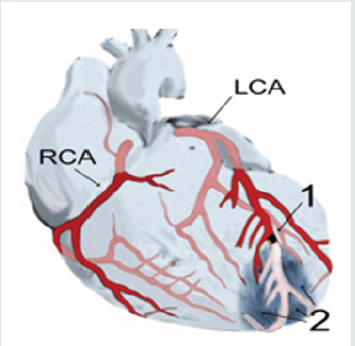
(Diagram of a myocardial infarction (2) at the tip of the anterior wall of the heart (an apical infarction) following the occlusion (1) of one of the branches of the left coronary artery (ACL), right coronary artery (RCA).

Figure 1.

Lupinepublishers-openaccess-cardiology

Development

Etiology

Acute myocardial infarction occurs in patients with ischemic heart disease, either in those who already knew they were suffering from and treated for this disease, or as an initial episode of the pathology. It is often preceded by a history of unstable angina, a name given to episodes of chest pain that become more frequent, more durable, that appear with lesser effort than in previous evolution or that do not give in with the usual medication. The myocardial (the heart muscle) suffers a heart attack when advanced coronary heart disease exists. This usually occurs when an ateroma plaque inside a coronary artery is ulcerated or ruptured, causing an acute blockage of that vessel. The crown of blood vessels that carry oxygen and nutrients to the heart muscle itself (coronary arteries) may develop athema plaques, which involves to a greater or lesser degree the flow of oxygen and nutrients to the heart itself, with effects ranging from an angina (when interruption of blood flow to the heart is temporary), to a myocardial infarction (when permanent and irreversible).

Thrombus and plunger

The presence of arteriosclerosis in a blood vessel causes narrowing in the vessel and more easily developing a thrombus in the vessel: a platelet clot, clotting proteins and cellular waste that ends up plugging the vessel. A plunger is a thrombus that has traveled through blood to a small glass where it is interlocked like a plunger.

Risk Factors

Risk factors for the onset of a myocardial infarction are closely related to risk factors for arteriosclerosis and include, but are not limited to: High blood pressure; (High or higher blood pressure than set parameters). High cholesterol; smoking; is more common in men than in women. Hypercholesterolemia or, more specifically, hyperlipoproteinemia, in particular elevated levels of low-density lipoprotein (LDL) and low levels of high-density lipoprotein (HDL); homozysteinemia, i.e. an elevation in the blood of homocysteine concentration, a toxic amino acid that rises with low or insufficient levels in the intake of vitamin B2, B6, B12 and folic acid; diabetes mellitus (with or without insulin resistance); obesity, which is defined through the body mass index (an index greater than 30 kg/ m2), abdominal circumference or waist/hip index; stress.
The main cause is coronary artery disease with insufficient blood supply, which causes tissue damage to a part of the heart caused by obstruction in one of the coronary arteries, often by rupture of a vulnerable ateroma plaque. Ischemia or poor oxygen supply resulting from such a blockage results in angina, which if rechanneled early, does not cause death of heart tissue, while if anoxia (lack of oxygen in a tissue) or hypoxia (decreased oxygen supply) is maintained, the heart injury occurs and eventually necrosis, i.e. infarction.
It is the most common, but not the only, cause of sudden cardiac death, through the arrhythmias. The picture is of cardiac arrest. However, in most cases there is electrical activity in the heart, the unemployment of which can be reversed with early defibrillation.

Clinical table

About half of heart attack patients have warning symptoms prior to the incident. The onset of symptoms of a myocardial infarction usually occurs gradually over the course of several minutes, and rarely occurs instantly. Any number of symptoms consistent with a sudden interruption of blood flow to the heart are grouped within acute coronary syndrome.

Chest pain

Sudden chest pain is the most common symptom of a heart attack, is usually prolonged and is perceived as intense pressure, which can spread or spread to the arms and shoulders, especially on the left side, back, neck, and even the teeth and jaw. Chest pain due to ischemia or a lack of blood supply to the heart is known as angina or angina, although painless heart attacks, or with atypical pains that do not coincide with what described herein, are not uncommon.

That is why it is said that the diagnosis is always clinical, electrocardiographic and laboratory, since only these three elements together will allow an accurate diagnosis. When typical, pain is described as a huge fist that twists the heart. It corresponds to angina but prolonged over time and does not respond to the administration of the medicines with which it was previously relieved (e.g. sublingual nitroglycerin) nor does it give way with rest. Pain is sometimes perceived differently, or does not follow any fixed patterns, especially in the elderly and people with diabetes. In heart attacks that affect the lower or diaphragmatic face of the heart it may also be perceived as prolonged pain in the upper abdomen that the individual could mistakenly attribute to indigestion or heartburn. Levine’s sign has been categorized as a classic and predictive sign of a heart attack, in which the affected person locates chest pain by strongly grasping his chest at the breastbone level.

Breathing difficulty

Dyspnoea or shortness of breath occurs when heart damage reduces heart output from the left ventricle, causing left ventricular insufficiency and, as a result, pulmonary edema. Other signs include diaphoresis or excessive sweating, weakness, dizziness (in 10 percent of cases it is the only symptom), palpitations, nausea of unknown origin, vomiting and fainting. The onset of these latter symptoms is likely to result from a massive release of catecholamines from the sympathetic nervous system, a natural response to pain, and hemodynamic abnormalities resulting from heart dysfunction.

Serious signs

More serious signs include loss of consciousness due to inadequate brain infusion, cardiogenic shock, and even sudden death, usually due to ventricular fibrillation.

In women

Women tend to experience symptoms markedly different from those of men. The most common symptoms in women are dyspnoea, weakness, fatigue and even drowsiness, which manifest up to a month before the clinical onset of ischemic infarction. In women, chest pain may be less predictive of coronary ischemia than in men.

Heart Attacks Without Pain or Without Other Symptoms

About a quarter of myocardial infarctions are silent, meaning they appear without chest pain and without other symptoms. These infarctions are usually discovered some time later during subsequent electrocardiograms or during an autopsy with no history of heart attack-related symptoms. This silent course is most common in the elderly, in patients with diabetes and after a heart transplant, probably because a donated heart is not connected to the nerves of the host patient. In patients with diabetes, differences in the threshold of pain, autonomic neuropathy and other physiological factors are possible explanations of the absence of symptomatology during a heart attack.

Diagnosis (clinical investigations)

Diagnosis of a myocardial infarction should be made by integrating clinical aspects of the individual’s current disease and a physical examination, including an electrocardiogram and laboratory tests indicating the presence or absence of cell damage of muscle fibers. For this reason, the semiology that the clinician must apply in the presence of precordial pain (by its characteristics and duration) should compel him to propose the diagnosis of acute myocardial infarction (AMI) with sufficient haste, since the delay in the approach results in the loss of a valuable time necessary to institute the available re-infusion method with the idea of recovering the greatest extent of myocardium since, as is well known, there is an inverse relationship between the time elapsed to start the procedure and the amount of muscle “saved”.

Diagnostic criteria

World Health Organization (WHO) criteria are those that are classically used in the diagnosis of a myocardial infarction. A patient is likely diagnosed with a heart attack if he or she has two of the following criteria, and the diagnosis will be definitive if he or she has the three Medical History of Ischemic Chest Pain that lasts longer than 30 minutes; electrocardiographic changes in a series of strokes, and increase or fall of serum biomarkers, such as creatine kinase type MB and troponin.
These WHO criteria were redefined in 2000 to give predominance to cardiac markers. According to the new provisions, an increase in cardiac troponin, accompanied by typical symptoms, of pathological Q-waves, elevation or depression of the ST segment or coronary intervention, is sufficient to diagnose a myocardial infarction.
The clinical diagnosis of IAM should then be based on the conjunction of the following three data: characteristic pain, suggestive electrocardiographic changes and elevation of enzymes, and it should be borne in mind that the latter may not be presented in a timely manner, so the first two changes should be taken into account to initiate infusion as soon as possible.

Physical exam

The overall appearance of patients with myocardial infarction varies according to symptoms. Comfortable patients or agitated patients with an increased respiratory rate may be seen. It is common to see a pale skin color, suggesting vasoconstriction. Some patients may have a mild fever (38–39 C), high blood pressure, or in some cases decreased blood pressure and the pulse may become irregular.

If heart failure occurs, high jugular venous pressure, hepatoyugular reflux, or swelling of the legs due to peripheral oedema may be found on the physical examination. Several abnormalities can be heard during auscultation, such as a third and fourth cardiac noise, pericardial rubbing, paradoxical unfolding of the second noise, and crackling on the lung.

Ecg. If a person has symptoms that are compatible with a heart attack, they will have an ECG (electrocardiogram) immediately, even in the ambulance that moves them. In fact, you will be attached to an ECG monitor for as long as you are in the Hospital, at least if you enter the room dedicated to the seriously ill heart or coronary unit. More than one ECG should be performed within a few hours since, in the first few hours, the result may be normal, even in the presence of a heart attack.

Cardiac markers

Cardiac enzymes are proteins from heart tissue that are released into the bloodstream as a result of heart damage, as is the case in a myocardial infarction. Until the 1980s, the enzymes apparatus aminotransferase and lactate dehydrogenase were routinely used for the assessment of heart damage. The disproportionate elevation of the MB subtype of the enzyme creatine kinase (CK) was then discovered specifically as a result of myocardial damage. Current regulations tend to favor troponin I and T units, which are specific to the heart muscle; it is even thought to begin to rise before muscle damage occurs. Elevated troponin in a patient with chest pain can correctly predict the likelihood of a myocardial infarction in the near future. A recent cardiac marker is the ISO enzyme BB of glycogen phosphorylase.
When heart damage occurs, levels of heart markers rise over time, so it’s customary to take blood samples and analyze levels of these enzyme markers over a 24-hour period. Because these heart enzymes don’t always increase immediately after a heart attack, those patients with chest pain tend to be treated assuming they have a heart attack until it is possible to evaluate them for a more accurate diagnosis.

It is very important to determine the extent of myocardial damage and based on this to make a prediction of the level of recovery of cardiac function that could be expected. Today, techniques are used that combine the use of markers such as troponin 1 with the advantages of cardiovascular MAGNETIC resonance imaging. For example, identification or mapping of troponin 1 (T1) by noncontrast magnetic resonance imaging allows for early identification of the severity of tissue damage in acute myocardial infarction. That is, if significant increases in T1 values are found through the use of magnetic resonance imaging, the likelihood of tissue recovery at six months of evolution decreases.

Angiography

In the most complicated cases or in situations where an intervention to restore blood flow is warranted, an angiogram of the coronaries may be performed. A catheter is inserted into an artery, usually the femoral artery, and pushed to the arteries that supply the heart. A radio-opaque contrast is then given and a sequence of x-rays (fluoroscopy) is taken. Clogged or narrowed arteries can be identified with this procedure but should only be performed by a qualified specialist.
Isotopic studies or cardiac catheterization (coronary spelling). As a diagnostic element, they arise when the acute phase has already passed. However, consideration may be to perform a kinegiocoronariography by cardiac catheterization, for the purposes of determining the clogged vessel(s) and to propose a disobstruction in acute, percutaneous angioplasty with or without stenting or, more rarely by endarterectomy.

Differential Diagnosis

Differential diagnosis includes other sudden causes of chest pain, such as pulmonary embolism thrombus, aortic dissection, pericardial effusion that causes cardiac tampering, tension pneumothorax, and esophageal tear

Treatment

A heart attack is a medical emergency, so it demands immediate attention. The main goal in the acute phase is to save as much myocardium as possible and prevent additional complications. As time goes on, the risk of damage to the heart muscle increases, so any time that is lost is tissue that has also been lost. When experiencing symptoms of a heart attack, it is preferable to ask for help and seek immediate medical attention. It is not advisable to try to drive yourself to a hospital.

Immediate care

When symptoms of a myocardial infarction appear, most patients wait on average for three hours, instead of proceeding as recommended: make a distress call right away. This prevents sustained damage to the heart, put in the way of expression: “lost time is lost muscle”. Certain positions allow the patient to rest while minimizing breathing difficulty, such as the half-seated position with the knees bent. Air oxygen access is improved if car windows are opened or the shirt collar button is released. If the individual is not allergic, an aspirin tablet may be given, however it has been shown that taking aspirin before calling an emergency medical service may be associated with unexpected delays. Aspirin has an antiplatelet effect and inhibits the formation of clots in the arteries. Soluble presentations are preferred, without enteric or chewable casings, to make their absorption by the body faster. If the patient cannot swallow, a sublingual presentation is recommended. A dose between 162 – 325 mg is generally recommended.

When you arrive at the emergency room, your doctor will likely give several of the following therapies:

a. Oxygen. It is usually given with nasal glasses at 2 or 3 liters. It may be the first step in the hospital or the ambulance itself.

b. Pain relievers (pain medications). If chest pain persists and is unbearable, morphine (1 milliliter-10 mg, or similar medications to relieve it (petidine-dolantine) is given.

c. Antiplatelet agents. They are medicines that prevent platelet aggregation in thrombus formation. The most used are aspirin in doses of 100-300 mg daily, and Clopidogrel.

d. Thrombolytics. These are medicines to dissolve the clot that prevents blood from flowing. Substances such as streptokinase or a “tissue plasminogen activator” are placed either in the vein or directly into the clot by means of a catheter (a long, flexible tube). This medication should be applied in the first six hours of initiating the pain, hence the importance of rapid care. Thrombolytics can only be administered in a specialized facility, usually an Intensive Care Unit, although extra hospital fibrinolysis has already begun by mobile emergency teams with Fibrinolytic TNKase® (tenecteplase) in order to initiate specific treatment as quickly as possible.

e. Nitrates. Nitroglycerin derivatives work by decreasing the heart’s work and therefore its oxygen needs. In angina they are taken in pills under the tongue or also in spray. They can also be taken in long-acting pills or put on slow-release patches on the skin. In the acute phase of a heart attack, they are often used venously (Solinithrin in intravenous infusion).

f. Beta blockers. They work by blocking many adrenaline effects on the body, in particular the stimulating effect on the heart. The result is that the heart beats slower and less strong, and therefore needs less oxygen. They also lower blood pressure.

g. Digital. Digital-derived drugs, such as digoxin, work by stimulating the heart to pump more blood. This is particularly important if the heart attack results in heart failure in the context of atrial fibrillation (quite common arrhythmia in the elderly) with rapid ventricular response.

h. Calcium antagonists or calcium channel blockers prevent calcium from entering myocardial cells. This decreases the tendency of the coronary arteries to narrow and further decreases the work of the heart and therefore its oxygen needs. They also lower blood pressure. They are not usually used in the acute phase of a heart attack, although they are used immediately afterwards.

Conclusion

After having carried out this thorough review we can conclude by expressing that as a highly deadly entity, in addition to the harmful damage that can be caused to humans, we must avoid the possible risk factors that may trigger such an event. Provide health promotion and prevention activities in the community in a clear and appropriate language that allows them to become aware of the danger they are subjected to by practicing inappropriate lifestyles and modes. As well as educating them in such a way that they can identify any symptoms or signs that make them suspect that they may be affected and therefore immediately go to their nearest doctor without self-medicating.

Read More about Lupine Publishers Journal of Cardiology Research & Reports Please Click on Below Link: https://lupine-publishers-cardiovascular.blogspot.com/

Monday, 30 January 2023

Lupine Publishers| Coronaviruses are Zoonotic, Meaning They are Transmitted Between Animals and People.

Lupine Publishers| Journal of Cardiology Research & Reports


Opinion

Common signs of corona virus infection include respiratory symptoms, fever, cough, shortness of breath and breathing difficulties. In more severe cases, infection can cause pneumonia, severe acute respiratory syndrome, kidney failure and even death. People who are suffering from prevailing chronic diseases such as cancer, heart disease, high blood pressure, diabetes, chronic kidney disease, etc. may die due to their weaker immunity system. Most of the people around the world died due to panic attack and due to extreme fear. A fear of not having vaccination for the virus infections has also taken a big death toll. Therefore, it is very important to build a stronger robust immune system and patients need to think positive and follow the positive approach to combat the disease.
We will discuss 3 things in these articles.
a. Understand energy concepts of the disease and healing of the same
b. How to boost our immune system either through a natural way or through energy healing system?
c. How does our subconscious mind contribute to the illness and death?

Understand Energy Concepts of the Disease and Healing for the Same

Covid 19 has a low vibration with a closed electromagnetic structure, with a resonance frequency of about 5.5 hz to 14.5 hz. In higher frequency range it cannot be active or survive above 25.5 hz. The virus normally attacks lungs, respiratory system, kidney and liver in few cases. The way to come out of virus infection is by boosting your immune systems through food or through natural energy healing methods. Energize your immune systems to recover faster.

A body of healthy person “vibrates” in the higher ranges. However, it might drop down even if the healthy person thinks negative. It is utmost important to maintain consistency. Our body and systems are in line with earth’s frequency. The earth behaves like a gigantic electric circuit. It’s Electromagnetic fields surrounds and protects all living things with a natural frequency called “Schumann Resonance”. (7.83 hz and it may vary too). Below steps needs to be followed for the energy healing of the respiratory systems and immune system.
How do we heal coronaviruses naturally using energy healing called “Reiki Healing” methods?
a. Heal your complete respiratory system including all organs such as nose, pharynx, larynx, trachea, bronchi and lungs regularly 3 times a day. Minimum 15 minutes of Reiki Distant healing needs to provided to all patients.
b. Heal patient’s ears, eyes, sinuses (maxillary, ethmoid, sphenoid and frontal) in case of sever congestion.
c. Heal body temperature in case of high fever daily 3 times a day
d. Energize Patient’s Immune system including healing thymus gland, bone marrow, White blood cells, bone marrow, spleen, complement system. Healers also needs to heal lymphatic system.
e. In case of infants, elderly aged patients, cancer patients, diabetic patients, healers needs to focus more time on healing their immune system and lymphatic system healing apart from respiratory systems.
f. If there is any issues with the kidney functioning, then healer may have to focus on healing complete excretory system g. Ensure patients drinks enough lukewarm water after completion of Reiki Distant healing.
h. After completion of Reiki Distant healing, patient needs to inhale hot water steam for 5 to 10 minutes. If required, they can use eucalyptus oil in hot water.

How to Boost Our Immune System Naturally Through Food and Nutrients?

Drink hot lemon water regularly. Take enough Zinc, beta carotene, Vitamin A, C, D, E and B6 in your daily diet.
Boost your immunity system by taking natural food. (vegetables, fruits, herbs and spices). For e.g. consuming garlic, ginger, turmeric, etc. in your regular diet Maintain Your blood PH Value to normal level (7.35 to 7.45). There are many research studies conducted around the world that no virus, bacteria or even cancer cells can survive when our PH value of the blood is normal or above normal level. It will thrive only PH value of the blood is acidic. Take enough sunlight in the morning or evening. This can help you to boost your immunity level especially blue rays in the sun light makes T Cells to move faster during combating process.
Ensure you get adequate sound sleep. This will help to boost your immunity level. Smiling and laughter are few natural remedies to enhance our immunity system and balance stress hormonal levels.

How Does Our Subconscious Mind Contribute to the Illness and Death?

Many people around the world are suffering from worries, fear, anger, nightmares, emotional disturbances, emotional exhaustion, stress, anxiety, used to taking chronic drugs for chronic diseases, insomnia and many psychological factors. These negative emotions lower patient’s immune systems as well as brings their body, organs, cells and systems to lower vibrations. This is the time virus or bacteria can attack people. For e.g. if people are suffering from extreme fear the frequency may drop down to 0.2 to 2.2 hz. Therefore, it is very important for all of us to raise our frequencies to through conscious behaviour or through various healing methods which can bring our mind to composed state. There is a proverb that “no poison can kill a positive thinker and no medicine can save a negative thinker”. Many of the people around the world died because of panic attack, fear of death, fear of not having vaccination for the disease, etc. It is how we feed our subconscious mind shapes up our recovery process from the illness. Therefore people needs to follow a positive life style, approach and remain strong by keeping our emotions and stress in control. Our subconscious mind is so powerful and 95% of the time we are using it and only 5% of the time we are using our conscious mind. When we are sleeping our subconscious mind is active. This is why it is important that we feed positive information to our subconscious mind. If we learn how to program it to take care of health or life challenges it would solve all our issues.

Read More about Lupine Publishers Journal of Cardiology Research & Reports Please Click on Below Link: https://lupine-publishers-cardiovascular.blogspot.com/


Monday, 19 December 2022

Lupine Publishers| The Global Scope of Sudden Cardiac Death

 Lupine Publishers| Journal of Cardiology Research & Reports


Opinion

Frequently there are people who, directly or indirectly, have suffered the loss of a loved one in circumstances that make it more surreal and painful; There are instances of individuals who, being well, on the street, will abruptly fall to the ground and cannot receive medical assistance; or that being young, healthy and strong, like some athletes, die without previous signs, leaving behind an impact of recognized importance on their friends and family. This phenomenon or process, known as sudden death (SD), as it occurs, is characterized by being a natural type of death, as it is not related, in its cause, to any violent event; it is unexpected in its presentation and it is quick in its installation, since it takes a short period of time from the start of the demonstrations, if any, to the diagnosis and certification of death. Sudden death can be related to diseases that cause it by affecting various systems; in the case of sudden cardiovascular death (SCVD) it is a natural death from cardiac causes, preceded by sudden loss of consciousness, which typically occurs within 1 hour after onset of acute symptoms, in an individual who is known to present a pre-existing heart disease, known or not by the patient, but in which the time and mode of death are unexpected. In the case of not being witnessed (it occurs in two thirds of the cases) it is considered sudden if the victim was seen alive 24 hours prior to the event. If life is maintained thanks to the use of artificial devices, the time from the moment of putting the patient under these supports is considered [1].
It appears, as a recognized entity, in the International Classification of Diseases and Health-Related Problems (ICD- 10), under different codes: (I.46.1: Sudden cardiac death, thus described; R95: Sudden infant death syndrome, (R96: Other sudden deaths of unknown cause) and represents, according to experts, one of the main challenges for healthcare systems in this century, this statement is justified by its high incidence [2]. It is known that cardiovascular deaths in global statistics represent an important cause of death worldwide, affecting age groups and gender [3]. But in the case of the SCVD, such is its magnitude that between 4 and 5 million events occur annually worldwide which means 10 events for every minute spent reading these lines [4]. In the United States, the SCVD becomes the first victimizer of the population, causing 400,000 deaths annually, with an incidence that exceeds deaths caused by cerebrovascular diseases, lung cancer, HIV-S IDA infection and breast cancer, just for name a few ones 1. In Cuba, based on the research work carried out during the last 25 years by the Research Group on Sudden Death (GIMUS) and the information published by the statistical yearbook of the Ministry of Public Health (MINSAP), is estimated the occurrence of 8021 sudden events, for 2019 which means 2 deaths per day and 1 episode every 65 minutes, with a rate of 71.6 x 100, 000 inhabitants representing a 7.8 % of natural deaths in that year [5]. But the great impact of the figures presented, has added by an element of a family, social and economic nature that transcends the personal sphere; is about the drama in its presentation, since it constitutes an important cause of years of life potentially lost when the event occurs abruptly, in subjects, often young, apparently healthy, in working-age groups [6].
Another element to consider in the approach to the global nature of the SCVD lies in aspects that clearly make it difficult for the scientific community to do projections (despite not escaping to any region of the world with that high number of deaths), including data and studies that make it possible to standardize the analyzes and the actions, since there is no total consensus regarding its definition and which is worse, although derived from its statistical registry. Special mention for the need to standardize the study of the marker, predictor and triggering factors that exist as well as that of coronary arterial disease, due to their etiological importance in this death [7].
The treatment of this health problem worldwide, based upon its etiopathogenical complexity and the diversity of population groups in which it occurs, goes beyond the field of study of any particular discipline, based on the fact that the relationship between disciplines in medicine is part of the disciplinary interaction between the sciences. This unquestionably leads us to Piaget, who points out that “interdisciplinarity ceases to be a luxury or an occasional product to become the very condition of progress” [6]. The complex nature of the problems currently facing the medical, science requires coherence of the knowledge - based on an interdisciplinary approach of problems from different areas of expertise to achieve their solution. This is a great challenge to face SCVD, resort to interdisciplinarity, which must be seen and understood as a process that allows to resolve controversies, exchange criteria, collate and evaluate contributions, integrate data and even reach new definitions; thus interdisciplinary cooperation is ultimately the rational alternative for addressing this and other issues, which go beyond the limits of traditional specialties.
An increase in the incidence and prevalence of cardiovascular diseases in the coming decades worldwide, forces us to review the current approaches from all the aforementioned aspects, without ever renouncing the strength that intersectorality represents. The circumstances surrounding intersectorality in the health sector are, to a large extent, present in the theoretical and practical approach that is needed to deal with sudden death; the gap between them is difficult to navigate, since in the first place it is necessary to achieve a clear definition by the health sector of the specific weight that each sector has in the epidemiological situation as a consequence and result, in order to properly insert them into the strategy and action plan, through programs and projects. The domain that the health sector has of the problem and its solutions is vital to achieve the participation of the other sectors, in addition to bearing in mind the importance of the participation of the sectors from the beginning, in the identification of problems in order to achieve a comprehensive reaction to them.
Here is the great challenge.

Read More about Lupine Publishers Journal of Cardiology Research & Reports Please Click on Below Link:

Wednesday, 2 November 2022

Lupine Publishers| Pulmonary Valvular Endocarditis in the Patient with Pulmonary Stenosis A Case Report with Real-Time 3DTEE& MDCT-640.

 Lupine Publishers| Journal of Cardiology Research & Reports


Abstract

Pulmonary valvular endocarditis is rare disease, the majority of reported cases previously showed isolated PV endocarditis without clear underlying predisposition factors. We report here a 52 ages female patient with endocarditis of pulmonary valve associated with sub valvular pulmonary aneurism, the underlying pulmonary valvular stenosis and RVOT stenosis were confirmed. The 2DTTE, 3D TEE and MDCT-640 were used to make exactly the diagnosis. Patient was under went an un complicated surgical procedure.

Keywords:Pulmonary valvular stenosis; TTE: Transthoracic Echocardiography; TEE: Transesophageal Echocardiography; RT- 3DTEE: Real-time-3DTEE; Endocarditis; Vegetation; MDCT: Multidetector Computed Tomography

Introduction

Pulmonary valvular stenosis is usually an isolated congenital anomaly and occurs in 7% to 12% of patients with congenital heart diseases [1]. and accounts for 80-90% of native outflow tract obstruction. Severe PS often associated with some degree of sub valvular stenosis resulting from RV hypertrophy. 3DTEE, MRI, Cardiac computed tomography can be used to better define the level of obstruction [2]. Bacterial endocarditis is a rare complication of the disease. Pulmonary stenosis with moderate to severe degree is stratified to intermediate to high risk of endocarditis [3].

Case Report

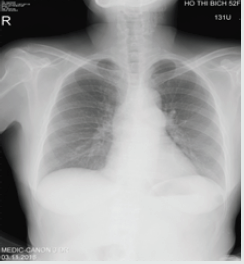
A female patient of 52 y.o presented at my hospital by persistent fever for one year, she had had this fever twice daily. Urine infection had been diagnosed and treated by Antibiotics in some hospitals, but the fever had been not interrupted. Finally, she came to MEDIC HCMC. Physical examination detected a 3/6 systolic murmur at the 3rd LICS like VSD. In her past history, no pathological finding has been noted. She was evaluated immediately by a chest X ray that demonstrated a slight prominence of the left second cardiac arch (Figures 1-4). The transthoracic echocardiography revealed a hypertrophic RV with normal TAPSE=20mm, normal LV size and function, EF=71%. Vegetation of 11x5mm in size that attached to the pulmonary valve, recorded from parasternal shorts axis view TTE.

Figure 1: Slighly prominent 2nd left cardiac arch was noted.

Lupinepublishers-openaccess-cardiology

The pulmonary cusps were doming and reduced opening degree, a severe pulmonary stenosis with max pressure Gradient=69mmhg, mean=42mmhg was confirmed by continue wave Doppler (Figures 5,6). Then Real-time 3DTEE was performed, and we found a mobile vegetation with greater size about 14x7mm associated with dilated pulmonary trunk of 35mm in diameter and a sub pulmonary valve aneurysm of 25x 36mm in measuring (Figure 7). CT angiography ( MDCT-640 ) with IV contrast medium Ultravist, slice thickness=0.5mm was indicated and detected a sub pulmonary valve aneurysm of 27x38mm and dilated pulmonary trunk: Annulus=27mm, Trunk=40-46mm, RPA=14mm, LPA=27mm. Furthermore, reduced RVOT size revealed: 9.5mm-10.2mm in diameter (Figure 8-11). Patient underwent uncomplicated surgical repair: opening RVOT, valvuloplasty and suture the perforated pulmonary cusp (this perforation was not seen prior to operation) with a favorable post-operative progress.

Read More About Lupine Publishers Journal of Cardiology Research & Reports Please Click on Below Link: https://lupine-publishers-cardiovascular.blogspot.com/

Wednesday, 14 September 2022

Lupine Publishers| Review of 10 years of practical RT-3DTEE (2010-2020)

 Lupine Publishers| Journal of Cardiology Research & Reports


Abstract

Background: Real-time three dimensional transesophageal echocardiography or RT-3DTEE, with advances in image processing and display, has increased the availability and utility of TEE to a variety of clinical settings as diagnosis and catheter intervention.

Patients & Methods: We study all Patients with RT-3DTEE, presented in MEDIC HCMC from October 2010 to May 2020.

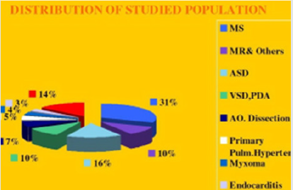
Results: From October 2010 to May 2020, in MEDIC HCM, we have already performed 1276 cases ofReal-time 3DTEE: Mitral Stenosis (31%), Atrial Septal Defects (16%) and other diseases of the heart.The display of three dimensional images of 3DTEE providing important information’s to assess the anatomical structures and the severity of diseases prior to perform interventional procedure.

Conclusion: Real-time 3DTEE overcomes the limitations of 2DTEE in making diagnosis and guiding the catheter intervention.

Keywords: Three Dimensional Transesophageal Echocardiography; Mitral Stenosis; Atrial Septal Defect

Background

The first utilization of a TEE device that promised to have clinical utility was reported in 1976 by Frazin and coworkers, who described the application of transesophageal M-mode echocardiography. The use of this technology was subsequently reported for the evaluation of ventricular function during supine exercise. The next success was the development of real-time, twodimensional TEE imaging, as first described in 1980 by Hisanaga, who developed a wide-angle mechanical sector scanner, while in 1982 Schluter reported on the use of transesophageal phased-array two-dimensional echocardiography. The incorporation of color mapping into a TEE device was first reported by investigators from Saitama Medical School in Japan. The same group was instrumental in early development of standard biplane, matrix phased -array biplane, and small pediatric probe. The current-generation probes are all multiplane devices. Rotating the small control wheel flexes, the tip to the left or to the right. But with the multiplane TEE transducer, this manipulation is rarely necessary. Rotation of the transducer refers to movement of the sector scan from 0 to 180 degrees. Transthoracic and transesophageal real-time three dimensional echocardiography is a significant advancement in technology. Advances in image processing and display, the addition of three dimensional imaging capability, and the portability of ultrasound system have increased the availability and utility of TEE to a variety of clinical settings as diagnosis, cardiac catheterization, operation and intensive care. Currently percutaneous interventions for many structural diseases increase more and more in our country. Interventional cardiologists are now treating a variety of lesions that previously required surgery as mitral stenosis, aortic stenosis, ostium secundum ASD, VSD, PDA, Coronary Fistula… Although Fluoroscopy and 2D TEE are usually used for procedural guidance, real-time three-dimensional TEE offers several important advantages over these modalities. Previously, Toshiba SSH 140 A with TEE biplan probe have been used to perform 2D TEE in our hospital. Since October 2010, transesophageal echocardiographies are made by X-MATRIX, iE 33 Philips machine.

Endpoints of study

i. Role of 3DTEE in the assessment of heart diseases.
ii. Advantages of 3DTEE in compare with 2DTEE.

Patients and Methods

a) Pts with 3D TEE at MEDIC HCM, Viet Nam, from October 2010 to May 2020.

Instruments: X-MATRIX Philips iE33, X-7 real-time 3D TEE probe.

Acquision mode including Live 3D or narrow sector, Full Volume or wide sector, Zoom or the smallest pyramydal size available for acquisition.

Techniques

i. Explain the procedure to the patient.

ii. The patient should not have had any intake of food or drink for at least 4-6 hours.

iii. Oral prostheses should be removed.

iv. The patient should be placed in the left lateral decubitus position.

v. Topical anesthetic and sedation.

vi. Introduce a bite block between the teeth.

vii. The probe is gently passed into the oral cavity over the tongue and guided into the larynx.

viii. The patient should be asked to swallow, the probe is gently introduced into esophagus.

b) Case series report study is applied for this topic.

c) The advantages of 3DTEE in compare with 2DTEE in diagnosis and evaluation of diseases prior to perform interventions.

Results

From October 2010 to May 2020, in MEDIC HCMC, we have already performed 1278 cases of 3DTEE: Mitral stenosis (31%) and other valvulopathie including Mitral Regurgitation, Aortic valvopathies, Atral Septal Defects (16%) and other shunts as PDA, VSD; then Myxoma and cardiac tumors, Endocarditis, studying prosthetic valves. The other complicated cardiopathies as Coronary artery fistula, Valsalva sinus rupture, Ebstein anomaly. The percutaneous balloon mitral valvuloplasty, transcatheter closure of ASD and surgery have demonstrated the precised diagnosis of 3DTEE (Figure 1).

Figure 1: Distribution of heart diseases detected by RT-3DTEE.

Lupinepublishers-openaccess-cardiology

Among 396 Pts with MS, mean age=45 (from 18-72 ages), the majority of patients were female (75,2%), Dyspnea is the first symptom to the consultation, sometime embolic events (7.5%) keep patients going to their cosultants, AF account 6,1%. Rheumatic Fever is predominant cause, with important fusion of commissures (70%), 2DTEE completed by 3DTEE is used to calculate the Wilkins score, providing information’s more exactly than 2D TTE alone, prior to perform Balloon Mitral Commissurotomy or surgery. MVA3D measured by Real-Time 3DTEE to compare with conventional two-dimensional planimetry MVA2D.The 3D Assessment was significantly smaller the 2D planimetry: MVA3D=0.95cm2±0.21; MVA2D=1.16cm2±0.24; mean difference=-0.21cm2, n=327, p<0,001. Some patients with high Wilkins score evaluated by 2DTEE still have responded well to Balloon Mitral Valvuloplasty. Real-time three dimensional transesophageal echocardiography provides important information’s regarding the involvement of rheumatic process on the mitral valve, particularly the symmetry length of commissural fusion. Furthermore RT-3DTEE also shows the thickening, the fibrosis and the calcification of the whole mitral commissures that cannot be visualized by 2DTEE. The 3D image allows superior visualization of the thickening of the mitral leaflet, particularly the commissures. The 3DTEE usually details the sub valvular apparatus not appreciated on 2DTEE while studying the leaflets. Because patients often presented late in Hospitals, their Wilkins score usually is high (68% with Wilkins score is superior to 8). Especially, LAA thrombus, even small size, furthermore, can be detected more clearly on RT- 3DTEE. Volume and mobility of LAA thrombus appreciated better on 3DTEE. Detection LA and LAA thrombus by RT-3DTEE is more sensitive than 2DTEE with X-plane mode and 3 D Zoom only are available in 3DTEE. Direct planimetry of mitral valve orifice by 3DTEE is the gold standard method now. The cropping function ensures that the orifice area is traced in a plan that is at the tip of the mitral valve (Figure 2-17).

Read More About Lupine Publishers Journal of Cardiology Research & Reports Please Click on below Link: https://lupine-publishers-cardiovascular.blogspot.com/

Tuesday, 2 August 2022

Lupine Publishers| Comparative Stratification of Cardiovascular Risk through Framingham and Globo Risk in Patients with Psoriasis of IAHULA November 2017-May 2018.

 Lupine Publishers| Journal of Cardiology Research & Reports


Abstract

Objectives: To know the frequency of Metabolic Syndrome in patients with Psoriasis who come to the consultation of the Dermatology Service of the Autonomous University Hospital of the Andes November 2017 to May 2018.

Materials and Methods: Observational analytical descriptive study. Patients with Psoriasis diagnoses who attended the dermatology office were selected, PASI, ATP-III, Framingham and GLOBORISK were applied.

Results: 55 patients, 55% men and 45% women, there was statistical correlation between older age groups and PASI index (p=0.023). The main modifiable cardiovascular risk factors were smoking, sedentary lifestyle and obesity, statistical correlation was found for sedentary lifestyle (p=0.047). The main non-modifiable cardiovascular risk factors were Hypertension and Diabetes showing both statistical significance (p=0.004), (p=0.0001). The ATP-III criteria showed statistical significance for Hypertension, glycemia, total cholesterol and low HDL (p=0.003, p=0.008, p=0.027, p=0.017). The frequency of metabolic syndrome represented 47.27% of the sample. The most affected gender was male (61.54%). Statistical correlation was found in the older age groups for the presence of Metabolic Syndrome (p=0.0001). The group with the highest frequency of metabolic syndrome was the one with 6 to 10 years of the disease (p=0.001). When applying the Framingham and GLOBORISK scores, there were higher scores in the patients as PASI was increased.

Conclusion: There is a higher frequency of Metabolic Syndrome in patients with Psoriasis, so it is recommended to establish measures aimed at reducing the burden of cardiovascular disease in these patients.

Keywords: Psoriasis; Metabolic Syndrome; Risk; Severity Index of the Disease

Introduction

Psoriasis has evolved conceptually over time, from being considered a skin disease in its first descriptions, currently we can define it as a chronic and inflammatory skin disease, without a defined, genetically determined, pathophysiologically autoimmune etiology, of intermittent evolution, at risk of skin, systemic and psychological comorbidities, which impact on quality of life [1]. The most characteristic feature of psoriasis is the hyperproliferation and altered differentiation of keratinocytes, in addition to immune, biochemical, and vascular abnormalities influenced by multiple environmental factors that can trigger or exacerbate its evolution, affecting between 2 and 3% of the population. World [2]. Psoriasis is estimated to affect approximately more than 125 million people worldwide, with a global prevalence of 2-3%. Being greater in those of Caucasian origin, it is variable in other races and exceptional among the indigenous people of the American continent and Australia. The white race has a prevalence of 0.1% and the incidence in the black race is low. In Germany there is a prevalence of the disease of 1.7%, in Sweden of 2.3%, the United States 2.1%, India 0.7%, Japan 0.5%. In Latin American countries it is working on it. Studies suggest that the prevalence in South America is 1% [3].

According to the global report of psoriasis published by the WHO in 2016, people with psoriasis are at high risk of suffering from various comorbid conditions, such as cardiovascular disease, diabetes, obesity, Crohn’s disease, myocardial infarction, ulcerative colitis, metabolic syndrome, strokes or liver disease. Patients with moderate/severe psoriasis, especially in the 4th decade of life, have an increased risk of having a heart attack and survival decreases 3 years for men and 4 years for women because of cardiovascular events [4]. Epidemiological studies have aroused great interest in the association of psoriasis with different cardiovascular, metabolic and immune comorbidities with which it shares inflammatory pathways or a common background [5], however, its association with metabolic syndrome has been increasingly recognized [6]. Metabolic Syndrome (MS) is a set of cardiometabolic risk factors that confer a higher risk of developing cardiovascular disease than that attributed to each component in isolation. It affects approximately 15 to 25% of the general population, its prevalence is increasing both in developed countries (United States and European countries) and in developing countries, accompanying the increase in obesity globally [7,8]. Metabolic Syndrome confers a significant burden of disease, for example, some studies show that MS confers twice the risk of coronary artery disease, [9] as well as increases the risk of cerebrovascular disease, fatty liver and certain types of malignancies such as lymphomas and squamous cell carcinomas [10].

Current recommendations suggest that patients with psoriasis should be evaluated to rule out the presence of MS, and if it is present, they should receive intensive treatment, with interventions in their lifestyle, weight loss, control of blood pressure, diabetes, and hypercholesterolemia [11]. There are multiple criteria used to define metabolic syndrome at an international Mildl, those proposed by the International Diabetes Foundation (IDF), those of the National Cholesterol Education Program Adult Treatment Panel III (NCEP ATP III) and others such as those of the Organization World Health Organization (WHO) and the American Association of Clinical Endocrinologists (AACE) that have in common grouping a series of abnormalities in the same individual [12-14].

It should be noted that most of the research papers use the ATP III criteria to compare results due to their practical utility; associating simplicity to carry out laboratory tests; and it also requires complying with three of the five parameters. There is evidence that this association (SM/PSORIASIS) is due to increased T cell immune activity, and increased production of tumor necrosis factor alpha. These classify psoriasis as the prototype of an inflammatory disease characterized by TH1 and TH17 activation, whose cytokines such as IL1, IL6, and tumor necrosis factor alpha are elevated in blood and skin mediators that could have an effect on adipogenesis, lipid metabolism, and insulin signaling. Hence, the entire inflammatory process in psoriasis has a great impact on obesity, diabetes, thrombosis, and arteriosclerosis, which, in turn, can influence the pathogenesis of psoriasis by promoting the proinflammatory state and increasing the propensity to develop the syndrome. Metabolic [15-17].

Given that Psoriasis is one of the main reasons for consultation in the dermatological area, and having no current data on the incidence of MS in Venezuela, it is necessary to establish the prevalence of MS in patients with Psoriasis and subsequently establish if there is a correlation between both pathologies. It is important to highlight that in the autonomous institute university hospital of the Andes edo. Merida Venezuela no research work has been carried out that has determined the cardiovascular risk profile or the frequency of metabolic syndrome in patients with Psoriasis who come to the Dermatology service consultation of said institution, therefore an observational study of cross section in the period established from November 2017 to May 2018. It will be of great contribution and serve as health information, in addition to contributing to the prevention of said pathology and thus reducing morbidity and mortality. The motivation to carry out this study arises; and in turn, to instill future generations to continue the present investigation.

Objectives

General

Stratification of cardiovascular risk using the Framingham and Globorisk scores in patients with Psoriasis who come to the Dermatology service consultation of the Autonomous Institute University Hospital of the Andes during the period from November 2017 to May 2018.

Specific

a. Know the demographic characteristics of the study population.
b. Describe the degree of severity of psoriasis lesions, according to the area index and severity of psoriasis (PASI).
c. Identify the Cardiovascular Risk factors in Psoriasis patients who come to the Dermatology service consultation at the Autonomous Institute Hospital Universitario de los Andes.
d. Identify the presence of the components of the metabolic syndrome, according to the ATP III criteria.
e. Establish statistical correlation between time of evolution of psoriasis, PASI index and SM.
f. Estimate cardiovascular risk according to the Framingham and GLOBORISK scales.
g. Establish correlation between cardiovascular risk/ psoriasis and metabolic syndrome/psoriasis.

Study design

Descriptive analytical observational study.

Population and sample

All patients who attended during the period from November 1, 2017 to May 31, 2018, with diagnoses of Psoriasis of both genders who attended the dermatology consultation of the Autonomous Institute Hospital Universitario de los Andes, and who met the inclusion criteria.

The inclusion criteria were as follows

a. Patients of both genders over 18 years of age.
b. Patients with a diagnosis of Psoriasis.
c. Informed consent signature.

Exclusion criteria

a. Immunological diseases, others.
b. Mixed connective tissue disease
c. Stage IV-V Chronic Kidney Disease
d. Neoplasm diagnosed.
e. Infectious processes in any form of presentation.

Data processing and analysis

Quantitative data were presented with measures of central tendency and dispersion (mean and standard deviation); the qualitative data were presented with absolute and relative frequencies (in percentages). The statistical association of qualitative data (bivariate analysis) was evaluated using the chisquare test, the statistical differences of quantitative data were evaluated using the Student’s t-test or ANOVA. The agreement between the Globo Risk and Framingham scales was evaluated through a Bland-Altman graph. Statistical significance was considered for values of p<0.05. Statistical analyzes and graphs were performed using SPSS version 21 (IBM Corporation, New York, US), Excel 2010 (Microsoft Corporation, Redmond, US) and GraphPad Prism version 5 (GraphPad Software Inc, La Jolla, USA).

Ethical component

The ethical components of this research work will be carried out based on the international ethical standards set forth in the Declaration of Helsinki [18] and that contemplated in the Code of Medical Deontology of the Venezuelan Medical Federation of March 20, 1985, in its Title V, Chapter 4, referring to research in human beings. It was requested to fill out the informed consent to the patients or their legal representatives when they are disabled, explaining in detail the purpose of the study; Likewise, the risk of complications that could arise with the taking of the sample, the confidentiality of the data obtained, the academic purposes and the non-intervention of the investigator in the treatment, as well as in the clinical evolution of the patient, will be explained. The economic resources for the realization of this study will be provided mostly by the author. This study has the support of the Autonomous Institute of the University Hospital of the Andes (IAHULA), the Multidisciplinary Laboratory of Clinical-Epidemiological Research (Lab-MICE) of IAHULA, the Department of Internal Medicine, the Dermatology department of IAHULA.

Results

Table 1 showed that the study sample was made up of 55 patients who attended the IAHULA Dermatology clinic, distributed by gender as follows; female gender n=25 with a respective percentage of (45.5%) and the highest proportion male gender n=30 (54.5%). The patients were stratified according to the PASI classification in 4 categories; inactive or asymptomatic forms, Mild, Moderate, and finally the severe and very severe forms in the last group as a last category to simplify their subsequent analysis. It was evidenced that the Moderate form of the disease according to the PASI index was the one with the highest frequency n=22, with a gender distribution of 10 (45.5%) for the female and 12 (55.5) for the gender. male. Regarding age groups, they were divided into 4 categories: those under 30 years, between 31 to 50 years, 51 to 60 years, and those over 60 years, respectively. The highest proportion of patients corresponded to the group over 60 years old (16), secondly those between 31 and 50 years old (14), thirdly between 51 and 60 years old (13) and lastly those under 30 years old (12) this correlation between the age of the patients and the PASI index being statistically significant.

Read More About Lupine Publishers Journal of Cardiology Research & Reports Please Click on Below Link: https://lupine-publishers-cardiovascular.blogspot.com/
https://lupine-publishers-biostatistics.blogspot.com/

Tuesday, 5 July 2022

Lupine Publishers| The Comparison between Two Sternum Closure Techniques after Coronary Bypass Surgery; Sterna-Band® (Peninsula) and Sternum Band (Ethicon®)

 Lupine Publishers| Journal of Cardiology Research & Reports


Abstract

Introduction: Recovery failures such as sternal dehiscence, sternal pain, sternal wound infections, osteomyelit and mediastinit can be seen after median sternotomy in coroner bypass surgery. In this study, two sternum band closure methods which we find more effective and secure than conventional sternal closure method are compared.

Method: In this prospective study that compares two sternal band closure techniques, 114 patients are divided in two groups as Group A (n=54) and Group B (n=60). This research compares the results for sternal dehiscence, sternal wound infections, sternal pain and mediastinit in these patient groups between December 2011 and July 2012.

Results: Out of the two, a meaningful recovery is achieved in Group A compared to Group B (p<0.05). No superficial woud infection, sternal dehiscence and mediastinit are found in patients of both band groups.

Conclusion: In this study, two different band closure techniques for sternotomy closure after the coroner bypass surgery are compared, and it is revealed that sternal pain index decreases meaningfully, compared to the other.

Keywords: Sternal band; Sternal dehiscence; Sternotomy; Pain index

Introduction

Median sternotomy is the most frequently used incision for cardiac surgery. Following cardiac surgery by median sternotomy, the sternum is closed by surgical steel wires. Potential postoperative sternal instability definition is dehiscence of sternal parts due to cutting of sternal cortical layers by sternum closing wires with usually inappropriate movements of patient. This may result ranging from increased postoperative pain to sternal wound infection and mediastinitis associated with higher morbidity and mortality and prolonged hospital stay and increased treatment costs [1-4]. Sternal bands used in our study are reported to prevent cutting of sternal cortex as tension on sternum is applied on larger and more balanced area, unlike to conventional sternal steel wires [4-6]. The aim of this study is to compare two different sternal band closing methods.

Patients and Method

In our clinic, 114 patients (60 males, 54 females; mean age 65,095 years) underwent to open cardiac surgery between December 2011 and July -2012 were prospectively reviewed (Figure 1). Sternal closing was performed by Peninsula Sterna –Band® (L.L.C. Livonia MI. USA ) in 54 patients consisting first group (group A) and by Ethicon Sternum Band (Norderstedt, Germany) used in 60 patients consisting second group (group B). Patients with preoperative renal insufficiency, left ventricle dysfunction, receiving anticoagulants, those who have hemorrhagic disorders were not included to the study. Preoperative demographic characteristics of patients are shown in Table 1. After cardiac surgery for closing sternum, in group A; two sternal bands were placed at inter costal spaces 3 and 4. Secondly standard 5/0 steel wires at inter costal spaces 1- 2- 5 and 6 were placed. In Group B, retrosternal sternal band at intercostals spaces 3 and 4 were placed and standard 5/0 steel wires at intercostals spaces 1- 2- 4-5 and 6 were placed.

Then subcutaneous and cutaneous tissues were closed standardly. Following post-operative hemodynamic stability, all patients were ex-tube at hour 4 to 6 and pain index scoring was obtained following mobilization during post-operative intensive care unit. Pain index was obtained by using pain scoring method Visual Analogue Scale (VAS) to evaluate the effect of both bands on post-operative pain. Patients were verbally asked to rate the presence, frequency and severity of pain on the sternum by a scale of 1 to 10. Pain index scoring was calculated by multiplying post-operative pain frequency by pain severity. Statistical analysis was performed by SPSS 15.0 version for Windows (SPSS Inc., Chicago, Illınois, USA) software. T-Test, Pearson Chi-Square test and Mann-Whitney test were used for data analysis (Figure 2). Statistical data was expressed as mean±standard deviation and significance was considered as p<0.05. The study was approved by Süleyman Demirel University Faculty of Medicine Scientific Study Projects Consulting Coordinating Board by project number B.30.2.SDU.0.20.05.07-507/5077 on 27.12.2011 and board decision no. 27/6.

Results

Table 2: Preoperative demographic characteristics.

lupinepublishers-openaccess-journal-cardiology

CABG: Coronary arteries bypass graft

Table 3: Preoperative demographic characteristics.

lupinepublishers-openaccess-journal-cardiology

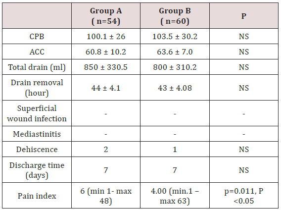
Pain index: Pain severity x Pain frequency

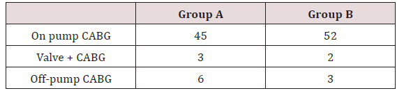
Pre-operative demographics were not statistically significantly different between two groups (Table 1). Surgery type of operated patients is shown on Table 2. When peri- and post-operative variables were evaluated, there was no statistical difference in respect of cardiopulmonary bypass time (CPB), aortic cross clamp time (ACC), post-operative drainage, the amount of blood utilized, administration of inotropes, drain removal time, superficial wound infection, mediastinitis, dehiscence and hospital discharge day ( p˃0.05), (Table 3).

Discussion

Complete closing of sternum following median sternotomy during open cardiac surgery, is a critical point in prevention of postoperative complications due to sternotomy. These complications include mainly dehiscence as well as a broad range of conditions such as pain, superficial wound infection and mediastinitis. Currently several sternal closing methods were described to prevent these complications [4]. Therefore, in our study we compared two different sternal bands used for sternal closing. In patients with sternotomy, sternal dehiscence is the most frequent complication and the rate is 0.3% to 5%. This is a mechanical problem associated with certain factors such as lack of attention of patient to protect the sternum post-operatively, COPD, obesity, osteoporosis, use of bilateral mammarian arteries, renal insufficiency, administration of steroids and re-operation [4]. The incidence of sternal wound infection is 0.4-5% and it is closely associated with dehiscence. Closing of sternum is an important factor for sternal dehiscence. During sternal closing done by conventional approach using steel wires 5/0 or 6/0, bone cortex erosion, fracture and consequently sternal dehiscence can occur due to compression of steel wires. Sternal dehiscence may lead to respiratory dysfunction, infection, increased pain and re-exploration (Figure 3).

Use of sternal band, especially retrosternal application at intercostals space 3 and 4 to transversely surrounding whole sternum, is the most important mechanism to reduce development of sternal dehiscence with its 6 times larger surface than steel wires and thus providing equal loading on sternum. In our study, sternal dehiscence and sternal wound infection were not detected in both groups. Postoperative pain is an acute pain gradually reducing by tissue healing accompanied also by inflammatory process [6]. Indeed, acute pain is a complex sensation besides its simple perceptional character. Difference in pain perception may be due to central process, fear, anxiety, depression and previous experience on pain perception. Certain patients may not describe the pain due to their higher pain tolerance or their introvert pain handling methods. Difference in pain perception of patients may be also due to post-operative analgesic administration methods. However, 30% of surgery patients don’t require postoperative analgesics [7]. In a prospective clinical study, localization, distribution and severity of postoperative pain were evaluated in consecutive 200 patients with cardiac surgery by median sternotomy [8]. Investigators determined the highest pain intensity on day 1 and lowest intensity on day 3. Although pain distribution was not different during post-operative period, there was difference in its localization [9]. Another factor affecting the pain severity is the age of patient. Young patients reported more severe pain than patients over 60 years (Figure 4).

Previous studies comparing sternal band and conventional sternal closing also showed that usage of sternal band was associated with decreased pain and this was explained by reduced development of sternal dehiscence [10-12]. However, in our study, we determined a difference between different methods of sternal bands in respect of pain development. When two groups of our study were compared, there was significant reduction in postoperative pain development in Group B compared to Group A [13]. At this point, we consider that reduced compression of band method on a smaller bone cortex area only at intercostals space 3 in Group B compared to band method of Group A, could explain lower pain. However, we think that the limited number of studies on pain in cardiac surgery and multi factorial nature of pain are limitations of our study [14-16]. Although our study is prospective and including low number of patients, we conclude that larger studies will provide more guidance by including psychological and anatomic-physical parameters such as difference in physical load on bone structure by the material utilized.

Read More About Lupine Publishers Journal of Cardiology Research & Reports Please Click on Below Link:  https://lupine-publishers-cardiovascular.blogspot.com/

Tuesday, 28 June 2022

Lupine Publishers| Understanding the Demographic and Clinical Characteristics of Heart Failure to Improve Management Practices: The Caribbean Perspective

 Lupine Publishers| Journal of Cardiology Research & Reports

Abstract

Heart Failure is a worldwide burden. The prevalence, incidence, mortality and morbidity rates reported show geographic variations, depending on the different aetiologies and clinical characteristics observed among patients with this complex syndrome. In this review we focus on the Afro Caribbean population with Heart Failure providing data review about the Heart Failure etiology based in previous Clinical Trials In addition we provide suggestions based in these Trials and our recent observational clinical studies that might be useful to improve our current Heart Failure Guideline-Directed Medical Therapy . Finally we highlight the need for more regional research.

Keywords: Caribbean region, Heart failure, Etiology, Management

Abbrevations: CHF: Congestive Heart failure; HF: Heart Failure; MI: Myocardial Infarction; SHF: Systolic Heart Failure; HT: Hypertension; DM: Diabetes Mellitus; HFNEF: Heart Failure with a Normal Ejection Fraction; DCM: Dilated Cardiomyopathy; NIDCM: Non-Ischemic Dilated Cardiomyopathy; IDCM: Ischemic Dilated Cardiomyopathy; LVSD: Left Ventricle Systolic Dysfunction; HFmrEF: Heart Failure with mid-range Ejection Fraction; HFrEF: Failure with Reduced Ejection Fraction; ARNI: Angiotens in Receptor Neprilysin Inhibitor

Introduction

There is no doubt that Heart Failure is a complex syndrome that results from any structural or functional impairment of ventricular filling or ejection of blood [1], moreover, Heart Failure is a disease in which there are striking population differences in almost every aspect of the disease. It has been recognized that the cause of Heart Failure is predominantly ischemic disease in nonblack but is related primarily to Hypertension in Blacks [2], accordingly, this statements should be confirmed in Afro-Caribbean patients because population differences exist that may be attributable to differences in social factors, genetics, environment , lifestyle, comorbidities, and complex interactions among this factors [3].

Heart failure research data in African American: can also them be applied in afro Caribbean patients?

The majority of the data on Heart Failure in African descendant has been done in African American population and that research should be validated in the Caribbean Region. The U.S. Office of Management and Budget [4] defines “Black” of “African American” as having origins in any of the Black racial groups of Africa. The term African Caribbean/Afro-Caribbean when used in Europe usually refers to people with African ancestral origins who migrated via the Caribbean islands [5]. The majority of Jamaicans (92.1%) identify as black [6] however there are other ethnic groups [7]: Mixed (6.1%), Asian (0.8%), others (0.4%). Much of Jamaica’s black population is of African or partially African descent with many being able to trace their origins to West Africa and the term African Caribbean must be restricted to an African descent person originating from the Caribbean. Although it seems reasonable to think in terms of African Descendant Data the Heart Failure clinical studies in African American should be validated in the Caribbean Region.

Disparities Between White and Black: Studies In African American Population

Recent research continues suggesting disparities between white and black populations: In a Random Cohort of 2,188 participants from The Reasons for Geographic and Racial Differences in Stroke Project - REGARDS-[8] without prevalent cardiovascular disease studied by race was showed that Plasma N-terminal pro b-type natriuretic peptide (NTproBNP ) levels are significantly lower in blacks as compared to whites however the contribution of endogenous suppression of Natriuretic Peptide system on cardiovascular disease in blacks remains to be established On the other hand , in relation with the etiology of patients with Heart Failure, when defined as a prior documented myocardial infarction or known epicardial coronary artery disease, ischemic heart disease appears to be present in just 35% of African- American patients and the exact mix of hypertensive heart disease and idiopathic dilated cardiomyopathy (as well as the influence of alcoholic cardiomyopathy) is not discernible from published data. Therefore, the contribution of hypertensive heart disease as the sole explanation of Left Ventricle dysfunction is likely to be between the 30% range reported in the SOLVD registry or the>50% incidence suspected in recently reported β-blocker trials [9] (Figure 1). Moreover in the Multi-Ethnic Study of Atherosclerosis --MESA-[10] , a cohort study of 6814 participants of 4 ethnicities (African American :27.8%) , African Americans had the highest incidence rate of Congestive Heart failure (CHF) and showed that the mechanisms of CHF also differed by ethnicity namely an interim myocardial infarction had the least influence among African Americans (Figure 2).

Figure 1: Heart Failure in African Americans: Incidence of Coronary Artery Disease. Pooled Data from major Trials in Heart Failure reporting probable cause of Left Ventricle Systolic Dysfunction. US Carv= =US Carvelidol Heart failure Trials Program; BEST=Beta Blocker Evaluation of Survival Trial; AA=African American; Non-AA=Non African American [9].

Lupinepublishers-openaccess-cardiology

Figure 2: Nelson-Aalen Plots of Cumulative Hazards for Congestive Heart Failure (CHF) among Multi-Ethnic Study of Atherosclerosis Study participants without an interim Myocardial Infarction [10].

Lupinepublishers-openaccess-cardiology

That is , as the Time course and pattern of development of heart failure (HF) can be primarily caused by myocardial infarction (MI), with pronounced remodeling and shape change leading to systolic heart failure (SHF) or HF primarily caused by hypertension (HT), with or without diabetes mellitus (DM), leading to heart failure with a normal ejection fraction (HFNEF) however both, patients with a history of MI and patients with HT may experience periods of HFNEF and finally Systolic heart failure (SHF) with a reduced ejection fraction: HFrEF [11] .More evidence about the Primary Cause of Heart Failure in African American can be also analyzed from the Baseline Characteristics of the African-American Heart Failure Trial [12] in which about 1000 patients showed a history of Ischemic Heart Disease in just 23% and also in the recent Prospective Comparison of ARNI with ACEI to Determine Impact on Global Mortality and Morbidity in Heart Failure (PARADIGMHF) Sub study [13] in which the Black participants (N=428) showed a history of Myocardial Infarction in just 17.3 % vs 48.3 % in the White participants (N=5544) , strengthening the Heart Failure mechanism with less influence of an interim of a myocardial infarction. From the few studies among Afro Caribbean population with Heart Failure is worth mentioning the study of Afro-Caribbean heart Failure in the United Kingdom [14]: compared with the white population, Heart Failure in Afro Caribbean Patients (n=211) was mostly associated with Non Ischemic Cardiomyopathy (87vs 59%), Dilated Cardiomyopathy (27.5 vs 10.8%) and Hypertensive Cardiomyopathy (12.3 vs 2.2%) and even more interesting is the fact that Cardiac Amyloidosis was confirmed in 11.4 vs 1.6% .

Heart failure in african-americans

As suggested for previous Clinical Trials Experience, Heart Failure Etiology [15] in African-Americans-AA:

a) Is much more associated with Non Ischemic etiology of Left Ventricular Dysfunction (LVD).

b) Hypertension is considered the putative disease process but dilated cardiomyopathies and Diabetes-related diseases are common.

c) Coronary Artery Disease remains a cause of LVD but is less common in AA than in whites.

Why African Americans, and extensively Afro-Caribbean, have more Heart Failure?

It has been proposed [16] the following mechanisms:

a) The impact of Modifiable Risk Factors.

b) Neuro hormonal imbalances and endothelial dysfunction.

c) Genetic Polymorphisms and

d) Socioeconomic Factors and Quality of Care.

It is clear that Hypertension remains as the most important risk factor: A Prevalence estimates for traditional CVD risk factors in Jamaicans showed [17]: hypertension, 25%; diabetes, 8%; hypercholesterolemia, 12%; obesity, 25%; smoking 15%. In addition, 35% of Jamaicans had pre hypertension, 3% had impaired fasting glucose and 27% were overweight. A higher proportion of women had diabetes, obesity and hypercholesterolemia while the prevalence of pre hypertension and cigarette smoking was higher in men. In the other hand, some race-specific differences in endothelial function and predisposition of AA to vascular diseases are [18]: (a) increased oxidative stress (b) decreased Nitrite Oxide availability (c) Exaggerated vasoconstrictor response and (d) attenuated responsiveness to Vasodilators and Nitric Oxide. In addition, Genetic Polymorphisms associated with the risk of Heart failure in AA has been considered [16] : (a) Beta 1 Adrenergic Receptor (b) Alpha 2c Adrenergic Receptor (c ) Aldosterone Synthase (d) G Protein (e) Transforming G Grow Factor Beta (f) Nitric Oxide Synthase (g) Transthyretin. This has been recently evaluated for Dungu [14]: In a comparison with white patients, the author found ATTR V122l and Cardiac Amyloidosis all types in 8.5 % vs 0.3 % and 11.4% vs 1.6 % respectively of Afro-Caribbean vs. White patients with Heart Failure living in the United Kingdom.

Clinical studies in afro-caribbean patients with heart failure living in jamaica

The available data of Afro Caribbean Heart Failure, including studies in Jamaica, is scarce. Tulloch Reid [19] brought the attention to cases with unexplained Dilated Cardiomyopathy and reported the association of HTLV-1 seropositivity and Unexplained Dilated Cardiomyopathy (DCM) in Jamaican patients. This author also reported the experience of 26 cases (45±11 years-old) with unexplained dilated cardiomyopathy at Kingston Public Hospital (personal communication). Lalljie [20] reported the experience in 100 Jamaican with heart failure patients: 49% had echocardiograms, of these 39% had ejection fractions (EF)>40%, 34 % had EF 21-40 % and 27% had EF<20%. Hypertensive heart disease was found in 54%, hypertensive cardiomyopathy in 14 % and ischemic heart disease just in 26 %. Accordingly and knowing that the majority of the data on Heart Failure in African descendant has been done in African American population and that these studies had to be validated in the Caribbean Region we planned some observational studies [21-23] in order to know:

a) The cardiovascular risk profile of patients with, angiographycally proven, Non-Ischemic Dilated Cardiomyopathy (NIDCM) vs Ischemic Dilated Cardiomyopathy (IDCM) (Figure 3).

Figure 3 : The presence or the absence of the most important traditional cardiovascular factors (Hypertension=HTN and Diabetes-DM), which have been described as strongly correlated with coronary artery disease, are not necessarily predictive of angiographycally-proven Non-Ischemic Dilated Cardiomyopathy (NIDCM) in an Afro Caribbean population with HFrEF [21].

Lupinepublishers-openaccess-cardiology

b) The demographic and clinical features of patients with left ventricle systolic dysfunction (LVSD) and differences between Heart Failure with mid-range Ejection Fraction (HFmrEF) and Heart Failure with Reduced Ejection Fraction (HFrEF) (Figure 4).

Figure 4 : The presence or the absence of the most important traditional cardiovascular factors (Hypertension=HTN and Diabetes-DM), which have been described as strongly correlated with coronary artery disease, are not necessarily predictive of angiographycally-proven Non-Ischemic Dilated Cardiomyopathy (NIDCM) in an Afro Caribbean population with HFrEF [21].

Lupinepublishers-openaccess-cardiology

c) The proportion and clinical features of patients with angiographycally proven Ischemic vs Non Ischemic Heart Failure with Reduced Ejection Fraction (HFrEF) from our Coronary Angiographies database (Figure 5).

Figure 5 : From a total of 380 patient’s angiographycally evaluated during the study period, 67 patients were included in the analysis. The prevalence (%) of patients with NIDCM was 55%. With the exception of age (59 vs 65, p<0.03) and confirmed myocardial infarction-MI (2 vs 25%, p<0.04) there were no significant differences (p<0.05) in the prevalence of CAD risk factors suggesting that among Afro-Caribbean patients with HFrEF, despite that both groups of patients were exposed to similar CAD risk factors, angiographycally proven NIDCM is more frequent that IDCM. This underlines the hypothesis that other factors may play an important role in the etiology of the Afro Caribbean Heart Failure [23].

Lupinepublishers-openaccess-cardiology

Lessons From the Clinical Studies on Afro Caribbean Patients with Heart Failure Living in Jamaica

From a broad perspective, these studies are suggesting that:

a) Although both groups (NIDCM vs IDCM) has been exposed over their lifetime to the same strong risk factors for CAD, they potentially and can develop the same outcome (DCM) but one group with angiographycally normal epicardial coronary arteries and the other with angiographycally abnormal coronary arteries.

b) Hypertension and Diabetes are the most important risk factors associated with Dilated Cardiomyopathy in this Afro Caribbean population.

c) Afro-Caribbean patients with heart failure and are mainly hypertensive with or without diabetes and grossly half of them develop LVSD due to non-ischemic causes.

d) They demonstrate a distinct etiological but similar clinical profile when they are classified in the category of HFmrEF and HFrEF

e) Among Afro-Caribbean patients with HFrEF, angiography ally proven NIDCM is more frequent (55vs.45%) that IDCM suggesting that despite both groups of patient’s exposure to the similar CAD risk factors, other factors play an important role in etiology of the left ventricle failure.

Understanding Afro Caribbean Heart Failure to Improve the Management Practices

According with the 2013 ACC/HFA Guidelines [1] the magnitude of benefit for Reduction in Mortality and Reduction in HF Hospitalizations demonstrated in RCTs is ,respectively, as follows: a) ACE Inhibitor or ARB: 17% and 31% b) Beta Blockers: 34% and 41% c) Aldosterone Antagonist: 30% and 35% and d) Hydralazine/ Nitrate (HYD/ISDN): 43% and 33%, accordingly the Pharmacologic treatment recommendation for persistently symptomatic African American patients with Stage C HFrEF, class III-IV, in order to reduce morbidity and mortality, and in addition to ACE Inhibitor, or ARB and in conjunction with Beta Blocker is Hydralazine/Nitrates (Class I, LOE A). Moreover, the 2017 ACC/AHA Guidelines Update [24] has recommended for patients with chronic HFrEF, to reduce morbidity and mortality the Angiotens in Receptor Neprilysin Inhibitor (ARNI) in conjunction with Beta Blocker (Class I, LOE B-R) and for patients with chronic symptomatic HFrEF, NYHA class II-III, who tolerate an ACE Inhibitor or ARB: the ARNI, to replace an ACE Inhibitor or ARB (Class I, LOE B-R) More understanding about the population of African Americans has been mentioned in the 2017 ACC Expert Consensus Decision Pathway for Optimization of Heart Failure with Reduced Ejection Fraction [25] which has stated that in African Americans Sacubitril/Valsartan and Ivabradine were tested in populations with few African Americans receiving HYD/ ISDN. Thus, for this population, there are no data for the efficacy or safety of ARNI in patients with an indication for HYD/ISDN.

Moreover, both HYD/ISDN and ARNI purportedly act via upregulation of cGMP pathways, which may increase the risk of hypotension. Additionally, the risk of angioedema with ACEI and ARNI is particularly high in African-American patients (0.5% with ACEI and 2.4% with ARNI); this risk, however, should not preclude initiation of these agents absent a documented history of angioedema. Two options would exist: A. Establish Guideline- Directed Medical Therapy (GDMT) with ACEI/ARB, beta blocker, and an aldosterone antagonist, then switch to ARNI; if stable, follow with HYD/ISDN if patient has persistent class III to IV symptoms with careful blood pressure monitoring or B. Establish GDMT with ACEI/ARB, beta blocker, and an aldosterone antagonist and then proceed with HYD/ISDN if persistent class III to IV symptoms; if stable, follow with ARNI substitution for ACEI/ARB with careful blood pressure monitoring. In the absence of randomized controlled data, it is reasonable to treat an African-American patient using either approach. However, the risk for hypotension with either strategy is uncertain. The treatment decision should be determined after an informed shared decision-making discussion with the patient, indicating the uncertainty of benefit.HYD/ISDN is available as a fixed-dose combination or as individual medications. The ACC/ AHA/HFSA guideline considers either as acceptable in this context.

Conclusion

Heart Failure is a complex syndrome. The prevalence, incidence, mortality and morbidity rates reported show geographic variations, depending on the different aetiologies and clinical characteristics observed among patients. In this review we focus on the Afro Caribbean population with Heart Failure providing data review about the Heart Failure etiology based in previous Clinical Trials In addition we provide suggestions based in these Trials and our regional clinical studies that might be useful to improve our current Heart Failure Guideline-Directed Medical Therapy There is a need to understanding more the epidemiology and mechanisms of Afro Caribbean Heart Failure in order to improve the current management practices from a Caribbean perspective. At this time there are no studies on the use of ARNIs in the unique Afro Caribbean population, accordingly clinical experience studies with regards to the tolerance and safety of sacubitril/valsartan to prospectively identify adverse events and discontinuation for adverse events in patients with HFrEF are required.

Read More About Lupine Publishers Journal of Cardiology Research & Reports Please Click on Below Link:  https://lupine-publishers-cardiovascular.blogspot.com/