Showing posts with label Journal of Anesthesia. Show all posts

Saturday, 2 September 2023

Lupine Publishers | Non-Invasive Ventilation in the Treatment of Acute Respiratory Failure with COVID-19

 Lupine Publisher | Journal of Anesthesia & Pain Medicine


Abstract

Decades have passed since the first reports of the successful use of mask non-invasive ventilation (NIV) in the treatment of respiratory failure. The aim of the study is determining the benefits of NIV in acute respiratory failure in patients with COVID-19. The study included patients admitted to the intensive care unit of the surgical clinic of the AMU from April 1 to May 1, 2020. NIV has visible advantages over mechanical ventilation. But it must be remembered that even in experienced hands, NIV is successful only in 75-90% of all cases, which depends on many factors, such as the severity of ONE, the training and experience of medical personnel. As with many types of therapy, operations, and technologies, improvement in the results of this method can be expected as experience is gained. High minute lung ventilation (>10 L/min) during NIV may predict non-invasive lung ventilation.

Keywords: Non-invasive ventilation; Acute respiratory failure; COVID-19

Introduction

Decades have passed since the first reports of the successful use of mask non-invasive ventilation (NIV) in the treatment of respiratory failure [1,2]. Attempts to use NIV with positive pressure in acute respiratory failure have been made earlier - in the 1970s and 1980s, but in general this experience was not very successful, because at that time devices for intermittent breathing with positive pressure were usually used (intermittent positivepressure breathing), which were poorly tolerated by patients and were usually intended for aerosol therapy [3]. The appearance of convenient masks for conducting ventilation with continuous positive airway pressure (CPAP) and new modes of respiratory support (especially pressure support mode) gave an impetus to the widespread introduction of NIV in clinical practice [4-6]. In 1990- 2000, the accumulation of experience with NIV and the encouraging positive results of this method in several studies allowed NIV to be assigned the first-line treatment place in acute respiratory failure [7-9]. The most important advantage of NIV in acute respiratory failure is the reduction in mortality, which may be associated with a reduced risk of nosocomial pneumonia and other hospital infections [10]. In addition, compared with invasive respiratory support, NIV is associated with less risk of damage and subsequent remodeling of lung tissue. Before using NIV, you need to pay attention to some important aspects of the method. The effectiveness of NIV depends on the correct assessment of its capabilities and limitations, while in order to avoid delays in the use of tracheal intubation and mechanical ventilation, in turn, the selection of a suitable patient, the participation of trained medical personnel and the timely detection of NIV failure are required [5,11,12]. Proper patient selection is a key factor in achieving NIV success. When COVID-19 is complicated by acute respiratory failure, patients with hypercapnia and moderate respiratory acidosis are most suitable, although a combination of respiratory and metabolic acidosis also lends itself well to NIV therapy [13]. Severe respiratory acidosis significantly increases the chances of patient intubation, especially at pH <7.20 [14-17], but in routine practice, as experience shows, in some patients NIV can also be successfully performed at low pH values of 7.10 [18]. A coma condition is also a contraindication to NIV. As a rule, none of these factors is an absolute contraindication to NIV, but they should be taken into account when deciding on the beginning of NIV, as well as when ascertaining the inefficiency of the method and the need for tracheal intubation. Known predictors of NIV success or failure include the patient’s neurological status (Glasgow scale), overall disease severity (APACHE II scale), and high tachypnea [19]. The “ideal” patient during the NIV should be a sufficiently communicative patient to provide conditions for applying and fitting the mask and synchronization with a respirator. Agitated and restless patients usually do not tolerate the NIV procedure. Most often in clinical practice, nasal or facial masks are used. The severity of the patient’s condition may be a factor determining the appropriate type of mask: for example, patients with less severe respiratory failure (DN) are better adapted to nasal masks, which are more leaking when used, while in more severe situations, oronasal masks are better suited [20]. On the other hand, with the modern choice of various models of masks, individual characteristics and preferences of patients are considered. Tight fitting of the mask to the patient’s face allows to minimize leakage and improve the patient’s synchronization with the respirator. At the same time, with excessively tight contact of the mask with the patient’s skin, ulcerations and necrosis may develop. In patients with agitation, anxiety, and high tachypnea, sedation may be prescribed to improve synchronization, but the risk of excessive sedation and respiratory depression should be remembered [19].

The Aim of the Study

Determining the benefits of NIV in acute respiratory failure in patients with COVID-19.

Material and Research Methods

The study included patients admitted to the intensive care unit of the surgical clinic of the AMU from April 1 to May 1, 2020.

The Results of the Study

Our experience with NIV has shown that most patients treated with NIV tolerate this procedure relatively well already at the initial stage. However, in several patients, during the first minutes or hours of NIV, no improvement (clinical indicators and gas exchange) is observed or the procedure is poorly tolerated, the proportion of such patients is usually about 15-35%. Experience shows that longer attempts to use NIV without achieving a noticeable improvement only delay the time of tracheal intubation and mechanical ventilation, which significantly increases the risk of worsening respiratory failure, an unfavorable outcome, including death. Using NIV, we came to the conclusion that, in most cases, NIV therapy failures are detected quite early - on the first day from the initiation of respiratory support, however, in some patients, NVL therapy failure manifests itself later - 24-48-72 hours after the initial improvement. Lack of improvement in consciousness or respiratory acidosis 24 hours after onset is NIV another predictor of NIV failure. Indications for the implementation of NIV are as follows:

a. Symptoms and signs of acute respiratory failure: a) severe shortness of breath at rest; b) BH> 25 / min, participation in the breathing of the auxiliary respiratory muscles, paradoxical breathing.

b. Signs of gas exchange disturbance: a) PaCO2> 45 mm Hg. Art., pH <7.35; b) PaO2 / FiO2 <200 mmHg. Art.

c. The exclusion criteria for NIV in ODN are as follows:

d. Stop breathing.

e. Unstable hemodynamics (hypotension, uncontrolled arrhythmias, or myocardial ischemia).

f. Inability to protect the respiratory tract (cough and swallowing disorders)

g. Excessive bronchial secretion.

h. Signs of impaired consciousness (agitation or oppression), the patient’s inability to cooperate with medical personnel. i. Facial trauma, burns, anatomical disorders that prevent masking.

Indications for the termination of NIV and the transition to intubation of the trachea and mechanical ventilation include the following:

i. The patient’s inability to carry the mask due to discomfort or pain.

ii. The inability of the NIV to improve gas exchange within 2 hours: an increase or preservation of hypoxemia, despite the high values of PEEP and FiO2.

iii. Inability to mask ventilation to ease dyspnea. iv. The need for endotracheal intubation to remove secretions or protect the respiratory tract.

v. Instability of hemodynamics and ECG, instability with the phenomena of ischemia or clinically significant ventricular arrhythmias.

vi. The increase in encephalopathy.

vii. During the study, the following advantages of non-invasive ventilation were identified:

viii. Prevention of “mechanical” and infectious complications associated with intubation, reducing the risk of developing infectious complications and mechanical damage (trauma to the larynx and trachea, stenosis, and bleeding from the upper respiratory tract).

ix. Preservation of natural protective reflexes of the upper respiratory tract.

x. Preservation of physiological cough, the patient’s ability to talk, swallow, eat, cough up sputum.

xi. Increase patient comfort.

xii. Reduced need for muscle relaxants, opioids, and sedatives.

xiii. The possibility of discrete use and weaning from the apparatus.

In our clinic, NIV was performed using Salvia Elisa ventilator respirators in CPAP+PSV mode through a face mask. Used standard masks from Drager (Germany) or Respironics (USA). To determine the parameters of the gas and acid-base composition of the blood, an ABL500 gas analyzer with an OSM3 oximeter (Radiometer, Denmark) was used. Indicators of the function of external respiration were recorded from the display of the respirator. All data were recorded immediately before the start of ventilation. The level of PEEP and pressure support was set individually, based on the specific clinical situation. The ventilation parameters required by patients were as follows: PEEP - from 5 to 12 cm of water column, PSV - from 0 to 14 cm of water. column, FiO2 - from 0.3 to 0.6. At the initial stage, auxiliary ventilation was carried out in a continuous mode. Further, a gradual decrease in respiratory support was carried out in accordance with the degree of clinical improvement, after which they switched to NIV sessions for several hours a day until it was completely canceled. The criterion for successful NIV was the improvement of the arterial blood gas composition and the ability to avoid endotracheal intubation.

Clinical Case

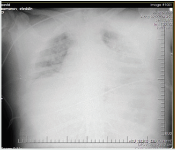
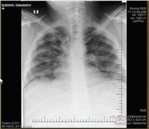
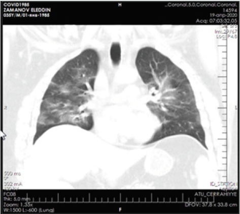
Patient - a man aged 34 years, with complaints of alternating chronic cough, temperature 39.2º C, chills, headache, and shortness of breath. During auscultation of the lungs, crepitus was observed, moist rales in the lower lobes of the lungs, oxygen saturation 90%, and respiratory rate 30-34 per minute. Admission laboratory tests included: WBC - 14,64 x 10⁹/L, LYM - 0,00 x 10⁹/L, albumin - 3.01 g/dL, PI - 50.5%, PT - 14.8 sec., INR - 1.47, Fibrinogen - 128 mg/L, GRP 47.4 mg/L, D-Dimer> 1500 ng/mL, Ferritin - 1178 ng/mL, P / F> 180. Chest x-ray and CT are shown in the following (Figures 1-4).

Figure 1: X-ray upon admission.
Lupinepublishers-openaccess-anesthesia-pain-medicine-journal

Figure 2: Radiograph after improvement of the patient.
Lupinepublishers-openaccess-anesthesia-pain-medicine-journal

Figure 3: CT scan of the lungs upon admission.
Lupinepublishers-openaccess-anesthesia-pain-medicine-journal

Figure 4: CT scan of the same patient.

Lupinepublishers-openaccess-anesthesia-pain-medicine-journal

Non-invasive ventilation was carried out by an oral-nasal mask with a ventilator ELISA. Installation and adjustment of parameters was carried out according to the general condition and according to blood gas data: BH <35, pH> 7.30, neurological dysfunction according to the Kelly scale> 3-5, a modified scale for determining the participation of auxiliary respiratory muscles <3 points. For hypercapnia, the following parameters were set Ps - 12, PEEP - 6 cm water column, FiO2 -30-40%, and with hypoxemia - Ps - 12, PEEP - 5 cm water column, FiO2 -50-60. The median treatment period with NIV was 6 days. The average daily treatment time with NIV on the first day was 16.5 hours, on the second day - 17.2 hours and on the third day 15.7 hours. The patient was discharged on the 14th day with improvement.

Conclusion

1. NIV has visible advantages over mechanical ventilation. But it must be remembered that even in experienced hands, NIV is successful only in 75-90% of all cases, which depends on many factors, such as the severity of ONE, the training and experience of medical personnel. As with many types of therapy, operations, and technologies, improvement in the results of this method can be expected as experience is gained.
2. High minute lung ventilation (>10 L/min) during NIV may predict non-invasive lung ventilation.

Read More About Lupine Publishers Journal of Anesthesia & Pain Medicine Please Click on Below Link:
https://lupine-publishers-anesthesia-pain.blogspot.com/

Friday, 14 July 2023

Lupine Publishers | Use of Regional Anesthesia in Patients with Multiple Sclerosis

 Lupine Publishers | Journal of Anesthesia & Pain Medicine


Abstract

Review Topic: Use of Regional Anesthesia in Patients with Multiple Sclerosis

Background: This article will focus on patients who have traditionally been considered contraindicated for the use of regional anesthesia. Specifically, patients with autoimmune mediated neurologic disorders. The selected pathophysiology of multiple sclerosis and regional anesthesia and their considerations via evaluation of research studies will be presented. Furthermore, suggestions for anesthesia care for these patients will be provided.

Project Aim: The purpose of this article is to and educate anesthesia providers on patients with multiple sclerosis and anesthesia implications for this population. There appears to be a lack of discussion and research in the United States regarding the use of regional anesthesia in these patients. After reading this article, the reader will be able to identify patients at an increased risk of nerve damage, evaluate them properly preoperatively, and objectively determine if the patient is considered a candidate for regional anesthesia.

Data Sources: The research information was gathered via an extensive literature review on Cumulative Index to Nursing and Allied Health Literature (CINAHL), PubMed, and EBSCOhost. Additional resources were also utilized for physiology and pathophysiology purposes: Clinical Anesthesia 7th edition and Nurse Anesthesia 5th edition.

Keywords: Multiple sclerosis; Nerve damage; Regional anesthesia; Contraindications; Demyelination; Local anesthetic; Autoimmune

Introduction

There are several clear benefits of regional anesthesia for surgical procedures. However, patients with pre-existing neurologic disorders create a unique challenge for the anesthesia provider considering the use of regional or neuraxial anesthesia. Specifically, patients with autoimmune mediated neurologic disorders that possess pre-existing neurologic deficits have been thought to be at a higher risk for neural compromise and therefore, are often excluded from the use of regional anesthesia [1]. In all cases where regional anesthesia is considered, the benefits must be weighed against potential complications; the greatest risk being a new or worsened neurologic deficit. With the advancement of regional techniques paired with ultrasound guidance, literature suggests that belonging to this patient population should not necessarily be a contraindication to regional anesthesia [2]. Due to the medicolegal concerns and historic avoidance of regional anesthesia in patients with neurologic disorders, there is a lack of discussion and studies in the United States on this topic. However, increasing support for the use of regional anesthesia for surgical procedures has produced a few studies within the United States and gained even more popularity internationally.
This article will explain the pathophysiology of multiple sclerosis, evaluate current anesthetic practices and recent literature on regional anesthesia for patients with this disorder and provide recommendations for further studies and practice guidelines. After reading this article the reader will be able to properly identify and assess patients at an increased risk for neurologic compromise, evaluate the risks and benefits of using regional anesthesia and determine if the patient is a candidate. The reader will also have a better understanding of the pathophysiology of multiple sclerosis and its anesthetic considerations to improve quality of care and perioperative outcomes.

Pathophysiology of Multiple Sclerosis

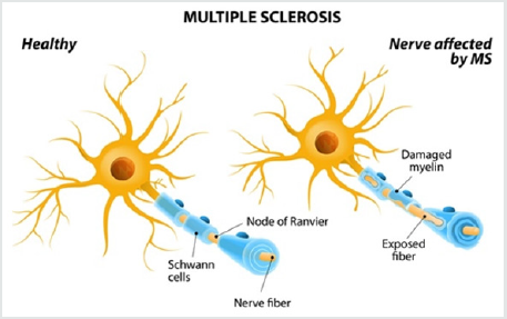
Multiple sclerosis (MS) is an inflammatory autoimmune disorder of the central nervous system (CNS). However, recent post-mortem studies have reported some degree of peripheral involvement [3]. Although genetic susceptibility is likely, there is no obvious genetic, environmental or infectious cause that has been identified. Additionally, there is no clear understanding of the immunopathogenesis of the disorder that determines site of tissue damage, variations throughout history, or the severity of disability Pathologically, MS possesses a unique combination of characteristics that include inflammation, demyelination and axonal damage in the CNS; followed by the formation of plaques resulting in further alteration of nerve conduction [4]. The innate immune system is thought to play a major role in the initiation of multiple sclerosis by modification of T and B- lymphocyte function. The adaptive immune system is also involved by activation of CD4 and CD8 cells. It is their polarization to Th1 and Th17 effector cells and cytokine formation that causes damage to the blood brain barrier. This allows autoreactive lymphocytes access to the CNS subsequently leading to the characteristic demyelination associated with the disease [3] (Figure 1).

Figure 1: Multiple Sclerosis.

Lupinepublishers-openaccess-anesthesia-pain-medicine-journal

Symptoms associated with MS are often varied with multifocal involvement, but there are two general clinical patternsexacerbating- remitting or chronic progressive. Progression of MS is caused by lack of remyelination and axonal degeneration which is irreversible and causes permanent neurological deficits [3]. Physical manifestations reflect the sites where demyelination has occurred. Although ascending spastic paresis of the skeletal muscles is often prominent, symptoms may also present as sensory deficits, autonomic dysfunction, emotional lability, bladder and bowel dysfunction and visual disturbances. MS exacerbations can be triggered by heat, stress, infections and pregnancy. Diagnosis of MS is made based on clinical features alone or in combination with oligoclonal immunoglobulin irregularities in the cerebrospinal fluid, prolonged latency of evoked potentials, and changes in white matter. There is currently no cure for MS and treatment is directed towards symptom control and slowing the progression of demyelination [4].

Regional Anesthesia Considerations

Management of anesthesia in patients with MS must include consideration of the impact that surgical stress will have on the natural progression of the disease. Surgical stress may trigger exacerbation of symptoms and an increase in temperature as little as one degree Celsius can result in complete block of conduction of demyelinated nerves. Due to the unpredictable and variable cycles of exacerbation and remission associated with MS, erroneous speculations that there is a relationship between certain drugs or anesthetic technique and exacerbations may be made during the perioperative period. Although there are no known interactions between the drugs used to produce general anesthesia and MS, the use of muscle relaxants can have negative effects on the patient. The use of a depolarizing muscular blocker, such as succinylcholine, is avoided due to the possibility of exaggerated release of potassium in patients with MS. Additionally, when nondepolarizing muscular blockade is used, resistance has been observed which may be explained by the proliferation of extra junctional cholinergic receptors because of upper motor neuron plaques [5]. When appropriate for the procedure, the use of regional anesthesia may allow avoidance of neuromuscular blockade and associated complications. Prior to determining if a patient with MS is a candidate for regional or neuraxial anesthesia, a thorough preoperative assessment must be done. History and pattern of disease, physical and neurological deficits, especially those impacting the respiratory system, oxygen saturation, medications, and previous triggers should be appreciated [4]. Patients with significant respiratory compromise or cognitive dysfunction may notably benefit from the use of regional anesthesia. Additionally, the efficient and prolonged analgesia associated with regional anesthesia may contribute to a less stressful and painful postoperative period decreasing the risk of exacerbations [3].

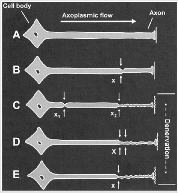
Peripheral nerve blocks (PNB) are far enough away from central lesions and are considered a safe therapy to use for anesthesia in the patient with MS. However, the addition of epinephrine to a PNB should be avoided to decrease further nerve strain through vasoconstriction. Ultrasound guidance for PNB placement is recommended to decrease risk of mechanical trauma as well as reduce the volume of local anesthetic used to prevent local anesthetic toxicity. Patients with MS are thought to be more sensitive to the effects of local anesthetics, especially when used centrally. Higher concentrations of oligopeptides are found in the CSF of patients with MS. Certain oligopeptides block sodium channel activity in the nerves and may be responsible for the profound muscle weakness associated with the disease. Local anaesthetics (LA) share physiologic properties with oligopeptides which may produce a synergistic reaction worsening MS symptom, especially when used for subarachnoid blocks. Therefore, spinal anesthesia should be used with caution. Despite spinal anesthetic concerns, epidural anesthesia is considered a safe alternative because the concentration of LA that approaches the subarachnoid space after an epidural injection is approximately one fourth of the epidurally administered concentration [3]., A main point of concern for anesthesia providers considering the use of regional anesthesia in this patient population is the proposed increased risk that the underlying condition may worsen, or a new deficit will develop. This phenomenon was first described in 1973 as the “double crush phenomenon” by Upton and McComas [1]. It was thought that a nerve with pre-existing neural compromise was more likely than a healthy nerve to incur damage at another point along the nerve. This is known as the double crush phenomenon, described by the photo below [1] (Figure 2).

Figure 2: Axoplasmic flow.

Lupinepublishers-openaccess-anesthesia-pain-medicine-journal

The double crush phenomenon refers to the coexistence of at least 2 injury insults or lesions along the same nerve. The theory originally suggested injury was a result from serial nerve compression or restrictions in axonal flow, however it since broadened to include conditions like neurologic disorders and metabolic, toxic or traumatic ischemic insults which may serve as the primary or secondary neural compromise. Patients with pre-existing neurologic disorders present with an assumed first crush as caused by their intrinsic disease thus, theoretically, predisposing them to a second crush. The second crush can be caused by many factors during the perioperative period, including intraoperative trauma or stretch, vascular compromise, hematoma formation, ischemia from tourniquet use or constrictive dressings, or from regional anesthesia related risks such as mechanical trauma from needle placement or LA toxicity [1]. Historically, patients with pre-existing neurological disorders were not offered neuraxial techniques due to fear of worsening neurological deficits. For decades, this has been standard clinical management of these patients because of a recommendation made in 1956 by Dripps and Vandam [6] .This standard of practice was continued because of provider and patient biases as well as the high risk of medicolegal concerns [6]. Despite the fears surrounding double crush phenomenon related to regional and neuraxial anesthesia worsening symptoms in patients with MS, there are several factors such as hyperpyrexia, stress and infection that are known to negatively affect the disease progression or exacerbation [5]. The cause of decline in neurologic function in patients with MS is not clearly understood and may simply be coincidental in the perioperative period regardless of the anesthetic technique used. However, because of the difficulty in establishing cause of second crush during surgery, many providers continue to avoid regional anesthesia [1].

Discussion

Much of the literature evaluating the safety of regional anesthesia in patients with MS is contradictory and sparse. Few studies have been completed and, those that have, have primarily been retrospective chart analyses. Although there is a great need for further investigation on this topic for definitive recommendations, current data suggests that risks previously associated with neuraxial blockade in patients with neurologic disorders may be less frequent than believed in the past [1]. Hebl, Horlocker and Schroeder conducted a retrospective chart review from the Mayo Clinic, spanning over twelve years from 1988 to 2000 [7]. They examined the effects of 139 patients with a history of a central nervous system disorder that received a neuraxial block for a surgical procedure. Prior to neuraxial blockade, motor weakness was the most frequent neurologic deficit among patients. Of the 139 patients, 130 had stable neurologic exams within six months of the block and 74 patients had active symptoms at the time of surgery. Despite over 50% of patients having active symptoms none of the 139 patients had documentation of postoperative deficits [7]. There were no new or worsening postoperative neurologic deficits found during the follow-up period which averaged sixty days (0.0% with a 95% CI). These results do not support the previous recommendations from Dripps and Vandam which have greatly influenced practice standards for the past fifty years [1]. Additionally, it was previously believed that LA dosing should be lowered in patients with MS and epinephrine additives should be avoided. However, only 11 of the 139 patients received reduced dosing and 72 patients had epinephrine added to their LA mixture and still no postoperative neurologic deficits were noted. Although these results cannot provide definitive recommendations of dosing or use of epinephrine, the study suggests that neuraxial blockade use with standard dosing in the patient with MS should not be considered contraindicated as it was previously thought to be The authors summarize that the decision to use neuraxial anesthesia techniques on patients with pre-existing neurologic disorders should be evaluated case by case and a thorough preoperative assessment is crucial. Many patients with advanced deficits affecting the respiratory and cardiovascular systems may benefit from the blunting of the sympathetic nervous system using regional techniques [7].

Ingrosso and colleagues evaluated the effects of peripheral nerve blocks for orthopaedic procedures in two patients with multiple sclerosis. Both patients were females ages 57 and 61 and had been diagnosed with MS for more than twenty years. Each patient had fractures isolated to the tibia and received a Bib lock. A Biblock consists of a standard femoral block combined with a sciatic nerve block to provide regional analgesia to the lower extremity. The blocks were performed identically using peripheral electrical nerve stimulation with insulated needles. The femoral nerve block and the sciatic nerve block each utilized 15ml 0.75% ropivacaine. Both blocks were observed and evaluated to be successful and set up rapidly. Each surgical procedure lasted an average of 75 minutes. The patients were evaluated immediately post operatively and at 30 days post procedure. Neither patient showed signs of exacerbation or progression of the disease at any point during evaluation. Although this study is clearly limited by sample size, it examined two patients at different stages of the disease that both showed positive outcomes and lack of disease progression after receiving a regional block. Before conducting this study, the authors only found 27 bibliographic references on the topic of patients with multiple sclerosis and regional anesthesia, with only 10 of those being in the last 10 years. The authors chose orthopaedic procedures of the lower extremities to avoid providing blockade to any typical central MS lesions to reduce influence of either supplies or drugs on those anatomical structures. The site of surgery allowed a total locoregional approach. Performing this type of block allowed avoidance of medullary block and emotional stress from general anesthesia. The prolonged postoperative analgesia provided from the Bib lock provided a stress-free postoperative course. The authors concluded that peripheral nerve blocks should be considered safe and should be the choice of anesthetic for the patient with MS when surgery allows for it [8].
A case report by Koff and colleagues describes a severe brachial plexopathy after a interscalene nerve block for a total shoulder arthroplasty in a patient with MS [9]. Despite known benefits associated with regional anesthesia in patients undergoing joint replacement, use in the patient with MS is not definitive. Although MS is a disorder of the central nervous system, there is data supporting peripheral involvement in 4-14% of patients [10]. Postoperatively, the patient reported dense sensory and motor block and was comfortable for the first hour. The next day, the patient was experiencing 5/10 pain with persistent flaccid motor block of his entire right arm. A neurology consults and electro myelogram were ordered. A diagnosis of brachial neuritis was made, and high dose steroids were prescribed. On post-operative day 11, complete paresis of the right arm remained, and new electromyography showed active denervation of all muscles in the arm with no voluntary motor recruitment. At 3 months, the patient showed signs of reinnervation of the muscles of the arm. However, after 8 months, the patient still had significant weakness in the right upper extremity, 50% loss of motion and visual atrophy of the arm as well as the pectoralis muscles of the right side [10]. Despite testing such as electromyography and MRI, it remains difficult to identify and differentiate an ethology when multiple factors were involved. Potential factors include direct trauma from the nerve block, neurotoxicity from LA, patient positioning such as extreme abduction and external rotation; all which can occur to any patient undergoing total shoulder replacement [11]. An additional confounding variable in diagnosing the cause of postoperative neurologic deficits in MS is that an exacerbation may be caused by many other non-traumatic factors such as hyperthermia, stress, and electrolyte abnormalities. Therefore, although the mechanism of this injury is not definitive, the pre-existing pathology of the peripheral nervous system may have been a contributing factor. Authors conclude that the decision to perform peripheral nerve blocks must be made on a case by case basis, with an understanding that the peripheral nervous system may be somewhat involved in the pathology of MS [9].

Conclusion

While it is certain that surgical stress negatively impacts the patient with MS, there are no definitive studies on the long term or immediate implications of nerve blocks. Further research is needed in this area, but recent studies show promise for positive outcomes for these patients. Theoretically, regional anesthesia/ analgesia would reduce pain and stress associated with surgery thus reducing the risk of exacerbation. Nonetheless, the theory of double crush phenomenon must be considered. Much of the literature supports the use of peripheral nerve blocks and epidural anesthesia/analgesia and reports that MS should not be considered a contraindication to these pain control modalities. However, spinal anesthesia should still be avoided due to proximity to the CNS and high concentrations of LA to central lesions. The decision to proceed with regional anesthesia in the patient with MS must be made on a case by case basis and careful considerations must be made regarding the risks and benefits. It is important for the anesthesia provider to tightly control temperature and stress during the perioperative period to exclude those as causes of disease exacerbation. Although more research needs to be done for definitive recommendations, overall evidence supports the safety and efficacy of regional anesthesia in patients with MS and presence of the disease should not be considered an absolute contraindication.

Conflict of Interest

There is no economic or conflict of interest present by the author

Disclosure

I have no conflicts of interest

Financial Support and Sponsorship

None

Read More About Lupine Publishers Journal of Anesthesia & Pain Medicine Please Click on Below Link:
https://lupine-publishers-anesthesia-pain.blogspot.com/
Saturday, 3 June 2023

Lupine Publishers | Unquestionable Wound Pain: Call for A Care-Plan Reform

 Lupine Publishers | Journal of Anesthesia & Pain Medicine


Opinion

Pain is a subjective experience and one that the medical healthcare profession will never fully be able to understand because the pain belongs to the patient. Ultimately, pain is whatever the patient experiences it to be, yet the traditional medical model approach is one that views the patient’s pain as something to be managed by the ‘professional’ [1,2]; who have been reported to often mismanage it [3]. Pain is in fact a personal encounter and one that is perceived differently both within and between individuals from one time to the next. This point must not only be acknowledged, but digested and acted upon, by healthcare providers working within the field of pain medicine. This paper concedes that pain is a biopsychological event that can only be fully managed by adopting a patient-centered and holistic approach to care. This proposition extends its focus to draw particular attention to those working in the field of wound care, due to the unique pain-related idiosyncrasies apparent among patients with acute and chronic wounds (from any origin: war/accident trauma; post-operative; foot/leg ulcers; burns/scars) that make them worthy of special attention in this matter.
In most recent years, wounds have been estimated to cost health services up to $96 billion globally [4] and this this figure is only set to rise due to self-compromising lifestyle and behavioral factors that lead to wound-promoting illnesses (for example obesity and diabetes) [5]. Pain has traditionally and consistently been dismissed as an overlooked aspect in wound care [6], yet pain has a detrimental and often devastating impact on wound healing [7] that negatively compromises an already weakened immune system [8]. Stress further perpetuates this deleterious cycle of events that tamper with effective wound healing, yet pain management does not take precedence in standard ‘wound care’ regimes. To illustrate, on reviewing the literature on various pain management guides, protocols and strategies specific to wound care published over the past twenty years (2000-2020), it became evident that, as with other types of pain, wound pain continues to be managed principally by pharmacological methods [9-12]. That is, guidance being offered from various commanding sources, from the World Health Organisation (WHO) to specialist wound and pain advocates [10-12] typically recommends a prescription care package of anti-inflammatory, non-steroidal medication (ibuprofen), which might be escalated to an oral pain killer (codeine) if the pain is greater and a stronger opiate analgesic (morphine) is offered when the pain becomes more severe. What this approach does is treat the symptom so is therefore less than optimum, because pain is more than just a symptom. Pain intensity, severity and duration are not only governed by physiological causes, but a psychological response. It has been reported that pain causes stress and stress negatively impacts healing [13]. Furthermore, pain perception (and therefore wound healing) is further compounded by anxiety; depression; and sleep deprivation as further by-products of stress [7].
Not only does this interacting biopsychosocial spiral of events impact on wound healing, but when it comes to pain from a wound, this topic becomes ever more complex due to the unique ways in which pain is manifested among patients with wounds. What makes wound pain distinct from pain caused by other illnesses (for instance cancer or arthritis), is that wound pain is consistently and blatantly visible to the sufferer. What this does is provides a constant reminder and red flag to notify the individual that there is something wrong. In fact, research has shown that wound pain is highest when dressings are removed [13], as opposed to being hidden by a dressing; and even when bandaged, the dressing remains a constant reminder of the fact that there is cause for concern. This form of biofeedback acts by conditioning a patient to a sense of heightened stress and unease that becomes amplified by an anticipatory response. This has been witnessed during such times when the wound becomes re-exposed, such as during dressing changes [13] and independently of pain sensation resulting from tissue disruption. A conditioned response therefore develops to prompt the physical system to experience pain. This anticipatory reaction explains pain perception as a bi-directional response. Not only can the physical body trigger warnings to the psychological self that there is a problem, but the mind can alert the body to feel pain. This mind-body dualism is magnified in the case of wound pain due to the high visibility of the source of the pain in question and this is precisely why pain regulation should be a focal point in any wound care plan due to its idiosyncratic qualities.
In order to bring pain medicine and treatment practice up to speed with contemporary integrated pain models [13], we need to consider holistic care packages that strategically intervene on both the physical and psychological levels of concern [6]. For example, empowering patients to self-manage their illness [1,14] both practically (e.g. wound dressing; patient-controlled analgesia) and mentally (e.g. cognitive reframing; stress management). Moreover, ‘treating’ the psychology should form an integrated part of any standard care package for wound patients [15], whereby continuous dialogue is used between patient and healthcare provider to monitor and reflect on aspects of the regimen. Patient pain diaries and monitoring of thoughts, feelings and behaviors can be achieved through use of self-support cognitive behavioral training for wound pain patients that facilitate restructuring negative coping strategies such as catastrophizing and fear self-statements [1].
Overall, it is aspired that this debate will provoke the attention of professionals working with patients with painful conditions to reconsider, firstly, that there is more to treating pain than a numbing of the symptom. As we appreciate, pain is an incredibly complex experience that cannot be managed reliably with medication alone. Secondly, wound pain is distinctive in that pain perception can be magnified by the unconcealed nature (or root cause) of the pain, which is unlike most other painful conditions. Finally, we wish readers to ponder over these germane issues right now, because despite witnessing scatterings within the literature of debates relating to the issues addressed here, it can be argued confidently, that all concerted efforts to get this message across to inform treatment plans thus far have failed, as we still do not see these viewpoints translated in to practice. We concede by reiterating that pain is subjectively owned by the patient and is therefore an un-questionable experience so can never be fully understood by a person outside of that experience; and call for a care-plan reform that guarantees a fully-integrated, biopsychosocial tailored package that responds to the voice of each patient.

Read More About Lupine Publishers Journal of Anesthesia & Pain Medicine Please Click on Below Link:
https://lupine-publishers-anesthesia-pain.blogspot.com/
Friday, 31 March 2023

Lupine Publishers | Estimation of the Predictive Factors of Myocardial Ischemic Preconditioning in Elective Surgical Patients

 Lupine Publishers | Journal of Anesthesia & Pain Medicine


Abstract

Introduction: the physiological response to the aggression produced by the surgical trauma provides the effective treatment capacity in case of complications. Surgical intervention causes endocrine, metabolic, autonomic, immunological, and hematological changes.

Objective: to estimate the predictive factors of the second window of myocardial ischemic preconditioning in the patients proposed for elective surgical interventions.

Material and Methods: A quasi-experimental study was carried out in the period January to December 2019 at the Maria Curie Cancer Hospital in Camagüey with patients who met inclusion and exclusion criteria in preoperative consultation in the research period, to whom preconditioning was applied. Ischemic two hours before surgery. Chi squared and logistic regression was calculated as appropriate.

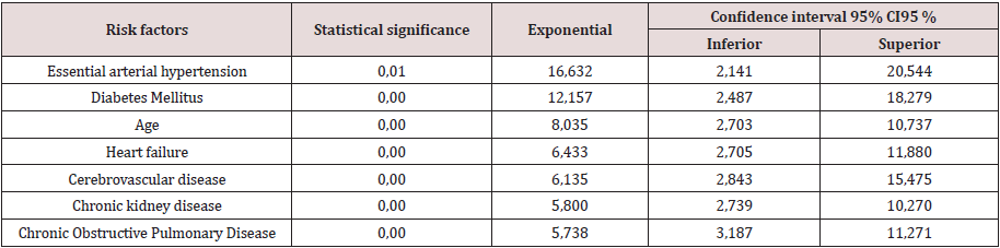
Results: Predictive factors such as essential arterial hypertension odds ratio 16,632, diabetes mellitus 12,157, age 60 years and over with odds ratios of 8,035, heart failure odds ratio 6,433, cerebrovascular disease odds ratio 6,135, and chronic kidney disease were estimated. Odds ratio 5,800 and chronic obstructive pulmonary disease with odds ratio 5,738.

Conclusion: the predictive factors of the second window of ischemic preconditioning were independent predictors of risk in elective surgical patients.

Keywords:Preconditioning; Elective surgical patient; Surgical stress; Predictive factors

Introduction

Knowledge of the physiological response to aggression produced by surgical trauma provides effective treatment capacity in the event of complications. Surgical intervention causes endocrine, metabolic, autonomic, immunological, and hematologic changes [1]. Somatic and autonomic afferent nerve impulses generated at the site of injury activate the endocrine response, while the inflammatory and immune response, mediated by hormones, begins to develop. and cytokines, secreted products of activated leukocytes, fibroblasts, and endothelial cells. The changes in the immune and endocrine systems in the face of surgical trauma, are objectified by the perioperative response of various markers. Surgical stress is a situation in which there is both an increase in the speed of generation of oxidizing species, free radicals (RL) and excited species, and a decrease in the activity of defense systems, resulting in higher concentrations, in the steady state of active oxygen species [2,3]. In these situations, the toxic effects of these RLs manifest and chemical reactions take place on lipids, proteins and carbohydrates inside the cells, which trigger irreversible damage and even death cellular. There are numerous diseases associated with the imbalance between oxidants and antioxidants, in the surgical patient from the preoperative period with personal pathological history, he becomes involved with the RL, through the physiological response of the diseases and the complexity of the surgical trauma as essential factors in the perioperative changes of hemostasis, the interaction of the endocrine and immune axis and of the drugs administered in the surgical anesthetic act [4]. The publication that describes the influence of anesthetics used during the perioperative period and the typical hormonal response generated by the surgical intervention and the patient is current. With modulation of adrenal response by the distribution of leukocytes and their immune functions. Anesthetics modify immune function by reducing the stress response and have a direct effect on immune cells [5]. The investigation was carried out with the objective of estimating the predictive factors of the second window of myocardial ischemic preconditioning in surgical patient’s elective for myocardial protection during major surgical intervention.

Material and Methods

General Aspects of the Study

This was a quasi-experimental investigation to estimate the predictive factors of the second window of myocardial ischemic preconditioning in elective surgical patients at the María Curie Cancer Hospital in Camagüey in the period from January to December 2019.

Definition of the Study Universe

The study population was delimited to 40 preconditioned patients 2 hours before the surgical intervention, an insufflated sphygmomanometer was placed at 200 mmHg for 5 minutes, after this time it is deflated, and 5 minutes are expected. This procedure is repeated 3 times. Blood draws are performed before and after the application of ischemic preconditioning. They are preconditioned and post conditioned patients who meet the inclusion and exclusion criteria.

Inclusion Criteria

Patients aged 20 years and over proposed for major elective surgical intervention.

Exclusion Criteria

Patients who do not offer their consent to participate in the research. Variables: age, sex, associated risk factors, antioxidant markers, discharge status, hospital stay, complications. The antioxidant markers before and after the preconditioning were determined: the concentration of reduced glutathione (GSH) by the method of Sedlak et al. [6] and malonic aldehyde (MAD).

Information Processing and Analysis Plan

Search and collection of information: A form was completed for each patient, complementing the information with data from the medical records.

Information processing: The database was compiled with the collected information, which was processed automatically using the SPSS statistical package. The information of the qualitative variables is summarized in descriptive statistics, related to the characterization of the patients operated on by the different surgical specialties, to determine the behavior of the activity of antioxidant and oxidant markers (preconditioning and postconditioning). The information resulting from applying the second window of ischemic preconditioning was processed according to the univariate and multivariate analysis for the study variables using the statistical program SPSS version 21. Implementing the chi-square test and logistic regression as appropriate. The understanding of the results obtained is facilitated through statistical tables and graphs, it was analyzed giving an answer to each proposed objective and comparing the results with those of other authors. Finally, after a synthesis work, conclusions and recommendations were issued [7].

Results and Discussion

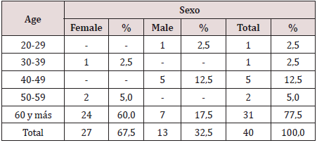
N 40 Source: clinical history as can be seen in Table 1, most of the surgical oncology patients belong to the group of 60 years and more, for 77.5%. The evolution of the age pyramid, thanks to the increase in life expectancy and better healthcare, places cancer diseases in the forefront of surgery, influenced by the life expectancy at birth of 80.2 years for women and 76 years for the men. Cuba is among the three countries on the continent with the largest aging population, in 2000 it exceeded 1.6 million older adults and in 2014 it represented 17.9% of the total population, in 2015 the Cuban population was 18.5% and in 2018 21% of the population aged 60 and over 6.7 During the aging process, the adult heart undergoes numerous biochemical, ultrastructural, functional and anatomical changes that reshape cell structure, function and adaptive responses to stress. Histologically, a decrease in the number of myocytes and progressive hypertrophy of these are observed in both ventricles. Also, studies in humans demonstrated aging-related abnormalities in cardiac metabolism, coronary flow reserve, endothelial function. Age is an independent predictor of risk in patients with coronary heart disease, this is explained by the lack of adaptation to acute myocardial ischemia [8]. Ischemic preconditioning is a mechanism by which repetitive episodes of ischemia induce greater tolerance in the myocardium to subsequent episodes of aging as a biological process with interindividual variability leading to the progressive loss of physiological functional reserve, the alteration of these functions is not evident. in basal situation but it manifests itself in moments of stress such as illness and perioperative.

Table 1: Distribution of patients with ischemic preconditioning according to age group and sex.

Lupinepublishers-openaccess-anesthesia-pain-medicine-journal

Table 2: Frequency distribution of patients with ischemic preconditioning according to personal pathological history.

Lupinepublishers-openaccess-anesthesia-pain-medicine-journal

N 40 Source: clinical history

In Table 2, twenty-one patients out of forty preconditioned have a diagnosis of essential arterial hypertension with 52.5% followed by diabetes mellitus 42.5% and cardiovascular disease 32.5%. Comorbidity is more frequent and both cerebrovascular disease and lung processes, kidney failure, hypertension, and diabetes justify part of the increased risk of the surgical patient. However, it seems useful to recall other pathophysiological mechanisms that explain the elderly’s reduced response capacity such as endothelial dysfunction, microcirculatory disorders (rarefaction), increased precapillary resistance, and decreased ability to develop collateral circulation as clear. Limiting residual flow in the risk area and perfusion flow in the remote area [9,10]. In the myocardium, the possible loss of ischemic preconditioning, the accelerated drop in the reserve of high-energy phosphates (lower tolerance to ischemia), calcium overload and senile myomalacia are factors to consider. The primary changes of arterial aging produce important secondary changes in the heart and other terminal organs, including the brain and kidneys. The vascular aging process is accelerated by the presence of primary cardiovascular disease, including high blood pressure and atherosclerosis, as well as by other risk factors such as diabetes, smoking, and obesity. Morphological changes include a decrease in the number of myocytes, a thickening of the left ventricular wall, and a decrease in both the density of the conduction fibers and the number of cells in the sinus nodes [11]. These changes are readily apparent in terms of elevated mean arterial pressure and increased pulse pressure. Increased vascular stiffness leads to significant secondary cardiac responses, but in young people the heart pump and blood vessels are optimally coupled for maximum efficacy. The mere fact of treating the topic of aging and optimizing the preoperative period of the surgical patient with the application of preconditioning due to ischemia is the objective of the research Table 3.

Table 3: Frequency distribution of patients with ischemic preconditioning according to personal pathological history.

Lupinepublishers-openaccess-anesthesia-pain-medicine-journal

N 40 Source: clinical history

Interpreting the adjusted odds ratio (OR) for the variables of the equation as follows: In the estimated Logistic Regression function, the variable essential arterial hypertension had a significantly different regression coefficient of 0 (p = 0.01) and adjusted OR of 16,632 (95% CI 2,141; 20.54). In this analysis, the risk of not modulating (oxidative enzymatic response) is approximately 7 times greater in patients with positive oxidative stress markers than in negative ones, that is, the enzymatic response after preconditioning is at the expense of oxidants. The diabetes mellitus variable had a significantly different regression coefficient of 0 (p = 0.00) and adjusted OR of 12,157 (95% CI 2.487; 18.279), which implies that the risk of presenting enzymatic response to diabetes mellitus patients Oxidant expense is approximately 12 times greater than in those without a history of diabetes mellitus.
A patient 60 years and older after ischemic preconditioning is 8 times more likely to have an oxidative enzymatic response with OR (8,035) and 95% CI 2,703; 10,737. The variable Heart failure had a regression coefficient other than 0 (p = 0.00) and adjusted OR 6,433 (95% CI 2,705; 11,880), which implies the risk of presenting the oxidative enzymatic response in patients with a history of heart failure. After ischemic preconditioning, it is 6 times higher than in patients without a personal pathological history of heart failure, with a margin of 2 to 11 times. The variable cerebrovascular disease had a regression coefficient other than 0 (p = 0.00) and adjusted OR 6,135 (95% CI 2,843; 15,475), which implies the risk of presenting the oxidative enzymatic response in patients with a history of cerebrovascular disease. After ischemic preconditioning, it is 6 times greater than in patients without a personal pathological history of cerebrovascular disease, with a margin of 2 to 15 times. The variable chronic kidney disease had a regression coefficient other than 0 (p = 0.00) and adjusted OR 5,800 (95% CI 2,739; 10,270), which implies the risk of presenting the oxidative enzymatic response in patients with a history of disease. Chronic renal disease after ischemic preconditioning is 5 times higher than in patients without a personal pathological history of cerebrovascular disease with a margin of 2 to 10 times. The variable chronic obstructive pulmonary disease had a regression coefficient other than 0 (p = 0.00) and adjusted OR of 5,738 (95% CI 3,187; 11,271), which implies that the risk of presenting chronic obstructive pulmonary disease in surgical patients the oxidative enzymatic response after preconditioning being 5 times greater than in those without a personal history of chronic obstructive pulmonary disease with a margin of 3 to 11 times.

In 1986, Murry CE et al. 12. described the concept of ischemic preconditioning (PI) in dogs and since then it has been evaluated in multiple animal and human models.
PI (development of tolerance to acute ischemia) is a cellular mechanism capable of delaying, but not preventing cell death; This protection is transitory and lasts from 1 to 2 hours in anesthetized animals. During a brief episode of ischemia, adenosine, bradykinin, norepinephrine, and opioids that activate G receptors are released locally, culminating in the opening of ATP-dependent potassium channels. The signals leading to the opening of these channels are not fully defined, but include activation of phosphatidylinositol- 3-kinases, protein kinase C, and mitogen-activated protein kinase (MAPK). Multiple aging-related abnormalities at various levels of this cascade were demonstrated in animal models [13]. A clinical trial published in JAMA demonstrates a reduction in the incidence of postoperative renal failure in patients receiving IP compared to the control group (37.5 versus 52.5%) and a decrease in the need for renal replacement therapy. However, no differences were found in terms of mortality and adverse cerebral and cardiovascular events (although they were not the primary objective of the trial) [14]. Also, in 2015 two essays were published both in the New England Journal of Medicine. These are the ERICCA14 trial and the RIPHeart trial [15].
The ERICCA trial, multicentre on more than 1,000 patients, showed no difference in the primary endpoint (death, stroke, acute kidney failure or acute myocardial infarction). No differences were found either in the subgroup analyzes or in the secondary objectives (troponin values, length of stay in the Intensive Care Unit and mechanical ventilation, incidence of delirium and new atrial fibrillation). No adverse effects were observed in the intervention group [16] In the RIP Heart multicenter trial of more than 1,600 patients, no differences were found in the primary endpoint (death, acute myocardial infarction, need for revascularization or stroke), nor in the secondary endpoints (troponin elevation, acute kidney failure, need for inotropes, length of stay in the Intensive Care Unit and quality of life after the procedure). But, although PI seems to have beneficial effects, what role does it play in the case of elderly elective surgical patients in the local context? The enzymatic response obtained was oxidative in most of the elderly patients, which corresponds to 90%, showing no benefit. In a 2007 myocardial revascularization surgery study, 57 patients were randomized to receive or not receive remote ischemia-reperfusion cycles after anesthetic induction. The results showed a reduction in troponin levels at 72 hours in the treated group compared to the control group. Other trials confirm the cardioprotective effect reflected in reductions in postoperative troponin T, I or CKMB values. However, not all trials were positive in this regard and do not confirm these results. The routine uses of inhalational anesthetics or beta-blockers, which demonstrated their cardioprotective effect in several clinical trials, is very likely to mask the beneficial effects of PI. In any case, in a subsequent meta-analysis carried out by d’Ascenzo et al. A decrease in troponin values was observed in the postoperative period once possible confounding factors were controlled, such as the use of such volatile anesthetics during surgery [17]. Studies on IP, of different quality and with different objectives, conclude similarly in IP, offering benefits in terms of decreasing the values of markers for myocardial injury and incidence of kidney damage in the immediate postoperative period, but it does not seem to improve the short and medium term results when survival and cardiovascular and cerebral adverse effects are analyzed. On the other hand, cardioprotective drugs routinely used as inhalation anesthetics, beta-blockers, and anti-calcium are sufficient, without remote ischemia providing additional benefits. It is also not clear which group of patients benefits from the technique, what is the most appropriate method to perform it, and even whether or not it has no adverse effects.

Conclusion

Research suggests that the mechanism of ischemic preconditioning is diminished in elderly patients, confirming essential arterial hypertension, diabetes mellitus, age, heart failure, cerebrovascular disease, chronic kidney disease, chronic obstructive pulmonary disease as independent cardiovascular risk factors in cancer patients.

Acknowledgement

Zaily Fuentes Díaz: review, analysis and bibliographical selection; statistical processing; preparation of the final report; review and correction of the report; review and final approval. Gonzalo Fabián Orga García: field or assistance work; bibliographic review, analysis and selection; survey application, statistical processing; preparation of the final report; review and correction of the report; review and final approval. Orlando Rodríguez Salazar: review and correction of the report; review and final approval.
Jorge Lozano Casanova: sample processing and final approval. Tania Puerto Pérez: statistical processing; review and correction of the report and final approval.

Conflict of interest

The authors declare that does not exist an interest conflict.

Read More About Lupine Publishers Journal of Anesthesia & Pain Medicine Please Click on Below Link:
https://lupine-publishers-anesthesia-pain.blogspot.com/

Thursday, 15 December 2022

Lupine Publishers | Ventilation and Oxygenation Management in the Traumatic Brain Injury Setting

 Lupine Publishers | Journal of Anesthesia & Pain Medicine


Introduction

Early management of ventilation and oxygenation in the patients with traumatic brain injury, can be of importance to reduce secondary injury risks and improve outcomes. Increasing in the intracranial pressure of the patients with traumatic brain injury is an important risk which these patients are faced with. Hypocapnia due to hyperventilation and vasoconstriction in the brain’s blood vessels as the result, causes reduction in blood flow. Therefore, the risk of secondary injury due to hypoperfusion and ischemia would increase. It is recommended to keep PaCO2 levels in the range between 35 and 40 in these settings. Minimizing the bag-ventilating of the patients with traumatic brain injury who have been intubated, is an important note which should be kept in mind to reduce the risk of hyperventilation in such patients. These patients should be placed on the mechanical ventilator at the earliest possibility. 7 to 8 liters/min can be a reasonable starting minute ventilation in these settings, because of the possibility of presenting hypermetabolic state in the patients with traumatic brain injury [1-5].
Permissive hypercapnia is inappropriate in these settings since these patients are at risk for Acute Respiratory Distress Syndrome. In such settings Capnography and ABG should be considered to correlate End-tidal CO2 with the Partial pressure of carbon dioxide. Normoxia is important in such settings to prevent secondary injury and improve the outcomes. The ABG should be checked in a time period about 15 to 20 minutes after intubation. Based on oxygen-haemoglobin dissociation curve in different cases, FiO2, PaO2 and O2 saturation ranges should be defined. Hemodynamic of the patients with traumatic brain injury should be managed appropriately. Hypotension and hypertension in these patients should be corrected to avoid further complications. It is important for the neurointensive care professionals to have knowledge about appropriate management of ventilation and oxygenation in the patients with traumatic brain injury.

Read More About Lupine Publishers Journal of Anesthesia & Pain Medicine Please Click on Below Link:
https://lupine-publishers-anesthesia-pain.blogspot.com/

Saturday, 22 October 2022

Lupine Publishers | Mechanical Ventilation Basic Terms and Physiology

 Lupine Publishers | Journal of Anesthesia & Pain Medicine


Terms and Physiology

There are some terms related to mechanical ventilation which the healthcare professionals who deal with the patients who are placed on the ventilator for various reasons, should have knowledge about them.
Based on the selected mode of mechanical ventilation there are “Control “variables like volume or pressure-controlled ventilation modes. Mechanical ventilation’s dependent variables are “ Conditional “ variables. “ Trigger “ initiates the inspiration. As a matter of fact, one breath can be time, pressure or flow triggered. End of inspiration and beginning of exhalation determinant would be the “ Cycle “. The mechanical ventilator can be time, volume and pressure cycled. Mechanical respiratory cycle’s resistive forces are named “ Airway resistance “ which its normal range is equal to or lower than 5 cmH2O. [1-2]
Atelectasis causes surface area’s gas exchange loss which is called “ De-recruitment “. Increasing PEEP can minimize Derecruitment which is among the most common cause of gradual hypoxemia in the patients who are intubated. Reopening the lung’s atelectatic or collapsed areas with pressure application and gas exchange surface area’s restoration, is called “ Recruitment “. Lungs elasticity or their ease to expand and stretch in response to volume or pressure changes is called “ Lung compliance “. Highly compliant lungs can be seen in the Obstructive lung diseases and Lungs with a low compliance can be seen in Restrictive lung diseases as an example. The weight which should be used in ventilator settings determination is “ Predicted body weight “ or “ PBW “. Height and sex are two factors which determine lung volumes and therefore they should be used in PBW determination. [3-4]

Read More About Lupine Publishers Journal of Anesthesia & Pain Medicine Please Click on Below Link:
https://lupine-publishers-anesthesia-pain.blogspot.com/
Friday, 27 May 2022

Lupine Publishers | Do You Really Want to Improve the Results of Treatment for Acute Pneumonia?

 Lupine Publishers | Journal of Anesthesia & Pain Medicine


Editorial

The question raised in the title of this letter is a natural consequence of the findings and conclusions that have been growing steadily in recent years regarding the results of treatment for acute pneumonia (АР). If you look at the publications of recent years in this field of medicine, it turns out that one of the main obstacles to progress in improving the results of treatment of this disease is the lack of appropriate methods for determining the pathogen. Thus, the lack of timely diagnostic information about the etiology of the disease excludes the possibility of targeted antibiotic therapy. In recent years, such regrets have become more and more relevant, playing the role of the main explanation for treatment failures .Continuing to narrow the unidirectional view of the problem and to pay attention only to the microbial factor as the main cause of the disease, such views are in fact another illusion, which, even in the case of its hypothetical implementation, will not make significant changes in the overall trend. This statement is easy to verify if you rely on well-known facts, and not use as arguments assumptions and guesses. First of all, it is necessary to remember the features of the initial period of clinical use of antibiotics. On the one hand, the first results showed their phenomenal effectiveness, and the greatest preference for many years was given to penicillin. On the other hand, bacteriological methods of verification of the pathogen at that time were significantly inferior in their speed and quality indicators to the methods of modern Microbiology. However, treatment of patients with AР for a long time was successfully carried out only by one penicillin and the question of the role of determining the causative agent of inflammation in those years did not have such a critical importance. I think it should be obvious to readers with medical education that such success in the use of antimicrobials in a dynamic and living biological environment could not continue indefinitely. The further course of events developed according to the so-called law of scissors, but the dynamics of these changes occurred very slowly, which made it difficult to monitor and timely assessment. On the one hand, each new generation of doctors was brought up in the spirit of priority and irreplaceability of antibiotics, gradually forming the image of a kind of panacea. One of the reflections of the new mentality was the wide spread of the principle “antibiotics alone” for the treatment of many diseases (including AP). Declaring this principle of treatment, no one was confused by the fact that the antibiotic in many patients is the only means of treatment and that the same drug is used in radically different processes. And these are indirect signs of the attitude to antibiotics as a panacea. As you know, the basic principle of antibiotics is only intended to suppress the pathogen, is not it? Other medicinal properties of these drugs do not have. All other changes that have already occurred in the focus of inflammation, and in the body in General, cannot be eliminated with the help of antibiotic therapy, no matter how powerful and long it was not. The latter task falls on individual protection and adaptation mechanisms and depends on the direction of additional assistance methods. The gradual narrowing of views on the nature of AР to the direct dependence of all sides of the disease on the type of the pathogen was accompanied by the formation of relevant normative documents recommending and prescribing the implementation of specific actions and prescriptions. Today, a specialist working on the basis of a license cannot go beyond these requirements, even if he had doubts about their effectiveness. In parallel with the deformation of the image of AР as an inflammatory process of nonspecific etiology in the lungs, there is another important process, the results of which are increasingly recorded in recent years. The symbionts of our body are also biological objects. Exposed to mortal danger, they have learned not only to survive, but also to create strains resistant to such aggression. To date, the group of resistant strains to antibiotics is already quite impressive, and its representatives are increasingly identified among the symbionts of healthy people [1]. In itself, the presence of such microflora in the body does not necessarily mean the fact of the disease, but in the case of the beginning of the last, conventional antibiotic therapy can meet with serious difficulties. In fact, we are talking about microbes that have always been found in the composition of human symbionts and that have only gained additional stability, not aggressiveness. Hardly anyone doubts the fact, that the emergence of such strains is the result of long-term and widespread use of antibiotics. In addition, the frequency of detection of such strains in a healthy population will increase as a result of continued antimicrobial therapy. At the same time, it is even theoretically impossible to stop this already started process, since such an initiative can lead to disastrous results. Therefore, it is necessary to realistically assess the current situation in bacteriology, which is not the worst option, and consider the gradual increase in the number of carriers of such strains among the healthy population as a future inevitable reality. It is known that the uncertainty of what is happening has always been one of the main prerequisites for the emergence of fears. In this regard, it is especially important to note that the author’s point of view is presented to the reader not for emotional impact, but only to develop their own rational assessments of already known facts. If for many years, the AР microbe is considered to be the main cause of the disease and the entire policy in the treatment of these patients was aimed only at suppressing this factor, the decrease in the effectiveness of treatment and the increase in the resistance of the microflora led scientists into complete confusion. In this case, modern ideas about the nature of the disease can indicate only one way out of this impasse - to speed up the process of determining the pathogen. However, the accumulated evidence shows the futility of trying to realize these hopes. Although the participation of various microorganisms in the inflammatory process of lung tissue differs significantly in its frequency, and the leaders of this list change periodically, it should be recalled that to date more than 100 microbes have already been established that can act as pathogens of AР [2]. In addition to these data, the literature on this problem always notes the fact that AР pathogens are not only bacteria, but also viruses, fungi, parasites. In this regard, it is especially advisable to pay attention to acute inflammation in the lungs, which is a consequence of a viral infection. According to the statistics of this disease, at least a third of patients in the world have the viral nature of AР [3,4]. These indicators cannot be ignored, as the continuation of the therapeutic principle of “antibiotics alone” in the AP reveals the following fact. All patients with viral etiology of the disease do not have targeted treatment, and antibiotic therapy is only an attempt to prevent the possible addition of a bacterial component. Therefore, successful treatment in such observations should be considered more as a merit of the patient’s body than as a triumph of medical efforts. At this stage, the etiology of AР is widely interpreted on the basis of the results of bacteriological studies of the microflora of the nasopharynx and oropharynx [5-7]. However, such studies require clarification of the breadth of their application, and the evaluation of their results raises logical objections. For example, it is known that bacteriological examination is usually carried out mainly in hospitalized patients. The etiology of AР in most patients undergoing outpatient treatment remains unknown. But, even among hospitalized patients, the duty of such studies refers to cases of severe forms of the disease, and positive results (including cultures of blood) are found in less than half of the cases [4,8]. However, even the interpretation of the positive results of the examination does not explain such a paradox as the presence of less virulent microflora in a number of AP observations compared with bacteriological examination of the upper respiratory tract in healthy people, when the simple presence of aggressive strains without any signs of the disease is revealed. Serological diagnosis is also not the result of studying the material directly from the area of inflammation. Allowing to identify trace reactions, this type of examination refers to indirect signs of contact of the body with a certain pathogen but is not an absolute proof of its presence at the moment directly in the inflamed organ. If we continue to discuss the role of the material in the reliability of the results, the most accurate method of determining the agent of AP, of course, is to study the content of the affected area. The possibility of practical implementation of such a study appears only in a limited group of patients with purulent pleural complications that develop at a certain stage of the process and previous treatment. However, even the results of this relatively small group of patients show that in 30 and more % of cases microflora was not detected [4,9] and there is no reasoned explanation for this fact. As a result, several hundred million AР cases are diagnosed each year in the world, but their etiology remains an open question, and the choice of antimicrobials has been and is being conducted empirically [4,8-10].

Despite the intuitive choice of antimicrobials, their use remains a very important support for the body. For most patients with AР, the suppression of an unexpected mutiny of the accompanying microflora is enough for the body to cope with inflammation in the future. However, in the most severe situations with the rapid and aggressive development of the inflammatory process, another aspect of the problem is revealed, which currently continues to be explained by the extremely high virulence of the pathogen. For example, among patients hospitalized with severe AP, mortality reaches 13-28%. Among the total number of hospitalizations with AP, septic shock is diagnosed in 30% of cases, and mortality in this group increases to 50% [4,11-13]. The statistics of these results seem, at first glance, quite understandable and logical: the more severe and aggressive the disease develops, the more difficult it is to eliminate the catastrophic violations that have arisen. However, the logic of such arguments collapses completely, if we take into account some very important, in my opinion, facts. First, if we consider shock as a septic complication of aggressive forms of AР, it is necessary to have conclusive evidence of this option, the cause of which is the penetration of pathogens into the bloodstream. However, the detection of bacteria in the blood is recorded only in 10-12% of all hospitalizations with AР without connection with the shock pattern [8,9]. That is, the latest figures reflect the frequency of diagnosis of bacteremia, which is not always accompanied by a clinic of shock. Secondly, interpretations of shock in AP as its septic variant consider this complication only from the point of view of the microflora and leave without attention such an important feature of inflammation as an individual reaction of the body to the process. It is the juxtaposition of these two causes (microorganism against microorganism) that creates the uniqueness of the manifestations of the inflammatory process even in the presence of the same pathogen. However, despite this long-known fact, the severity of AР in recent years is increasingly attributed solely to the properties of microflora. Finally, the main feature of the nature of the shock in the clinic, the AP determines, from my point of view, the undeniable fact that this intense process is the only inflammatory disease of nonspecific etiology of all known, which occurs in the vascular system of the pulmonary circulation. Rapid change of stages of the process makes it difficult to adapt the body to new conditions. In the most aggressive cases, the body gives us a clear signal of its catastrophic situation. Demonstration of shock in AP is a reflection of the body’s efforts to facilitate the work of pulmonary vessels, which could not provide the necessary blood flow and synchronous operation with a large circle of blood circulation [14]. The causes and pathogenesis of this form of shock differ significantly from other variants and require a different treatment solution. So, this form of shock has been highlighted by us in a separate section as pulmonal shock. Understanding the pathophysiology of current disorders involves eliminating the reflex effect of the source of inflammation in the lungs on pulmonary blood flow [15-18]. Instead, shock in AР is compared with its manifestations of another origin, and such patients begin to receive standard therapy in the form of intravenous injections with the addition of hormones and vasopressors [4,8,9,11,13,19-22]. Intravenous fluids increase blood flow to the pulmonary vessels and further disrupts blood circulation in the small circle, stimulating further hemodynamic changes. In parallel, the processes of exudation in the area of inflammation are stimulated. In response to such aggression, the body of many patients stops responding contrary to expectations and then begins the use of vasopressors and hormones. The result of such therapeutic efforts in pulmonal shock in patients with AР is the loss of many patients, and the frequency of pleural complications among hospitalized patients reaches 60% or more [4]. The above brief considerations, together with some known facts and figures, allow us to present only the contours of the modern problem of AР treatment and draw the attention of readers to another direction of its solution. It is important to note another fact that a non-standard approach to solving this problem has already been tested in the clinic and its results allowed us to note that early pathogenetically justified assistance with AР guarantees the prevention of a complicated course of the disease [23,24]. Therefore, the question that was raised in the title of this letter has a real basis. In order to answer this question in the affirmative, it is necessary first of all to radically reconsider the understanding of the nature of the disease and the mechanisms of its development. Without this step it is impossible to move on. Do you really want to improve the results of treatment for acute pneumonia? So, go ahead and improve!

Read More About Lupine Publishers Journal of Anesthesia & Pain Medicine Please Click on Below Link:
https://lupine-publishers-anesthesia-pain.blogspot.com/
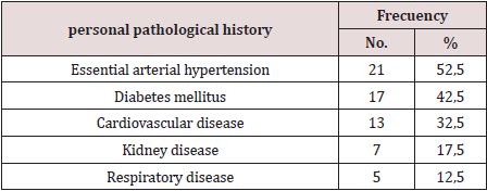
Saturday, 9 April 2022

Lupine Publishers | Estimation of Pulmonary Artery Catheter Length: A Retrospective Study

 Lupine Publishers | Journal of Anesthesia and Pain Medicine


Abstract

Background: Over-insertion of a pulmonary artery catheter (PAC) can cause serious complications, such as pulmonary embolism and pulmonary artery rupture. Careful management is essential.

Methods: This study investigated relationships between patient parameters and appropriate PAC insertion length. A function on the chest X-ray display was used to measure the curve. In Study 1, we investigated concordance using intraclass concordance coincidence (ICC) between recorded insertion length of the PAC and actual inserted length. We reviewed recorded insertion data from patients’ anesthesia records and measured actual inserted length. In Study 2, we measured and estimated the appropriate insertion length using chest X-rays taken just after surgery. Multiple logistic regression was performed to investigate relationships between patient background parameters and appropriate insertion length.

Results: For Study 1, recorded insertion length was 41.2±2.7cm and actual inserted length was 41.9± 3.1cm. ICC (2,1) was 0.867, which shows a high correlation. For Study 2, height and cardiac width on preoperative chest X-rays were significant factors determining appropriate PAC length. The multiple regression equation for appropriate PAC length (z) with height (x) and cardiac width (y) was calculated as z(cm) = 0.1x +0.8y+18.1. Height and cardiac width were significant factors determining appropriate PAC insertion length.

Conclusions: We calculated an equation for the relationship between height and cardiac width to obtain the appropriate PAC insertion length.

Keywords: Pulmonary artery catheter; Right pulmonary artery; Chest X-ray

Abbreviations: ICC: intraclass concordance coincidence; PAC: pulmonary artery catheter; PCWP: pulmonary capillary wedge pressure.

Background

The pulmonary artery catheter (PAC) is used for many diagnostic applications, including measuring pulmonary artery pressure, cardiac output, and mixed venous oxygen saturation [1]. Over-insertion may cause serious complications, such as pulmonary embolism and pulmonary artery rupture. Careful management is essential. Approximately 90% of injuries occur at the medial and inferior branches of the right pulmonary artery, because a PAC inserted from the right internal jugular vein will most often enter the right pulmonary artery [2]. There are numerous reports of complications arising from pulmonary artery ruptures following PAC insertion. Such injury has a lower frequency (at 0.03-0.7%) than infections, pulmonary embolism, pericarditis, and valvular vegetation as complications after cardiovascular surgery; however, it is a serious problem, with a mortality rate of 37% [3]. When inserting a PAC, the tip is recommended to be within 2cm of the catheter silhouette on standard anteroposterior chest film [4]. PAC manufacturer instructions state that the best position for the tip is at the hilum of the lungs in the right pulmonary artery (see Additional file 1, p. 148). Position is determined by confirming pressure and waveform detected through the PAC tip. When waveform obtained from the tip of the PAC with the balloon inflated shows correct pulmonary capillary wedge pressure (PCWP), the tip must be in the pulmonary artery, and the balloon is deflated. Final confirmation of position is performed by chest X-ray postoperatively. The purpose of this study was to estimate appropriate PAC length from chest X-rays and investigate relationships between the size of the body and heart and appropriate PAC length. A function on the chest X-ray image display was used to measure the curve and evaluate PAC length. We hypothesized that the size of the heart or height and weight correlates with appropriate PAC length.

Methods

This study was a retrospective observational study of cardiovascular surgery patients in 2013. The study protocol was approved by the Ethics Committee of Osaka Medical College (Reference No. 1591Rin-47), which waived the requirement for informed consent because of the retrospective design of the study. Participants were patients in whom a PAC was inserted from the right internal jugular vein to the main or right pulmonary artery and who underwent surgery using cardiopulmonary bypass in the supine position. Emergency surgery and descending aorta replacement surgery were excluded. Induction of anesthesia was performed by two anesthesiologists. General anesthesia was induced by administration of propofol, midazolam, rocuronium, remifentanil, and sevoflurane. After endotracheal intubation, the right internal jugular vein was punctured at the cricoid cartilage level, the PAC sheath was inserted, then an 8Fr PAC (CCO/CEDV thermodilution catheter; 777HF8®; Edwards Lifesciences, Irvine, CA) was inserted through the sheath. A central venous catheter was inserted on the cranial side of the PAC insertion point. Intraclass concordance coincidence (ICC) was calculated in 20 cases in Study 1 to show concordance between recorded insertion length and actual inserted length. In Study 2, multiple regression analysis was performed to investigate relationships between appropriate insertion length and the parameters. Statistical analyses were performed using JMA9® (SAS Institute, Tokyo, Japan).

Study 1

A function on the X-ray imaging display was used to measure the curve. A chest X-ray was taken in the anteroposterior view immediately after surgery, with the patient in the supine position under general anesthesia. For Study 1, there were only 20 patients in whom the PAC was inserted from the cricoid cartilage, which was assumed to be at the level of the sixth cervical vertebra, over the study period. Anesthesiologists read the scale of the insertion length of the PAC just after surgery and recorded the data on anesthetic records as ‘insertion length’. A chest X-ray was taken concurrently. The actual inserted length was measured with the display function using the postoperative chest X-ray. We confirmed the grade of concordance between recorded PAC insertion length collected from anesthesia records and actual inserted length. Using the curve measurement function, we measured PAC length from the middle of the sixth cervical vertebra to the tip of the PAC. The recorded insertion length was reviewed against anesthesia records. This included ~5.7cm of the connector of the sheath; therefore, 5.7cm was subtracted from reviewed data to obtain the correct length. To investigate concordance of the two parameters, we calculated the ICC. ICC is a descriptive statistic that can be used when quantitative measurements are made on units that are organized into groups. It describes how strongly units in the same group resemble each other. ICC [1,2] means that each subject is measured by each rater, and raters are considered representative of a larger population of similar raters. Reliability is calculated from a single measurement. It was determined that >0.7 ICC [1,2] meant high concordance between recorded insertion length and actual PAC length.

Study 2

Patient backgrounds were reviewed from patient records and chest X-rays taken preoperatively. Cardiac width was the transverse diameter of the heart in the posterior-anterior view in a standing position as the widest diameter of the cardiac silhouette on each side of a central perpendicular line. Thoracic width was the widest diameter of the chest in the posterior-anterior view taken at a distance between the internal surface of the ribs on the right and left sides, superior to the costal attachment of the diaphragm [5]. Cardiothoracic ratio was calculated as the ratio of cardiac width divided by thoracic width. At our institute, over the period in which Study 2 was conducted, there were 395 patients who underwent cardiovascular surgery, including pediatric cardiovascular surgery and vascular surgery, amongst others. There were 149 cases of adult surgery, not including abdominal aorta replacement and stenting. Elective surgery that was performed through median sternotomy was included. Of these 149 cases, 121 met inclusion criteria for Study 2. Appropriate insertion lengths of the PAC were estimated and measured in these 121 patients. The curve measuring function on the display was used with a chest X-ray taken just after cardiac surgery in the operating room. Actual inserted length was measured from the sixth cervical vertebra to the tip of the PAC. In cases where the PAC did not reach the hilum, we estimated the course of the PAC and added the distance from the inserted length to the hilum.

Figure 1.

Lupinepublishers-openaccess-journals-anesthesia-pain-medicine-journal

We determined that the appropriate position of the PAC tip was the intersection of the PAC in the superior vena cava and the PAC tip in the right pulmonary artery. (Figure 1) shows how the inserted length was measured. The white line shows the inserted PAC and the black line shows estimated distance from the tip to the hilum. The actual inserted length was 48.2cm and the estimated added length was 2.1cm. The appropriate insertion length was therefore calculated as 50.3cm. When the PAC exceeded the hilum in the X-ray, the appropriate length was determined as the length from the sixth cervical vertebra to the hilum of the lung.

Results

Table 1: Patient backgrounds and measurements in Study 2 (n=121).

Lupinepublishers-openaccess-journals-anesthesia-pain-medicine-journal

PAC: pulmonary artery catheter

Table 2: Results of multiple logistic regression in Study 2.

Lupinepublishers-openaccess-journals-anesthesia-pain-medicine-journal

β: standardized partial regression coefficient: r: correlation coefficient; **p<0.01

For Study 1, recorded insertion length was 41.2±2.7cm and actual inserted length measured using chest X-ray was 41.9±3.1cm. ICC (2,1) was 0.867, which shows a high correlation. For Study 2, (Table 1) shows patient backgrounds and measurements. There were nine cases in which the PAC tip was inserted exceeding the right hilum. Height and cardiac width on preoperative chest X-rays were significant factors in determining appropriate inserted PAC length (P<0.05) (Table 2). The multiple regression equation for appropriate PAC length (z) with height (x) and cardiac width (y) was calculated as z (cm) = 0.1x+0.8y+18.1.

Discussion

We hypothesized that PAC length is affected by the size of the body and heart. Our results aligned with values previously reported, despite differences in pulmonary artery pressure, cardiac diseases, and loop formation depending on the catheter. When using average values for Japanese people with normal blood pressure (male height: 168.6cm; female height: 156.1cm; cardiac length for both males and females: 13.8cm) in our equation, the appropriate insertion PAC length was calculated as 46.0cm for men and 44.8cm for women. Participants were patients with cardiac diseases requiring surgery, and cardiac width was, on average, 15.1cm, which is greater than the Japanese average. This may explain why the appropriate insertion length in our study was greater than average values. PAC insertion length for obtaining PCWP is commonly determined as 45-55cm [4]. Researchers in Japan and India, where average height is shorter, reported the appropriate insertion length as ~40cm, with length tending to be greater in valvular disease patients [6,7]. Studies have reported that PAC length from the right internal jugular vein to the PAC tip for obtaining PCWP is ~44± 6cm and 42-48cm, and no correlation between height and appropriate PAC length was noted by the Japanese researchers [8,9]. Another study suggests that PAC length for obtaining PCWP is commonly <50cm. When the inserted PAC length is >50 cm, PAC insertion should be retried, or the position assessed using chest X-ray or fluoroscopy as there may be sagging or loop formation [9]. According to one manufacturer (Edwards Life Sciences), the recommended PAC tip position is at the hilum. At this position, pressure waveform shows PCWP when the balloon is inflated. At this stage, the balloon should be deflated and pulled back ~2-3cm slowly to avoid sagging and loop formation in the right atrium and ventricle. Pulmonary artery rupture by PAC occurs rarely but is a critical complication. Pulmonary hypertension, anticoagulation therapy, old age, low body temperature, displacement of the PAC tip, balloon overinflation, or heart surgery are reported risk factors for rupture [3]. Cardiovascular surgery includes multiple factors that can cause rupture, therefore, careful observation of PAC insertion length is needed. Although there is a lack of data on the effectiveness of PAC in cardiac surgery [10], parameters taken from the PAC are relied on and it remains a standard procedure in many practices.

Conclusion

Height and cardiac width were significant factors determining the appropriate PAC insertion length. We calculated an equation for the relationship between height and cardiac width to obtain the appropriate PAC insertion length.

Ethics approval and Consent to Participate

The study protocol was approved by the Ethics Committee of Osaka Medical College (Reference No. 1591Rin-47), which waived the requirement for informed consent because of the retrospective design of the study.

Read More about Lupine Publishers Journal of Anesthesia Please Click on Below Link:
https://lupine-publishers-anesthesia-pain.blogspot.com/