Showing posts with label Journal of Cardiology. Show all posts

Monday, 16 October 2023

Lupine Publishers | Factors Influencing Stereotaxic Pulmonary Vein Isolation

 Lupine Publishers | Journal of Advancements in Cardiology Research & Reports

Abstract

Background – Catheter ablation of atrial fibrillation (AF) is performed to restore and maintain a sinus rhythm. Remote magnetic navigation system (RMNS) allows an efficient and safe procedure. Left atrial (LA) anatomic barriers of this device are not well known. Aims – This study was aimed to evaluate clinical, echocardiographic and cardiac computed tomography (CCT) anatomic LA characteristics as predictors of stereotaxic AF procedure duration. Methods – From February 2015 to April 2016, 102 symptomatic and drug refractory AF patients were consecutively enrolled in an observational, prospective trial when first AF ablation. AF Radiofrequency (RF) was performed with a RMNS using Niobe ES. Clinical endpoints and LA characteristics were reported, prospectively by a transthoracic and transesophageal echocardiography, and CCT scan. Results – Mean patient age was 5912 years old, 77% male, mean CHA2DS2VASc of 1.31.3 and mean LA surface of 236.5cm2. Procedure duration of 97.232.9 minutes and fluoroscopy duration of 13.47.9 minutes were recorded. Persistent versus paroxysmal AF (p<0.05), previous flutter ablation (p<0.01), LA dilation (p<0.05), narrow LA ridge (p=0.01), small surface area and high eccentricity of the left inferior pulmonary vein (LIPV) (p<0.01) are correlated to an increased procedure duration. Previous flutter ablation (p<0.01), persistent AF (p<0.05), LIPV eccentricity (p<0.05) and ridge width (p=0.05) were found to be independently associated with procedure duration. Conclusion – Our study is the first analyzing predictors of stereotaxic procedure duration. Narrow LA ridge, small and flattened LIPV were independently correlated with an increased procedure duration. Yet neither co-morbidity nor cardiomyopathy was associated to procedure changes.

Keywords: Atrial fibrillation ablation; pulmonary vein isolation; remote magnetic navigation; procedure duration; anatomic characteristics

Introduction

Radiofrequency ablation (RF) is a treatment of choice for atrial fibrillation (AF) because of a positive risk/benefit ratio compared to antiarrhythmic drugs [1-3].
Even if significant advances have been made over the past years regarding RF, pulmonary vein isolation (PVI) notably, several limitations remain to be overcome, such as the management of recurrences usually due to pulmonary veins (PV) reconduction, the high level of X-ray exposure and a significant risk of complications [4-6].
AF ablation is carried out in expert centers with high patient volumes. It is one of the most common procedures in electrophysiology departments (30 to 50% of total procedures) due to its prevalence and the recent guidelines [1-3]. The management of end cavity ablation in challenging clinical settings may lead to tedious and risky procedures [6,7]. and the evolvement of AF ablation indications lead to increased procedures per operator [8]. Consequently, operators are facing an increased X-ray exposition, fatigue and lack of concentration [4-6], leading to extended procedures and an increased complications risk [6, 9].
Recently, a remote magnetic navigation system (RMNS) was introduced as a way to ensure stable catheter positioning, to provide adequate tissue contact, and to reduce patient and physician X-ray exposure [10-19]. Stereotaxic procedure is supposed to reduce the usual drawbacks when manual RF, tamponade and X-ray exposure notably [10-20]. New robotic technologies seem to be as effective as manual RF [6,13,20]. Despite of ongoing RMNS improvement in order to enhance remote navigation with fast computing hardware and new motion controllers, factors influencing PVI using RMNS are not well understood [13]. Consequently, the assessment of both strengths and weaknesses of the RMNS regarding AF ablation is clinically relevant.
The aim of this study was to itemize clinical and anatomical factors influencing stereotaxic PVI duration when AF ablation.

Methods

Study population

The current trial was an observational, prospective, and blinded endpoint-assessment trial. This monocentric trial included 102 consecutive patients hospitalized for a first procedure of AF ablation in the electrophysiology department of the University Hospital of Saint-Étienne (France) from February 2015 to April 2016. All patients underwent AF ablation by a single experienced operator accustomed to stereotaxic procedures.
Inclusion criteria were: first ablation procedure due to symptomatic and drug refractory AF, be over 18 years old and collection of an oral consent. Pregnant women were excluded as patients with left atrial appendage (LAA) thrombus. The local ethics review committee approved the study.
The following data were prospectively collected: demographic patients data, comorbidities, AF background and AF therapeutic management.

Pre procedure imaging

Conventional transthoracic echocardiography (TTE) and transesophageal echocardiography (TEE) were systematically performed 24-72 hours before the ablation procedure with a commercially available system (Vivid E9, GE Healthcare, France). During TEE study, LAA was carefully analyzed to detect left atrial thrombi and spontaneous echo contrast [21].
Cardiac computed tomography (CCT) was performed using 256-slice (Somatom Definition Flash, Siemens Medical Solutions, Erlangen, Germany) scanner technologies with similar protocols in patients in a supine position during suspended end-expiration, 24h to 72h prior of the AF ablation procedure. Post-processing of cardiac computed tomographic images was performed on a dedicated advanced image processing workstation (Aquarius intuition, Tera recon, Foster City, CA). Reconstructed cardiac computed tomographic images were reviewed and interpreted by an experienced independent investigator blinded to clinical and echocardiographic data. The anatomy of PVs, LAA and left atrium (LA) ridge were assessed. CCT procedure and CT-scan data are reported in the supplementary appendix.

Ablation procedure

The procedure is detailed in the supplementary appendix. LA mapping was performed using CartoÒ 3 System (Carto® 3 system, Bio sense Webster, CA), an electromagnetic system allowing realtime Advanced Catheter Location™ and visualization of both ablation and circular mapping catheters (NaviStar® and Lasso catheters®). Once the map was completed, 3D computed tomography scan was performed in order to optimize LA reconstruction.
The RMNS (Niobe™ EPOCH, Stereotaxis Inc., St Louis, MO) employs a steerable magnetic field remotely guiding a flexible catheter [6, 9–13]. Two giant computer-controlled 1.8-ton magnets are positioned at opposite sides of the fluoroscopy table. A magnetic field of 0.08 to 0.1 Tesla is generated allowing a 3D navigation thanks to three small magnets incorporated in parallel in the RF catheter tip. The magnetic field is applied to a theoretical cardiac volume of 20cm x 20cm. Catheter movements depend on direction changes of the two magnets in relation to each other. A computerized motor drive system (Cardiodrive®, Stereotaxis Inc., St Louis, MO) advances or retracts the catheters, whilst its spatial orientation requires a computerized work station (Navigant® 2.1, Stereotaxis Inc., St Louis, MO). A constant application of the magnetic field maintains contact between the catheter tip and endocardial tissue throughout the cardiac cycle. The new generation RMNS results in faster control of the catheter, leading to potentially reduced navigation duration [22].

Procedure and fluoroscopy parameters

Skin to skin total duration was recorded for all patients. The following parameters were also recorded: X-ray duration (sec), X-ray (Gy) and indexed X-ray (Gy x cm²) procedure time. These parameters were divided in different periods: setting up, mapping and ablation period (including left and right PVI).

Statistical analysis

Continuous variables were presented as mean±SD, or median+IQR as appropriate. Categorical variables were expressed as percentage. Linear uni and multi-variate models were generated to predict procedure duration. Characteristics of each model were given at the regression parameter for each variable (b), with its 95% confidence intervals and p-value. The multiple linear regression model was built in a backward stepwise manner, selecting theoretically impacting covariates (defined by p<0.05 in the univariate analysis) to predict procedure times and X-ray patient exposure, to maximize the goodness of fit expressed as R². All analyses were performed using R (R Foundation for Statistical Computing, Vienna, Austria, http://www.R-project.org).

Results

Patients

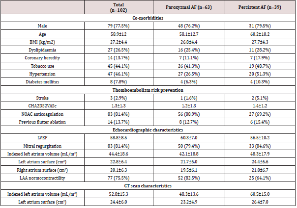
Population data are summarized in (Table 1). One hundred and two consecutive patients were prospectively included, divided into 63 paroxysmal AF (62%) and 39 persistent and long-standing AF (38%). The population characteristics were as follows: mean age of 59±12years old, 77% of male, a body mass index of 27±4.5 kg/m2, mean CHA2DS2VASc of 1 [1-2], anticoagulated with non-vitamin K oral anticoagulant (81%) and without cardiomyopathy. Mean LA surface was 23±6.5 cm2 and LAA normocontractility was mostly assessed.

Table 1:Patient characteristics.

Lupinepublishers-openaccess-cardiology

Continuous variables are presented as mean±SD. Categorical variables are expressed as number (percentage). AF=atrial fibrillation; BMI=body mass index; LAA=left atrial appendage; LVEF=left ventricular ejection fraction; NOAC=non-vitamin K antagonist oral anticoagulant; sAoVTI=sub aortic velocity time integral.

Procedure features

Procedure parameters are summarized in (Table 2). A 100% acute PV isolation success was reported. Mean ablation procedure time was 97±33 minutes with a mean RF time of 66±31 minutes. Total X-ray duration was 13.4±7.9 minutes. Fluoroscopic use was mainly related to the setting up period (58%), compared to 23% of ablation procedure duration. Ablation and total duration are not different whether paroxysmal or persistent AF. Three acute complications occurred: a pericardial effusion without tamponade and two medically-treated inguinal haematomas.

Table 2:Procedure parameters.

Lupinepublishers-openaccess-cardiology

Continuous variables are presented as mean±SD. AF=atrial fibrillation.

Univariate analysis

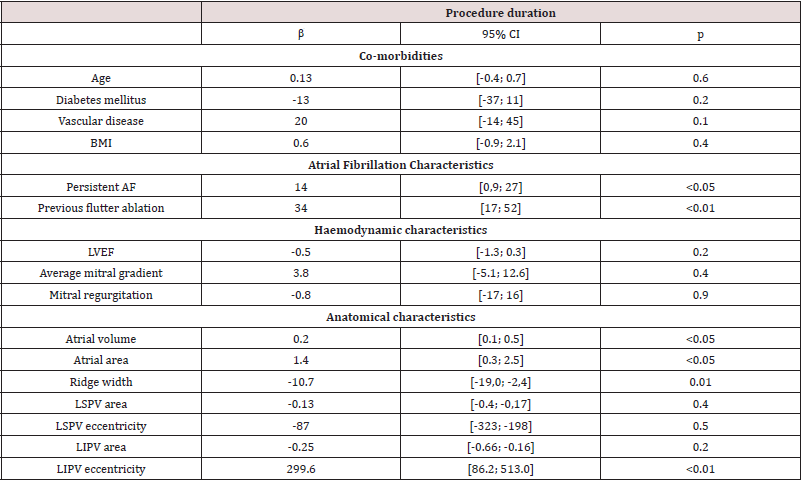
Clinical characteristics impacting the ablation duration No co-morbidity was associated with an increase of the AF ablation procedure or X-ray exposure duration (Table 3). On the other hand, persistent versus paroxysmal AF (p<0.05), and previous flutter ablation (p<0.01) were both risk factors associated with a with procedure duration. The left ventricular function was not associated with a change in ablation procedure duration parameters (Table 3): neither LVEF (p=0.2) nor sAoTVI (=0.6). Furthermore, mitral valve disease, whether mitral regurgitation (p=0.9) or stenosis (p=0.4), was not correlated to longer procedures.

Table 3:Impact of clinical, hemodynamic and anatomical characteristics on the procedure duration.

Lupinepublishers-openaccess-cardiology

Univariate analysis. AF=atrial fibrillation; BMI=body mass index; LVEF=left ventricular ejection fraction; LIPV=left inferior pulmonary vein; LSPV=left superior pulmonary vein.

Anatomical characteristics impacting the ablation duration LA dilation, assessed by LA area (p<0.05) and LA volume (p<0.05), was associated to an increased procedure duration (Table 3). This association was found during the mapping poeriod of the procedure (p<0.01), but not for the setting up and ablation ones (Table 1), supplementary appendix). On the other side, wider LAA ridge was correlated with a shorter fluoroscopy duration (p=0.01). Smaller LIPV surface area (p<0.01) and higher LIPV eccentricity (p<0.01) were correlated with longer RF duration (Table 2), supplementary appendix).

Multivariate analysis

Through multivariate linear regression analysis with relevant clinical and echocardiographic features, a previous flutter ablation (p<0.01) and persistent AF (p=0.03) were found to be independently associated with total procedure duration. Furthermore, left LA fluoroscopy duration was independently influenced by LIPV eccentricity (p<0.05) and LA ridge width (p=0.05).

Discussion

Major findings

This prospective observational study suggests that no comorbidity and cardiomyopathy was associated to longer stereotaxic PVI procedures. LA dilation was correlated with increased setting up but not ablation duration. Persistent AF and previous atrial flutter are independently associated to an increased procedure duration. Only LA ridge width and LIPV anatomy influenced significantly and independently the ablation duration.

Stereotaxic procedure duration

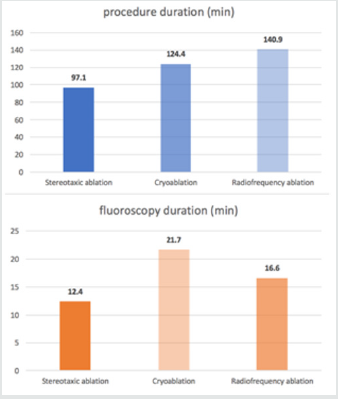
Due to the improving indications of AF ablation, all dedicated EP departments deal with an increased daily ablation procedure. Thereby, different challenges appear: [1] decrease procedure duration leading to reduce physician fatigue and X-ray exposure and [2] optimize the management of consecutive daily procedures. As an indicator shown in (Figure 1), the procedure duration seems to be significantly shorter in the current study using stereotaxic system, compared to cry balloon and manual RF ablation in FIRE and ICE trial [23]. Indeed, manual RF ablation seems to approximately 50% longer than stereotaxic procedure, while fluoroscopy duration is 75% longer with cry balloon and 33% longer with manual RF than stereotaxic ablation. In addition to a shorter procedure, stereotaxic ablation reduces the operator tiredness and increases its accuracy by allowing a seated and comfortable procedure. However, it is important to keep in mind that all ablations were performed with an experienced operator, after a usual learning curve.

Figure 1: Procedure and fluoroscopy duration with stereotaxic ablation compared to cryoballoon and radiofrequency ablation in FIRE and ICE trial [23].

Lupinepublishers-openaccess-cardiology

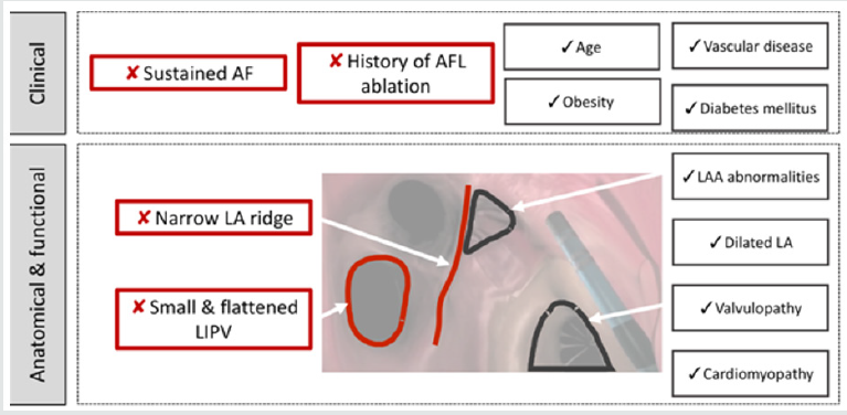
Factors influencing procedure duration (Figure 2) Clinical characteristics impacting the ablation duration. Age, sex, diabetes mellitus, vascular disease and obesity are not correlated with an increased manual RF procedure duration [24], which is consistent with our trial. Our study was the first highlighting the lack of association between body weight and procedure duration. In addition, AF radiofrequency seems to be safe despite of overweight: there is not a higher hemorrhagic and infectious complications incidence reported [25]. Yet several AF risk factors such as obesity and sleep apnea, seems to be important to maintain a sinus rhythm after AF ablation [26]. Persistent AF was associated to a longer PVI procedure, suggesting the presence of more severe LA architectural abnormalities when persistent AF. Indeed, persistent AF seems to be associated to a much more severe atrial cardiomyopathy compared to paroxysmal AF [27]. Furthermore, previous ablation of atrial flutter RF was an independent risk factor of longer ablation procedure. The interrelationship between AF and atrial flutter is still unclear [28]. But this finding suggests that patients with previous atrial flutter get a complex atrial cardiomyopathy including fibrosis [24] and atrial dilation. It could lead to a longer and less efficient IVP procedure [29].

Figure 2: Central figure: Clinical, hemodynamic and anatomical factors influencing the stereotaxic AF ablation duration.

Lupinepublishers-openaccess-cardiology

This figure summarizes results of the univariate analysis. Image corresponds to an endocavity view of the left atrium with a septal view. AF=atrial fibrillation; AFL=atrial flutter; LIPV=left inferior pulmonary vein; LA= left atrium; LAA=left atrial appendage.

Hemodynamic characteristics impacting the ablation duration neither systolic left ventricular dysfunction nor mitral disease were correlated to a longer procedure. Since 50% of AF patients get heart failure and 25% of heart failure patients get AF [30], the efficacy and safety of the AF ablation has to be proved. Indeed, antiarrhythmic drugs fail AF patients with heart failure [31], and AF ablation could be a therapeutic key, as suggested by CASTLE-AF trial [32]. In this study, catheter ablation decreased hospitalization rate and mortality, increased left ventricular function, over midterm follow up [33]. The stereotaxic ablation seems to be an interesting strategy when heart failure, regarding the safety of the procedure and the absence of LV dysfunction and LA dilation impact in procedure duration.
Left atrial architectural features impacting the ablation duration In our study, LA dilation led to a longer procedure, with an increased mapping duration, yet no effect on stereotaxic ablation duration was reported. Stereotaxic procedure seems to overcome anatomical difficulties such as LA dilation, preventing technical difficulties because of an efficient navigation.
On the other side, a narrow ridge was a predictor of longer ablation using RMNS. The left lateral ridge is known as a uniform width or muscular thickness being narrower and thicker at the antero-superior level [34]. This area acts as a fibrillary process due to the presence of the vein of Marshall, an electrical gap between left PV and LA [35]. This area constitutes a preferential zone of PVs reconnection [36]. Manual RF procedures often fail to complete ablation line in LA ridge [37]. This study also pointed out a tough ablation in this area despite stereotaxic accurate navigation.

Clinical implications

AF catheter ablation is an efficient treatment to achieve a rhythm control strategy, regarding recent guidelines [8]. But this procedure is still challenging and needs to be more efficient and safer. RMNS may be an interesting way to achieve this goal. In this study, all procedures allow an acute PV isolation, associated to a short procedure and fluoroscopy time. Furthermore, RMNS allows an efficient navigation during PVI, regardless LA dilation and left ventricular dysfunction.
But RMNS accuracy could be improved. The Stereotaxis Magnetic Navigation System allows precise navigation with a spatial resolution of 1 degree of omni-directional deflection and 1 mm for catheter advancement and retraction, as opposed to the manual catheter manipulation and catheter movements which highly depends on the operator. RMNS leads to a stable cathetertissue contact during cardiac motion, unlike manual RF ablation [38]. This study suggests that a narrow LA ridge and a small and flattened LIPV increase overall procedure time. The knowledge of these anatomical limitations ta achieve IVP may help the engineers to work on RMNS improvements.

Study limitations

The major limitation of this observational study was its lack of randomization. However, this study included consecutive patients, prospectively, in order to prevent bias analysis. In addition, this monocentric trial was based on IVP procedures performed by a single operator. It avoids the inter-observer variability but limits the exptrapolability of the data. Finally, a new trial should be designed to test LA anatomical predictors of AF recurrences and PVs reconnection with RMNS compared to manual RF and cry balloon procedures.

Conclusion

Our study proposed to highlight predictors of stereotaxic procedure duration. Both narrow LA ridge, small and flattened left inferior PV are independently correlated with increased procedure duration. But no co-morbidity and cardiomyopathy were linked to a procedure change.

Supplementary Appendix

Read More About Lupine Publishers Journal of Advancements in Cardiology Research & Reports Please Click on the Below Link:

https://lupine-publishers-cardiovascular.blogspot.com/

Monday, 21 August 2023

Lupine Publishers | Iron-Based Nanoparticles, an Accurate and Powerful Sniper Targeting SARS- Cov-2

 Lupine Publishers | Journal of Cardiology Research & Reports


Abstract

For the past few months, the world has been facing another coronavirus disease, COVID-19, caused by the SARS- CoV-2, giving the rise of a pandemic. In the vast majority of infected individuals, SARS-CoV-2 causes a mild ailment, but in some subjects, it progresses to severe disease or even death, with some groups being at high risk. However, SARS-CoV-2 is not the first coronavirus that caused serious, sometimes fatal, disease. Almost 20 years ago, SARS-CoV and later MERS were coronaviruses that led to severe diseases but did not result in pandemics. Some of the therapeutic lessons learned during the SARS-COV and MERS epidemics are being used now to treat COVID-19 patients, such as the still debated use of Chloroquine. However, there were other important preclinical studies performed around the time of SARS-COV and MERS epidemics that may also be useful applications in the COVID-19 context. This review highlights the benefits that could be gained by revising the non-conventional therapeutic approaches used in the previous coronavirus epidemics in improving the detection, treatment, and prevention tools and developing patient treatment follow-up strategies. Specifically, this review discusses the utilization of iron oxide nanoparticles due to their attractive properties. It also highlights the therapeutic opportunities and future directions of the iron oxide nanoparticles to be eventually employed in the current coronavirus pandemic.

Keywords: SARS-Co V-2; COVID-19; Nanomedicine; Iron Oxide Nanoparticles

Abbreviations:ACE Inhibitors= ACEIs; Acute Lung Injury= ALI; Acute Respiratory Distress Syndrome= ARDS; Angiotensin Converting Enzyme-2= ACE2; Angiogenesis II= AngII; Angiotensin Receptor Blockers= ARB; Antisense Oligonucleotide= ASO; Black, Asian and Minority Ethnic= BAME; Cardiovascular Diseases= CVDs; Computed Tomography= CT; Diabetic Melitus= DM Intensive Care Unit= ICU; Interferon-α= IFN-α; Interferon-β= IFN-β; Iron Oxide NPs= IONPs; Mas Receptors= MasR Middle East Respiratory Syndrome Coronavirus= MERS-CoV; Nanoconjugates= NCs; Nanoparticles= NPs; Nanozymes= IONzymes; Nitric Oxide= NO; Norwegian University of Science and Technology= NTNU; Phosphodiesterases-5= PDE5; Poly (amino ester) with carboxyl groups (PC)-coated magnetic NPs= pcMNPs; Prorenin receptor- Ang II type 1 receptor= PRR-ACE-Ang II-AT1R; Pulmonary Arterial Hypertension= PAH; Reactive Oxygen Species= ROS; Receptor Binding Domain= RBD; Renin Angiotensin System= RAS; Reverse Transcription Polymerase Chain Reaction= RT-PCR; Royal Gwent Hospital= RGH; Severe Acute Respiratory Syndrome Coronavirus= SARS-CoV; Spike protein= S; Transmembrane Protease Serine 2= TMPRSS2; Tumour Necrosis Factor= TNF; World Health Organization= WHO

Introduction

Outbreaks caused by infectious diseases represent a tremendous challenge to humanity, with coronaviruses accounting for a relevant part of them[1]. Coronaviruses are a single-stranded positive-sense RNA family that can affect several vertebrate hosts. In the past, these viruses were known to cause minor to mild upper respiratory tract diseases with symptoms similar to the common cold in many cases. However, in the last 17 years, three aggressive human coronavirus pathogens appeared as we witnessed the emergence/re-emergence of zoonotic diseases causing epidemics and pandemics. These include the Severe Acute Respiratory Syndrome Coronavirus (SARS-CoV)1 with 8098 infected cases and 774 [2] deaths, the Middle East respiratory syndrome coronavirus (MERS-CoV) with 2494 infected cases and 858 [3] deaths, and the most recent outbreak of the novel coronavirus (SARS-CoV-2 (COVID-19) which was first reported in Wuhan-China in December 2019 and was considered a pandemic on March 11th 2020 [4]. As of November 20th, 2020, there are 57,236,335 reported cases, of which 39,722,802 recovered and 1,365,634 died[5]; the current pandemic originated enormous global health, social and economic crises1. SARS-CoV-2 succeeded in crossing species barriers to infect humans and can now be effectively transmitted from one person to another1. So far, 7 coronaviruses affecting humans are known: SARS-CoV, MERS-CoV, and SARS-CoV-2 causing symptoms that start with influenza-like signs that either stays mild and disappears or escalates to severe lung injuries with multiorgan failure, acute respiratory distress syndrome (ARDS), and acute lung injury (ALI); the remaining 4, namely HKU1, NL63, OC43, and 229E, are associated with mild respiratory symptoms [4, 6]

The death rate of SARS-CoV-2 increases by age and the presence of other comorbidities1. SARS-CoV-2 mechanism of action affects three host components: blood, inflammation, and cellular components. In the blood component, the viral proteins “ORF10 and ORF3a” coordinate to attack the heme part of the hemoglobin (1-beta chain), which results in the dissociating of the iron to form porphyrin, decreasing the amount of the functional hemoglobin, which leads to the development of anemia and respiratory distress symptoms [7]. In the inflammation component, it was noticed that the overproduction of proinflammatory cytokines -such as IL-6, IL-1β, and tumor necrosis factor (TNF)- could cause a SARS-CoV-2- induced cytokine storm in addition to the accumulation of the fibrin and thrombin [8]. The sudden cytokine increase elevates the risk of developing vascular hyperpermeability and multiorgan failure, eventually leading to death. The inflammatory component of the SARS-CoV-2 includes a significant upsurge in the reactive oxygen species (ROS), triggering redox imbalance, mitochondrial and lysosomal dysfunction, and rendering the cells even less resistant to infection. Such a sequence of events can result in serious and permanent long-term cellular damage, which accelerates the immune system’s aging process and affects tissues/organs (i.e. lung) [8]. In addition, the inflammatory component is responsible for the formation of the ground-glass-like figure of the COVID-19 lung. This indicates that developing immunomodulators or therapeutic strategies to target the inflammatory component, ROS, and the overactive cytokines could be one of the SARS-CoV-2 therapeutic options [7, 9]. The cellular component is the most crucial one in the pathogenesis of SARS-CoV-2, which is why we will be discussing it in depth. So far, angiotensin-converting enzyme-2 (ACE2) receptors have been considered as the primary cellular entry point for three strains of coronavirus: NL63, SARS-CoV, and the novel SARS-CoV-2 [10]. ACE2 receptors are expressed ubiquitously in the blood vessels, heart, lung, kidneys, gut, brain, testis [11], and salivary glands [12]. It is expressed in rodents and humans, and present bound to the cell membrane with low levels in the plasma [11].
The nasal epithelium is now considered the portal entry for the SARS-CoV-2 infection and transmission, where higher viral loads were found in symptomatic and asymptomatic patients. The virus’s entry depends on the binding of the spike (S) protein to a specific cellular receptor, which then initiates a series of cellular proteases. A crucial and limiting factor for the viral entry and the initial infection is the ACE2 receptor facilitated by host cell-derived serine protease, the transmembrane protease serine 2 (TMPRSS2). It is worth noting that TMPRSS2 could be used as an alternative entry route for the SARS-CoV-2 using cathepsin B/L [13, 14]. The S protein’s high affinity towards the ACE2 is now considered an important determinant factor for the viral replication rate which in turn determines the severity of the disease [15]. Datasets for multiple human tissues, including airways, were retrieved from published/unpublished databases that could be found on European Genome–phenome Archive (https://www.ebi.ac.uk/ega/home), GEO [16], and MedGen. These data were analyzed by both Vieira et al. [17] and Deprez et al. [18]. Results showed a high level of ACE2 expression in nasal secretory cells, multiple types of epithelial cells across the airway, and the alveolar epithelial type II cells13. Looking back at the history and importance of the ACE2 receptors, we will find that the renin-angiotensin system’s (RAS) classical view has dramatically changed since the discovery of the ACE2 receptors. Since then, ACE2 was classified as a membrane-bound receptor; it cleaves the angiogenesis II (AngII) to generate the active peptide Ang (1–7). The cellular actions of the Ang (1–7) are then mediated by Mas receptors (MasR) [19] to perform cardio- protective effects.

ACE2 Expression Modulators

ACE2 expression is modulated by the following factors: age, ethnicity, sex, and the existence of other health comorbidities: in SARS-CoV2, these factors have an impact on the susceptibility and severity of the disease11. An in vivo study evaluating the age effect on the expression of several cardiac markers between two groups of 24 month-old and 12 month- old mice showed that 24 month-old ones expressed significant enhancement of the prorenin receptor - Ang II type 1 receptor (PRR-ACE-Ang II-AT1R) axis with a reduction in the ACE2/ Ang (1–7)/MasR axis [20]. Global studies from China, Italy, Spain, UK, and the USA showed that elder people have low expression of ACE2 in the lung11 and therefore are more susceptible and fragile to the disease, which could be attributed to the virus ability to further lower the ACE2 baseline expression in the ACE2 low producing cells [11]. This might be attributed to the fact that aging is a major factor in the development of cardiovascular diseases (CVDs) due to endothelial cell dysfunction and inflammation [21]. Also, the fetal dataset retrieved and analyzed from GEO16 revealed the low to no expression of the ACE2 with no co-expression of the TMPRSS2, which might partially explain why SARS-CoV-2 represents a low risk to young generations [13, 14, 22]. In terms of ethnicity and sex, it was shown that the diverse genetic basis in the Black, Asian, and Minority Ethnic (BAME) groups could indeed affect ACE2 functions. Also, it has been shown that other ethnic groups have some structural variations of the ACE2, that confirmed its protective effects due to its low binding affinity to the viral S protein [11]. This could be the reason why people from the BAME origins have high SARS- CoV-2 susceptibility due to the increased affinity of their ACE2 variation to the viral S protein showing more disease severity, which might contribute to the death rate [23-25]. Additionally, Baumer et al. [26] showed that 35% of the SARS-CoV-2 cases that were admitted to the intensive care unit (ICU) and 35% of the deaths in the Royal Gwent Hospital (RGH), Newport, Wales, were of BAME ethnic descent. Another study showed that men had a higher chance of getting SARS-CoV-2 as their cells express a higher percentage of ACE2 compared to women [23, 24].

Risk factors such as elder age and comorbidities (namely, CVDs, Diabetic Mellitus (DM), obesity) are known to worsen SARS-CoV-2 severity, eventually leading to death. Patients with the above-mentioned risk factors share one feature that worsens their SARS-CoV-2 condition, which is the involvement of the endothelial cells in the progression of the SARS- CoV-2 with endothelial cells having different expression profiles of the ACE2 in patients with different comorbidities. The up or downregulation of the expression of this receptor can either benefit or harm the SARSCoV- 2 patients depending on which group they belong to; As it determines the patient’s response to the viral invasion. ACE2 is generally expressed by endothelial cells and plays an important role in vasodilation, anti-hypertrophy, and anti-fibrosis [27]. Under normal circumstances, the downregulation of ACE2 leads to an increased function with the ACE/AngII axis inducing antiapoptotic, thrombotic, proinflammatory, and vasoconstriction effects; while the upregulation of the ACE2/ Ang (1–7)/MasR axis exhibits various cardiovascular protective effects including the vasodilatory, anti-proliferative, anti-atherogenesis, antithrombosis, and antifibrotic effects. Dysregulation between these two axis was reported in SARS-CoV-2 infected patients from high-risk groups. An example of the dysregulation between the two axis was reported in pulmonary arterial hypertension (PAH); with idiopathic or heritable PAH patients expressing an increased level of AngII levels with markedly decreased levels of phosphorylated status (Ser-680) of ACE2 whereas, PAH due to congenital heart disease showed decreased levels of serum ACE2 and Ang (1–7). In contrast, DM and other CVDs such as hypertension are considered high-risk factors for the severity of SARS-CoV-2 due to the significant increase in the expression of the ACE2 receptors increasing the viral entry points [28]. Lately, evidence has emerged, linking obesity to the poor prognosis of patients with SARS-CoV-2. This is attributed to comorbidities associated with obesity; these comorbidities are caused by the endothelial dysfunction leading to hypertension and inflammation [29, 30]. The downregulation of the ACE2 in PAH and old age groups might seem beneficial at first glance, but it also means that once infected, the virus will cause a further deficiency in the low ACE2 bassline, which will worsen the condition. In this category, using the ACE inhibitors (ACEIs) and Angiotensin Receptor Blockers (ARB) could be harmful [24], unlike in DM and other CVDs where they might help as ACE2 is highly expressed in these conditions [31,32]. Surprisingly, respiratory allergies, asthma, and controlled exposure to allergen were not considered high risks for developing severe SARS- CoV-2 despite having a low expression of ACE2 [33]. The fact that PAH, despite having a decreased level of ACE2 expression, is still considered a high SARS-CoV-2 risk group indicates that there might be other factors beyond ACE2 expression, modulating the response of the respiratory allergies, asthma and controlled exposure to allergen groups to SARS-CoV-2 that we are not aware of.

Therapeutic Lessons from Past Coronaviruses

The fact that SARS-CoV-2 shares several similarities with SARSCoV as it belongs to the β-coronavirus genus, and its receptorbinding domain (RBD) resembles the SARS-CoV one indicates that they may share the same ost-cell receptor6. As such, some medications used to treat SARS-CoV were considered for SARSCoV- 2, like Chloroquine. Chloroquine is an immune modulator used in treating parasitic malaria infections; it inhibits the binding of the parasite to the ACE2 receptors, with some studies showing the antiviral activity of the drug against SARS-CoV-2 [34]. The fact that this medication can act on the ACE2 receptors promoted its use in SARS-CoV and SARS-CoV-2 [35] as it works either by inhibiting the viral fusion through the cell membrane, preventing the glycosylation of the host cell’s receptors, or preventing the assembly of the virus in the endoplasmic reticulum [34]. It also inhibits the autophagy process [36, 37]. Ribavirin is another SARSCoV antiviral drug that was employed for SARS-CoV-2 treatment [38]. The Ritonavir combination with lopinavir, other SARS-CoV antiviral drugs were also evaluated as a therapeutic option. Due to the involvement of inflammation in COVID-19, there have been signs of the benefit of using interferon-α (IFN-α) and interferon-β (IFN-β) treatments. Furthermore, some MERS-CoV in vitro and animal in vivo experiments demonstrated that combining the ribavirin and INFs treatments improved the clinical outcomes [39]. However, translating these findings to be used in the routine clinical treatment of SARS-CoV-2 infected patients needs further investigations [39]. The fact that ACE2 is expressed by various cardiovascular cells also indicates that any imbalance between the ACE axis can harm the heart, with the increase in the ACE/AngII being associated with heart failure while ACE2/ Ang (1–7)/MasR can prevent such event. Formulating ACEIs and ARB can protect from cardiovascular complications. However, their role in the lung is still unknown and requires further investigation to understand better if they can be used to ameliorate the ALL induced by SARS-CoV-211. SARS-CoV-2-related endothelial cell dysfunction was shown to be accompanied by a reduction in nitric oxide (NO) production. NO was shown to significantly inhibit the viral replication cycle by inhibiting the synthesis of the viral proteins and RNA [40], which gave the scientists another possible therapeutic angle by improving the bioavailability of NO. This is done by either supplementing patients with NO or improving the intrinsic NO bioavailability by inhibiting its degradation through the use of phosphodiesterases-5 inhibitors (PDE5) such as Sildenafil [41]. The NO bioavailability can be improved by packaging it in nanocarriers, which increased the interest in combating the virus by implementing nanomedicine tools. Nanomedicine offers many attractive properties that can overcome the current limitations and surpass the present conventional therapeutic and detection tools.

Current SARS-CoV-2 Nanomedicine Applications

Viruses are often considered nature’s devil nanocarriers. They package their genetic materials, protect it from the immune system, prolong its presence in the biological system, deliver it to the host cell, and then transfer it from infected cells to other destinations. They are nature’s evolutionary work of art with many unique features, making them smart, attractive, capable, yet dangerous nanoparticles (NPs). Many researchers in the nanomedicine field proposed the use of virus-based NPs as gene therapy tools [42]. Viral nanocarriers are double-edged swords that should be studied thoroughly for their benefits/damages. Indeed, to win the battle of COVID-19, we need to think and implements tools that reassemble the virus’s nature. Therefore, the use of nanomedicine to face SARS-CoV-2 is now being evaluated. The nanomedicine community today, more than ever, can significantly contribute to this battle with nanomaterials being developed and thoroughly investigated. Governments/countries, research/academic institutes, health organizations, and charity foundations are providing substantial funds to aid in developing nano-formulations that could be used as diagnostics tools, nanocarriers for therapeutics (drugs and vaccine) as well as developing non-conventional nano-therapies. Ongoing nanomedicine research includes the development of a rapid pointof- care diagnostic tool that will enable us to screen a higher number of persons to capture the silent careers and improve the detection threshold to detect the virus at the early infection stages.
Having such a detection tool will aid in the viral surveillance monitoring to identify regions with increased infection rate using a time effective procedure. In addition, creating a low-cost detection nanocarrier means that low-income communities will have access to such detection tools. Another angle by which we can fight SARSCoV- 2 is reusing existing therapeutic agents while innovating new ones. By understanding the basic interaction between the SARSCoV- 2 and the host cell, we can develop a nanocarrier that can either block the interaction or act as a new host for the virus. We can also use that interaction to our benefit and design a nano-vaccine. Despite being at the preliminary stages, antiviral nanomedicine therapies are promising, cost-effective, and have high-quality properties that could open new avenues in the prevention, diagnosis, and treatment of COVID-19. The physical size of SARS-CoV-2 makes the relevance of nanotechnology clearer, which is also supported by the antiviral research using nanomaterials [43]. Due to the global pandemic and emerging need for developing prevention and treatment strategies for COVID-19, the World Health Organization (WHO) adopted the strategy of repurposing existing nanomaterials to develop drug/ vaccine nano-therapies, detection tools, and antiviral coatings.

Nano-Detection Tools

Current Nano-based diagnostic tools suggested for SARS-CoV-2 include a nano-based-colorimetric bioassay that was developed using gold-NPs; these NPs were capped with a thiol-modified antisense oligonucleotide (ASO) that is specific for N-gene (nucleocapsid phosphoprotein) of the SARS-CoV-2. Capping the gold-NPs is important to ensure the efficient detection of SARSCoV- 2 in COVID-19 patients within 10 minutes [44]. Another study reported the use of polymer-stabilized multivalent gold-NPs functionalized with sialic acid derivative to make it interact with the viral spike glycoprotein [45]. Reverse Transcription Polymerase Chain Reaction (RT-PCR)-based methods are considered SARS-CoV2 gold standard detection tool. However, in their systematic review, Rodriguez et al. [46] showed that this detection tool has falsenegative rates of 2-33%46 in repeat sample testing. Also, Cohen and Kessel [47] showed that the SARS-CoV-2 RT-PCR detection tool has false-positive rates 0-16% [47] in repeat sample testing. To overcome the false positive/negative results, a study showed the possibility of increasing the RT-PCR precision using fluorescentlabeled- NPs that are conjugated to viral RNA specific probes [48]. Additionally, another group developed a Nano plasmonic sensor chip that has an extraordinary time efficiency (<15 min) and sensitivity (LOD = 370 vp/mL) as it detects the entire SARSCoV- 2 virus [49]. Finally, the Norwegian University of Science and Technology (NTNU) formed a collaboration with St. Olavs Hospital to develop an iron oxide nanoparticle-based detection Tools [50].

Nano-Based Vaccines

Nano-based vaccines are also drawing attention in facing the current pandemic. The use of nanotechnology in the development of vaccines is called ‘Nanovaccinology’; Nanovaccinology is considered an alternative and effective tool that can substitute the conventional vaccines. This is attributed to:
i) their tailorable surface properties and improved stability,
ii) immuno-stimulatory properties,
iii) high payloads,
iv) tunable sizes, which determines the cellular uptake rate, and
v) controllable drug release kinetics [51, 52]. Materials used in the synthesis of the NPs, their surface chemistry, and size are important factors in determining which cells will be activated and the potential immune response that will be triggered [53, 54]. They will also determine the vaccine release rate, pharmacokinetic properties, biodistribution, and the bioavailability of the immunogenic agents. Vaccines are developed from:
i) inactivated/killed pathogens (first-generation vaccines), ii) synthetic
peptides (second-generation vaccines),

iii) DNA vaccines (third-generation vaccines), and/or iv) liveattenuated microorganisms [53]. The efficacy of these conventional vaccines depends upon using appropriate delivery systems, therefore conjugating them with NPs can improve their efficacy. Nano-based vaccines can benefit from the added NP’s properties in guiding the vaccine to the immune cells enhancing the antigen uptake and therefore boosting the host’s immunity, and due to the high cellular uptake of some NPs, this can lead to the induction of humoral and cellular responses50. A study showed the possibility of using iron oxide NPs (IONPs), which are currently being employed to treat anemia as nanovaccine due to their in vitro antiviral activity [55]. Additionally, other promising nanomaterials are being studied with 4 COVID-19 nano-based formulations currently in trials, ClinicalTrials.Gov [56]. One nano-vaccine that is currently being investigated employs lipid nanoparticles to carry viral mRNA [57]. Additionally, other studies showed the effectiveness of using Chloroquine NPs [58] as well as the currently ongoing inhaled NO NPs trial [59]. Furthermore, scientists in the nanomedicine field are currently trying to promote nanomaterials in treating and preventing pneumonia caused by SARS-CoV-243. Nanoconjugates (NCs) Based Stem Cell Therapy Patients suffering from SARS-CoV-2 can develop virus-induced lung injuries and, to some extent, present abnormalities in liver function. The fact that some patients suffered from life-time damages in these organs led to considering the use of the Nanoconjugates (NCs) Based Stem Cell Therapy [60]. The main two challenges facing stem cell therapy are i) the low cell retention and survival rate, which in turn affects their repair capacity, and ii) the difficulty monitoring cellular behavior and fate [61]. Due to the NPs superior physical and chemical properties, NCs Based Stem Cell Therapy can overcome these limitations. In this approach, NPs are loaded/conjugated with functional agents (e.g., dye, gene, targeting ligands….etc) that could be easily taken up by the desired or studied stem cell type to genetically engineer them61. Alternatively, NPs could be used to selectively label stem cells to monitor their behavior and determine their fate; additionally, cells can be labeled with NPs that are surface modified with materials (targeting ligands) that could enhance their retention rate in the desired tissues [61]. NCs were shown effective in autoimmune disorders, CVDs, cancer, …..etc. and are currently being investigated to overcome SARS-CoV-2 induced organ damages [60]. Nano-coating tool NPs from the metals and metal oxides such as iron oxide [62], zinc oxide[63], silica (SiONPs) [64], gold (AuNPs) [64], silver (AgNPs) [65], and cuprous oxide (CuONPs) [66], possesses antibacterial and antiviral properties on their own. For that purpose, surfaces and equipment are coated with them to prevent any bacterial or viral contaminations. Especially ventilators, which are important equipment that are used for treating patients with sever SARS-CoV-2; this will help in better managing ventilator-associated pneumonia [67, 68]. Nowadays, the SARS-CoV-2 nanomedicine research is interested in implementing inorganic NPs of specifically the iron oxide nanoparticle to be used as a detection tool, therapeutic and theragnostic agents in this pandemic for the many attractive properties they have39.

Could iron oxide NPs be the magic bullet for SARSCoV- 2?

From the above ongoing SARS-CoV-2 nanomedicine research, we chose to discuss the super magnetic and iron oxide NPs as they have great potential in helping us fight SARS-CoV2. Previous results evaluating iron oxide NPs showed that they have:
i) anti-inflammatory effects on human endothelial cells and human smooth muscle cells [69],
ii) anti-inflammatory effect on mouse macrophages, and
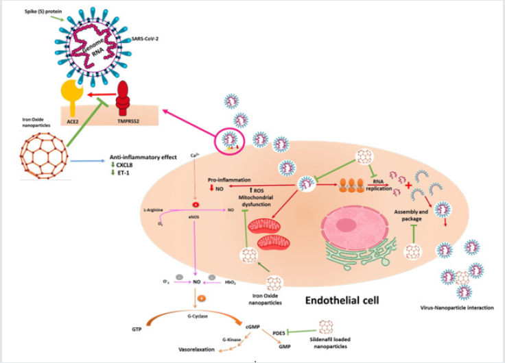
iii) showed no toxicity to endothelial cells from different origins [70]. With one prototype being loaded with the PDE5 inhibitor, Sildenafil69which can be used as a SARS-CoV-2 therapeutic option. We understand the importance of the vasculature component to the recovery from SARS-CoV-2, particularly the endothelial and inflammatory cells [70]. Therefore, we and others emphasized on the use of this ironbased nano-formulation for the detection and future treatment of COVID-19 disease by benefiting from the current knowledge of SARS-CoV-2 viral invasion, replication and survival cycles as demonstrated in (Figure 1).

Figure 1: Iron oxide nanoparticle’s potential therapeutic and detection properties

Lupinepublishers-openaccess-cardiology

In terms of SARS-CoV-2 detection and COVID-19 therapeutic option, there is an interest in iron oxide NPs to be further investigated. SARS-CoV-2 current detection method relies on using RT-PCR; despite being accurate, this method is hampered by the sample processing steps. Zhao et al. developed a rapid, inexpensive, and safe detection procedure by using the poly (amino ester) with carboxyl groups (PC)-coated magnetic NPs (pcMNPs). The pcMNPs, combine the lysis and binding steps into one step, enabling purifying the viral RNA from several samples using manual or automated methods within ≥ 20 min [71]. In support of these findings, another study by Chen et al. showed that iron oxide NPs offer a better viral detection tool [72]. In terms of anti-inflammation/antioxidant effects, iron oxide NPs were shown to have enzyme-like activity, making them classified as nanozymes (IONzymes). Nano-enzymes are favored over natural enzymes as their activity can be modified by tailoring the nanoparticle’s size, shape, and surface properties. The IONzymes, being the most typical nano- enzyme, can perform two enzymes like function, the peroxidase, and catalase activities. The nanoparticle’s composition, surface modification, and the environment pH determines which enzymatic activity IONzymes will perform73. For example, at acidic pH conditions, they exhibit peroxidase-like activities. Because of this activity IONzymes are used as biomarker detection tools in several diagnostic immunoassays that are capable of detecting the presence of Ebola, diabetes and certain tumours [74]; in addition, IONzymes have antibacterial effects and improve wound healing process74. Under neutral conditions,
IONzymes exhibit catalase-like activities, reducing the ROS, therefore improving the anti-inflammatory processes [74]. Recent studies showed that SARS-CoV-2 infection is accompanied by a significant increase in the ROS activity, which indicates the therapeutic benefit of using IONzymes in the management of SARSCoV- 2 infection [8, 75]. This catalytic activity was previously shown effective in the peroxidation of the viral lipid envelope, inactivating the enveloped viruse [73]. Furthermore, the antioxidant effect of the iron oxide NPs can be further enhanced through the surfacefunctionalization of the NPs with naturally occurring antioxidant such as gallic acid [76] and dextane conjugated trypsin [77]. In terms of their cardiovascular benefit, a study conducted by Duan et.al showed that iron oxide NPs could be used in treating CVDs associated with oxidative stress as they functioned as an autophagic-related antioxidant in HUVECs [78]. In addition, iron oxide NPs were shown to have serine protease inhibition activity, indicating that they might inhibit the TMPRSS2 that is required in the binding of the SARS-CoV-2 to the ACE2 without disturbing the ACE/AngII and ACE2/ Ang (1–7)/MasR axis balance. This inhibition can provide an extremely valuable option in the treatment and prevention of the SARS-CoV-277. The exact interaction between the TMPRSS2 and the iron oxide NPs, however, still needs further investigation. In addition, iron oxide NPs were shown to possess antiviral effects against H1N1 influenza A virus, as they inhibit the virus from binding to host cells (i.e. lung epithelial cells) [79]; also, a recent study reported that iron oxide NPs can induce membrane lipid peroxidation in synthesized liposomes, ending the viral replication cycle, which makes it a universal antiviral strategy [73]. Furthermore, it was shown that iron on its own is considered an attractive component to the virus as it is essential for viral survival, cellular invasion, and replications [7]. This explains the fact that anemia is associated with SARS- CoV-2 as the virus attacks the hemoglobin and breaks its down [7].

Therefore, thinking like a virus and creating attractive NPs for the virus might be the way to go. Having an intact nano-formulation that is hard for the virus to breakdown yet can attract the virus and interact with it might help contain the virus and prevent its rapid spread. Iron oxide NPs can attract the virus due to the presence of the iron, they are available in many forms, their stability can be tailored to range from days to months, and they possess antiviral activity with the possibility of loading/tagging them with antiviral drugs. The viral attraction to the iron oxide nanoparticle is currently being used as an antiviral tool and for rapid viral detection [71, 72]. Iron oxide NPs were shown to affect the cellular component of the SARS-CoV-2. As we know, the involvement of the endothelial cell dysfunction was proven to worsen and complicate the SARSCoV- 2 infection. Therefore, using NPs that can be taken up by the endothelial cells, reducing its dysfunctionality would be highly beneficial. So far, iron oxide NPs have been studied to mark and track endothelial cells. Some of them were shown to have no toxicity and reduce the endothelial cell’s inflammatory markers (CXCL8 and ET-1) [70]. A particular prototype, nanoMIL-89, was loaded with the PDE5 inhibitor sildenafil (generating Sil@nanoMIL-89) and investigated as an alternative tool in PAH treatment. This prototype was shown to have the above-mentioned anti-inflammatory effects on the endothelial cells, it also reduced the vascular smooth muscle cell proliferation as in PAH these cells undergo an increased proliferation rate70 and finally prolonged the Sildenafil half- life [69]. As we know, PAH is considered one of the high-risk groups in SARS-CoV-2, and developing a tool with several therapeutic properties could reduce the risk of disease worsening. Furthermore, Xiong et al. have shown and discussed the cardioprotective property of the iron oxide NPs [80]. Finally, some iron oxide NPs were shown to accumulate in the lung [70]. The lung is the most vulnerable organ to the SARS-CoV-2 infection as it has a wide alveolar epithelial cell surface expressing ACE2 receptors that is prone to viral invasion. Once infected, the lung will lose its elasticity due to the reduction in the surfactant quantity; and the consequences of the imbalance in the ACE/AngII axis and ACE2/ Ang (1–7)/MasR axis. Leading to an impairment in the gas exchanges and fibrosis, eventually causing severe bilateral peripheral pneumonia giving the lung its COVID-19 ground glass figure detected by the Computed Tomography (CT) scan 11. The high affinity of the iron oxide NPs to the lung, without causing lung oedema or showing lung toxicity in vivo, makes it interesting and valuable tool to be investigated in this pandemic [81,82].Their accumulation in the lung can be used in treating pneumonia. Caaman o and Morales showed in their study that due to their antibacterial effect, iron oxide NPs improved the antibacterial activity of erythromycin67, which, if tailored, could be used in treating COVID-19 related pneumonia. In addition, it was shown previously that iron oxide NPs inhibited the influenza A virus entry to the epithelial cells, which are the main SARS-CoV-2 entry host [79]. The possibility of aerosolizing these NPs to be inhaled adds more weight to this application, especially for patients under ventilation. Because the inhalation route will provide direct access to the most affected organ (i.e. the lung), increasing the local concentration which will improve the drug’s efficacy and avoid the systemic side effects at the same time [39]. In our opinion, NPs are an exciting adjuvant strategy that should be considered particularly when developing vaccines for infectious diseases that so far do not have effective ones, such as SARS-CoV-2. With proper research, nanomedicine could provide enormous potential for SARS-CoV-2 prevention, diagnosis, and treatment. This requires the interdisciplinary collaborations between virologists, biologists, chemists, engineers as well as consulting clinicians to implement nanomedicine in: i) developing affordable and rapid SARS- CoV- 2 diagnostic tools to be globally available (e.g. nano-antiviral sensors), (ii) developing nano-formulations (e.g. iron oxide NPs) that can prevent the viral replication and interfere with the RNA synthesis, iii) using nanomaterials that can prevent the interaction between the virus and ACE-2, and finally (iv) to use that knowledge in developing new Nano-based vaccines. Several NPs are currently being studied to be used as a detection tool, with iron oxide NPs attracting the COVID-19 scientific research. The fact that they have antimicrobial effects makes them potential strong candidates in developing contamination-safe equipment and tools.

Conclusion

Life will never be as we knew it before. This pandemic has proven that to win this fight against COVID-19, we need to focus our efforts on improving our scientific community and research capabilities. It showed us that building human capacity in research is the only way to win this battle, and that enriching the knowledge of our communities is our best weapon. Our communities must come together in sticking to the guidelines until we learn to coexist again naturally. Furthermore, our scientists, including virologists, biologists, chemists, engineers, clinicians, and healthcare workers from all over the world, need to concentrate their efforts on translating and deploying advances in the diagnosis/ treatment/prevention strategies, including nanomedicine, to the frontline.

Acknowledgements

This publication was made possible by the post-doctoral research award [PDRA3-0324-17001 and PDRA4-0129-18003] awarded for NAM and IM, respectively from the Qatar National Research Fund (a member of The Qatar Foundation). The contents herein are solely the responsibility of the author.

Conflict of Interest

All authors declare no conflict of interest

Read More About Lupine Publishers Journal of Advancements in Cardiology Research & Reports Please Click on the Below Link: https://lupine-publishers-cardiovascular.blogspot.com/


Tuesday, 27 June 2023

Lupine Publishers | Scanning Electron Microscopic Investigations to Clarify the Role Played by the Endocardium in the Origin of Dilated Cardiomyopathy(DCM)

 Lupine Publishers | Journal of Advancements in Cardiology Research & Reports


Abstract

Dilated cardiomyopathy (DCM) is a cardiac disease characterized by dilatation and impaired systolic function of the left or both ventricles. The etiology of DCM is multifactorial, and many different clinical conditions can lead to the phenotype of DCM. During recent years, the pathophysiology of DCM has been under intensive investigation, and, thereby, the knowledge of DCM has increased rapidly. However, the pathophysiological mechanisms, by which morphological modifications eventually result in clinical heart failure, are complex and not yet totally resolved. Better knowledge of the morphological background and disease-originating mechanisms would probably help us to focus early treatment on the right subjects and potentially also develop new treatment options in the affected patients. This study aimed to investigate the pathophysiological origin of DCM from a morphological point of view. Therefore, scanning electron and polarised light microscopic investigations on explanted hearts from DCM patients were carried out to determine the morphology of the endocardium. Tissue samples were taken from 4 male (average age; 72.21/years) and 2 female DCM patients (63.14years). The study population included patients suffering from DCM who were listed on transplant waiting lists while being clinically categorized as stage NYHA III-IV. Patients‘ hearts were explanted for cardiac transplantation and the explanted hearts were examined by scanning electron microscopy and polarised light microscopic investigations. The endocardial layer was partially desquamated from the basement membrane and showed isolated island-like cell formations. Areas of loosened cells connected to each other and to the basement membrane, abrasion of the endothelial cells, formation of filiform and lamellar Lambl’s excrescences, locally well-defined elevations above the intact endothelium, calcium deposits and hyperplasia of collagen fibers were detected. There were also formations resembling fungal micelles.

Keywords: DCM; Electron Microscopy; Thoracic aortic aneurysm; Electron microscopy

Introduction

Dilated cardiomyopathy (DCM) is a cardiac disease characterized by dilatation and impaired systolic function of the left or both ventricles. DCM causes considerable morbidity and mortality. The etiology of DCM is multifactorial, and many different clinical conditions can lead to the phenotype of DCM. During recent years it has become evident that genetic factors play an important role in the etiology and pathogenesis of idiopathic DCM [1]. The pathophysiology of DCM has been under intensive investigation, and, thereby, the knowledge of DCM has increased rapidly. The genetic background of the disease seems to be relatively heterogeneous, and the disease-associated mutations affect mostly whole families and only a few separate patients. Disease-associated mutations have been detected for example, in genes encoding sarcomere, cytoskeletal, and nuclear proteins, as well as proteins involved with regulation of Ca2+ metabolism. However, the pathophysiological mechanism, by which morphological mutations eventually result in clinical heart failure, are complex and not yet totally resolved [2-5]. Better knowledge of the pathophysiological background and disease-originating mechanisms would probably help us to focus early treatment on the the most acute subjects and potentially also develop new treatment modalities and improve cardiac outcome in the affected patients.

Most of the trials investigating the origin of DCM are focused on genetic disorders. However, in addition to genetic and pathophysiological investigations, morphological analyses of the internal structure of the heart are becoming increasingly important and may contribute to an explanation of the origin of the occurrence of DCM [6,7]. It is well known that genetic factors play an important role in the etiology and pathogenesis of idiopathic DCM [2, 8-10]. The origin of DCM as an endocardium-based disease has, so far, never been described. For this study, we investigated and now describe the morphology of the endocardium of DCM patients using polarised light microscopy in addition to conventional scanning electron microscopical techniques. Especially, we were interested in identifying a potential origin of DCM.

Materials and Methods Tissue Samples

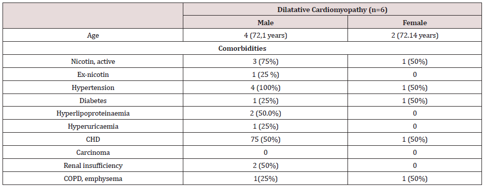
Tissue samples from explanted hearts were taken from DCM patients undergoing routine cardiac surgery (cardiac transplantation) at the Department of Thoracic, Heart and Vascular Surgery, University Hospital of Goettingen, Germany. The biopsies were taken from 4 male (average age; 72+2 years) and 2 female DCM patients (63+1 years). The study population included cardiac transplant patients suffering from endstage DCM (NYHA III-IV). Table 1 presents summarized data of the basic patient characteristics.

Table 1:Baseline characteristics of the study population.

Lupinepublishers-openaccess-cardiology

Continuous variables are presented as mean + standard deviation. Categorical variables are presented as an absolute percentage. Abbreviations: CHD: Coronary Heart Disease, COPD: Chronic obstructive pulmonary disease

Scanning electron microscopy

In order to reveal the morphology of the explanted hearts, scanning electron microscopy was performed in all tissue samples. Specimens taken from the endocardium were fixed for 6 hours in a solution containing 2.5% glutaraldehyde and 0.2m Mol cacodylate. Afterwards, samples were dehydrated in a series of increasing concentrations of alcohol. After critical point drying, all samples were sputtered with gold-palladium. Samples were visualized using the digital scanning microscope (Zeiss DSM 960, Germany).

Results

Scanning electron microscopical findings

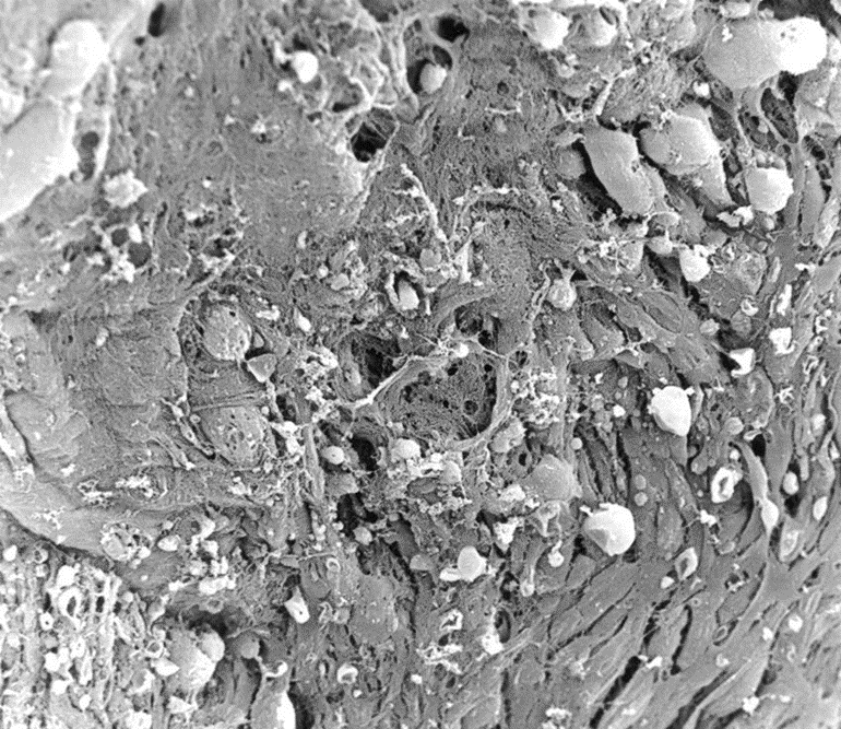
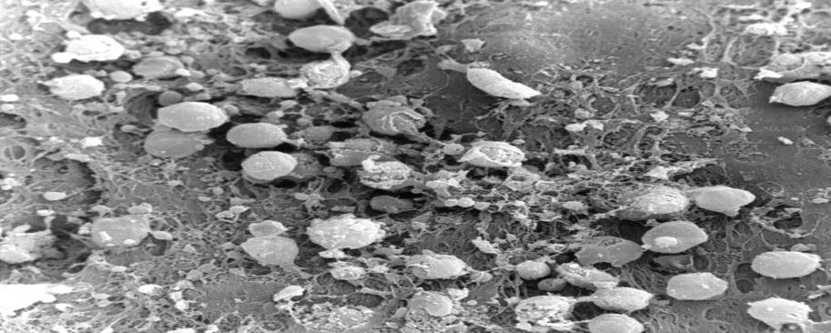
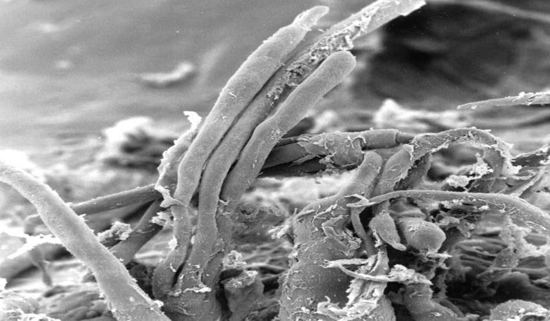
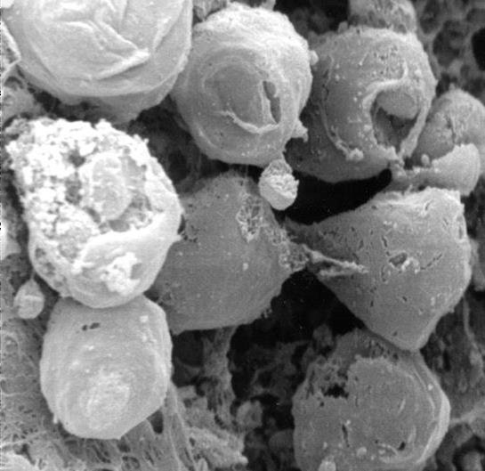
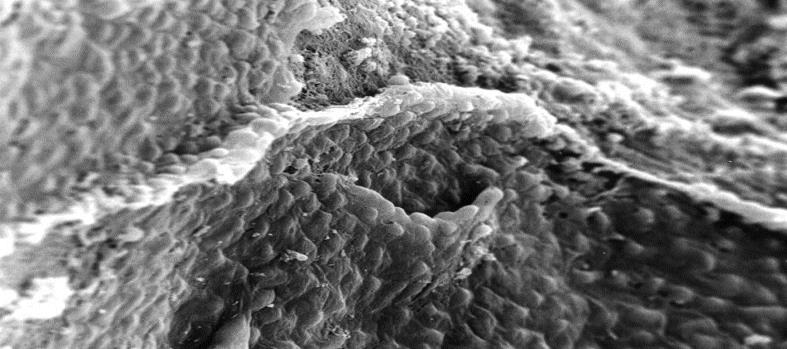
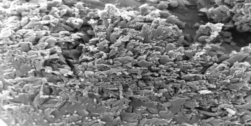
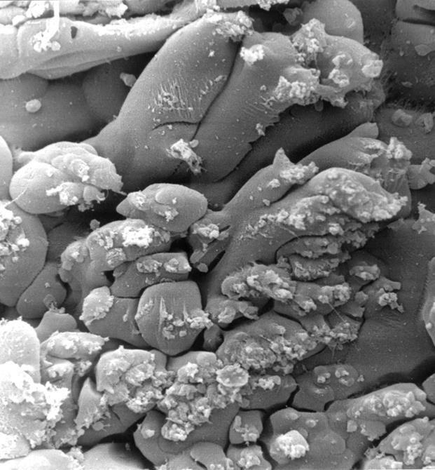
In all the tissue materials investigated, the endothelial cells were mostly desquamated. The remaining endothelial cells showed a loose binding to each other. At higher magnification, the endothelial cells appeared swollen and took on a foamlike appearance. The endothelial cells also showed similar metaplasia and loose binding to each other as in other endocardium parts. In addition, we found diffusely distributed sites of columnar endothelial cell formations in all tissues, some of which had lost their intercellular junction (Figures. 1,2). In such islets the endothelial cells had, in places, agglomerated to form columnar structures (Figure 3). These structures obviously represent a transitional form to Lambl’s excrescences. The Lambl excrescences occurred in a filiform and a lamellar form. Often the excrescences did not have an endothelial coating (Figure 4). Hyperplasia of the collagen fibers was clearly visible in such excrescences in different layers. The ratio of filiform to lamellar forms was approximately 80% in all specimens. Very often, crater-like „punched“ defects were visible on the surface of the myocardium, which showed an endothelial coating at higher magnification (Figure 5). On such altered endothelial cells, micellelike structures were visible (Figure 6). At higher magnification the micelle-like structures are better visible (Figure 7).

Figure 1: Electron microscopical view of endocardium of explanted hearts with DCM, 500 x magnification: desquamation of the endothelial cells with a loose binding to each other.

Lupinepublishers-openaccess-cardiology

Figure 2: Electron microscopical view of endocardium of Hearts with DCM, 1000 x magnification: at higher magnification, the endothelial cells appeared swollen with a foamlike appearance.

Lupinepublishers-openaccess-cardiology

Figure 3: Electron microscopical view of endocardium of heart with DCM, 200 x magnification: filiform Lambl’s excrescences.

Lupinepublishers-openaccess-cardiology

Figure 4: Electron microscopical view of endocardium of heart with DCM, 3000 x magnification: dendothelial desquamation on Lambl’s excrescences.

Lupinepublishers-openaccess-cardiology

Figure 5: Electron microscopical view of endocardium of heart with DCM, 330 x magnification: crater-like„ punched“ defects.

Lupinepublishers-openaccess-cardiology

Figure 6: Electron microscopical view of endocardium of heart with DCM, 200 x magnification: micelle-like structures.

Lupinepublishers-openaccess-cardiology

Figure 7: Electron microscopical view of endocardium of heart with DCM, 1000 x magnification: higher magnification of micelle-like structures.

Lupinepublishers-openaccess-cardiology

Discussion

Multiple etiologies of DCM, including ischemic, genetic, toxic, viral, inflammatory, autoimmune, and, last but not least, idiopathic causes are known? [11-17]. In developed countries, the incidence of DCM is 5- 10patients/100,000 (15-17) with a prevalence of 36 patients/100,000 people [18,19]. Future demographic changes such as an aging population and greater life expectancy are expected to increase the incidence of DCM even more. Thus, DCM will likely play a growing economic and medical role in the coming years. [20,21]. Pathophysiologically, DCM is characterized by dilatation and impaired systolic function of the left or both ventricles. So far, the origin of this disease is thought to be a disorder of the cardiac muscle in which myocyte weakness leads to ventricular dilatation and heart failure [22,23]. However, until now, it has not yet been considered that the origin of DCM may be endocardium-based. Therefore, we performed the presented study to investigate the morphological role of the endocardium and its role in the origin of the development of DCM. In the myocardium of DCM, we were able to detect two differing findings. On the one hand, there are changes in the endothelium and the extracellular matrix, which we know in a very similar form from aortic and mitral valves explanted due to degeneration [24]. On the one hand, there is a desquamation of the endothelial cells, which lose their junctions to each other. They are also swollen. The collagen fibers are hypertrophied and have lost their helical structure. In addition, lamellar Lambl` excrecences which we have been able to demonstrate in a similar form in degenerated aortic and mitral valves [24] are frequent, Craterlike defects of the endothelium have rarely been observed. On the other hand, we were able to detect fungal micelle-like strictures in all examined tissues, which mainly occurred at the edge of severely destroyed mycardium. The increased activation of matrix metalloproteinases in pathologically altered human endocardium emphasizes the crucial role of the extracellular matrix in the development of this disease [25-29]. The detection of involvement of the endocardium tissue in DCM suggests that pathophysiological processes similar to the degeneration of heart valves play a major etiological role in the development of DCM. Parallel to assumed pathophysiological processes of the heart valves, collagen substance transition disorders based on endothelial dysfunction could lead to pathologically increased stress on the extracellular matrix, which causes a similar response of the endothelium and the extracellular matrix as in the degeneration of aortic and mitral valve disease [30,31]. Further investigations are necessary to clarify the role of a possible infection aetiology.

Conclusion

In this study of explanted hearts of DCM patients, several morphological modifications of the endocardium and extracellular matrix are similarity of these alterations to the degeneration of aorta and mitral valves, which suggest that similar pathophysiological changes, such as possible disturbances in the synthesis of collagen fibres play an important etiological role in the development of DCM. The detection of micelle-like structures requires further clarification.

Conflict of Interest

I hereby declare that there were no financial or other interests in the execution and evaluation of this work.


Read more about Lupine Publishers Journal of Advancements in Cardiology Research & Reports Click on the below link: https://lupine-publishers-cardiovascular.blogspot.com/


Wednesday, 26 May 2021

Lupine Publishers| Review and Hypothesis about Gout

 Lupine Publishers| Advancements in Cardiovascular Research (ACR)


Introduction

The first identification about gout as clinical entity was made by the Egyptians in the year 2640 b.C. (Schwartz 2006). For many centuries it was not unveil the mystery of the origin of the illness. Gout is the unique pathology that belongs to the human race. When uric acid is deposited in the articulate tissue produce an intense inflammation, basic element in the development of gout. Interesting is the evidence of ultra sonographic signs of monosodium urate crystalline articulate deposits in 25% of clinically asymptomatic hyperuricaemic subjects (more than 8mg/ dL) [1], and approximately 9% of the joints without clinical signs of flogosis [2]. Large epidemiological studies have now demonstrated that gout is an independent risk factor for incident coronary heart disease, [3-6] heart failure, [7] stroke [8] peripheral artery disease [9] and death cardiovascular [10,11]. But, several meta-analyses have concluded that hyperuricaemia is an independent risk factor for coronary heart disease [12,13] and also, for the development of hypertension [14,15]. The standard diagnostic goal remains the identification of the typical birefringence of crystals of uric acid under polarized light microscope in the synovial fluid and in the aspirated material from the tophi [16,17]. Hyperuricaemia with or without urate deposit (modern denomination of the formerly called gout) is currently one of the most frequent dysmetabolic diseases. [18,19].

Over the last decades, to uric acid has been attributed a possible role of cardiovascular risk prevalently the left ventricular hypertrophy for an increase in inflammatory mediators, such as tumor necrosis factor alpha and activation, and the rennin angiotensin system, increase in interstitial fibrosis of the myocardium and producing endothelial dysfunction [20,21]. Uric acid being a small molecule, is able to penetrate within the vascular wall cells, stimulating inflammatory activity leading to smooth muscle cell hypertrophy and favoring the endothelial dysfunction process [22]. An increase in arterial stiffness has also been demonstrated [23]. The antioxidant effect seems to be attenuated by the increase in its plasma concentration to turn into a pro-oxidant effect when it exceeds the 6 mg / dL level. In recent years, scientific evidence has suggested the possibility of a pathophysiological correlation between the action of xanthine oxidase and the genesis of cardiovascular damage in patients with chronic hyperuricaemia with and without uric acid deposits [24,25].

Uric Acid

Uric acid is an organic heterocyclic compound. It is the endproduct of the metabolism of purine nucleotides that are the principal constituents of cellular energy stores, such as ATP, and components of DNA and RNA. Urate ions appear as monosodium urate with a solubility limit in plasma of about 6.8 mg/dL at 37°. When the crystals precipitate by excessive solute, they are coated with polypeptide or protein molecules [26,27]. Precipitation of uric acid can occur for other factors such as the state of tissue hydration, pH, cation concentrations and the presence of proteoglycans, collagen and chondroitin sulphate. Uric acid is a molecule with an effective antioxidant action (protective: intracellular localization) and a potent pro-oxidant effect (negative impact: extracellular localization) in relation to the micro-environment in which it is located [28]. This dual role has been described as the “uric acid paradox”.

Plasma Proteins

Acid-bearing drugs bind to albumin. Those with basic function are bound to globulins. They are subdivided into three fractions: alpha, beta and gamma. The first two cover transport functions, while the third includes the immuno globulins involved in the body’s defense processes.

Synovial fluid

The synovial fluid under normal conditions is a viscous light yellow and clear. The fluid contains few proteins and cells but is rich in hyaluronic acid (about 300mg/dL) synthesized by type B synoviocytes which provides high viscosity. The larger molecules, such as immunoglobulin’s and complement, are found to be in lower concentrations, made due to the physiological function of the filter. The protein content of normal synovial fluid ranges from 25-30% of the total plasma protein content, with a very lower proportion of the higher molecular weight proteins such as alpha- 2-globulin, haptoglobin and fibrinogen (which belongs to beta globulins) because the synovial membrane is impermeable to high molecular weight proteins. Fibrin and fibrinogen are normally not present in synovium. During the inflammatory process an increase of the total proteins is observed as a consequence of the increase of the permeability of the synovial vessels.

Personal Experience

Cardiovascular

About three decades ago, working as a general practitioner and cardiologist at the “Dr. Julio Méndez” Municipal Sanatorium (Buenos Aires, Argentina), hyperuricaemia was a factor of concern and discussion about its possible action on the cardiovascular system. We had no doubts about its role in the development of joint gout but we did not have certain data about its action on the arteries. The question was whether hyperuricaemia is an independent risk factor for cardiovascular disease. We knew that the uric acid is transported by very low density lipoproteins which would make them more easily precipitated, prevalently if they find an acid medium inside the arteries. Moreover, the acidity could be caused by the depolymerization of mucopolysaccharides and the presence of bivalent cations would concurrently precipitate the circulating proteins (uric acid carriers), we thought that with the contact of uric acid with chondroitin sulfuric acid of the arteries (similar to the articulate cartilage component), could develop the inflammatory cascade. Hypertension frequently coexists and contributes with hypoxia facilitating the inflammatory action of uric acid and lipoproteins.

Therefore, uric acid would not in itself be a coronary risk factor, but would act in combination with hypertension, hyperlipidaemia and/or hyperglycaemia. During the time I drove of the Ischemic Heart Disease team, in order to know the relationship between hyperuricaemia and cardiovascular disease, 40 patients both gender was enrolled with ischemic heart disease studied with electrocardiogram, cycloergometry, echocardiogram and coronary angiography. The male group (35) had a mean age of 56 years, with a history of myocardial infarction [27] and myocardial ischemia [8], and in the female group (5) had a mean age of 64 years only with myocardial ischemia. All patients had a hyperuricaemia 10% higher than normal level for each gender. Only one patient had hyperuricaemia, the rest of the sample had two or more cardiovascular risk factors: high blood pressure (55%), hereditary history of cardiovascular disease (50%), smoking (43%), Obesity (38%), hyperlipidaemia IIa (38%), hyperlipidaemia IV (38%), diabetes mellitus (30%), gout (39%), and sedentarism (18%).

Conclusion

Apparently hyperuricaemia alone not be a risk factor but, in combination with hypertension, hyperlipidaemia and/or hyperglycaemia, could develop the inflammatory cascade acting on the endothelium. No woman had myocardial infarction or articulate gout. The level of uricaemia was not related to the severity of cardiovascular disease. Interestingly, the group older than 60 years with hyperuricaemia had greater risk. Hyperuricaemia, hypertension, hyperlipidaemia and age greater than 60 years in males would be associated with a higher severity of cardiovascular risk, and would favor the development of atherosclerosis [29].

Joints

There were also several doubts about the development of joint gout in patients with serum acid levels below 7mg/dL, and why the lack of clinical symptomatology in other patients with severe reduced glomerular filtration rate with exceptional levels of uricaemia such as 17mg/dL. Monosodium urate is deposited only if the medium is acid as occurs in the urine, and in contact with the chondroitin sulfuric acid in the joints. A possibility of producing a gouty acute attack with non-high levels of uricaemia could indicate that the uric acid introduced into the joint would not have an element that would function as a shock absorber. In the same line of thought, it was possible to arrive at the conjecture that during the acute inflammation, and for the benefit of the local vasodilatation, there could be a passage of elements that had a damping action. The question was: What are the elements that are not found in large quantities in normal synovial fluid and increase during inflammation? It was first thought of the higher molecular weight plasma proteins as the alpha and beta globulins as well as fibrinogen. An investigation was carried out in order to clarify these doubts It was decided to take samples of the synovial fluid of the knee from gouty (20) and non-gouty (20) patients. Hyaluronidase to synovial fluid was added in order to decrease its own viscosity. Once liquefied, a sample was taken to perform a protein electrophoresis. Technically it was not possible to measure fibrinogen intraarticulate. The next step was to compare the amounts of alpha, beta, and gamma proteins of the two patient groups. It was observed that in patients with acute joint gout fractions of alpha and beta globulins were always significantly lower than those of health patients. (Unpublished data) We hypothesized that patients with less alpha and beta globulins in the synovial liquid were more sensitive to developing joint gout.

Conclusion

The acute attack of gout, without a high uric acid level, could be caused by the low presence of high molecular weight proteins in the synovial fluid. If the disease is heritable, the reduction of alpha and beta proteins could be inherited. The self-limitation of the gouty attack could occur by the entry of proteins of high molecular weight, secondary to the vasodilatation produced by the acute inflammation, which would function as buffer. Other proteins and/ or substances could also be buffers such as fibrinogen.

Read More About  Advancements in Cardiovascular Research (ACR) Click on Below Link:

https://lupine-publishers-cardiovascular.blogspot.com/


Thursday, 18 April 2019

Journal of Cardiology Research- Lupine Publishers

Coronary Cameral Fistulae a Scarce Entity by  Ranjan Modi in ACR - Lupine Publishers


Coronary artery fistulae are communication between coronary arteries and other structures like cardiac chamber (coronary cameral fistula) or a vein (coronary arteriovenous fistula) [1]. Coronary fistulae account for 0.2 to 0.4% of the congenital cardiac abnormalities. Spontaneous closure occurs in 23% of small fistulae, primarily those arising from left coronary system in which conservative management may be appropriate [2]. Though surgery still remains the main stay of treatment, interventional closure seems to be an appropriate choice. We present a 20 day old male baby born to non consangious marriage. The neonate was full term delivery in the hospital. Prenatal history was uneventful with no history of any drug intake during pregnancy and regular antenatal care visits with complete immunizations. The baby presented with history of feeding difficulty since birth, no history of recurrent chest infections, no suck rest suck cycle and no cyanosis. The patient had tachycardia with heart rate of 140/min and tachypnoea with respiratory rate of 36/min on examination, blood pressure of 90/60 mmHg - Lupine Publishers.


https://lupinepublishers.com/cardiology-journal/fulltext/coronary-cameral-fistulae-a-scarce-entity.ID.000101.php

https://lupinepublishers.com/cardiology-journal/pdf/ACR.MS.ID.000101.pdf 
https://lupinepublishers.com/cardiology-journal/abstracts/coronary-cameral-fistulae-a-scarce-entity.ID.000101.php 

For more Lupine Publishers go through the below link
https://www.lupinepublishers.com/


For more Cardiology Open Access Journal articles Please Click 
Here: https://www.lupinepublishers.com/cardiology-journal/