Showing posts with label Journal of Infectious Diseases. Show all posts

Friday, 17 June 2022

Lupine Publishers | Relation of Falooda Ice Cream Loving with Protein in Urine

 Lupine Publishers | Journal of Immunology & Infectious Diseases


Abstract

The colour of urine is yellowish that contain toxic substances that is elevated from the body through urethra. Protein perform very important function in our body that is necessary for building up hair, nail. We obtained protein from the diet that we eat in our daily life such as meat, eggs and milk and great source of protein. But high levels of protein in urine may be harmful for body and cause many diseases that normally occur due to improper function of kidneys. The people that have abnormal levels of protein may be suffering hypertension, Amyloidosis and heart diseases. Eating too much falooda ice cream cause abnormal levels of protein because it contains dairy products. Urinalysis is used to detects the ingredients present in the urine. It was conclusive that falooda ice cream does not correlate with proteinuria.

Keywords:Amyloidosis; Lupus; Protein; Ice cream; Frothy Urine

Introduction

Urine is yellowish shape fluid or liquid waste that contains 95%water and reminder contains of hormones ammonia, and proteins, salt and minerals. Urethra is present that discharged the urine from body. If colour of urine is pale yellow to gold, then everything is normal. Protein performs many important functions in our body such as body needs protein to build hair, nail, bone and muscles [1]. When blood pass through the kidneys then waste products and excess water are filter out. If high amount of protein is present in urine this is called proteinuria. There are a lot of causes of abnormal amount of protein in urine such as stress, fever, dehydration, strenuous exercise. Those people that elevated protein in urine than normal have many medical conditions like Amyloidosis, heart disease, hypertension, lupus, multiple myeloma. Similarly, symptoms related with proteinuria are shortness of breathing, Dry skin, fatigue, frothy urine, trouble sleeping, hiccups and vomiting [2,3]. Urinalysis is a test for detecting the protein in urine. Urine sample is tested with dipstick. When large amount of protein is present in urine then colour of strip paper will be change. Higher amount of protein in the urine is a sign of chronic kidney disease. There are some ways that one can avoid by adopting them. By avoiding dairy products, canned food, by eating 5 to 6 serving of fruits, less eating of meat, fish and eggs. When people eat too much meat eggs and dairy products so there is a chance of proteinuria. Because of all of these are source of protein. Too much protein in urine may damage kidneys and proximal renal tubules. Normally up to 145mg total protein found in urine per day. More than 250mg per day is known as Frank proteinuria. In normal pregnancy, renal blood flow increased, and additional protein lost in urine [4]. This protein loss should not be exceeding 270 mg in 24 hours. If more than 270 mg per day is found, then there is development of preeclampsia. Falooda ice cream is a sweet drink made with ice cream, kulfi, sabja seeds and boiled semiya. This is mostly found in every street corners of India and it is very cold and sweet drink. There are many healthy drinks for summer such as aim panna, Punjabi lassi and masala chaos. As ice cream available in many flavours similarly, falooda is available in many flavours such as kesar falooda and rose falooda. Falooda ice cream is most popular in Pakistan and India. People drink it during summer when sun burns everything with its high intensity. Falooda ice cream has very cooling properties because sabja seeds present in it. The colour of falooda ice cream may be rose or orange colour. There are other several ingredients that are present in it. Such as dry fruits, kulfi, Almonds, pistachios, semiya and boiled milk. Falooda ice cream is beneficial for health because too much healthy ingredients are present in it. So, people should eat it at least 3 times per week. Falooda ice cream provide cooling effects to our body and brain. The objective of present study was to associate the falooda ice cream with urine protein [5].

Methodology

Urinalysis is used to detects the ingredients present in the urine. But here we performed this test to detect the levels of protein into urine. For which take a container put in which fresh sample of urine and dipped the strip into container for 4 seconds [6-8]. After that hold strip the colour of strip will be change. Matched strip with the given labelling chart and notes the value of protein.

Project Design

There were 100 participants or subjects in this survey their participation were from the Bahauddin Zakariya university. The main goal of this survey was asked to the students about the ice cream whether this is harmful or beneficial for health [9]. A few of students said that if it is eaten in high quantity this can be harmful because of healthy ingredients. But mostly students were agreed to this point that it can be beneficial, and they also said, that excess of everything is bad so people should eat this food or drink in limited amount.

Results and Discussion

This table shows that males that have negative value are 19 % while those males who have positive value are also 19%, All of these males love the falooda ice cream. Similarly, females having negative value are 13% and those females which have positive value are totally 8% all these are also falooda ice cream loving [10]. These males and females said, that falooda ice cream is energetic drink especially during summer (Table 1). This tables shows males which have negative value are 11% they do not like ice cream. While those males which have positive values are 10% these males also not falooda ice cream loving. While females having negative values are 12% that do not like falooda ice cream. Similarly females having values positive are only 8%. About their view of point by eating falooda ice it can cause many diseases such as obesity because it contains too much dairy products (Table 2).

Table 1: Relation of falooda ice cream loving to the protein in urine

lupinepublishers-openaccess-immunology-Infectious-disease-journal

Relation of not falooda ice cream loving with protein in urineResults of Changing Hyperparameters and the Elapsed Times.

lupinepublishers-openaccess-immunology-Infectious-disease-journal

Conclusion

It was concluded that there is no relation between falooda ice cream loving and protein in urine.

Read More About Lupine Publishers Journal of Immunology Please Click on Below Link:
https://lupine-immunology-infectious-disease.blogspot.com/

Friday, 13 May 2022

Lupine Publishers | Is There any Type of Affiliation between Falooda Ice Cream Loving and Urine pH?

 Lupine Publishers | Journal of Immunology


Abstract

Urine pH measured the alkalinity or acidity levels of urine if value of urine pH is less than 7 it may be considered acid if value of urine pH is more than 7 then it considered alkanity. Urine pH levels is directly linked with the diets such as eating citrus fruits and legumes cause high levels of urine pH. There are some drugs available that treat urinary infection such as kanamycin and neomycin. Falooda ice cream contains healthy ingredients but overeating it can cause abnormal levels of urine pH. Falooda ice cream has many health benefits such as gives energy to the body and maintains glucose level.

Keywords: Kanamycin; Pulmonary Ventilation; Ice cream; Sabja and Semeia Seeds; Urinary Tract Infection

Introduction

urine pH is defined as the measurement of alkalinity or acidity of urine and ranges from 1 to 14 [1]. p stands for power and H for hydrogen means measurement of concentration of hydrogen ion in the body. If value of pH is 7 then it is neutral below 7 it considered acidic and above 7 it considered basic. Person with high value or range of urine pH is more alkaline so it causes urinary tract infections and kidney stones [2-3]. Kidney take blood from the body in which uric acid and other harmful waste are present after filtering send it into urine and it leave your body. If there is too much uric acid in urine it can cause formation of crystal in joints and this leads to painful problem called gout [4]. People who eat citrus fruits, legumes, and vegetables have high level urine pH. Bacteria in our body play an important role they protect us from infection, some bacteria are harmful for us because they are responsible for urinary tract infection, they split urea into ammonia and make urine more alkaline. Renal stone formation is connected to urine pH. There are some medications and diets are available that change the PH of urine because these medications prevent formation of kidney stones. Some drugs such as streptomycin, kanamycin and neomycin are available that treat urinary tract infections [5]. During sleeping pulmonary ventilation decreased which causes respiratory acidosis, so first waking urine is more acidic. People who eat diets rich in cranberry juice and meat have urine acidic. PH test paper measures alkaline acid condition of urine. If reading is below 7 it is acidic if reading is on higher scale it will be alkaline. Urine pH is related with diets so by eating proper diets one can maintains urine pH. Falooda ice cream is a refreshing chilled drink or dish which is pleasing to both brain and body [6]. Falooda ice cream is famous in India and Pakistan due to its cooling properties. Ingredients that makes tasty and yummy to the falooda ice cream are cream, basil seeds, semeia, dry fruits, falooda and dairy products. Due to its cooling properties people like it during summer days when sun burns everything. The preparation is very easy. Boiled milk, soaked sabja seeds and semiya. Add milk, rooh afza, cream, dry fruits in blender and blend them for 5 to 10 minutes. Then pour this blended mixture into tall glass then add pineapple chunks, sugar syrup, jelly and served it immediately. Falooda ice cream provides energy because milk is present in it and it is source of vitamins and minerals. Mostly people do not like it because it contains too much sugar which cause heart attack and other diseases and high blood pressure. The people who want to get energy should drink or eat it 3 to 4 times per week [7]. Due to presence of sabja seeds it is very useful for skin and health. People with high levels of sugar should not eat falooda ice cream because there is lot of glucose or sugar in it. People also served it to the guest during summer because everyone wants to drink or eat it in the hot days. The objective of present study was to correlate the falooda ice cream loving with urine pH.

Materials and Methods

Gloves, strip, container, fresh urine sample and labelling chart are the following materials that are required during the urine tests. First take a sample of urine into container and container should be clean then hold a strip and dipped it into urine for 1 to 2 seconds until colour of strip will change than before. Picked up the labelling chart and matched it with the colour of strip it will tell the value of pH. whether pH. of urine above 7 or below 7. If value of pH. equal to 7 then it will be neutral [8].

Project Design

For checking the effects of falooda ice cream on the urine pH we designed this project. Near about one hundred students who had completed this survey and give the opinions about the falooda ice cream. These subjects were undergraduate and belonged to the university of Multan Bahauddin Zakariya [9]. Some students said, that falooda ice cream is a light food by eating it during summer one can keep the mind and body cool. But there were a few students who are not agreed about the benefits of falooda ice cream. They said, falooda ice cream is a fatty food and has high levels of sugar due to which it may be harmful for health by eating too much. This table shows urine pH level of males and females who are falooda ice cream loving so Males having value of pH 5are 0% and other Males having value of pH 6, 7, 8, 15%, 6%, and 3% respectively. Similarly, females who have values of pH5 in their urine are 4%. those females having values 6, 7, 8, are 24%, 8%, 4% respectively (Table 1). This table shows the values of males and females who do not love the falooda ice cream. Males which have value of pH. 5 in their urine are just 2% and other those have value 6, 7, 8 and are 3%, 4%, 2% and respectively. And females with value of 5 pH. are also 4%. Similarly, those females who have value of pH. 6, 7, 8, are 19%, 1% and 1% respectively (Table 2).

Table 1: Affiliation of Urine pH to the people who love falooda Ice cream.

lupinepublishers-openaccess-immunology-Infectious-disease-journal

Table 2: Affiliation of Urine pH to the people who do not love falooda Ice Cream.

lupinepublishers-openaccess-immunology-Infectious-disease-journal

Conclusion

This was concluded that there is no affiliation between falooda ice cream loving and urine pH.

Read More About Lupine Publishers Journal of Immunology Please Click on Below Link:
https://lupine-immunology-infectious-disease.blogspot.com/

Saturday, 19 March 2022

Lupine Publishers | Microbes and ReA

 Lupine Publishers | Journal of Immunology & Infectious Disease


Mini Review

Reactive arthritis (ReA) belongs to a group of diseases under spondyloarthritis (SpA). It is a systemic disease triggered by an infection at a distant site, other than joint. The infectious triggers are mostly genitourinary, gastrointestinal or respiratory tract. An association has been established between arthritis and micro-organisms like Shigella, Yersinia, Salmonella, Campylobacter and Chlamydia [1]. The role of microbiomes in inflammatory arthritis and autoimmunity has been proposed decades ago. In genetically susceptible patients, infection activates Th17 mediated immune response which causes arthritis and systemic inflammation [2]. The interplay between HLA B27 gene and microorganism has also been studied for the pathogenesis of reactive arthritis. HLAB27 is a major histocompatibility complex, strongly associated with SpA. Around 45-90% of patients with ReA had HLA B27, which predicts the progression and severity of disease [1]. Several theories have been postulated for the pathogenesis of ReA. These include molecular mimicry, arthritogenic peptide, Toll like receptors (TLRs) [2]. The virulence factors such as invasion, protein tyrosinase, cytoxin, adhesion present in microbes facilitates their persistence in the gut.

The bacterial epitope presented by MHC class I is recognized by cytotoxic T lymphocytes. T cells then cross react with self-antigen derived peptides [3]. Through molecular mimicry between bacterial protein and HLAB27, HLA B27 presents its own peptides similar to bacterial antigens leading to autoimmunity [4]. Other genes like HLA-B39, HLA-B60, and HLA‐DR1 are implicated in HLA B27 negative patients. Bacterial LPS helps in virulence of bacteria and modulates immune system. The intraarticular LPS stimulates proinflammatory cytokines like TNFα, IL1 and IL6, enhances lymphocyte entry into synovium, activation of IL8 from chondrocytes leading to enhanced leukocytes recruitment in synovium and persistent phagocytosis. Chondrocytes produce collagenases, nitric oxide and proteases which lead to degradation of cartilage [3]. Though there are clear evidence of association between various pathogens and the occurrence of inflammatory arthritis, it would not be an overstatement to say that the exact pathogenetic mechanisms remain unclear. Clinical observations have suggested that the clinical features and natural course of the inflammatory arthritis varies to some extent depending on whether the trigger is genitourinary or gastrointestinal. Precision medicine in the subject would require further research on molecular mechanisms that will help to individualize the treatment of these patients.

Read More About Lupine Publishers Journal of Immunology Please Click on Below Link:
https://lupine-immunology-infectious-disease.blogspot.com/

Friday, 28 January 2022

Lupine Publishers | A New Multidisciplinary Strategy for Using Novel Tunable Immune Response (TIR) Biomaterial Platform Technology as the New Alternative for Treating Immuno-based Diseases

 Lupine Publishers | Journal of Immunology & Infectious Diseases


Opinion

Innovation in technology is the key element to modernize medical treatment and will have a significant impact on all phrases of our lives, particularly the healthcare upon aging population. Many diseases have the common root on immunology, particularly inflammation, and the current advanced immunotherapy has all based on the biological approach like cytokines to alter the immune response for achieving better therapeutic outcomes. At here, I would like to propose a new multidisciplinary strategy as a new alternative for treating immuno-based diseases via the use of a novel family of tunable immune response (TIR) biomaterial platform technology. The theoretical base of this new alternative strategy is to modulate the immune cell (i.e., macrophage) associated inflammation via the feeding of different types of L-arginine (Arg)-based biodegradable polymers to macrophage to control the direction of the dual Arg metabolic pathways by macrophage. It is well-known that macrophage can uptake Arg and metabolize it either through the iNOS or Arginase pathways. The iNOS pathway would lead to the production of NO, and hence cytotoxic (M1 pro-inflammatory phenotype), while the arginase pathway would lead to the formation of polyamines and L-proline, and hence tissue regeneration/repair (M2 anti-inflammatory phenotype). It is my opinion that the use of the macrophage’s Arg metabolic pathways to tune immuno-responsive character of macrophage is a better strategy than the conventional use of cytokines for either classical or alternative macrophage activation. This is because

I.Cytokines have a much shorter life span and can be easily deactivated in real clinical applications, while the Arg-based Tunable Immune Responsive (TIR) biomaterial platform technology strategy can have a very long lifespan and shelf life, i.e., lower healthcare cost and more sustained efficacy.

II.The Arg-based TIR biomaterials can be formulated into a variety of physical forms like nanoparticles, fibrous membranes, melt-spun fibers, 3D microporous hydrogels, films, coating etc., while the cytokines can’t be formulated into any form, except their native form. The capability of having a variety of physical forms can definitely facilitate and expand their clinical applications. For example, TIR-based nanoparticles can couple with drugs to provide a synergistic therapeutic effect toward disease treatment. Such an integration of macrophage-proactive TIR-biomaterials with drugs will open a new dimension of immunotherapy.

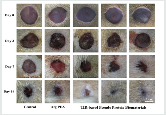
This new family of TIR-capable biodegradable biomaterials have a nickname, “TIR-capable Pseudo-protein biomaterials” because they are built from 3 building blocks, amino acids, alcohols and acids, and possess both proteins and non-protein properties. Because of the enormous variety of these 3 building blocks, these pseudo-protein biomaterials can be tailored designed for meeting specific clinical needs, hence have become a platform biomaterial technology. The 1st generation of these new pseudo-protein biomaterials has been licensed and approved as the coating material for a new drug-eluting stent (Slender IDS®) for human commercial use in Europe in 2016 to treat the blockage of coronary artery. Very recently, Chu has teamed up with the Institute of Chinese Medicine in the Hong Kong Baptist University to pursue the use of the newly developed pseudo-protein nanotechnology as the delivery vehicles for Chinese medicine like gambogic acid to treat the most challenging and difficult triple negative breast cancer (TNBC). Their animal data are very encouraging and promising. This infusion of a new western pseudo-protein nanotechnology from Cornell with ancient Chinese medicine is believed to be able to modernize Chinese medicine-based treatment significantly for a far better therapeutic efficacy and lower toxicity. My lab has recently also collaborated with the National Engineering Research Center for Tissue Restoration and Reconstruction and School of Materials Science and Engineering in the South China University of Technology, Guangzhou to test the feasibility of this new TIR-based pseudo-protein biomaterials for treating diabetic burn wounds in diabetic-induced mice as diabetic patients are well-known to have the inherent difficulty to heal their wounds timely. Burn wounds are well-known for their excessive inflammatory character. Therefore, a combined diabetic burn wound appears to provide the most challenge animal model to test the potential of my lab newly developed strategy of TIR-based pseudo protein biomaterial technology for immunotherapy of a variety of diseases. The animal trial data show that this novel TIRbased pseudo-protein biomaterials can indeed timely promote the healing of the 3rd degree burn wounds in diabetic-induced mice, and the mechanism behind this very promising animal data is the TIR-based pseudo-protein biomaterials induced transformation of the highly inflammatory M1 macrophage phenotype in the diabetic burn wound bed into anti-inflammatory M2 macrophage phenotype. Figure 1 shows the wound healing performance of a diabetic 3rd degree burn wound mice after the topical treatment by my lab TIR-based pseudo-protein biomaterials.

Friday, 21 January 2022

Lupine Publishers | Does there is Relevancy between Falooda Ice Cream loving and Urine Nitrites?

 Lupine Publishers | Journal of Immunology & Infectious Diseases


Abstract

Naturally source of nitrites are vegetables like cabbage, spinach and vegetables, it is form of nitrogen. High levels of nitrites in urine is sign of urinary tract infection. Some bacteria are responsible for urinary tract infection such as proteus and klebsiella, but doctor also prescribed some antibiotics against bacteria. Women are more suffering in urine tract infection than men. Positive test for nitrites is called nitrituria. For measuring the levels of nitrites in urine urinalysis is done. Falooda ice cream looking beautiful with different layers that make it healthy drink but a few of people try to avoid by eating it because it contains high levels of sugar.

Keywords: Nitrites; E. Coli; Antibiotics; Urethra; Diabetes; Urinary tract infection

Introduction

Nitrites is form of nitrogen, which contains two oxygen atoms. Nitrites is naturally found in vegetables like cabbage, celery, carrot and spinach. The presence of nitrites in urine may be harmful mean sign of urinary tract infection. The presence of nitrites in urine is due to bacterial infection in urinary tract. Urinary tract infection can occur in urethra, kidneys, ureters and bladder. Some bacteria have ability to convert the nitrates into nitrites due to presence of specific enzyme. The presence of nitrites in urine can be diagnosed with urinalysis test. The bacteria that are responsible for urinary tract infection, proteus, klebsiella, pseudomonas. But most common bacteria are E. coli in which urease enzyme is present that acidifies the urine. The symptoms of urinary tract infection include blood in urine, cloudy urine, strong smelling urine and burning with urination. Urinary tract infection is most common in pregnant women and may be dangerous. Urinary tract infection can cause premature delivery, headache, abdominal pain and high blood pressure in pregnant women if left untreated. If test for the nitrites in urine is positive it is called nitrituria. While negative nitrites test happens with dilute urine or low colony forming unit. A urinary tract infection is most common in women aged 20 to 50 years than man. There are many ways that one can prevents from nitrites in urine or urinary tract infection. Such as by drinking plenty of water bacteria can be flash out that is present in urinary system. Cranberry juice and apple cider vinegar also treat the urinary tract infection. Doctor also prescribed some medication for the treatment of nitrites in urine. Doctor prescribed antibiotics on the basis of what kind of bacteria cause infection. Patient should also take enough sleep and adopt personal hygiene. There are many drinks and foods that keep the body cool and fresh, similarly falooda ice cream is one of the most popular drink that sweet in taste and delicious. Falooda ice cream is a rich source of energy, one glass contains 218 calories. This is sweet dish that served to the people during hot days. This is made in tall glass which give beautiful appearance. The main ingredients of falooda ice cream sabja or basil seeds and semeia that is good for skin and hair have cooling properties. Other ingredients that are present in it like cream, milk, falooda, rooh afza and sugar. Sabja seeds help in weight loss and lowering the high blood pressure. Falooda ice cream has many health benefits, it provides energy to the body, keep body cool during hot days. There are many flavours of falooda ice cream like rabdi, rose syrup, royal, and kesar. People can make it at home easily. First, soaked basil seeds, dry them also soaked semeia seeds, boiled milk. Put them into blender and blend them. Then pour into tall glass add dry fruits, falooda, Almonds, ice cream and rooh afza and served it. If you want energy, then drink cold and sweet falooda ice cream because it contains vitamin and minerals due to presence of dairy products. People should drink or eat it 3 to 4 times per day. But patients of diabetes should avoid it because it contains too much glucose or sugar and cause heart disease and diabetes. The objective of present study was to correlate the falooda ice cream with urine nitrites [1,2].

Materials and Methods

For measuring the levels of nitrites in urine urinalysis is done. First of all, person will need an empty and clean plastic container so that filled it with fresh sample of urine. A strip and gloves are also required. By wearing the gloves dipped the new strip into container and stirred it into urine sample for 2 seconds. The colour of strip will change than before as it will dip into container. Draw out strip from the sample so that measured the levels of nitrites into urine. In the last discard the gloves, strip and container [3-6].

Project Design

There were 100 subjects who completed this survey, but main goal of this survey was asked to the students of Baha Uddin Zakariya university about falooda ice cream loving. Mostly students said that by eating falooda ice cream one can prevent from the hypertension and get too much energy. But the subjects who disagreed it said, that falooda ice cream lead to obesity and cardiovascular diseases (Table 1) [7].

Table 1: Relation between falooda ice cream and urine nitrites to the people that love falooda ice cream.

lupinepublishers-openaccess-immunology-Infectious-disease-journal

Table 2: Relation between falooda ice cream and urine nitrites to the people that do not love falooda ice cream.

lupinepublishers-openaccess-immunology-Infectious-disease-journal

This table shows males that have negative value of nitrites in their urine are 27% which are falooda ice cream loving and those who have positive value of nitrites are 13 % which are also falooda ice cream lover. Similarly, females having negative value are 22% and females with positive value are 8% which like the falooda ice cream. About the opinions of these males and female’s preparation of falooda ice cream is very easy and not cost expensive dish having many ingredients that prevent us from different diseases (Table 2).

This table shows that males with negative values are 17% and with positive values are only 4 % All of these males are not loving the falooda ice cream. Similarly, females having negative values are 6 %which are not like falooda ice cream. While females having positive value of nitrites in their urine are just 3% which are not loving falooda ice cream [8-10].

Conclusion

This was concluded that there is no relevancy between urine nitrites and falooda ice cream loving.

Read More About Lupine Publishers Journal of Immunology Please Click on Below Link:
https://lupine-immunology-infectious-disease.blogspot.com/

Monday, 18 October 2021

Lupine Publishers | The Effect of Some Natural Fermenters on Growth, Heamatology and Escherichia Coli Infection Control in Weaned Pigst

 Lupine Publishers | Journal of Immunology & Infectious Diseases


Abstract

The present study was carried out to evaluate the activities of the Lactobacillus species (LAB) isolated from “burukutu” and pig hindgut with patented probiotics on body weight, hemogram and E. coli infection control in weaned pigs. Sixty (60) large white breed of weaned pigs, aged between 30-35 days old with an average weight of 5 kg. They were randomly allotted to five replicate groups with six pigs each. Group A1 were administered 0.8 ml (6x10⁶cfu) of lactobacillus isolated from “burukutu” and inoculated into wet feed, Group A2, 0.8 ml lactobacillus from “burukutu” (6x10⁶cfu) inoculated in dry feed. Group B1, had, 0.8 ml (6x10⁶cfu) of lactobacillus bacteria isolated from hindgut of pigs and inoculated into wet feed; Group B2, 0.8 ml (6x10⁶cfu) dry feed containing lactobacillus from Pig hindgut. Group C1, 0.8 ml (6x10⁶cfu) fermented liquid feed inoculated with Bacillus subtilis and Bacillus pumilis; Group C2, 0.8 ml (6x10⁶cfu) dry feed inoculated with Bacillus subtilis and Bacillus pumilis. Group D1, 0.8 ml (6x10⁶cfu) fermented liquid feed inoculated with Lactobacillus acidophilus; Group D2, 0.8 ml (6x10⁶cfu) dry feed inoculated with Lactobacillus acidophilus. Group E1, fed wet basal diet; Group E2, fed dry basal diet. Animals in the various treatment’s groups were infected orally with 6 ml (1x10¹⁰cfu·ml-1) of E. coli bacteria. Weekly body weight, hematological values and faecal shedding of E. coli post infection were evaluated. WBC, lymphocytes, neutrophils and total protein values were significantly high (p<0.01) in the treatment groups from week 4 to week nine. The wet form of the feed in each treatment produced significantly heavier animals (p<0.01) with an average weight of 25 kg. All dietary treatments showed significant reduction (p<0.05) in E. coli counts. The present study has demonstrated that lactobacillus species isolated from “burukutu” and pig-hindgut was able to improve weight gain and performance of weaned pigs. Therefore, isolated bacteria from “burukutu” and pig hindgut can serve as potent probiotics and growth promoters.

Keywords: Burukutu; Pig Hindgut; Lactic Acid Bacteria; Escherichia Coli Infection Body Weight; Haematology

Introduction

The neonatal period is a critical time in piglet ontogeny, because the gastrointestinal tract (GIT) and immune system are not yet fully developed [1]. These in efficiencies make piglets vulnerable to invasion by pathogenic microorganisms and with their low resistance to diseases affect the whole process of individual development [2]. Supplementation with Lactic Acid and Bacteria (LAB) in neonatal piglets can regulate the formation of the piglet gut microflora, thus benefiting the health of piglets [1,3] Currently, there is growing attention on fermented pig feed because it improves growth performance [4-6] and influence the bacterial ecology of the gastrointestinal tract (GIT), in particular members of the family Enterobacteriaceae, including Salmonella spp [6,7]. A considerable amount of research has been conducted to select beneficial strains of lactic acid bacteria for fermented liquid pig feed production for example, [6,8] tested 146 strains of bacteria for their ability to control Salmonella. Bacterial species often used for inoculating feed to produce fermented liquid feed are Lactobacillus plantarum and Pediococcus spp [8]. In recent years, reports have described the beneficial effects of LAB, such as regulation of the intestinal microflora, inhibition or prevention of pathogens in the gastrointestinal tract, enhancement of intestinal mucosal immunity and maintaining intestinal barrier function [9,10]. The aim of this study, therefore, was to determine the invivo effect of some selected fermenters, on weight gain and haemogram of weaned pigs.

Materials and Methods

Study Area

The study was carried out in Samaru Campus of A.B.U, Zaria, Sabon Gari Local Government Area of Kaduna State and Located on Latitude (11°11´N, 07, 38´E, 686m above sea level) in the Northern Guinea Savannah with temperature ranging from 13.8° to 36.7° C. and an annual rainfall of 1092.8mm [11]. Agriculture in Zaria can be divided into two types: Rainfall type (from May to October) and irrigation farming in the dry season (from November to April). Dry season farming is the second most prevalent Agricultural activity in Zaria with vegetables being the common produce, but in some cases, fruits are sandwiched among cereal crops [12].

Ethical Approval

Approval for the conduct of this research was obtained from the ethical clearance committee for animal use and care of Ahmadu Bello University Zaria; with the approval number: ABUCAUC/2016/005.

Source of Experimental Animals

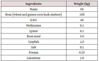
A total of 60 Large white breed of weaned pigs aged 42-60 days old with average body weight of 5.5 kg were sourced from breeders in Hayin Mallam, a small village (settlement) in Samaru, Sabon-Gari Local Government Area of Kaduna State and popularly known as ‘Pig-City’. Forty-eight (48) of them were sheltered in the piggery pen of the Division of Agricultural colleges Samaru, Zaria. Whilst the remaining 12 weaners which will serve as the control group, were housed separately in the Piggery pens of the Department of Veterinary Medicine, Faculty of Veterinary Medicine, A.B.U Samaru, Zaria, Kaduna State these separation was done, so as to avoid cross contamination of diseases. The Pigs were fed ad libitum with water and commercial Pig Weaner diet (Table 1) containing 21% crude protein and 3606 kcal ME/kg.

Table 1: Feed composition for weaned pigsy.
lupinepublishers-openaccess-immunology-Infectious-disease-journal

The ingredients composing the feed were mixed thoroughly and bagged in 100kg grain bags to ease transportation and stored at room temperature in the feed store of the Department of Medicine, Faculty of Veterinary Medicine, Ahmadu Bello University, Zaria. Prior to the feeding with the experimental feed mixes, the following base-line parameters were determined for the experimental animals: For haematological parameters, white blood cells (WBC) especially neutrophils, lymphocytes, basophils and eosinophils were evaluated. These parameters were determined using the automatic blood analyzer (CELL-Dyn 3200, Abbott Lab, Abbott Park, IL). Blood Samples were centrifuged at 6.67g at 40C.

Body Weight

All the pigs in each group were weighted weekly, using a salter’s scale hung on a beam to support the animal. The following mix ratios of feed versus fermenters were used in mixing wet and dry feed forms for feeding of the experimental animals: 8 kilograms of the feed was weighed using a kitchen scale and poured into a pair of 10liters plastic buckets into which 0.8mls of the overnight culture of lactobacillus bacteria was added and thoroughly mixed. Then, 8 liters of distilled water was added to one of the buckets and stirred with a clean spatula while the feed-mix in the second bucket was left dry. This procedure was repeated for other fermenters. However, the control feed-mix was constituted without inclusion of any of the fermenters used for the other experimental feed-mixes.

Experimental Design

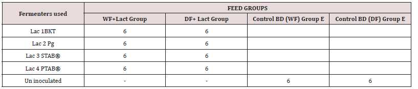
Table 2: Experimental grouping of the weaned pigs based on dietry treatment
lupinepublishers-openaccess-immunology-Infectious-disease-journal

The weaned pigs were tagged and randomly allocated into 5 groups of 6 pigs each with each group having a replicate (Table 2).

Keys

WF+Lact = Wet Feed plus Lactobacillus bacteria

DF+Lact = Dried Feed plus Lactobacillus bacteria

Control BD (WF) = Basal diet in wet form

Control BD (DF) = Basal diet in dry form

Lac BKT = Lactic acid bacteria isolates from “burukutu”

Lac Pg = Lactic acid bacteria isolates from hindgut of pigs

Lac PSTAB®= Bacillus pumilis/Bacillus subtilis a (patented probiotic)

Lac PTAB®= Lactobacillus acidophilus a (patented probiotic)

Diets and Feeding Regimen

A grower diet was formulated for the experimental weaned pigs (Table 1). Then two dietary treatments were formulated with or without bacteria inclusions. 1. Dry feed (DF) supplied as meal and 2. Liquid feed supplied as wet form (WF). The liquid feed was prepared by mixing meal and water in a 1:4 ratio. The WF was prepared by mixing the feed with water in a 20 liters well labeled tank and agitated at 27°C (room temperature) before inoculating all the four treatment groups with lactobacillus from the different sources (BKT, Pig gut, Patented bacteria PSTAB and Patented bacteria PTAB). The control group was fed only the basal diet without LAB inclusion. For the dry feed, it was prepared by mixing the basal feed in four different 20 liters well labelled tanks which were agitated at 27°C (room temperature) before inoculating all the four treatment groups with lactobacillus from different sources (BKT, Pig gut, Patented bacteria PSTAB and Patented bacteria PTAB). The same procedure was applied for feed preparation like the first, but the only difference was that water was not added to the feed. For animals in the control group, only the basal diet was offered in dry and wet forms without LAB inclusion. The animals in all the groups were initially fed for a period of three weeks without the treatments (pre inoculation feeding). Then the various bacteria, isolates were added accordingly into their various rations as described by previous researchers [11,13]. The appropriate amounts of feed were consumed within approximately 30 min once daily in the [14]. according to their body weight so that the whole ration fresh water was also provided ad lib.

Lactobacillus Enumeration and Isolation

After three weeks treatment period, faecal samples were collected from the rectum of all the animals in each treatment groups to ascertain the level of lactobacillus shed in the faeces. Approximately five (5)g of feces was collected from the rectum of each animal in the treatment group with a sterilized spatula, and with the animal properly restrained. The fecal samples were properly labelled and immediately transported to the Microbiology Laboratory of the Department of Veterinary Medicine, Ahmadu Bello University Zaria, and processed, using the following steps as described by [15]:

a) Samples of feces were suspended in quarter-strength Ringer’s solution, homogenized with a classic blender (PBI International, Milan, Italy) and poured on plates containing prepared Rogosa agar (Oxoid Ltd.) which were properly labelled according to the treatment groups for total count of lactobacilli. After anaerobic incubation at 370 C for 24 hours, 10 colonies were randomly selected from plates containing the last sample dilution (10-8).

b) Isolates were cultivated in MRS broth (Oxoid Ltd.) at 370 C for 24 hours and re-streaked into MRS agar. The total Lactobacillus counts were carried out on MRS-agar plates. Numbers of colony forming units (CFU) was expressed as log10 CFU per gram.

c) Microscopy observation and Gram staining’s performed to confirm isolates.

Source of E. Coli for Infection of Pigs

Feces were collected from a diarrhoeic weaner pig in a piggery in Hayin Mallam in Samaru, placed into a sterile polythene bag and transported to the bacterial zoonoses laboratory of the Department of Veterinary Public Health and Preventive Medicine, Ahmadu Bello University Zaria. Ten (10g) of the feces was inoculated into 90mls of broth (Tryptone soya broth), incubated at 37ºC for 24hours for enrichment. A loop full of the broth culture was streaked on EMB agar medium and incubated for 24hours at 370 C. Colonies characteristic of E. coli (green metallic sheen) were tested using the method of characterization as mentioned in Bergey’s Manual of Systematic Bacteriology [16].

Serotyping of Pathogenic E. Coli

A loop full of sterile normal saline was placed on a clean glass slide and a colony from the plate was picked and emulsified to form a homogenous mixture then a drop of the polyvalent phase3 sera (Oxoid, UK) was added and gently rocked for about five seconds until E.coli antigen were detected by agglutination reaction.

Infection of The Weaners with E. Coli Isolate

The fed treatment LAB groups were challenged with E. coli. Each weaner received 6 mls (1x10¹⁰CFU·ml-1) orally, of the E. coli strain solution using a 10mls syringe that was held at the back of their oral cavities at 5.00 pm on day of infection according to the method described by [17].

Faecal Sampling and Bacteriological Analysis of Challenged (Infected) Pigs

Faecal samples were collected from each weaned pig 15 hours after the challenge at 8.00am of the following day and serially diluted (0.1 in 9.9mls). Two tubes at 10-4 and 10-6 were selected and spread plated on EMB-agar (Oxoid, UK) for Enterobacteriaceae enumeration and incubated at 37°C for 24-48 h, as described by [5]. Number of colonies forming units (CFU) was expressed as log10 CFU per gram to obtain the number of colonies shed on a daily basis and this was done for a period of two weeks and results collated. Similarly, the control groups were also infected at the same time and observed for clinical signs of colibacillosis such as diarrhea, rough hair coat and lethargy and those showing such clinical signs were treated with Vetcotrim® bolus antibiotic (Kepro company UK, 5g/50kg per os).

Results

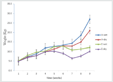
Figure 1: Mean weekly body weight gain of weaned pigs fed BKT lactobacillus isolates in wet and dry feeds. A wet = wet feed + BKT bacteria Isolate A dry = dry feed + BKT bacteria Isolate E wet = wet feed only E dry = dry feed only
lupinepublishers-openaccess-immunology-Infectious-disease-journal

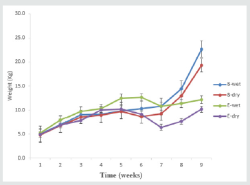
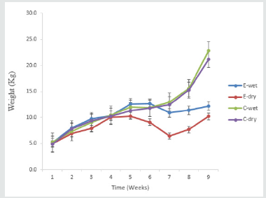
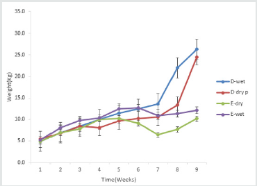
There was a significant (p<0.05) progressive increase in mean weekly body weight in animals fed BKT bacteria inoculated wet and dry form of feed when compared with the control animals fed basal diet in wet and dried form. The group fed BKT in wet feed form produced significantly heavier animals compared to those fed dry feed with BKT bacteria. From week four, there was a gradual increase in weight in all the treatment group and the control group (p<0.05) but however, from week seven an exponential increase in weight was observed in the treatment group and a marked decrease in weight in the control group until week nine when the experiment was terminated (Figure 1). The group fed PGUT bacteria isolates showed a significant exponential (p<0.05) (Figure 2) increase in weight which was as compared to the control group fed with basal diet in both wet and dry form. However, the group fed PGUT bacteria in wet form produced significantly (p<0.05) heavier animals as compared to those fed inoculated dry feed. Also, there was a steady weight gain between week four and seven in the group fed PGUT bacteria in wet feed. And this increased significantly (p<0.05) from week seven to nine, while the group fed dry feed with PGUT bacteria isolate showed a gradual decrease in weight between week and six but increased rapidly from week seven to nine. The control group fed wet feed only showed a gradual increase in body weight from week four to five then plateaued between week five and six and gradually decreased till week nine while the control group fed dry feed only, group started showing a decrease in body weight from week four to seven before it increased steadily up to the ninth week (Figure 2). The group fed with PSTAB bacteria in wet and dry form of feed showed an exponential (p<0.05) increase in body weight as compared to the control group fed basal diet in wet and dry form. Though, animals in the treatment wet group where slightly heavier than those in the treatment dry group. There was a gradual increase in body weight in the treatment group from week four to seven which later progressed until week nine. While the control group showed a slow increase at week four which progressed steadily to week nine though much lower than the treated groups (Figure 3). From week four, a gradual increase in body weight was observed in all the treatment and the control groups but from week seven, the treatment groups showed a significant (p<0.05) increase in body weight as compared to the control. Also, for control animals, a decrease in weight was observed from week five to week seven with a gradual increase from seven to nine-week (Figure 4).

Figure 2: Mean weekly body weight gain of weaned pig fed lactobacillus isolates from pig gut in wet and dry feed. B wet = wet feed + PGUT bacteria Isolate B dry = dry feed + PGUT bacteria Isolate E wet = wet feed only E dry = dry feed only
lupinepublishers-openaccess-immunology-Infectious-disease-journal

Figure 3: Mean weekly body weight gain of weaned pig fed lactobacillus isolates containing Bacillus subtilis and Bacillus pumilis in wet and dry feed. C wet = wet feed + PSTAB bacterial Isolate C dry = dry feed + PSTAB bacterial Isolate E wet = wet feed only E dry = dry feed only
lupinepublishers-openaccess-immunology-Infectious-disease-journal

Figure 4: Mean weekly body weight gain of weaned pigs fed lactobacillus acidiphilus formulated in pig food supplement (tablet) in wet and dry forms. D wet = wet feed + PTAB bacteria Isolate D dry = dry feed + PTAB bacteria Isolate E wet = wet feed only E dry = dry feed only
lupinepublishers-openaccess-immunology-Infectious-disease-journal

Mean WBC Count

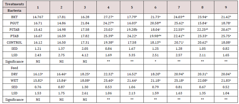
Results of the effect of probiotic bacteria on weekly mean WBC counts of weaned piglets from pre feeding to nine weeks is presented in (Table 3). At week 4 WBC was significantly higher in piglets fed with BKT isolates (p<0.01) when compared to other treatments and the control groups. Also, there was no significant difference between PGUT, PSTAB and PTAB, the lowest was observed in the control group. At 5 weeks, PTAB produce high WBC count (p<0.01) when compared to other treatment and the control. The result in 6 weeks revealed that BKT, PGUT and PTAB isolates produced similar WBC counts at (p<0.05) while PSTAB gave the least WBC count that was similar to control group (p<0.01). At week 7, PGUT and BKT had similar values of WBC but significantly higher than the control (p<0.01). Similarly, there was no significant differences observed from the treatments PSTAB, PTAB and the control group. At week 8 BKT, PSTAB and PTAB significantly produce more WBC count (p<0.01), though similar WBC values were found in PTAB and control group, PGUT produced the least values. At 9-week PTAB significantly produce more WBC values (p<0.01). Then any other treatment and the control. However, BKT and PSTAB gave similar result that was significantly higher (p<0.05) than PGUT and control treatment. The feed form as presented in (Table 3). The result showed that from week 4-9 piglets fed wet feed form significantly produce more WBC counts than the dry form (p<0.001). However, WBC did not defer significantly from week 1-3.

Effect of Probiotics on Haematological Parameters of Weaned Pigs Fed Actobacillus Fermented Feed

Table 3

Mean Hemoglobin Value

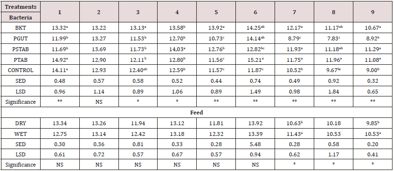
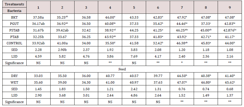
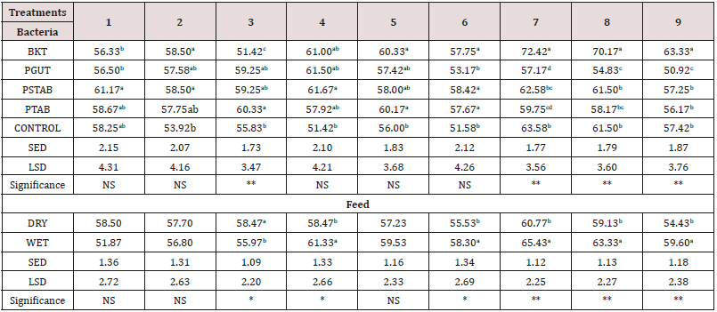
The result for mean Hgb values for all the animals fed wet and dry feed forms in the treatment and control groups were all within the normal range for piglets (swine).The group fed with isolates from PTAB in dry feed produced the highest Hgb value which was significantly higher (p<0.05) than the other treatment group and the control group (Table 4). (Table 5) The effect of probiotic bacteria on the weekly mean neutrophil values from 1st week to 9th week of the study. From 1st, 3rd and 5th week of the study, there was no significant difference (0<0.05) among the treatment groups and the control. However, at week 4 BKT and PSTAB isolates were significantly produced higher mean neutrophil values (0<0.01) than PTAB, PGUT and the control group which was statistically similar. At week 6, BKT and PSTAB were statistically similar and produced higher (0<0.01) weekly mean neutrophil values than PTAB, PGUT and the control group. At week 7, BKT significantly produced the highest (p<0.01) neutrophil value which was similar to PSTAB and the control group. PTAB gave the lowest neutrophil value at this week. At week 8, a similar trend was observed as in the case of week 7 but the lowest neutrophil value was produced by PGUT. At 9 weeks, BKT significantly gave the highest neutrophil values (p<0.01) when compared to their treatment groups that are similar. However, the control group significantly produced a high neutrophil value (0<0.05) than PTAB which gave the last neutrophil value. The feed form as presented in Table 5 showed that from 7-9 weeks piglets fed wet form of food significantly produced more neutrophil values (p<0.05 and p<0.001). However, neutrophil values did not differ significantly from week 1-3. (Table 6) The effect of probiotic bacteria on the weekly mean lymphocyte values from 1st to 9 weeks of the study. From 1st, 2nd, 4th and 6th week of the study, there was no significant difference (0<0.05) among the treatment and control groups. However, at week 7, 8 and 9, BKT treatment group produced the highest lymphocyte values (p<0.01) which was statistically higher than PSTAB, PTAB and a PGUT treatment groups which were statistically similar. The feed form as presented in Table 6 showed that from 3-9 weeks, piglets fed wet food forms significantly produced more lymphocyte values (p<0.05 and p<0.01). However, weekly lymphocyte valued did not differ significantly at weeks 1, 2 and 5.

Table 3: Effect of probiotics on the WBC values of weaned pigs for a period of nine weeks of pre feeding, feeding and challenged with E. coli infection.
lupinepublishers-openaccess-immunology-Infectious-disease-journal

Table 4: Effect of probiotics on the hemogram (HgB) of weaned pigs for a period of nine weeks pre feeding, feeding and challenged with E. coli infection.
lupinepublishers-openaccess-immunology-Infectious-disease-journal

** = Significant at 1% and * at 5% level respectively. Mean within a column of any set of treatment followed by different letter are significantly different at 5%level while means followed by similar letters in column are not significantly different NS = Not Significant SED = Standard deviation BKT = Bacteria from burukutu PGUT = Bacteria from pig intestine PSTAB = Patented bacteria PTAB, Patented bacteria

Table 5: Effect of probiotics on the hemogram (Neutrophils) of weaned pigs for a period of nine weeks pre feeding, feeding and challenged with E. coli infection.
lupinepublishers-openaccess-immunology-Infectious-disease-journal

** = Significant at 1% and * at 5% level respectively. Mean within a column of any set of treatment followed by different letter are significantly different at 5%level while means followed by similar letters in column are not significantly different NS = Not Significant SED = Standard deviation BKT = Bacteria from burukutu PGUT = Bacteria from pig intestine PSTAB = Patented bacteria

Table 6: Effect of probiotics on the (Lymphocytes) of weaned pigs for a period of nine weeks pre feeding, feeding and challenged with E. coli infection.
lupinepublishers-openaccess-immunology-Infectious-disease-journal

** = Significant at 1% and * at 5% level respectively. Mean within a column of any set of treatment followed by different letter are significantly different at 5%level while means followed by similar letters in column are not significantly different NS = Not Significant. SED = Standard deviation, BKT = Bacteria from burukutu, PGUT = Bacteria from pig intestine, PSTAB = Patented bacteria, PTAB, Patented bacteria

Discussion

This study has shown that there was a significant increase in weight of animals in the treatment groups fed probiotic bacteria in the wet and dried form of the feed as compared to the control wet and dried treatment groups. The observed increase was more pronounced in groups fed the wet form of the feed than those fed the dried form. This finding underscores the benefit associated with feeding diets in a liquid form, and the need for pigs to be provided with water and feed simultaneously [14,18] and in this way, the weaned pigs do not need separate learning for feeding and drinking behavior’s [7,19] Again, [19], demonstrated that the dry matter intake of the newly weaned pig can be increased by providing fermented liquid feed. All the groups that received wet forms of the diet with inclusion of probiotics from locally brewed drink (burukutu) showed a better growth as compared to those that received patented bacteria (PSTAB, PTAB) and that from pig (PGUT). The observed overall increase in weight in the BKT wet fed and PTAB wet groups as compared to the other groups (PSTAB and PGUT) indicates that, the probiotic strain used in this two treatment groups produce good acidity which is required to ferment liquid feed as reported by [6].that the fermentation of a nutritionally balanced feed will improve performance by increasing feed intake and gut health with resultant nutrient utilization. [6], had also reported a 22.3% improvement in weight gain and a 10.9% improvement in feed efficiency in the use of fermented liquid feed as compared to dry feed. This study has also shown that, probiotic inclusions in the dry form of feed in the BKT dry and PSTAB dry treatment groups also produced heavier animals than PTAB and PGUT groups, while the control as well as the PGUT inoculated dry feeds produced low weights. This observation agrees with reports that probiotics may not have positive effects on average daily weight gain and feed conversion of pigs [11,19]. As well as the reports of [8] that indicated that Lactobacillus fermentum and Lactobacillus ingluviei were associated with weight gain in animals while Lactobacillus plantarum was associated with weight loss in murines similarly, Lactobacillus gasseri was associated with weight loss both in obese humans and in pigs. The serum profile data indicated that there was no significant difference in white blood cell count, neutrophil, lymphocyte and hemoglobin concentration at the pre feeding period. No bacteria and feed form interaction were noted for white blood cell count, neutrophil, lymphocyte and hemoglobin concentration. This could be due to good hygiene provided for the piglets so that these blood parameters which are related to immune system were unaffected. There was an increase in WBC count only when probiotics was fed to the treatment groups and during challenge with pathogenic E.coli strain from week four of this study, This observation agrees with the report of [12] which states that microbial infection or the presence of foreign bodies or antigens in the circulatory system increases WBC counts. On the contrary, [12] observed an increase in WBC counts in a study of unchallenged postweaning piglets (weaned at 21 days of age) indicating increased total WBC following weaning (and age) respectively. Therefore, the increased total WBC in our weaned pigs could be attributed to the effects of the stress induced by weaning and challenge with E. coli. Lymphocyte counts of the pigs increased at weeks 4 and weeks 7 as observed in the groups fed probiotics inoculated feed and challenged with E. coli infection, which was significantly different from the control Groups challenged with E. coli infection. [13,20] and [13] had reported that the exact mechanisms of immune modulation by probiotics have not been fully explained but they may stimulate different subsets of immune system cells of which macrophages and lymphocytes are inclusive [3,21,22]. Mentioned that probiotics containing lactic acid producing bacteria enhance immune responses and defense activities against undesirable microorganisms and such protection has been partially attributed to increase innate immune response. These could have been the reason for the increase in lymphocyte counts in this study. Neutrophils are majorly responsible for phagocytosis of pathogenic microorganisms during the first few hours after their entry into tissues. There was however a significant increase in the neutrophil values in all the treatment groups and the control indicating the possible response of the neutrophils to the foreign bodies. The significant increase in total protein as observed in this study in all the treatment groups may be due to improvement in appetite and feed utilization by the animals. This concurs with the findings of [23], that increase in total protein indicate a high conversion of nutrients that can be correlated with immune stimulator through the action of probiotic flora. However, our results were parallel with that reported by [9] who reported no significant difference in the levels of serum albumin and globulin in probiotic treated calves, however, they observed a significant increase in the levels of serum total proteins. The findings in this study are also in harmony with that recorded by [14], in probiotic treated kids [24]. reported that there was no effect of complex probiotic feeding on total protein and albumin levels [1,4] also didn’t observe significant effect on total protein and albumin levels with supplementation of rations with probiotics in Broiler chickens.

Conclusion

The findings from this study have shown that: The addition of probiotics basal diets has promising effects on the performance of weaned piglets in terms of increasing body weight gain and improving the immune system thus reducing the severity of infection by pathogenic microorganisms. BKT inoculated feeds both in the wet and dried form, were found to be better than the patented bacteria as a probiotic as it increased the body weight as well as improved the bodies defence by increasing the immune system cells like WBC. Thus, locally sourced probiotics have like BKT and PGUT the potential of being patented for use in the Pig Industry in Nigeria.

Read More About Lupine Publishers Journal of Immunology Please Click on Below Link:
https://lupine-immunology-infectious-disease.blogspot.com/


Saturday, 28 August 2021

Lupine Publishers | Inadvertent Sulfonylurea Overdose and Treatment with Octreotide: A Case Report

 Lupine Publishers | Journal of Immunology & Infectious Diseases


Abstract

Suicide attempts using hypoglycemic agents are uncommon but are associated with a high level of morbidity and mortality. A fifty-eight-year-old-gentleman with no history of diabetes was presented in a state of shock to the emergency room with the ingestion of 12 tablets of 2 mg glimepiride. The patient was in persistent state of hypoglycemia. In such cases of sulfonylurea toxicity, goal of therapy depends on the adequate glucose supplementation to maintain normal blood glucose level. Octreotide is an effective drug in preventing rebound hypoglycemia after sulfonylurea ingestion. Octreotide in combination with dextrose should be considered for first-line therapy in the treatment of sulfonylurea-induced hypoglycemia.

Introduction

The most common complication of sulfonylurea overdose is hypoglycemia [1]. The toxicity is caused secondary to the exertion of the sulfonylurea pharmacological properties. It reduces the glucose level by release of insulin from beta cells of the pancreas [2]. Early symptoms of hypoglycemia from sulfonylureas are characterized by weakness, hunger, diaphoresis, pallor, palpitations, sinus tachycardia, headache, irritability, and tremor. If hypoglycemia remains untreated, neuroglycopenia may develop resulting in impaired concentration and judgment, confusion, blurred vision, drowsiness, and amnesia. Further progression can result in seizures or coma, and possibly death [3]. Conventional therapy of hypoglycemia with intravenous dextrose infusions may only temporarily correct blood sugar levels as sulfonylurea and active metabolite levels may remain high for a prolonged period of time resulting in persistent hypoglycemia [4]. Although octreotide use has been advocated as a first line therapy, indications and dosing are not firmly established. It has also been identified that the use of octreotide may reduce the incidence of recurrent hypoglycemia that is seen with dextrose-alone therapy. Our case report discusses a patient with severe hypoglycemia resulting from suicide attempt by ingesting 24 mg of glimepiride and highlights the response to treatment with octreotide after failed attempts to correct the patient’s hypoglycemia with dextrose.

Case Report

A fifty-eight-old male with no previous co-morbidities was presented to was presented to the emergency room of Northwest General Hospital, Peshawar in the month of January 2019. The son reported that the patient is a non-diabetic. He ingested his spouse’s 12 tablets of 2 mg glimepiride as a suicide attempt.In the emergency room, the patient was tachycardic, tachypneic and diaphoretic. Initial glucose meter reading was found to be 36 mg/dL and a blood glucose level on the metabolic panel was 49 mg/dL. Patient serum creatinine was 2.1 mg/dL. While in the emergency room, the patient received 50% dextrose intravenously eventually requiring an intravenous infusion of 10% dextrose. Despite this treatment, the patient remained persistently hypoglycemic with blood sugars less than 80 mg/dL. He was admitted to the medical intensive care unit for close monitoring. Despite giving high dose of dextrose infusion, his blood sugars remained low. A decision was made at this point to administer 50 micrograms of octreotide subcutaneously. Two hours later, the patient’s blood sugar started to improve and the intravenous 10% dextrose was discontinued. Eight hours later, the patient received another dose of 50 micrograms of octreotide and his blood sugars started improving. The dose was continued for two days and he remained consistently euglycemia. Once stable, his insulin levels and C-peptide levels were done which were normal. Other parameters including complete blood picture, U&Es, liver function tests and urine analysis were within normal limit. Following initial correction of his low blood sugar, the patient was encouraged orally. Once stable he was discharged on normal blood sugars with advise to see a psychiatrist.

Discussion

Glimepiride is a second-generation sulfonylurea indicated for diabetes mellitus type 2. After the intake, the drug is completely absorbed and the maximum concentration is reached in 0.7-2.8 h. It is primarily metabolized in the liver, first to its active metabolite via the cytochrome P450 and then to its dehydrogenated inactive metabolite [5]. Glimepiride was generally associated with lower risk of hypoglycemia compared to other sulfonylureas and it should be used in caution with hepatic and renal disease. It has a narrow therapeutic index. Glimepiride overdose is associated with hypoglycemia [1]. Onset of hypoglycemia may be delayed up to 12 h, and duration may be prolonged for days after overdoses. Patients who are hypoglycemic experience dizziness, weakness, headache, confusion, lethargy, slurred speech, coma, and seizures. Other clinical effects include tachycardia, palpitations, nausea, and diaphoresis. Protracted hypoglycemia can result in death [2]. Several case reports are published on sulfonylurea overdose in adults. Potential for hypoglycemia associated with dosage increases is well described, especially in older patients, sometimes with fatal outcomes. Review of national poison center data found 14 sulfonylurea-associated fatalities reported between 1992 and 1996 in adults aged 18 to 79 years. Eleven cases were the result of suicides and involved contestants [6]. A potential complication of treatment of sulfonylurea-induced hypoglycemia with intravenous dextrose is recurrent hypoglycemia. Dextrose administration results in hyperglycemia which in turn potentiates insulin release from the pancreas leading to recurrent hypoglycemia. Re-administration of dextrose perpetuates this cycle, resulting in high dextrose requirements and the need for frequent monitoring of blood glucose levels.By contrast, octreotide, a synthetic octapeptide analogue of somatostatin, effectively suppresses insulin secretion and has a very benign adverse effect profile. The long-acting, synthetic somatostatin analog, octreotide, is FDA-approved for the treatment of acromegaly, metastatic carcinoid symptoms, and vasoactive intestinal secreting tumors. It has also been used for the cessation of upper gastrointestinal bleeding and to correct refractory hypoglycemia caused by sulfonylurea overdoses. Octreotide can be administered either intravenously or subcutaneously, with both routes having equivalent bioavailability. It appears to abolish the need for hypertonic dextrose infusion, thus avoiding the need for a central line, close observation, and complications relating to fluid and electrolyte disturbances. This obviates the need for prolonged ICU admission. Octreotide is now regarded as a first line antidote for sulfonylurea poisoning with the role of dextrose confined to rapid restoration of euglycemia in the already hypoglycemic patient and maintenance of euglycemia until such time as octreotide can be sourced and administered [7].

Patients who developed hypoglycemia with therapeutic doses of sulfonylureas have been given supplemental dextrose and octreotide, with subsequent correction of the hypoglycemia. There are numerous case reports describing treatment of sulfonylurea overdoses with octreotide. In 2002, Carr and Zed described glyburide overdoses in two patients with refractory hypoglycemia despite dextrose 50% boluses and 10% infusions who demonstrated fewer hypoglycemic episodes and lower dextrose requirements with octreotide 50 μg every 8 h for three doses. These authors provide a summary of six previously reported cases that demonstrate the benefits of octreotide therapy [8]. Subsequent to Carr and Zed’s report, there have been 13 case reports on the treatment of sulfonylurea-induced hypoglycemia with octreotide, including two cases in young children [9]. As octreotide represents the definitive management of sulfonylurea induced hypoglycemia, efforts should be made to administer it as soon as possible. If available in the remote area, it can be safely commenced according the administration regime described above. If not immediately available, efforts should be made to move the antidote to the patient as part of the management plan.

Read More About Lupine Publishers Journal of Immunology & Infectious Diseases Please Click on Below Link:
https://lupine-immunology-infectious-disease.blogspot.com/

Saturday, 10 July 2021

Lupine Publishers | Chediak-Higashi Syndrome

 Lupine Publishers | Journal of Immunology & Infectious Diseases


Abstract

Chediak-Higashi syndrome (SCH) is a rare autosomal recessive genetic disorder characterized by oculo-cutaneous albinism, immunodeficiency responsible for recurrent infections, predisposition to bleeding, And late neurological deterioration. The pathognomonic sign is the presence of giant intracytoplasmic granules in most of the cells of the organism but often they are identified in peripheral blood. In 85% of cases, CHS patients develop the accelerated phase characterized by an Hemophagocytic Lymphohistiocytosis syndrome (HLH) responsible for a high mortality rate. The only current effective treatment of haematological and immunological abnormalities remains allogeneic bone marrow transplantation, but without impact on skin manifestations or subsequent neurological deterioration. It is all the more effective as it is performed before the onset of an HLH syndrome.

Introduction

Chediak-Higashi syndrome (SCH) is a rare autosomal recessive genetic disorder characterized by oculo-cutaneous albinism, immunodeficiency by cytotoxic activity of T lymphocytes and natural killer cells responsible for recurrent infections, predisposition to bleeding, And late neurological deterioration. According to the International Union of Immunological Societies, the SCH is a primary immunodeficiency by immune dysregulation belonging to familial lymphohistiocytic haematophagocytosis syndromes (HFH) with hypopigmentation [1]. The LYST-CHS1 (Lysosomal Trafficking Regulator Gene) gene was identified on the long arm of chromosome 1 in 1q42-q43 [2,3]. This gene encodes the CHS protein whose exact function remains imprecise. About 500 cases have been reported [4,5]. The diagnosis is oriented by the clinical signs and facilitated by the study of the microscopic aspect of the hair which highlights the presence of pigment aggregates; But the pathognomonic sign of the disease is the presence of giant intracytoplasmic granulations in most cells of the organism [6], especially in peripheral blood or bone marrow. Approximately 85% of patients develop an acceleration phase characterized by a syndrome of lymphohocytic hemophagocytosis (HLH), which occurs during the first decade, rarely present at the onset of the disease [7]; It is fatal in the absence of treatment [8]. Currently, the only effective therapeutic option is bone marrow transplantation, which improves haematological and immune abnormalities, but does not prevent subsequent neurological deterioration. The prognosis remains poor in the absence of a bone marrow transplant, the death often occurring before the age of ten years.

Patients and Methods

They are four children followed in the pediatric department of the CHU Mustapha of Algiers for Chediak-Higashi syndrome between 2014 and 2017. The diagnosis was focused on the clinical manifestations, the presence of giant intra-leukocyte granulations. A genetic study in three children confirmed the diagnosis.

Results

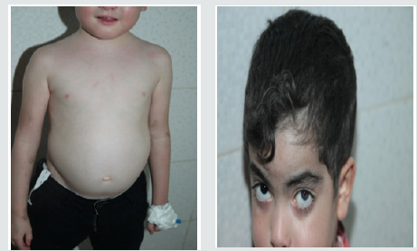
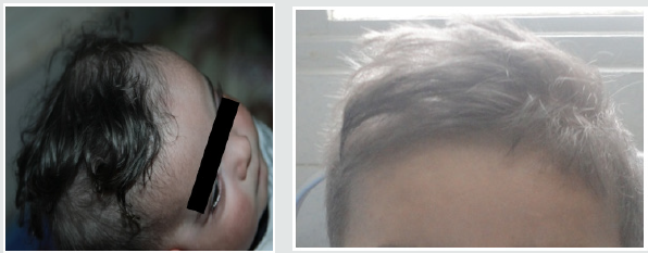
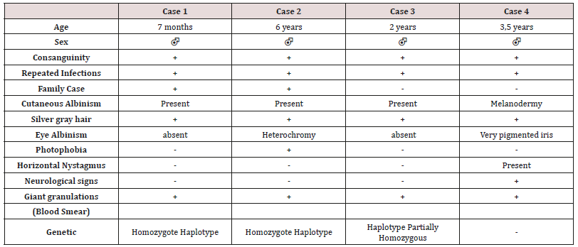
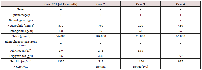
These are three boys and one daughter with an average age of diagnosis of 3.2 years (7 months - 6 years). Consanguinity is found in all cases as well as a family form (2 brothers). Three patients have a history of repeated infections. Children who were vaccinated did not report any particular incidents. Clinically, oculocutaneous albinism is present in 3 children (Figure 1) and melanoderma with a highly pigmented iris in 1 case (Table 1). Silver gray hair is present in all patient (Figure 2). The peripheral blood smear allowed to make the diagnosis by showing the intra-cytoplasmic giant granulations in all the patients. Microscopic study of the hair found deposits of melanin in irregular clods in the hair shaft in favor of Chediak-Higashi syndrome (Figure 3). A syndrome of lymphohistiocyte hemophagocytosis (HLH) (Table 2) is present in 3 cases and then in one case 8 months after the diagnosis.

Figure 1: Hypopigmentation of the skin Clinically, oculocutaneous albinism is present in 3 children hypo pigmentation of the left iris

lupinepublishers-openaccess-immunology-Infectious-disease-journal

Figure 2: Silver-gray appearance of the hair (Silver gray hair is present in all patients).

lupinepublishers-openaccess-immunology-Infectious-disease-journal

Figure 3: Deposition of melanin in the hair shaft Microscopic study of the hair found deposits of melanin in irregular clods in the hair shaft in favor of Chediak-Higashi syndrome

lupinepublishers-openaccess-immunology-Infectious-disease-journal

Melanoderma with a Highly Pigmented Iris in 1 Case All patients with signs of lymphohistiocytic activation were put on HLH 2004 protocol. The evolution is marked by a good tolerance of the treatment. One child received an allogeneic marrow transplant but died six months later. The remaining three patients are still alive and clinically stable.

Discussion

Autosomal recessive genetic disease very rare, Chediak-Higashi syndrome affects all races and all age groups. Less than 500 cases have been reported [4,5]. Most often they are reported cases or small series published. Prevalence is difficult to determine due to reported cases more than once, and other unreported cases. The largest published series (15 cases) was reported in Japan over a period of ten years [9]. Inbreeding in all our patients is reported in 50 to 85% of cases [5]. The LYST / CHS1 gene of the disease encodes the cytosolic CHS protein whose function remains imprecise. It would play a role in the exocytosis of proteins from late multivesicular endosomes. More than sixty mutations have been reported in the literature (false sense, nonsense, deletion, insertions) [10]. Mutations in the LYST / CHS1 gene lead to an abnormal function of the CHS protein with an impairment of intracytoplasmic transport, protein sequestration in giant intracytoplasmic structures and blocking of the secretory function, in particular that of leucocytes and melanocytes. Phenotype / genotype correlations were reported, thus a deletion-type mutation correlated with the early and fulminant onset of the acceleration phase, while a missense-like mutation correlated with a better prognosis with no acceleration phase and neurological deterioration [11]. Clinical signs begin after birth, or before the age of 5 years. Oculocutaneous albinism (AOC) is an important sign of diagnostic orientation, present in three of our patients, characterized by hypopigmentation which generally affects the skin, the hair and the eyes. It is related to pathological aggregation and an unequal distribution of melanosomes. The AOC may be present from birth, and concern the three organs or some of them, total or partial or even absent [12]. Sometimes hyperpigmentation as in one of our patients can exceptionally be seen, delaying the diagnosis [13,14]. Most patients have photosensitivity. Some patients have an atypical phenotype with an attenuated form, or the AOC is subtle or absent and probably unknown [11,15]. Rarely other skin lesions are observed as hyperhydrosis, erythema multiforme. The hypopigmentation of the hair gives them a blonde, gray or white color, often with a silvery or metallic luster. The eyes are blue in color and hypo pigmentation of the iris may be associated with decreased pigmentation of the retina, and ocular manifestations such as photophobia, decreased visual acuity, nystagmus, and strabismus Frequent and recurrent infections are common in childhood. Often severe, they are related to a defect in T cell cytotoxicity, NK function, and a decrease in the chemotactic and bactericidal activity of the granulocytes [16,17]. Pyogenic infections are the most frequent, especially in the skin, upper airways and mucous membranes. The most frequently isolated germs are staphylococcus aureus, β-hemolytic streptococci and pneumococcus. The involvement of the oral cavity manifests in the form of gingivitis, gingival haemorrhage, early falls of the teeth. Mouth ulcers have been described. Periodontitis has been identified as a manifestation of immune dysfunction. The bleeding tendency in these patients is related to a deficiency in the storage pool of dense granules and a defect in platelet aggregation. Haemorrhagic manifestations are usually benign and usually do not require treatment. The accelerated phase of the disease is the most important and dangerous complication of the SCH. It is responsible for a high rate of mortality within a few months [18]. It can occur at any age but especially during the first decade (85%) [19]. Our patients developed this accelerated phase before the age of 6 years. Rarely, it is the first manifestation [7,20]. Its early onset is associated with the existence of a deletion-like genetic mutation [11] and has a collapsed or absent activity in cytotoxic LTs [21]. It is manifested by a syndrome of lymphohystiocytic haematophagocytosis (HLH) whose factors triggering this acceleration are not clear. The role of EBV infection found in one of our patients was raised without this relationship being established [22]. The diagnosis of accelerated phase disease is based on the criteria of the Histiocytic Society 2004 [23]. Neurological manifestations occur in about 50% of cases and may occur at any time in childhood or adulthood, they are variable: peripheral neuropathy, coma, convulsions, ataxia, cognitive disorders, impaired balance, movement abnormalities and mental alterations. The bone marrow transplant does not prevent their subsequent appearance [24]. The diagnosis of SCH should be early, often done at around 6 years of age, but in about 25% of cases the diagnosis is delayed after age 10 [12] for our patients. The average age at diagnosis was 3.2 years. It is suspected on the clinical elements, facilitated by the microscopic study of the hair which shows aggregates of melanin pigments found in all our patients, this aspect allows the differential diagnosis with other types of cutaneous hypopigmentation. But the pathognomonic sign of the disease is the presence of giant intracytoplasmic granules in most of the cells of the organism [6] but often they are identified in peripheral blood as was the case of our patients or in the bone marrow. The diagnosis is confirmed by a genetic test for the mutation LYST. Antenatal diagnosis of the disease is possible in the cells of the chorionic villi, amniotic fluid, leucocytes of the fetal cord [25]. As with Chediak-Higashi disease, other genetic immune deficiencies are accompanied by partial oculocutaneous albinism, such as Griscelli’s disease and Hermansky-Pudlak syndrome. The distinction can be made unambiguously by the different appearance of pigment clusters in the hair sheath, much finer in the case of CHS, and especially by the presence of giant intracytoplasmic granules observed only in the CHS. However, in some cases of myeloid leukemia, one can see giant granulations called pseudo Chediak- Higashi anomaly [26]. The treatment of Chediak-Higashi disease is multidisciplinary and is based on the management of complications of the disease, treatment of the “accelerated phase” or HLH and especially the transplantation of hematopoietic stem cells. Symptomatic treatment of Chediak-Higashi disease is based on effective antibiotic therapy against infections and transfusions of blood derivatives to fight anemia and hemorrhagic complications. Eye disorders should be corrected. The eyes and skin should be protected from UV rays. Vaccinations are generally well tolerated as was the case for our patients and must be done. Hygiene and oral health care are Primordial. The occurrence of neurological symptoms and their progression must be dealt with early enough by a rehabilitation specialist. In the case of an acceleration phase (HLH, a treatment combining corticosteroids, VP16, cyclosporin and intrathecal injections of MTX (HLH 2004) [23] is introduced to achieve remission, which occurs in 75% of cases [27], but relapses are frequent and response to treatment decreases over time. When transplantation is achieved, transplantation is recommended. In SCH patients with HLH by EBV the addition of Rituximab could improve treatment [28]. In the case of refractory HLH, another therapeutic option including a monoclonal anti-CD52 antibody (Alemtuzumab) [29] is possible as a second line treatment before bone marrow transplantation. Allogeneic bone marrow transplantation (BMT) is the only current effective treatment that heals hematologic and immunological abnormalities, but has no effect on oculocutaneous albinism or subsequent neurological deterioration [9,24,30]. The pre-graft conditioning regimen comprises a combination of etoposide, busulfan, cyclophosphamide [31]. Reduced intensity of pre-graft conditioning with fludarabine, melphalan, and alemtuzumab resulted in increased survival in primary or family HLH with lower toxicity [32,33]. Bone marrow transplantation is most effective when performed before the accelerated phase occurs [31]. Patients with a profound decrease in the cytotoxic function of T lymphocytes (CTL) have a high risk of developing lymphocytic syndrome (HLH), so their screening may be an indication for early marrow transplantation [21]. The overall survival rate after marrow transplantation is 60-70% (30-32) The prognosis remains poor in the absence of a bone marrow transplant, death occurs frequently during the first decade by infections or development of an accelerated phase HLH [34] About 10 percent of patients who survive in early childhood will develop severe neurological disorders in adolescence and early adulthood [35].

Table 1: Clinical, Cytological and Genetic Data

lupinepublishers-openaccess-immunology-Infectious-disease-journal

Table 2: Signs of Hemophagocytosis LymphoHistiocytic (HLH)

lupinepublishers-openaccess-immunology-Infectious-disease-journal

Conclusion

Chediak-Higashi syndrome is a rare disease, the diagnosis is suspected in a child with oculocutaneous albinism with recurrent infections. The majority of clinical forms are early “infantile” lethal in the absence of treatment. A minority of patients present with an “attenuated” form of the disease will survive after childhood but develop an associated neuro-degerative disease. In all cases early diagnosis should be posed by a simple examination, peripheral blood smear which shows the presence of giant intracytoplasmic granules pathognomonic of this condition. The only current effective treatment of haematological and immunological abnormalities remains allogeneic bone marrow transplantation, but without impact on skin manifestations or subsequent neurological deterioration. It is more effective when it is performed before the onset of an HLH syndrome. In the event of the occurrence of accelerated phase (HLH), a treatment according to the HLH 2004 protocol is instituted in order to obtain a remission before the bone marrow transplant. The prognosis of infant form is poor, with death occurring frequently in the first decade of life through infections or development of HLH. The search for predictive factors for the development of HLH may help in the indication of early bone marrow transplantation.

Read More About Lupine Publishers Journal of Immunology Please Click on Below Link:
https://lupine-immunology-infectious-disease.blogspot.com/