Showing posts with label Neurological Disorders Journal. Show all posts

Wednesday, 4 January 2023

Lupine Publishers| Paraparesis caused by Aneurysmal Bone cyst (ABC)

 Lupine Publishers| Journal of Neurology and Brain Disorders


Abstract

Through the study of a case of spinal aneurysmal bone cyst managed in the Department of Neurosurgery at Hassan II University Hospital of Fez we focus our discussion of various aspects of this rare disease.

Our study concerns a male patient admitted to the emergency department for spinal cord compression that necessitated a spinal MRI. It showed T4 vertebral compression, hypointense T1, hyperintense T2 with hematic fluid levels causing spinal cord compression. The patient underwent surgical treatment with good clinical outcome. Histological study revealed a spinal aneurysmal bone cyst.

Keywords: Aneurysmal cyst; MRI-surgery; Embolization

Introduction

The aneurysmal bone cyst is a benign lesion with an aggressive character. It represents 1.4% of primary bone tumors, its spinal localization remains uncommon (3-30%) and spinal syndrome is the most frequent reason for consultation [1]. Plain radiography and CT have a significant role in its diagnosis, but MRI remains the investigation of choice. The management of spinal aneurysmal bone cyst remains controversial.

Case

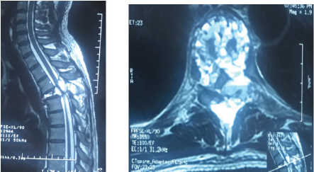
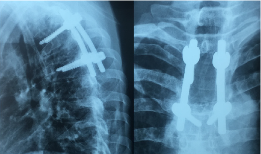
A 40 years old man, chronic smoker (a packet/day) was admitted to the emergency for a spinal syndrome for 2 years that was complicated 2 months before admission by the appearance of a grade D paraparesis, hypoesthesia with a sensory level at xiphoid process and urinary incontinence. The spinal MRI showed compaction of T4 vertebra with a heterogenous hyperintensity on T2 and STIR, a hyperintensity on T1 with heterogeneous enhancement of hematic fluid levels causing spinal cord compression at same level (Figure 1). The management consisted of performing a laminectomy of T3-T4 with pedicle screw and rod fixation of T3 and T5 vertebra (Figure 2). The postoperative course was marked by improved clinical signs. The Histopathology report came back in favor of aneurysmal bone cyst.

Figure 1: Spinal T2 weighted MRI showing (a) sagittal view with T4 compression and (b) Axial view with fluid level spaces.

Lupinepublishers-online-journal-of-neurology-and-brain-disorders

Figure 2: Spinal X-ray Lateral and anteroposterior showing post-operative pedicle screw and rod system.

Lupinepublishers-online-journal-of-neurology-and-brain-disorders

Discussion

Aneurysmal Bone Cyst (ABC) is a benign cystic lesion composed of blood-filled spaces, devoid of coating endothelial, muscle or elastic, surrounded by fibroblasts, described for the first time by Jaffe and Lichtenstein in 1942 [2]. Its incidence is 0.14 per 100,000 individuals with a female predominance (ratio 1.16) [3]. For Lichtenstein [4], the ABC is a secondary bone lesion due to hemodynamic imbalance resulting in a significant capillary dilatation, resulting from an increase in venous pressure. This increase in venous pressure might be due to venous thrombosis or abnormal arteriovenous communication. Biesecker et al [5] measured the intracystic pressure which was identical to that of arterioles and concluded that this hypertension is due to an arteriovenous fistula following a secondary lesion. Some authors [6-8] think that the appearance of an aneurysmal bone cyst would be secondary to impaired intraosseous vascular flow in the phase of consolidation following trauma. However, several cases of familial aneurysmal bone cysts [9,10] have been reported, hence orienting towards a genetic origin and indeed the genetic and immunohistochemical studies [11] have demonstrated a translocation on chromosome [7].

Aneurysmal bone cyst is most frequently found in the long bones especially the metaphyseal region [12,13], while the spinal location remains rare and affects the cervical, thoracic, lumbar and sacrum, with no case of coccygeal bone ABC reported [14]. Clinically, spinal ABC may be asymptomatic [1], but usually it manifests itself as a spinal disorder, with or without spinal cord compression signs. MRI remains the examination of choice which shows a welldemarcated bone lesion, hypointense on T1, hyperintense on T2 with a peripheral border taking contrast, defining spaces with fluid level [4]. Surgery that involves wide excision represents the preferred treatment of choice for ABC with a recurrence rate of 25% and multiple complications such as deformation and growth disorders [15]; For these reasons, selective arterial embolization was introduced into the management of this entity that proves similarly effective and with fewer complications, representing the treatment of choice in cases where the patient shows no deficit or unstable associated fractures [16]. Radiation therapy, which was an important pillar in the therapeutic arsenal, was gradually abandoned because of the risk of exposure and development of radiation-induced sarcomas [17]. Other therapeutic protocols have been proposed: the intralesional injection of calcitonin, and doxycycline Ethibloc [17-20], however they have not proven effective as with other therapeutic methods. Currently, research [21-23] has focused on the interest of Denosumab, a monoclonal antibody used in the treatment of giant cell tumors, and the intrinsic stimulation by the mesenchymal stem cells and have both shown promising results [24,25].

Conclusion

Aneurysmal bone cyst is a benign disease with unpredictable course, and thus what makes its treatment a challenge especially in locations where access is difficult. Surgery and embolization currently remain the treatment protocol of choice. Research on Mesenchymal stem cells and Denosumab have demonstrated promising results in the therapeutic management of ABC.

Read More About Lupine Publishers Journal of Journal of Neurology and Brain Disorders Please Click on Below Link: https://brain-disorders-lupine-publishers.blogspot.com/

Tuesday, 4 October 2022

Lupine Publishers| 3rd Nerve Palsy After Microsurgical Clipping of Basilar Top Aneurysm

 Lupine Publishers| Journal of Neurology and Brain Disorders


Abstract

Postoperative oculomotor nerve palsy is a rare complication that occurs in the setting of micro¬surgical aneurysm clipping. While a number of theories have been postulated to explain the development of postoperative oculomotor nerve palsies, the underlying pathophysiology of such complications still remain to be elucidated. In this report, we present a case of postoperative isolated ipsilateral oculomotor nerve palsy after clipping of basilar tip aneurysm which we believe may be attributed to periperative oculomotor nerve manipulation related neuropraxia.

Keywords:Oculomotor Nerve Palsy; Basilar Apex Aneurysm; Postoperative

Introduction

Oculomotor nerve is the third cranial nerve that enters the orbit through the superior orbital fissure and controls muscles that drive most movements of the eye and raise the eyelid. Oculomotor nerve is derived from the basal plate of the embryonic midbrain. Cranial nerves IV and VI also participate in the control of eye movement. Oculomotor nerve palsy is an eye condition resulting from damage to the third cranial nerve or a branch there of. As the name suggests, oculomotor nerve supplies the major¬ity of the muscles that control eye movements. Therefore, damages to oculomotor nerve will render affected individuals unable to move his or her eye normally. Unilateral oculomotor nerve palsy is often encountered in the setting of ipsilateral aneurysms located at the Posteri¬or Communicating Artery (PcomA), Internal Carotid Artery (ICA) or their junction (PcomA/ICA). There have been some reports on ocu¬lomotor nerve palsy as a result of aneurysms in basilar tip, anterior artery or anterior communi¬cating artery. In the present report, we describe a case of basilar apex aneurysm presenting with ipsilateral postoperative oculo¬motor nerve palsy that is thought to be attrib¬uted to peroperative oculomotor nerve handling followed by neuropraxia.

Case Report

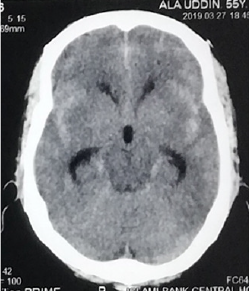
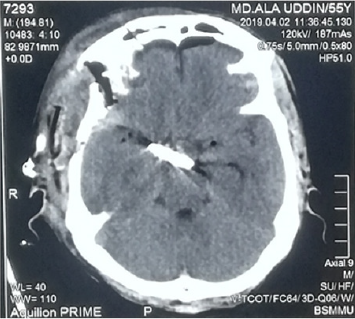
A 55years old hypertension male with no other significant past medical history or contributing family history complained about sudden severe headache for one hour followed by several episodes of vomiting and unconsciousness for 05 hours on admission at our hospital. Physical examination results were within nor¬mal limits except for considerable neck stiffness, positive Kernig sign and Brudzinski’s sign. Computed Tomography (CT) scan demonstrated diffuse subarachnoid hemorrhage, which was especially concentrated in ambient, sylvian fissure, interpeduncular and suprasellar cisterns with intraventricular extensions and mild triventricular hydrocephalus (Figure1). Computed tomography of the head show¬ing diffuse subarachnoid hemorrhage especially concentrated in ambient, sylvian fissure, interpeduncular and suprasellar cisterns with intraventricular extensions and mild triventricular hydrocephalus. CT Angiography (CTA) revealed a large saccular basilar tip aneurysm (approximately 10.7×9.5×10.5mm) (Figures 2A & 2B). The aneurysm was micro surgically clipped (Figures 4) via the right Orbitozygomatic approach (Figures 3A & 3B). Postoperatively the patient developed complete right third nerve palsy characterized by the presence of dilated pupil, ptosis and downward deviation and abduction of the eyeball. CT scan revealed no postoperative intracranial hematoma and the surgical clip was in the proper location. However, compression of the basal cisterns and assessment of the ventricular system were noted (Figure 5). Postoperative CTA showed no existence of another aneurysm or vasospasm (Figure 6). The patient was discharged 02 weeks after the surgery. At this point, his mydriasis and eye lid drooping were still present to a lesser extent. At one-month follow-up, the patient’s pupil was slightly contracted and reflexed reluctantly to direct and indirect light stimulation.

Figure 1: Computed tomography of the head show¬ing diffuse subarachnoid hemorrhage especially concentrated in ambient, sylvian fissure, interpeduncular and suprasellar cisterns with intraventricular extensions and mild triventricular hydrocephalus.

Lupinepublishers-online-journal-of-neurology-and-brain-disorders


Figure 5: CT scan revealed no postoperative intracranial hematoma and the surgical clip was in the proper location.

Lupinepublishers-online-journal-of-neurology-and-brain-disorders

Figure 6: Postoperative CTA showed no existence of another aneurysm or vasospasm and the surgical clip was in the proper location.

Lupinepublishers-online-journal-of-neurology-and-brain-disorders

Figure 7: On postoperative day two, the patient developed a complete right third nerve palsy.


Discussion

There are many proposed hypotheses pertain¬ing to the etiology of oculomotor nerve palsy before and after aneurysm surgery. Direct com¬pression by the aneurysm is the most classic and common cause. Other causes include direct injury to the third nerve intraoperatively [1-6], microvascular ischemia [7-9], focal hemato¬ma formation [3], vasospasm [8-12], anomaly of the vessels along the oculomotor nerve [10], elevated intracranial pressure and herniation, compression by intracranial structures other than aneurysms [7], and undetermined ori-gins [11]. In the present case, compression by aneurysm may not be possible because of the anatomi¬cal distance between the two entities. Postoperative CT, CTA decrease the chances of hematoma formation, vasospasm and elevated intracranial pressure. Although we cannot rule out the possibility of ischemic injury to the oculomotor nerve. Small vessel ischemic injury to oculomotor nerve usu¬ally exhibits pupilsparing [9] whereas our patient demonstrated a blown pupil. So, we believe that, probably from coarse dissection of the cavernous sinus dura and as well as direct anterior clinoidectomy or from heat of the low power drill may be responsible for the 3rd nerve palsy of our patient.

Conclusion

The novel use of intramuscular cerebrolysin and citicoline in a patient with kernicterus was safe and effective.

The Neuroreparative Effects of Cerebrolysin have Been Attributed to [3-5]:

a) Inhibition of apoptosis.

b) Improving synaptic plasticity and induction of neurogenesis.

c) Augmenting the proliferation, differentiation, and migration of adult.

d) subventricular zone neural progenitor stem cells, contributing to neurogenesis.

e) Induction of stem-cell proliferation in the brain.

Citicoline (cytidine diphosphate choline) is a mononucleotide made of ribose, pyrophosphate, cytosine and choline is a watersoluble naturally occurring substance that is generally grouped with the B vitamins. It is also considered a form of the essential nutrient choline. It is a safe substance with generally minor side effects which may include digestive intolerance after oral administration [6]. An accumulating research evidence suggests that citicoline is endowed with interesting pharmacological properties that can make it useful in the treatment of various disorders that has no universally accepted effective treatment including neurological conditions such as Parkinson’s disease, brain ischemia, hemorrhagic stroke, Alzheimer’s disease; and ocular condition such as glaucoma, nonarteritic ischemic neuropathy and amblyopia [6].

The Neuro-Protective Effects of Citicoline Were Attributed to the Followings [6]:

a) Preservation of cardiolipin and sphingomyelin

b) Preservation of arachidonic acid content of phosphatidylcholine and

c) phosphatidylethanolamine.

d) Partial restoration of phosphatidylcholine levels.

e) Stimulation of glutathione synthesis and glutathione reductase activity.

f) Reduction of phospholipase A2 activity.

g) Increasing glucose metabolism in the brain.

h) Increasing cerebral blood flow.

Reducing oxidative stress and preventing excessive inflammatory response in the brain by inhibiting the release of free fatty acids and reducing blood brain barrier breakdown. Enhances cellular communication by increasing the availability of neurotransmitters, including acetylcholine, norepinephrine, and dopamine. Lowering increased glutamate concentrations and increasing the decreased ATP concentrations induced by ischemia. Citicoline increases dopamine receptor densities, and therefore could improve memory impairment resulting from poor environmental conditions. Citicoline could also improve focus and mental energy and could be useful in the treatment of attention deficit disorder. Citicoline has also been shown to improve visual function in patients with glaucoma [7].

Conclusion

The novel use of intramuscular cerebrolysin and citicoline in a patient with kernicterus was safe and effective.

Read More About Lupine Publishers Journal of Neurology and Brain Disorders Please Click on Below Link: https://brain-disorders-lupine-publishers.blogspot.com/

Tuesday, 12 July 2022

Lupine Publishers| The Novel Use of Cerebrolysin and Citicoline in the Treatment of Kernicterus

 Lupine Publishers| Journal of Neurology and Brain Disorders


Abstract

Background: There is no effective therapy for many neurological disorders associated with significant neurological damage such as kernicterus. Patients with Kernicterus (Chronic bilirubin encephalopathy) which is a neurological dysfunction resulting from exposure of the brain to severe hyperbilirubinemia mostly during the neonatal period continue to experience significant disability. There is no known therapeutic intervention that can obviously improve this condition. The aim of this paper is to describe a novel therapeutic approach which in two months produced marked improvement of the neurological dysfunction caused by kernicterus.

Patients and Methods: A girl with kernicterus was not speaking and not saying any word. She was lacking the balance (coordination) without obvious muscle weakness. She was unable to maintain the sitting posture on a chair for few minutes. She was unable to maintain straight standing posture when supported on chair at all. She had difficulty in holding things. The girl was treated with a novel therapeutic approach including two courses of intramuscular cerebrolysin and intramuscular citicoline.

Results: After treatment the first month of treatment, speech development was initiated, and she was saying few words. She was able to sit normally on the chair and maintaining the sitting posture indefinitely. She was able to maintain more straight stable standing posture without holding a chair and with the ability to hold things at the same time indicting improved coordination. She also developed improved ability to hold small things like a pen. After the second month of treatment, the girl was able to stand alone and was making few steps slowly holding furniture. After treatment the second course of treatment, the girl was able to stand alone and walk rapidly holding furniture. Treatment was not associated with any side effects.

Conclusion: The novel use of intramuscular cerebrolysin and citicoline in a patient with kernicterus was safe and effective.

Keywords:Cerebrolysin; Citicoline Treatment; Kernicterus

Introduction

Kernicterus is a neurological dysfunction resulting from exposure of the brain to severe hyperbilirubinemia mostly during the neonatal period. In kernicterus, hyperbilirubinemia results in deposition of bilirubin in the grey matter of the brain causing neurotoxicity associated with mass-destruction of neurons by apoptosis and necrosis resulting in irreversible brain damage, and chronic neurological disorder. Clinical manifestations of kernicterus may include severe motor disability with inability to walk. There is no effective therapy kernicterus [1,2]. The aim of this paper is to describe a novel therapeutic approach which in two moths produced marked improvement of the neurological dysfunction caused by kernicterus.

Patients and Method

A girl was first seen at the age of about five years during November,2018 because of delayed development with lack of body control, abnormal movements, and no speech. Her birth weight was about three Kilogram, and she developed severe neonatal hyperbilirubinemia of 26 mg/dL, and was treated by phototherapy and exchange transfusion. The family consulted many physicians in Iraq, and when no obvious improvement could be achieved nor any hope of improvement was given, the family took their daughter to India. During March,2017, the girl was seen by Dr Rakesh Kumar Jain who was a senior consultant at the neurology center of Fortis Memorial Research Institute in Gurgaon India. Dr Jain performed a 21-channel electroencephalography on the girl which showed intermittent short and sharp waves from bilateral focal regions with no ictal events. The electroencephalography report stated that drug induced EEG showed normal sleep pattern with intermittent bilateral frontal sharp waves. Dr Jain also performed a study of the short latency auditory evoked potential on the girl. The report of this study stated that the stem auditory evoked response showed normal latencies of waves with hearing threshold of 20 dB bilaterally. Dr Jain sent the girl to the Modern Diagnostic Research Center for brain MRI which was performed at the 22nd of March, 2017, by Dr Sneha Thakur, a consultant radiologist. The brain MRI detected no significant abnormality. Dr Rupal Gupta, a consultant ophthalmologist at the Fortis Memorial Research Institute examined the girl on the 30th of March, 2017, and prescribed eye glasses for the girl. After visiting many physicians in Iraq and India, the only beneficial therapeutic intervention for the girl was the correction of her refractive error with eyeglasses.

When the girl was seen at the pediatric neuropsychiatry clinic at the Children Teaching Hospital of Baghdad Medical City during November,2018, she was not speaking and was not saying any word. The girl was lacking the balance (co-ordination) without obvious muscle weakness. She was unable to maintain the sitting posture on a chair for few minutes and was also unable to maintain straight standing posture when supported on chair at all (Figure 1). She also had difficulty in holding things.

The girl was treated with a novel therapeutic approach consisting of two courses.

The First Course Included

Cerebrolysin 5ml given by intra-muscular injections every third day on the morning, and she received 20 doses over two months. Citicoline 500 mg given by intra-muscular injections every third day on the morning, and she received 20 doses over two months. Cerebrolysin and citicoline were given on two different days.

The Second Course Included

Cerebrolysin 5ml given by intra-muscular injections every fifth day on the morning, and she received 10 doses over fifty days. Citicoline 500 mg given by intra-muscular injections every fifth day on the morning, and she received 10 doses over fifty days. Cerebrolysin and citicoline were given on two different days.

Results and Discussion

After treatment the first month of treatment, speech development was initiated and she was saying few words. She was able to sit normally on the chair and maintaining the sitting posture indefinitely (Figure 1). She was able to maintain more straight stable standing posture without holding a chair and with the ability to hold things at the same time indicting improved coordination (Figure 2A). She also developed improved ability to hold small things like a pen (Figure 2B). After the second month of treatment, the girl was able to stand alone and was making few steps slowly holding furniture. After treatment the second course of treatment, the girl was able to stand alone and walk rapidly holding furniture (Figure 3). Treatment was not associated with any side effects. In this paper, we have reported that the novel use of intramuscular cerebrolysin and citicoline in a patient with kernicterus was safe and effective. The use of cerebrolysin and citicoline was beneficial in the treatment of various childhood neuro-psychiatric disorders including developmental and pervasive developmental disorders, brain atrophy, kernicterus, and cerebral palsy [3-5]. Cerebrolysin is a peptidergic medicine which contains mainly biologically active neuropeptides including brain-derived neurotrophic factor, glial cell line-derived neurotrophic factor, nerve growth factor, and ciliary neurotrophic factor. It has a nerve growth factor like activity on neurons, and growth promoting efficacy in different neuronal populations from peripheral and central nervous system. Cerebrolysin has a direct neurotrophic effect, and obvious neuroprotective properties against many types of lesion in vitro and in vivo. The therapeutic effects of cerebrolysin have been considered to be similar to the pharmacological activities of naturally occurring nerve growth factors. The safety, tolerability, and efficacy of neuroreparative cerebrolysin therapy have been established in clinical trials included adults with stroke and Alzheimer s disease.

The Neuroreparative Effects of Cerebrolysin have Been Attributed to [3-5]:

a) Inhibition of apoptosis.

b) Improving synaptic plasticity and induction of neurogenesis.

c) Augmenting the proliferation, differentiation, and migration of adult.

d) subventricular zone neural progenitor stem cells, contributing to neurogenesis.

e) Induction of stem-cell proliferation in the brain.

Citicoline (cytidine diphosphate choline) is a mononucleotide made of ribose, pyrophosphate, cytosine and choline is a watersoluble naturally occurring substance that is generally grouped with the B vitamins. It is also considered a form of the essential nutrient choline. It is a safe substance with generally minor side effects which may include digestive intolerance after oral administration [6]. An accumulating research evidence suggests that citicoline is endowed with interesting pharmacological properties that can make it useful in the treatment of various disorders that has no universally accepted effective treatment including neurological conditions such as Parkinson’s disease, brain ischemia, hemorrhagic stroke, Alzheimer’s disease; and ocular condition such as glaucoma, nonarteritic ischemic neuropathy and amblyopia [6].

The Neuro-Protective Effects of Citicoline Were Attributed to the Followings [6]:

a) Preservation of cardiolipin and sphingomyelin

b) Preservation of arachidonic acid content of phosphatidylcholine and

c) phosphatidylethanolamine.

d) Partial restoration of phosphatidylcholine levels.

e) Stimulation of glutathione synthesis and glutathione reductase activity.

f) Reduction of phospholipase A2 activity.

g) Increasing glucose metabolism in the brain.

h) Increasing cerebral blood flow.

Reducing oxidative stress and preventing excessive inflammatory response in the brain by inhibiting the release of free fatty acids and reducing blood brain barrier breakdown. Enhances cellular communication by increasing the availability of neurotransmitters, including acetylcholine, norepinephrine, and dopamine. Lowering increased glutamate concentrations and increasing the decreased ATP concentrations induced by ischemia. Citicoline increases dopamine receptor densities, and therefore could improve memory impairment resulting from poor environmental conditions. Citicoline could also improve focus and mental energy and could be useful in the treatment of attention deficit disorder. Citicoline has also been shown to improve visual function in patients with glaucoma [7].

Conclusion

The novel use of intramuscular cerebrolysin and citicoline in a patient with kernicterus was safe and effective. 

Read More About Lupine Publishers Journal of Neurology and Brain Disorders Please Click on Below Link: https://brain-disorders-lupine-publishers.blogspot.com/
Monday, 9 August 2021

Lupine Publishers| Bruxism and Enuresis: Common Entities But Understood

 Lupine Publishers| Online Journal of Neurology and Brain Disorders (OJNBD)


Editorial

In the population in general, but especially in Paediatrics, there are a couple of sleep disorders that deserve different attention, due to the lack of medical culture to address them, these two conditions are bruxism and enuresis. The first one is very, very frequent to find it around us, either with any person that surrounds us, and it is very frequent in women, since it will increase the appearance of headaches in the population in general.

This is defined as the involuntary habit of squeezing or grinding the teeth, especially during sleep; the people in general, but more serious still, the doctors or dentists, have the rather erroneous idea that it is by parasites, and every time they arrive with said symptom to consult they take their “paque-desparacites” without asking anything else; and is that we speak of a phenomenon or condition that has a very definite origin in high levels of anxiety, which leads to a wear on the gums, temporomandibular joint dysfunction, headache or headaches that are very resistant to treatment and depression and anxiety for a chronic bad sleep.

It is thought that only the famous “guard-occlusal” prescribed in dental offices is the answer, but this is far from true, since it must be accompanied by exercise that allows relaxation, and if you hurry me, swimming is fantastic; supportive psychotherapy in many patients; sleep hygiene measures should be indicated, and always the review by a maxillofacial specialist; I always say this phrase to my patients who define the basis of their treatment “should be far from the person who is now and reinvent themselves” as this derives from their temperament and personality.

Enuresis is defined as the persistence of urine or urine without control beyond the age at which bladder control is reached (in a range of 4 to 6 years of age), and occurs both day and night, although the latter by far it is the most frequent, and it is catalogued within the group of parasomnias, which as we already mentioned are activation disorders or sudden sleep arousal. What causes enuresis? Although anatomical alterations in the urinary tract must be ruled out, mainly and by far they are psychological, since it is the face of the depression in many children, that aside they present with anguish and feelings of guilt, believe me that it is quite painful for a child who lives This phenomenon, a situation that if we do not treat it with the maturity and stature required, will mark the life of this being for the rest of his life.

Initially, the management of anti depressants may be indicated, but much more should be treated emotional aspects, with supportive psychotherapy, rewarding behaviour of parents, which means not hitting or mortifying the child, and more than what is found, it is better to talk to him, explain that it is not his fault, and that he would receive some prize or encouragement if he could avoid it; Limit your fluid intake until certain time of day, exercise and our sleep hygiene measures.

Read More About Lupine Publishers Online Journal of Neurology and Brain Disorders (OJNBD) Please Click on Below Link:  https://brain-disorders-lupine-publishers.blogspot.com/


Tuesday, 3 August 2021

Lupine Publishers| The Construction and Validation of the Revised Arabic Scale of Obsession Compulsion (ASOC)

 Lupine Publishers| Online Journal of Neurology and Brain Disorders (OJNBD)


Abstract

Background: Recent surveys estimated the prevalence rates of obsessive-compulsive disorder (OCD) more than the earlier surveys. Moreover, in the general non-clinical population, many studies found high incidence of obsessions and compulsions (OC).

Objective: To develop and validate a revised version of the Arabic Scale of Obsession – Compulsion (ASOC) as a trait scale, suitable for research studies.

Methods: Samples of 150 non-clinical under graduates were recruited. The last version of the revised ASOC comprised 20 short statements, plus five items as fillers. Three subscales of OC were used as criteria from the MMPI, and the Symptom Check List, (SCL), as well as the Obsessive-Compulsive Inventory (OCI).

Results: Cronbach alpha reliabilities reached .882 (men), .910 (women), and .897 (total group). The correlation coefficients of the ASOC with the MMPI, SCL, and OCI scales were .759, .783, and .885, respectively. A principal component analysis retained one high – loaded factor labeled Obsession-compulsion. The loading of the ASOC unto this factor was .948, indicating very high factorial validity.

Conclusion: The ASOC has good psychometric characteristics, i.e., high internal consistency, and concurrent, and factorial validity.

Keywords: Arabic scale of obsession-compulsion; Minnesota multiphase personality inventory; Symptom check list; Obsessioncompulsion inventory; Reliability; Validity; Egypt

Abbrevations: OCD: Obsessive-Compulsive Disorder; OC: Obsessions and Compulsions; ASOC: Arabic Scale of Obsession- Compulsion; MMPI: Minnesota Multiphase Personality Inventory; SCL: Symptom Check List; OCI: Obsessive-Compulsive Inventory; DSM: Diagnostic and Statistical Manual of Mental Disorders

Introduction

The fourth edition of the Diagnostic and Statistical Manual of Mental Disorders (DSM) classified obsessive-compulsive disorder (OCD) under the category: anxiety disorders. More recently, in the fifth edition of the DSM [1], however, OCD becomes the first item in a separate category under the name: Obsessive-Compulsive and Related Disorders. It includes OCD, body dysmorphic disorder, hoarding disorder, trichotillomania, excoriation, substance / medication – induced obsessive-compulsive and related disorders. OCD is characterized by the presence of obsessions and / or compulsions. Obsessions are recurrent and persistent thoughts, urges, or images that are experienced as intrusive and unwanted, whereas compulsions are repetitive behaviors or mental acts that an individual feels driven to perform in response to an obsession or according to rules that must be applied rigidly (p. 235) [1]. In earlier surveys, the prevalence of OCD in the general population was 0.5% [2], but more recent surveys estimated the 12 – month prevalence of OCD in the United States as 1.2 %, with a similar prevalence internationally (1.1% - 1.8 %) [1]. In Egypt, a study in 1991 showed an incidence of OCD at 2.3 % [3].

On the other hand, a number of research studies indicated the high incidence of obsession (about 80 %) in the general nonclinical population as well as the similarity between normal and pathological obsessions [4-6]. Furthermore, the form and content of the obsessions did not differ between normal’s and OCD patients. Nevertheless, obsessions of patients occur more frequently, last longer, are more intense, disrupt their lives, arouse more discomfort and resistance, and are difficult to dismiss. The same results applied well to compulsions [4]. Therefore, it seems suitable to consider normal and abnormal obsessions and compulsions (OC) on the basis of the quantitative and dimensional approach.

Egypt, as a developing country, like the rest of the Arab countries, are in great need of psychological tests and questionnaires. In 1992, Abdel-Khalek [7] developed the Arabic Scale of Obsession Compulsion (ASOC), and in 1998, he developed an equivalent English version of this scale [8]. Several studies were published using this English version [9-16], as well as the Arabic form. Moreover, a Spanish form of the scale is available [17].

Twenty-six years have passed since the publication of the Arabic form of the ASOC. Furthermore, its author found some aspects to be improved as follows: (a) the first version consists of many items (32), and a short form is badly needed to avoid the participant’s boredom and carelessness, and to save his or her time, (b) some items are long statements and it is preferable to use short ones, (c) the response alternatives were dichotomous (Yes/No), and the Likert format has psychometric advantages, and (d) the old scale contained 28 % negative items scored “No” for OC (e.g., I do not like strict discipline and too much accuracy). Some authors stated that negatively worded items often turn out to be harder to understand or more complicated to answer than positively worded items [18]. Other authors concluded that negatively worded items impair response accuracy [19], so there is a need to depend only on positively worded items. The aim of the present research was to develop a revised version of the ASOC as a trait scale to be used in research in the general population, and to estimate its psychometric properties.

Material and Methods

Participants

A convenience sample of 150 undergraduates enrolled in different faculties in University of Alexandria, Egypt took part in this study (74 men; 76 women). Their ages ranged from 17 to 25 years (M age= 20.95, SD= 2.01). They were non-paid volunteers, and neither disturbed clinical cases nor diagnosed institutionalized patients, but, rather were presumably healthy individuals. That is, they were not selected from hospitals or clinics. However, no psychiatric assessment was conducted to support that these participants had no mental illness.

Methodology

Psychometric Scales

The Arabic Scale of Obsession-Compulsion (ASOC)

Construction of the revised scale: The 32 items of the original ASOC were shortened and the negative wording changed to positive to avoid the problem of the double negative when the participant answered these items. Five new items were added. The 37 statements were brief and written in standard, modern, and simple Arabic. A sample of 150 undergraduates responded to the 37 items based on a 5 –point Likert scale. Then, the corrected item-resetof- test score correlations (i.e., the item-remainder correlations) were computed. All the correlations were statistically significant. Because the aim was to develop a 20 item scale, the items with highest correlations with the remainder were retained.

Response alternatives: Each item of the ASOC is answered on a 4-point Likert-type scale as follows: 1 (No), 2 (Some), 3 (Much), and 4 (Always). The total score could range from 20 to 80, with higher scores indicating higher OC. The ASOC was intended to be used as a trait and not a state scale, inasmuch as the instructions refer to the term “in general”.

Response set: Because of the psychometric problems in the negatively worded items, and many persons face difficulty in responding to them, particularly with double negative, it was decided to use only the positively worded statements. To control acquiescence response bias and other response sets, to some extent, five filler items were randomly added with a normal, positive, and non-OCD content without considering them in the total score. Examples of the filler items are as follows: “I am happy with my life style”, “I feel optimistic about the future”, and “I am satisfied with myself”.

Scoring: The ASOC consists of 25 items but five items are fillers and must be excluded from the computation of the total score (Items number: 1, 5, 12, 17, and 20). The remain 20 items are positive indicators of OC. The algebraic sum of the participant’s scores on the 20 items represents his or her total score on the ASOC.

The MMPI Psychasthenia Scale

The Minnesota Multiphase Personality Inventory (MMPI) Psychasthenia Scale [20] was used to test the concurrent validity of the ASOC.

The SCL-90-R Obsessive-Compulsive Scale

The Symptom Check List-90-revised (SCL-90-R) [21] OC subscale was administered to estimate the validity of the ASOC.

Obsessive-Compulsive Inventory

The OCI [22] was used also to estimate the concurrent validity of the ASOC.

Procedure

The four scales were administered anonymously in Arabic to participants in group sessions of small groups in their classrooms, during regular university hours. The time of administration ranged from 15 to 30 minutes. Participants provided verbal agreement to offer themselves as subjects after the objectives of the study were briefly outlined. Assurances were made that anonymity would be maintained. Graduates studying for Master’s Degree in Psychology carried out the administration of the scales.

Results

Reliability

The corrected item-total correlations of the 20 ASOC items ranged between 0.26 and 0.71. cronbach’s alphas were 0.882 (men), 0.910 (women), and 0.897 (total sample), indicating high internal consistency.

Read More About Lupine Publishers Online Journal of Neurology and Brain Disorders (OJNBD) Please Click on Below Link:  https://brain-disorders-lupine-publishers.blogspot.com/
Lupine Publishers| The Prevalence of Suicidal Ideation among People Living With HIV and Aids Attending Art Clinic at Adult Centre of Excellence University Teaching Hospital, Lusaka

Lupine Publishers| Online Journal of Neurology and Brain Disorders (OJNBD)


Abstract

Background: Suicidal ideation has long been associated with HIV infected populations worldwide. It has been found that HIV does not only attack the immune system of an individual but also the nervous system leading to psychological dysfunction of an individual. Objective: To establish the prevalence of suicidal ideation among people living with HIV and AIDS. Method: A cross sectional quantitative design was adopted. Systematic random sampling method was used to select the sample. The total sample comprised of 280 participants. A social demographic questionnaire and Suicidal Risk Screening Scale (SRSS) were used to collect data. Results: The study findings from the SRSS test revealed that (n=193, 69%) of the participants had lower suicide risk while (n=87, 31%) fell into the higher suicide risk category. The study therefore showed that the prevalence of suicidal ideation was 31%. Conclusion: Suicidal ideation was prevalent among people living with HIV and Aids.

Keywords: Suicidal ideation; Human immunodeficiency virus; Psychological dysfunction

Introduction

IHuman Immunodeficiency Virus (HIV) was first reported in 1981 and has since become a major worldwide epidemic. HIV attacks the immune system of the body and causes the Acquired Immune Deficiency Syndrome (AIDS) [1]. When an individual has HIV the body becomes susceptible to diseases since the immune system has been reduced and can neither fight infections nor protect the person from diseases. This condition tends to trigger different psychological challenges among the people who are infected [2] found that since 1990, 271 or nearly 2% of approximately 14,000 people living with HIV who died in the United Kingdom had taken their own lives, and the proportion of deaths due to suicide had increased in the period since effective HIV treatment became available. It is generally believed that non-adherence to treatment is an expression of suicidal thoughts. The evidence from literature reveals that those who make suicidal attempts have seriously thought about doing so earlier. Sub-Saharan Africa has one of the highest global prevalence rates of HIV and AIDS. There are an estimated 24.7 million (23.5-26.1 million) People Living with HIV and AIDS (PLWHA) in sub-Saharan Africa [3]. The infection is more prevalent in Africa among developing countries and South Africa is considered to be one of the world’s worst affected by HIV and AIDS and about 5.7 million people are affected and one in three pregnant women are living with HIV and AIDS [4].

Zambia being a developing country has not been spared from prevalence of HIV, and the adult HIV prevalence rate stood at 14.3% in 2007 [5] and is still high at 12.5% [3]. Furthermore, the knowledge of HIV status has major implications for individuals who are positive with the infection. A study conducted in South Africa among PLWHA revealed that 24% who were tested had suicidal ideation [6]. In South Africa, many suicides and attempted suicides go unreported, but available statistics are alarming, with prevalence rate of between 17-25 per 100,000 of the population and an attempted suicide ratio of about 1:20 [7]. Suicide accounts for about 9.5% of non-natural deaths in young people and 11% in adults in the country, with the average age of suicide being 35 years and for suicide attempts 20-29 years followed by the 10- 19-year age group. Consequently suicidal ideation, attempts, and completions remain alarmingly common among people living with HIV and AIDS (PLWHA), despite a recorded decline in suicide rates since the advent of Highly Active Antiretroviral Therapy (HAART) in the 1990s to levels comparable with those of other chronic disease afflicted populations [7].

Statement of the problem

Suicidal ideation is one of the public concerns among people living with HIV and AIDS. Suicidal thinking may occur among people living with HIV and AIDS, triggering harmful impacts on the quality of life, treatment adherence, and disease progression [8]. The HIV and AIDS infection attacks the immune and nervous systems leading to psychological dysfunction. Prolonged conditions of HIV and AIDS subject people to suicidal ideation and attempted suicide and in some cases lives have been lost [8] and [7]. Suicidal ideations are significantly common among persons living with HIV and AIDS compared to non-infected controls and have been reported in most cases to be associated with psychiatric disorder [9]. Yet the psychosocial factors contributing to these psychiatric disorders remain unreported, particularly in the developing country context and mostly, in sub-Saharan African countries [10]. In Zambia, there has been no evidence of research publications on the prevalence of suicidal ideation among people living with HIV and AIDS. Therefore, this research sought to investigate the prevalence of suicidal ideation among people living with HIV and AIDS.

Objective of the study

To investigate the prevalence of suicidal ideation among people living with HIV and AIDS attending ART clinic at Adult Centre of Excellence at UTH in Lusaka.

Literature Review

Introduction

High rates of suicide and accidental or violent death have also been described in HIV infected populations including in those receiving effective ART [11]. The extent to which HIV infection is also associated with increased risk of suicidal ideation is not well documented. Hence, most HIV-related studies focus on suicide as an endpoint [12]. As such this has resulted in less studies focussing on the aspect of prevalence suicidal ideation. According to [13,14]. Suicidal thought involves a range of suicidal behaviours, which sometimes may be fatal or non-fatal. In a study of British private households, some researchers found differences in the risk pattern of suicidal thoughts compared to completed suicide [15]. In that study the incidence of suicidal thoughts was seen to be over 200 times greater than the incidence of suicide. Therefore, from the literature given, people living with HIV and AIDS tend to be more subjected to negative thoughts which lead to poor quality of life with suicidal thoughts.

Neurological changes

The researchers [16] stated that the nervous system is the worst impaired system in HIV condition after the immune systems. This dysfunction has an effect on the psychological aspect of PLWHA which makes them vulnerable to self-harming. Several studies have reported that, HIV infection has an effect on neuropsychological functioning ranging from mild to severe [17]. Hence, neurological changes are some of the causes of suicidal ideation as these substrates also affect the ability to initiate action or thought and regulate mood, and they promote persistent perseveration dysfunctions [18,19]. These neurological changes may bring about negative thoughts towards physical appearances and psychological dysfunctions.

In addition, the prevalence of self-harm or suicidal ideation will tend to persist in the condition of repeated suicidal thinking among individuals living with HIV and AIDS. Therefore, Emotional difficulties along with the tendency for perseveration may influence the ability to think about problems while impairing the ability to think logically. In fact, according to [20] adults who have a ruminative personality style are more likely to contemplate suicide. These adults are likely to become stressed up on negative issues as they go into deeper thoughts with anxiety. Consequently, more severe side effects of ART, detectable HIV viral load, and a critically low T-helper CD4 cell counts may also be related to suicidal ideation. As individuals have low CD4 cell counts, they are subjected to poor health conditions in which contracting of multiple infections cannot be prevented and suicidal ideation is likely to persist on their psychological well-being [21].

Read More About Lupine Publishers Online Journal of Neurology and Brain Disorders (OJNBD) Please Click on Below Link:  https://brain-disorders-lupine-publishers.blogspot.com/

Monday, 2 August 2021

Lupine Publishers| IgG4-Related Disease Misdiagnosed as Cholangiocarcinoma

Lupine Publishers| Online Journal of Neurology and Brain Disorders (OJNBD)


Abstract

Immunoglobulin G4-related disease (IgG4-RD) is an increasingly recognized immune-mediated condition comprised of a collection of disorders that share particular pathologic, serologic, and clinical features [1,2]. These disorders were previously thought to be unrelated [3-5]. The commonly shared features include tumor-like swelling of involved organs, a lymphoplasmacytic infiltrate enriched in IgG4-positive plasma cells, and a variable degree of fibrosis that has a characteristic “storiform” pattern. In addition, elevated serum concentrations of IgG4 are found in 60 to 70 percent of patients with IgG4-RD.

IgG4-related sclerosing cholangitis (IgG4-SC) is a characteristic type of sclerosing cholangitis, with an unknown pathogenic mechanism. Patients with IgG4-SC display increased serum IgG4 levels [6] and dense infiltration of IgG4-positive plasma cells with extensive fibrosis in the bile duct wall [7]. Circular and symmetrical thickening of the bile duct wall is observed in the areas without stenosis that appear normal on cholangiography, as well as in the stenotic areas [8]. IgG4-SC has been recently recognized as an IgG4- related disease. IgG4-SC is frequently associated with autoimmune pancreatitis (AIP). IgG4-related dacryoadenitis/sialadenitis and IgG4-related retroperitoneal fibrosis are also occasionally observed in IgG4-SC [9-12]. However, some IgG4-SC cases do not involve other organs. IgG4-SC is most common in elderly men. Obstructive jaundice is frequently observed in IgG4-SC.

A number of diseases, such as, Cystic fibrosis, Chronic obstructive Choledocholithiasis, Biliary strictures (secondary to surgical trauma, chronic pancreatitis), Anastomotic strictures in liver graft, Neoplasms (benign, malignant, metastatic), Infections, hypertonic saline instillation in the bile ducts, Post-traumatic sclerosing cholangitis, Systemic vasculitis, Amyloidosis, Radiation injury, Sarcoidosis, Systemic mastocytosis, Hypereosinophilic syndrome, Hodgkin’s disease, may easily be confused with IgG4- related sclerosing cholangitis, or coexist in a patient [13]. In this case, report 57 years male patient presented with jaundice, fatigue, weight loss, oral moniliasis and right sided neck swelling. He was misdiagnosed as Cholangiocarcinoma.

Keywords: IgG4-Related disease; Cholangiocarcinoma; Neck swelling; Jaundice

Abbreviations: IgG4-RD: Immunoglobulin G4-Related Disease, AIP: Auto Immune Pancreatitis, MRCP: Magnetic Resonance Cholangio Pancreatography, CT: Computed Tomography, ERCP: Endoscopic Retrograde Cholangio Pancreatography, PSC: Primary Sclerosing Cholangitis

Introduction

IgG4-related disease is a newly recognized fibro inflammatory disorder. Tumefactive lesions, storiform fibrosis, IgG4-positive plasma cells infiltration and frequent but not always elevated serum IgG4 level characterize it [14]. IgG4-related sclerosing cholangitis (IgG4-SC) is the most common extra pancreatic manifestation of IgG4-related disease, and it has become the third distinct disease entity of sclerosing cholangitis [15]. The clinical and radiological abnormalities seen in IgG4-SC may resemble those seen in cholangiocarcinoma. IgG4-SC frequently keeps accompany with concurrent autoimmune pancreatitis (AIP). Only few cases were reported to be diagnosed with IgG4-SC in the absence of AIP, with a male preponderance [16].

Case Report

This is a 57 years old male patient, diabetic but not hypertensive, Presented with right submandibular swelling, epigastric fullness anorexia, jaundice, itching, fatigue, weight loss, oral moniliasis and Tea colored urine for 10 days, Laboratory tests showed normal white blood cell count, 5930/Ul, elevated serum bilirubin, total 6.3mg/dl (0.4 - 1.4), direct 4.1mg/dl (< 0.4), high alanine aminotransferase (ALT) 222 U/L (3-30) and high aspartate amino transferase (AST) 114 U/L (10-35). Under the tentative diagnosis of obstructive jaundice. He underwent abdominal ultrasonography and dilatation of bilateral intrahepatic bile ducts was noted. Magnetic resonance cholangiopancreatography (MRCP) was performed. A short segment of stenosis in both hepatic ducts with marked poststenotic dilatation was revealed. Then, he underwent triphasic abdominal computed tomography (CT), which showed small hilar hepatic mass with dilated intra hepatic biliary radicals.

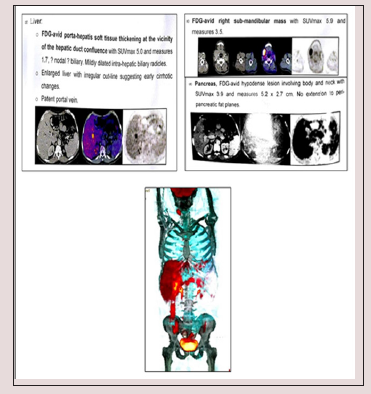
Figure 1:


PET/CT study showed hypermetabolic pancreatic body lesion, hypermetabolic porta-hepatic soft tissue thickening and metabolically active right sub-mandibular mass (Figure 1). Cholangiocarcinoma was highly suspected and internal drainage was done with two stents by Endoscopic retrograde cholangiopancreatography (ERCP) (Figure 2). Pancreatic body mass biopsy showed chronic pancreatitis and inflammatory pseudo tumor. The tumor markers were checked and showed CEA 2.75mg/ ml (<3.4) and AFP 6.2mg/ml (<8). The serum total bilirubin was declined to 1.86mg/dl after ERCP. Salivary gland excision biopsy was done which showed extensive fibrosis, macrophages and dens plasma cell infiltration, multinucleated giant cells of foreign body type and the plasma cell were positive for IgG4 monoclonal antibody with no evidence of malignancy. The IgG4-positive plasma cells account for 80 - 100 per high-power field despite normal total IgG and IgG4 serum level. The patient diagnosed as IgG4 related disease and received rituximab 1g twice with 2 weeks interval in addition to oral prednisolone 40mg/d which was tapered gradually with marked improvement clinically and laboratory.

Discussion

The IgG4-associated cholangitis (IAC) is one of the IgG4 associated sclerosing disease. In fact, the IgG4 associated sclerosing disease had been reported to involve many organs, causing IgG4 associated sclerosing pancreatitis, cholangitis, retroperitoneal fibrosis, sialadenitis, lymphadenopathy, thyroiditis, nephritis, pneumonia, prostatitis, and some inflammatory pseudotumors [17]. Overlapping of these IgG4 associated sclerosing diseases is common. They are characterized by an elevated serum IgG4, extensive IgG4-positive plasma cells and T-lymphocyte infiltration in the involved organs and well responded to steroid therapy. The pathogenesis of IgG4-associated sclerosing disease remains undetermined [18].

Diagnosis of IAC requires a high index of suspicion. The differential diagnoses include primary sclerosing cholangitis (PSC), cholangiocarcinoma, pancreatic cancer and benign traumatic biliary stricture. The cholangiographic appearance of IAC is not specific. The stricture of bile duct in IAC might be in lower end of common bile duct when combined with autoimmune pancreatitis (AIP). Some were multiple and may be in the intrahepatic or the hilar hepatic bile duct and very similar to that of Primary sclerosing cholangitis (PSC) [19-21]. When the stricture is solitary and had no other combined pancreatic disease, it will be difficult to differentiate from carcinoma. Our case had a stricture in hepatic duct and normal pancreas, which led to misdiagnosis of cholangiocarcinoma preoperatively.

The elevated serum IgG4 is a hallmark of IAC, but it is not diagnostic for the disease. Not all IAC cases have high serum IgG4 [22]. On the contrary, some cases of PSC and other diseases might have high serum IgG4. It is difficult to differentiate cholangiocarcinoma from IAC by present imaging studies [23]. Use of IgG4 immunostaining on cytology specimens is not recommended because the density of IgG4- positive cells in the tissue cannot be determined from these specimens. Mild tissue IgG4 immunostaining can occur in other diseases [24]. Therefore, endoscopic brush cytology could not help to make a diagnosis of IAC, but a malignant result of cytology could exclude IAC. Preoperative diagnosis is sometimes difficult, especially when serum IgG4 is not high. Histological examination of the surgical specimen is needed to make a final diagnosis in some rare cases. The optimal steroid treatment regimen of IAC is not defined. Most patients respond initially to steroids but relapse is not uncommon 17. In patients with IAC, careful observation for relapse of cholangitis or other possible IgG4 associated sclerosing diseases is mandatory both during and after withdrawal of the steroid therapy. Though surgery is not indicated in patients with IAC, surgery had been performed in a great proportion of patients for the difficulty in making a precise diagnosis preoperatively before.

Read More About Lupine Publishers Online Journal of Neurology and Brain Disorders (OJNBD) Please Click on Below Link:  https://brain-disorders-lupine-publishers.blogspot.com/


Wednesday, 28 July 2021

Lupine Publishers| The Relationship of the Trigemino-Cardiac Reflex to Sleep Bruxism

  Lupine Publishers| Online Journal of Neurology and Brain Disorders (OJNBD)


Abstract

The trigemino-cardiac reflex is well researched in medicine. A newly proposed classification based upon the region stimulated elucidates sleep bruxism’s paradoxical effect on the trigemino-cardiac reflex, characterized by tachycardia, hyperpnea and hypertension. This article discusses this complex relationship and the resulting signs and symptoms reported in sleep bruxism, and how it meets these new criterion.

Keywords: Trigemino-cardiac reflex; Trigeminal cardiac reflex; Masseter inhibitory reflex; Sleep bruxism

Abbreviations: TCR: Trigemino Cardiac Reflex; MIR: Masseter Inhibitory Reflex; SB: Sleep Bruxism; GG: Gasserion (trigeminal) Ganglion; GERD: Gastro-Esophageal Reflux Disorder; OSA: Obstructive Sleep Apnea; RF: Reticular Formation; TMD: Temporomandibular Dysfunction; HR: Heart Rate (pulse); MABP Maximum Arterial Blood Pressure; 5-HT: Serotonin

Introduction

The trigemino-cardiac reflex (TCR) is a unique and powerful brainstem reflex that has received a great deal of research interest. Sleep bruxism (SB) is sleep disorder that affects the TCR as well as other brainstem reflexes via stimulation of the brainstem; at the level of the gasserion ganglion (GG). This paper will discuss the unusual relationship of the TCR and SB in addition to how well sleep bruxism meets a new proposed classification system for TCR activation.

Literary Research

Relevant literature was identified through searching PubMed; Research Gate; Google scholar database and Mendeley web library using the search terms “trigemino-cardiac reflex”; “sleep bruxism”; “GERD”; and “masseter inhibitory reflex”.

Discussion

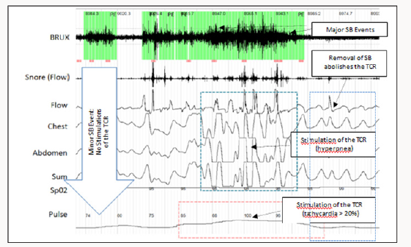
Sleep disorders are an increasing health problem in all countries that have a direct effect on quality of life and productivity/safety of workers [1-3]. Sleep bruxism (SB) is a movement type sleep disorder characterized by transient tachycardia; tachypnea and hypertension occurring slightly before; during or slightly after the bruxism event; resolving immediately after the event ceases [4].

This increase in sympathetic activity has been shown to result from stimulation of the TCR at the level of the gasserion ganglion (GGa) [5-7]. SB is seen to occur concurrently with sleep apnea; occurring immediately before or after apnea events; but may also be seen on EMG studies independently of OSA [8]. SB also results in micro-arousals from sleep; classifying it as a true sleep disorder. Epworth Sleepiness Scale scores of 4 to 9 are characteristic of SB whereas scores of 10 and higher are suggestive of OSA4. The daytime sleepiness (males) and tiredness (females) seen in SB have similar deleterious effects on alertness; productivity and quality of life to OSA. In addition; SB is associated with the inception of chronic myofascial pain [9] affecting the orofacial region; tension and migraine type headaches; and temporomandibular dysfunction syndrome (TMD) affecting the temporomandibular joints [9-10].

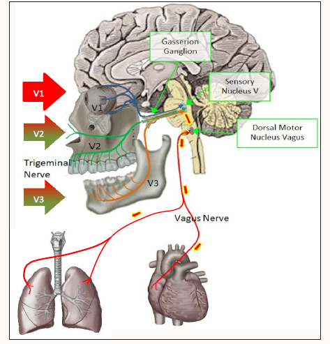
The TCR is a powerful brainstem reflex that manifests as a sudden onset of hemodynamic influences on heart rate (HR); blood pressure (MABP) and has been associated with cardiac arrhythmias; asystole; apnea and gastric mobility [11]. It is an oxygen-conserving reflex that was first discovered in 1999 [12]; with considerable research ensuing [13-17]. The reflex may be activated by mechanical or chemical stimulation of the trigeminal nerve at any course along its distribution. Stimulation of the TCR results in neuronal signals being transmitted via the trigeminal nerve to the GGa; continuing to the sensory nucleus of the trigeminal nerve (V5) in the brain stem (mesencephalic nucleus). Signals are then transmitted polysynaptically through the reticular formation (RF); via short internucial fibers; to the dorsal motor nucleus of the vagus nerve (X). This pathway is considered as an afferent to the TCR (Figures 1-3). Parasympathetic neurons comprise much of reflex; arising in the motor nucleus of V5. Stimulation of V5 results in bradycardia; hypotension; as well as apnea and gastric hypermobility [17]. The reflex is of utmost importance during surgical procedures adjacent to the branches of the trigeminal nerve as the TCR can inadvertently be stimulated compromising the surgical procedure [18-21]. In procedures near or in the GGa; the opposite effect may be encountered: tachycardia; tachypnea and hypertension and gastric hypomobility [22,23].

Figure 1:

Figure 2:

Figure 3:

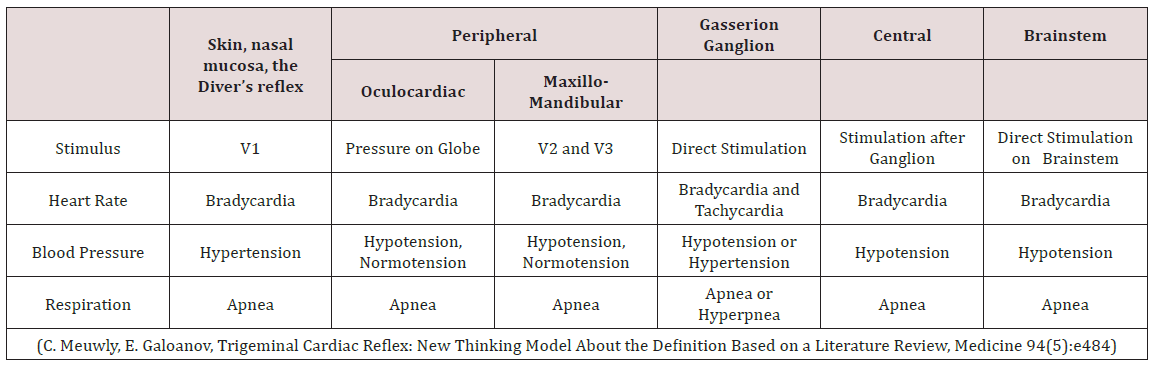
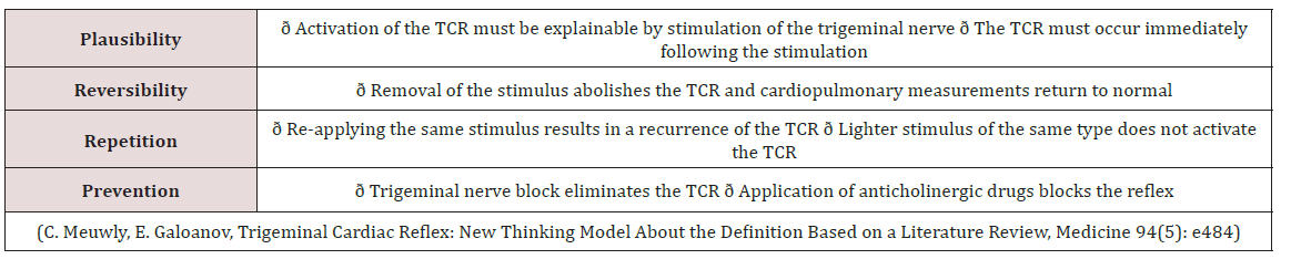
Meuwly; Galoanov et al have proposed a new classification for the TCR based upon where the trigeminal nerve is stimulated (Table 1) [17]. The classic “diver’s reflex” results from stimulation at the level of the first branch of the trigeminal nerve and the typical TCR response of bradycardia; hypotension and hypopnea. It is a cutaneous stimulation of the trigeminal nerve; on the ocular and nasal skin regions. The peripheral classification includes the actual orbit of the eye (by compressing the orbit); the maxillary branch (V2) and the mandibular branch (V3). Only the maxillary and mandibular stimulation regions result in the paradoxical sympathetic response of tachycardia; tachypnea and hypertension (Figures 1-3). In this classification; the only region of stimulation of the TCR occurs at the level of the GGa; all other regions react as expected. This stimulation at the level of the Gasserion ganglion can result in paroxysmal brady or tachycardia; hyper or hypotension and apnea or hyperpnea; depending upon the stimulus. A recent study described this phenomenon and concluded from the research that HR was the most significantly affected and the most useful measurement in the determination of TCR [24]. A diagnostic criterion that is generally accepted is of a heart rate decrease (or increase) of 20% or more from baseline; for a positive TCR diagnosis [17]. Table 2 lists other cause-effect relationship criterion proposed to diagnose and differentiate TCR proposed by Meuwly et al [17]. Sleep bruxism meets the first five of seven criteria of Table 2. GGa nerve blocks have an elevated risk profile and do not meet ethical standards for trials; and anticholinergic drugs fail to suppress the TGR effects of SB. By utilizing this additional criterion as well as the decrease (or increase in the case of SB) of HR by greater than 20% ; discrimination of other sympathetic and parasympathetic disorders that could affect HR and mimic the TCR is possible; rendering a more accurate diagnosis of this condition [24].

Table 1: The Extended Classification of the Trigeminocardiac Reflex.

Table 2: Additional Criterion based upon Cause and Effect (2 Positive Criterion = Positive TCR Stimulation).

Research has also found a genetic commonalty in SB sufferers: a polymorphism of the HTR2A gene on chromosome [13-25,26]. This gene codes for 5-HT (serotonin) receptors in the brain and gut. The masseter inhibitory reflex (MIR); another brainstem reflex; is located in the mesencephalic nucleus and has the MIR reflex nucleus affected by this polymorphism [27,28]. The resulting hypersensitivity to serotonin results in loss of inhibition by the MIR as well as activation of central pattern generators involved with chewing. In SB; extreme muscle contractions of the masseter; temporalis and suprahyoid muscle groups result [29]. The HTR2A polymorphism; seen in SB; was found to occur frequently in other conditions including obstructive sleep apnea; tension headaches; migraine headaches (without auras) and a myriad of psychiatric disorders [30-33]. Interestingly; OSA has headaches as a commonly reported symptom; often upon waking (assumed to be due to hypercapnia). In recent studies of the TCR it was demonstrated that; at the connection at the level of the reticular formation and nucleus ambiguus; there appeared to be an endogenous modulation. It was also found that the 5-HTR1A and 5-HTR2A serotonin reception genes were involved in mediation (stimulation; depression) of the connections (antagonists altered the TCR) [17]. With the 5-HTR2A polymorphism known to exist in SB; it is reasonable to assume that this and other regions could also be similarly affected (potentially resulting in hypersensitivity of the TCR).

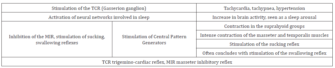
During SB bursts; there is an ensuing cascade of events that occurs (Table 3). There are a number of cranial reflexes involved including the TCR; the MIR; the sucking reflex 4 (which can result in trauma of the buccal mucosa and development of linea alba lesions); and the swallowing reflex [34-36]. Scalloped borders of the tongue are also a common finding in SB; resulting from activation of the genioglossus muscle pressing the tongue forcibly against the teeth [37,38]. In other research it has been shown that there is a commonalty between SB and gastro-esophageal reflux disorder (GERD); supporting SB’s influence on the vagus nerve [39,40]. This can manifest as tooth erosions on the inner or lingual surfaces of the teeth and increase susceptibility to dental caries [41].

Table 3: The Sleep Bruxism Cascade.

With the suppression of the MIR; the forces generated during SB events far exceed those of normal chewing [29]. Damage to the teeth is common including:

a) Sensitive; loose or broken teeth [44]

b) Abfraction lesions [44]

c) Accelerated periodontal disease [45]

d) Formation of mandibular tori [46]

e) Eagle’s syndrome (calcification of the stylohyoid ligaments) [47]

f) Elongation of the coronoid processes [48]

g) Excessive compression of the TMJ; often initiating or accelerating degenerative changes [49-51].

As expected; these forces can be a contraindication to some dental procedures including porcelain crowns and inlays [52,53] and dental implants [54,55].

Conclusion

When considering how SB affects the TCR and MIR; the range of signs and symptoms observed during SB events can be readily interpreted. Restless leg syndrome (RLS) is another movement type sleep disorder that results in activation of the TCR similar to SB. Tachycardia; hyperpnea and hypertension (TCR stimulation); shown to also occur in RLS; has been shown to be a risk factor for heart disease in a number of studies [56,57]. To date SB has not been studied to the extent of RLS and it is not known at this time if SB is also a risk factor for heart disease. With the significant effect of SB on the TCR; Further research is certainly warranted.

Read More About Lupine Publishers Online Journal of Neurology and Brain Disorders (OJNBD) Please Click on Below Link:  https://brain-disorders-lupine-publishers.blogspot.com/

Monday, 26 July 2021

Lupine Publishers| Is Treatment Resistant Focal Epilepsy Less Frequent in Veterans?

 Lupine Publishers| Online Journal of Neurology and Brain Disorders (OJNBD)


Abstract

Rationale and objective: Epileptic seizure disorders have become an increased source of concern in veterans given their relative high exposure to traumatic brain injury (TBI). 40% of adults with focal epilepsy are expected to develop treatment-resistant epilepsy (TRE), which can be amenable to treatment with epilepsy surgery. Yet, epilepsy surgery in veterans with epilepsy (VWE) is performed less frequently than in non-veterans with epilepsy. One possible explanation may be that when seizures begin after the age of 50, seizure freedom is likely to occur in 70% of patients. The purpose of this study was to examine whether the frequency of treatment-resistant epilepsy was different in veterans and to identify potential variables that may account for this difference.

Methods: In this retrospective study we included 157 veterans followed in the outpatient clinic of the Miami Epilepsy Center of Excellence Veterans Health Administration. Data collected from the medical records included age at onset of epilepsy, etiology, seizure type and epilepsy syndrome, response to pharmacotherapy, presence of psychiatric co morbidities (classified as mood disorders, generalized anxiety disorder, post-traumatic stress disorder, polysubstance abuse and other), antiepileptic regimen and adherence to medical treatment.

Results: Among the 157 patients, the mean age was 56.7 (±15.4) years and 140 (88.5%) were males; 119 patients (75.7%) had focal epilepsy presenting with complex partial with or without secondarily generalized tonic-clonic (GTC) seizures. TRE was identified in 25 patients (15.9%; 95% confidence interval: 11.0% to 22.5%); being a woman (p<0.01) and having focal epilepsy (p=0.04) were the only two significant variables associated with the development TRE.

Conclusion: In this study, the prevalence of TRE in this cohort of veterans was lower than that reported in the general epilepsy population. These findings need to be replicated in a larger study that includes the 16 VA Epilepsy Centers of Excellence.

Keywords: Focal epilepsy; Treatment resistant epilepsy; Major depressive episode; Traumatic brain injury; Posttraumatic stress disorder

Abbrevations: TBI: Traumatic Brain Injury; TRE: Treatment Resistant Epilepsy; VWE:Veterans with Epilepsy; GTC: Generalized Tonic Clonic; ILAE: International League against Epilepsy; PWE: Patients with Epilepsy; AEDs: Antiepileptic Drugs; PTSD: Post Traumatic Stress Disorder

Introduction

Epileptic disorders have become an increased source of concern in veterans given the relative high exposure to traumatic brain injury (TBI), particularly because of the increased number of veterans who have suffered from serious TBI in the course of the wars in Aphganistan and Iraq. Furthermore, TBI can often result in the development of treatment-resistant focal epilepsy (TRE), for which epilepsy surgery can at times be one of the potential treatments. Yet, the use of epilepsy surgery among the 16 VA Epilepsy Centers of Excellence during fiscal year 2016 revealed only 8 surgical procedures in veterans with epilepsy (VWE) among a total of 5.980 unique patients seen during 2016 [1]. The purpose of this study was to investigate whether TRE in VWE followed at one Epilepsy Center of Excellence differed from that reported in the general population of patients with focal epilepsy and if so, to identify potential causes for such difference.

The international League against Epilepsy (ILAE) proposed the new definition of TRE as a failure to reach seizure remission after adequate trials of two tolerated, appropriately chosen and used antiepileptic drugs (AEDs) [2]. It is estimated that between 30% and 40% of patients with epilepsy (PWE) fail to reach seizure freedom, despite multiple trials with AEDs [3]. Several variables may contribute to the development of intractability, including lack of response to the first AED, specific syndromes, symptomatic etiology, family history of epilepsy, psychiatric co morbidity, high frequency of seizures, and early age at onset of epilepsy [4,5]. These observations suggest that prognosis of the seizure disorder can often be determined in the early stages of the disease. Recognition of these variables has a direct bearing on the management of these patients, as they may ensure an early referral for a pre-surgical evaluation and when possible, may shorten the medico-social and economic burden of intractable epilepsy [6]. On the other hand, some studies have suggested that late-onset epilepsy (beginning after the age of 60) may be more responsive to medical management than epilepsy diagnosed during adolescence and early adulthood [7-9].

Methods

This was a retrospective cross-sectional study. We reviewed the medical records of every consecutive patient with epilepsy followed at the outpatient epilepsy clinic of the Miami VA Healthcare System (VAHS) Epilepsy Clinic over a period of 24 months (January 2010 to January 2012). The study was approved by the Miami VAHS institutional review board. Two Board-certified epileptologists confirmed the diagnosis of epilepsy using the ILAE criteria (2010), based on the clinical history, neuro imaging data and electrographic recordings. The data extracted from the medical records included demographic variables (age, gender, ethnicity) epilepsy-related variables (seizure type(s) and epilepsy syndrome (focal or generalized), etiology of the seizure disorder (unknown, remote symptomatic [tumor, stroke, TBI, or “other”] or idiopathic), age of onset of epilepsy, co morbidities (medical, cognitive and psychiatric co-morbidities [classified as mood and /or anxiety disorder, post-traumatic stress disorder (PTSD), and polysubstance abuse/dependence, other] AED treatment, were recorded. We used the ILAE definition of TRE cited in our Introduction [2]. We defined epilepsy “in remission” as the absence of seizures in the last two years from the time the patient was evaluated at the epilepsy clinic. Of note, any patient with persistent seizures and/or in whom the clinical semiology of their seizures was not typical of an epileptic seizure underwent a diagnostic video-EEG monitoring study to establish if these events were epileptic or non-epileptic events and if epileptic seizures, to identify the type of seizures.

In this study 15 of the 25 patient with TRE underwent video- EEG monitoring, including all the women in this case series. The clinical characteristics of seizures of the 10 patients with TRE who did not have a video-EEG monitoring study were typical of epileptic seizures. Patients who were thought to have psychogenic non-epileptic seizures were excluded from the study. In addition, 9 patients who had persistent seizures but had only undergone one optimal trial with an AED were excluded. A total of 157 patients with epilepsy were included in this study. Demographic, epilepsy-related variables and data of psychiatric co morbidities were extracted from the medical record and included: age, gender, ethnicity, race, seizure type (focal, generalized), whether the seizure disorder was idiopathic, Symptomatic or unknown), age at onset of epilepsy, list of current AEDs and dosage (s), and response to each AED trial. Data of psychiatric co morbidities included mood disorder, anxiety disorder, post-traumatic stress disorder (PTSD), polysubstance abuse and other psychotic spectrum disorders such as schizophrenia, schizoaffective disorder and psychosis NOS.

Data Management and Analysis

Age was treated as a continuous variable and t tests for independent groups were used to compare data between TRE and seizure free group. Categorical variables were summarized by accumulated percentages. Chi-square or Fisher exact tests were used to compare the categorical variables for association between seizure freedom and clinical factors. Where the expected counts were less than 5, Fisher exact tests were performed. For all analyses, p≤ 0.05 was defined as statistically significant. Statistical analyses were performed using SAS software version 9.2 (SAS Institute, Cary, NC, USA).

Results

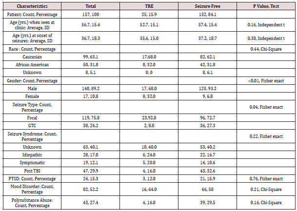
Among the 157 patients included in our analysis 25 patients met criteria of TRE and 132 were seizure-free. (Table 1) summarizes the data pertaining to the demographic, epilepsy-related and co morbidities variables among the seizure-free and TRE patients. The prevalence of patients with TRE was lower (15.9%, 95% confidence interval (CI): 11.0% to 22.5%) than that reported in multiple studies, which ranges between 30% and 50%) (2, 10-12). Had we included in our analysis the 9 patients with persistent seizures that had been tried on one AED, the percentage of patients with TRE would have only increased to 21.6% (95% CI: 15.9% to 28.7%), still below the expected rate. Two variables were significantly associated with the development of TRE in our patients: female gender (47%) and focal epilepsy (92.0 %). See (Table 1) for statistical analysis.

Table 1: Characteristics of Study Cohort (N=157).

TRE: Treatment resistant epilepsy; SD: Standard deviation, GTC: Generalized tonic clonic

TBI: Traumatic brain injury, PTSD: Post traumatic stress disorder

Percentages are reported to one decimal digit

Discussion

Epilepsy has become a neurologic disorder of great concern among veterans, given the large number of soldiers returning from the wars in the Middle-East where they suffered TBI [10]. This study was developed to assess the cross prevalence of TRE among outpatient veterans followed in a VA Epilepsy Center of Excellence and compare it to that published in populations of non-veteran PWE. To our surprise, the prevalence of TRE was lower in our patients than that published in studies conducted in non-veterans [11,12]. The second surprising finding was the observation that TRE was significantly more prevalent among women veterans. At this point, we cannot explain the reasons for a lower prevalence of TRE in our patients and our findings will need to be replicated in larger prospective studies. Of note, neither the cause of epilepsy, nor the age of onset of the seizure disorder failed to account for a better seizure control in our cohort, as suggested by several published studies that had suggested that elderly patients are more likely to have good outcomes [13,14].

In PWE, those with symptomatic or cryptogenic epilepsy are more likely to be medication resistant than those with idiopathic epilepsy [8,15]. In our study, the etiology of seizures did not play a factor in developing intractability. Traumatic brain injury was the most common cause of symptomatic epilepsy but failed to represent a predictive factor of intractability-perhaps because there are few cases of penetrating head injury, which is most commonly associated with medial intractability. Initial seizure frequency has been identified as a significant prognostic factor for TRE in the literature [16]. Unfortunately, we were not able to investigate this issue in a reliable manner because most of the patients were diagnosed somewhere else several years prior to our evaluation and the information was not found in the retrospective review of the medical records. This study has several limitations. First, this was a retrospective study. Second, we relied on the patients’ self (and or family members’) reports of their epileptic seizures. Yet, patients may have seizures and not be aware of their occurrence, which could result in an under-estimation of seizure frequency. Third, our study was based on data from a single Epilepsy Center of Excellence. While these are the centers where veterans with TRE are referred for treatment in the VA system, our data will need to be replicated in a larger study that includes all 16 Epilepsy Centers of Excellence in the USA.

Conclusion

The findings of this study may suggest that TRE may be less frequent in VWE than in other published cohorts and that women veterans may be at greater risk. These findings have to be considered as preliminary but deserve a careful investigation in a larger study that may include the other 15 Epilepsy Centers of Excellence of the VA and which can be compared with non-veteran populations.

Read More About Lupine Publishers Online Journal of Neurology and Brain Disorders (OJNBD) Please Click on Below Link:  https://brain-disorders-lupine-publishers.blogspot.com/