Background: The ruminant claw horn lesions seriously diminish histological structure and functional efficiency of the digital
cushion of the dairy cows.
Objective: To investigate histological and histochemical properties of the digital cushion in the dairy cow with claw horn
lesions, such as white line disease, sole damage and hemorrhage.
Results: The results showed that digital cushion loses its normal histological structure in claw horn lesions.
Conclusion: Further biochemical, physiological and feeding
studies on the experimentally induced claw horn lesions are
necessary to understand the nature of the events playing roles involving
animal feeding in the development of the lesions, to develop
new animal feeding strategies aiming to protect and maintain the normal
structure of the digital cushion.”
Keywords:Protein Degradability; Goats; Intensive Feeding; Nitrogen Balance; Digestibility
Introduction
Lameness is one of the most important problems negatively
affecting milk yield and animal welfare in modern dairy farming
in high-performance animals. The cows with laminitis produce
lower amount of milk [1-5]. Moreover, cyclic reproductive changes
of these cows slow down [6] or cease because of anoestrus [7]
or cystic ovarian disease [8]. Lameness is a major animal welfare
problem because that it is a painful condition and causes to
behavioral changes of the affected cow. A lame cow tends to display
low mobility [9], to spend decreased daily feeding time and dry
matter intake [10] and lying behaviour may be indicative [11]. In
the dairy cow, claw horn lesions [CHL] constitute 65% of the total
lesions causing to lameness [12,13]. Although possible roles of risk
factors, such as claw conformation and trimming, animal welfare,
management, feeding and housing have been evaluated in detail
by previous researchers [14-17], pathophysiology of CHL is still
unclear. Previous researchers [18,19] have suggested that CHL is
arisen from traumatic lesions of the supportive tissues of the claw.
Around the time of calving, increase of hoofase enzyme, oestrogen
and relaxin secretions results in loosening and increased mobility
of suspensor apparatus of third phalanx [19]. The bovine digital
cushion locating under the third phalanx has a complex structure,
which major part is constituted of fat tissue. The cushion comprises
three cylindrical segments [axial, medial and abaxial segments] of
adipose tissue surrounded by a thick connective tissue trabecules,
those arranged in parallel to each other [20,21]. The pads of the
bovine digital cushion play crucial roles in reducing the body
load transferred to surface of sole via absorbing of substantial
forces acting within the claw [22]. The biomechanical function
of the digital cushion in distributing and attenuation of the load
transferred to the base of flexor process of the distal phalanx
have been well appreciated by previous researchers [21,23,24]. The aim of the present study was to determine histological and
histochemical properties of the digital cushion in the heifers and
primiparous, second and third parturitions, and multiparous
dairy cows managed under similar feeding, housing and welfare
conditions. The foot was chosen from the animals with CHL, such as
white line disease, sole damage and haemorrhage, in order to define
the histology and histochemical changes in the digital cushions of
the animals.
Material and Methods
Animals
This experiment was approved by the Ethical Committee of
Experimental Animal Production and Research Centre (2011/023)
of Veterinary Faculty of Selçuk University. In the study, the
120-cadaver foot (totally 240 hooves) of Holstein heifers and
cows were used as materials. The animals were culled because
of moderate to severe CHL and lameness in one or more hooves.
The claws of the animals with similar feeding, housing and welfare
conditions were subjected to a detailed clinical examination prior
to slaughter. Information on the age, number of parturitions, calving
history and stage of lactation were and recorded, in addition to
the clinical findings. Among the reasons given for culling were
insufficient milk yield, lameness and infertility. The animals were
divided into 5 groups, each consisted of 6 animals, as
group I: heifers,
group II: primiparous cows,
group III: cows with second parturition,
group IV: cows with third parturition,
group V: multiparous cows with more than three calving.
The first group animals were 20 months old averagely; other
groups were between 2.6-6.2-year-old. After slaughtering, all the
foot was cleaned, and lesions, such as discoloration, sole ulcer and
hemorrhages were recorded, frozen at -20°C and kept until use for
histological procedure.
Dissection of the Hooves and Obtaining Tissue Samples
Each foot was thawed. The soft tissues of the sole and heel of
each hoof were separated following the horn had been removed,
in order to fully expose the axial, middle and abaxial pads of the
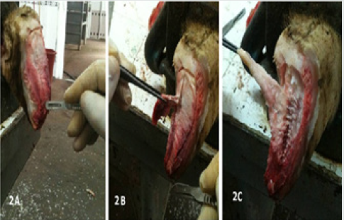
digital cushion (Figures 1A-B & Figures 2A-C) [18]. Axial pad of
the cushion was totally removed, fixed in 10% phosphate buffered
(0.1M pH 7.4) formal-saline and used for histological procedures.
Figure 1: The study area, green - places of mass appearance of painted lady butterflies (Vanessa cardui L.) in 2019.
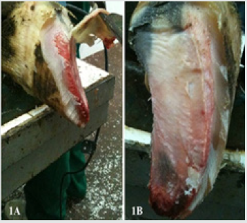
Figure 2: Separating the corium (2A), dissecting pads of the digital cushion (2B) and totally removing the pads (2C).
Histology and Histomorphometry
The tissue samples were subjected to routine histological
tissue processing. Briefly, the tissue samples were washed with tap
water overnight, dehydrated in ethylene series, cleared in xylene
and immersed in paraffin. The sections taken at 6μm were stained
as follows; hematoxylin-eosin for investigating general histology
and histological changes [25], safranin-O (SO) for histochemical
properties of ground substance, alcian blue at pH 2.5 (AB 2.5) for
staining acid mucins and mucosubstance, alcian blue at pH 4.0
(AB 4) for demonstrating carboxylated glucose aminoglycans in
the ground substance, trichrome stain for Type I collagen fibres
(Coll I) and other histological detail, Verhoef’s elastic fibre (EL)
stain for EL fibers [26]. The specimens were investigated under
light microscope equipped with digital imaging system and digital
images were recorded. The images were analyzed with digital
image analysis software (BS200 PRO-2005).
Statistical Analysis
The data obtained from the digital image analyses were
analyzed statistically with one-way variance analysis (ANOVA) and
Tukey’s pair wise comparison tests by using SPSS software 14.01
(Release 14.01 Licence No: 9869264).
Results
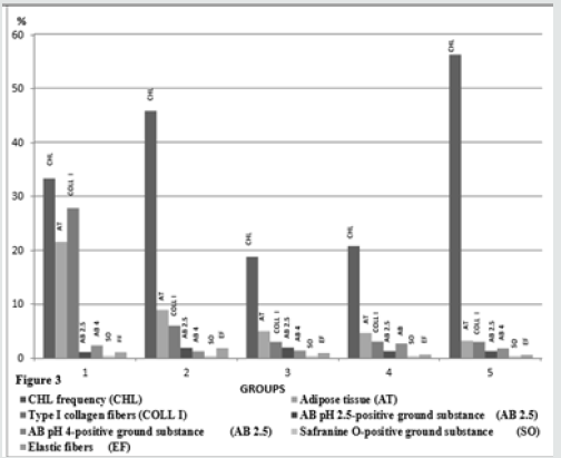
Figure 3: CHL incidence and histometrical results of the groups.
Clinical findings of the animals prior to slaughter and
results of postmortem gross examination of the foot Welfare and
management of the animals included into the study were far from
than satisfactory. The animals were housed on the concrete floor,
under poor hygiene conditions, and claw trimming was irregular.
The animals were mainly fed with corn silage, alfalfa, vetch and hay.
Findings of the postmortem examinations of the foot showed that
the animals suffered from CHL, such as sole ulcers, hemorrhage
and folding, white line disease and sole erosions in one or more
hooves. The highest percentage of the claws with CHL was in the
multiparous cows, in the group 5. The groups 2 and 1 the group
followed it respectively. The lowest lower CHL frequencies were
found in the groups 3 and 4, and the frequencies of these groups
were quite similar (P>0.05, Figure 3).
Gross Macroscopic Findings of the Cushion Segments
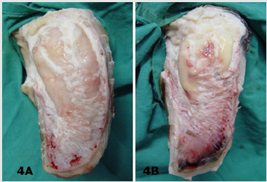
In most of the claws with CHL, segmental structure of the
digital cushion was diminished. The digital cushion tissues of the
heifers (Group 1) were pale and softer, whereas in the cows with
the increasing the parturition number, the tissues gained yellowish
colour and relatively more brittle and fragile. The cushion segments
were also smaller in the animals of the groups 4 and 5, aged and
multiparous cows (Figures 4A & 4B).
Figure 4: Macroscopic appearance of digital cushion
tissues of a heifer from the group 1 (4A) and a cow from
the group 5 (4B). The cushion pads of the group 5 animal
are smaller and yellowish in colour.
Histological Findings
In general, the digital cushion pads of the animals were mainly
formed of a loose connective tissue and unilocular adipose tissue.
Adipocytes appeared as large cells with unstained cytoplasm in all
of the used staining procedures (Figures 5-9). Fibrous connective
tissue septa surrounded the loose connective and adipose tissues.
There were striking structural differences in the histology of the
digital cushion of heifers and calved cows. Loose connective tissue
in the heifers, was replaced with adipose tissue in the primiparous
cows, and fibrous connective tissue rich in collagen fibres in
the third lactation cows. In the heifers and primiparous cows,
adipocytes were seen as groups, whereas in the cows of the groups
3, 4 and 5, the cells were observed as individual cells and the cell
groups consisting of small numbers of the adipocytes. Adipose
tissue percentages of the groups are given in the Table 1. The highest
adipose tissue percentage (21.55%) was in the heifers, group 1, and
the value was significantly (P<0.001) higher than those of the other
groups (Figure 3). The groups 3 and 4 displayed similar (P>0.05)
adipose tissue percentages. The multiparous animals (group 5) had
the lowest adipose tissue percentage (Figure 3). Type I collagenous
fibres were seen as thick undulating bundles of the fibres darkly
stained with aniline blue in trichrome stained sections (Figures 5A-
5E).
Figure 5: Collagen fibre bundles (arrows) and adipocytes
(asterisks) are seen in the sections from lateral claw cushion of the
right forefoot of the heifer from the group 1 (5A), in the medial claw
cushion of the left hindfoot of the animal from the group 2
(5B), in the lateral claw cushion of the left forefoot of the animal
from the group 3 (5C), medial claw cushion of the left hindfoot
of the animal from the group 4 (5D) and lateral claw cushion of the left
hindfoot of the animal from the group 5 (5E) are seen.
In contrast to a definite decrease in the adipose tissue rate, the
connective tissue gradually increased from the group 1 to the
group 5. Crossmon’s trichrome. Magnification bar: 100μm.
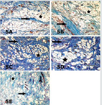
Figure 6: Sections of the digital cushion stained with AB pH
2.5 from the medial claw of the left forefoot of the animal from
the group 1 (6A), the lateral claw of the right forefoot of the animal
from the group 2 (6B), the lateral claw of the right hindfoot
of the animal from the group 3 (6C) are seen. Staining intensity of the
ground substance (arrows) in different sections is quite
similar. Asterisks depict the adipocytes. AB pH 2.5 stain. Magnification
bar: 100μm.
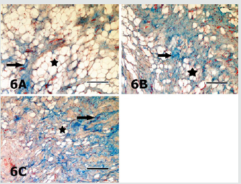
Figure 7: Sections of the digital cushion stained with AB pH 4 from the medial claw of the left hindfoot of the animal from
the group 1 (7A), the medial claw of the left hindfoot of the animal from the group 4 (7B), the medial claw of the left hindfoot
of the animal from the group 4 (7C) and the medial claw of the right hindfoot of the animal from the group 5 (7D) are seen.
Slight differences in staining intensities of the ground substance (arrows) in different sections are seen. Asterisks depict the
adipocytes. AB pH 4 stain. Magnification bar: 100μm.
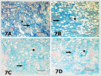
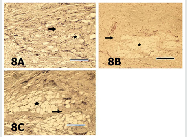
Figure 8: Sections of the digital cushion stained with
safranin O from the lateral claw of the left hindfoot of the animal from
the group 1 (8A), the lateral claw of the left hind foot of the animal
from the group 2 (8B) and the lateral claw of the right fore
foot of the animal from the group 3 (8C) are seen. Staining intensities
of the ground substance (arrows) in different sections are
quite similar. Asterisk depicts the adipocytes. SO stain. Magnification
bar: 100μm.
The heifers had significantly (P<0.001) higher type I collagen
fibre percentage (27.82%) than those of the other groups those
had quite similar (P>0.05) percentages (Figure 3). In AB pH 2.5
stained specimens, the AB positivity was mainly located in the
ground substance around the connective tissue fibres, embedded
in-between the adipocyte groups (Figures 6A-6C). The positivity
was also stronger around the individual adipocytes and the groups
constituted of small numbers of these cells. Percentage of the AB pH
2.5 stained ground substance was highest (2.08%) in the group 3
and group 2 followed it (1.96%), whereas the groups 1, 4 and 5 had
quite similar (P>0.05) and significantly (P<0.01) lower values than
those of the groups 2 and 3 (Figure 3). AB pH 4.0 mainly stained
the similar ground substance regions with AB pH 2.5, around the
connective tissue fibres, located around the adipocyte groups
(Figures 7A-7D). Results of the histometrical investigations of the
AB pH 4.0 stained specimens showed that the group 1 and group
4 had similar (P>0.05), and significantly higher (P<0.01) positivity
percentages than those of the groups 2, 3 and 5 (Figure 3). In SO
stained specimens, the positivity was very weak (Figures 8A-8C)
in all groups. The positivity percentages of the groups were quite
similar and there were no significant differences (P>0.05) between
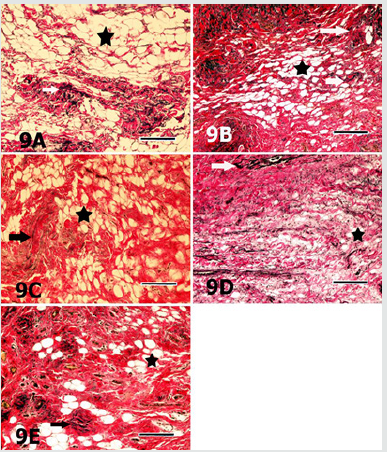
the groups (Figure 3). Elastic fibres were mainly found in-between
the connective tissue trabecules surrounding adipocyte groups
and tunica adventitia of the blood vessels, and seen as brownishblack,
branching coarse and gently undulating fine fibres, in the
sections stained with Verhoeff’s elastic fibre stain (Figures 9A-
9E). The results of digital image analyses showed that the group
2 had the highest (1.89%) elastic fibre percentage and the value
was significantly (P<0.05) higher than those of the other groups.
The groups 1and 3, and 4 and 5 had quite similar elastic fibre
percentages (P>0.05, Figure 3).
Figure 9: Sections of the digital cushion stained with
Verhoef’s elastic fibre stain from the medial claw of the right hindfoot
of
the animal from the group 1 (9A), the medial claw of the left hindfoot
of the animal from the group 2 (9B), the lateral claw of left
forefoot of the animal from the group 3 (9C), medial claw of the left
hind foot of the animal from the group 4 (9D) and the lateral
claw of the left hind foot of the animal from the group 5 (9E) are seen.
Elastic fibres (arrows) are mainly located in connective
tissue trabecules surrounding the adipocyte groups (asterisks).
Verhoef’s elastic fibre stain. Magnification bar: 100μm
Discussion
In the present study, statistically significant (P<0.01)
differences were found between the CHL frequencies of the groups.
Particularly, high incidences of CHL in the primiparous cows
(45.80%, group 2) and multiparous (56.20%) cows (group 5) were
striking. The differences possibly have arisen from higher incidence
of CHL in more than one or all foot of the animals in these groups.
Previous researchers [27,28] have reported that the histological
development and functional maturation of the digital cushion are
not completed before the age of three, and also incidence of sole
lesions, such as sole ulcerations and white line disease are relatively
higher in the first lactation because that the cushion tissues is
less elastic and rich in saturated fatty acids. In accordance with
the previous researchers [27,28] Incidence of CHL was higher in
primiparous cows in the present study. The roles of the additional
loads and forces exposed at late pregnancy, and structural and
biochemical changes in the regional tissues, in addition to trauma
of both corium of sole and the cushion tissues in the pathogenesis
and occurrence of CHL have been discussed in detail by previous
researchers [17-19,29,30]. Moreover, collaborative effects of
improper, inadequate feeding and housing practices are mentioned
among significant disposing factors. In coordination of anatomic
structures of the hoof [bones, joints, ligaments and bursae] leads to
CHL, mainly sole ulcers and white line disease. Thus, hoof problems
occur more commonly among the cows kept in cubicles or concrete
yards or straw yards than in the cows kept in cowsheds. Previous
researchers [18,31,32] have reported that lameness incidence
increases in multiparous cows with increase of ageing and calving,
especially after third lactation, due to increasing the amount of the
loose connective tissue while reducing adipose tissue, decreasing
body condition score [BCS], thinning of the cushion segments. In
the present study, a gradually decline was found in adipose tissue
percentages of the groups, with ageing and increasing parturitions.
Loosening of suspensor ligament system of the hoof results in
increasing in mobility of bones, and this situation disposes the sole
corium and sole horn to traumatic lesions, and consequently plays
significant roles in the development of CHL, such as sole ulcers,
white line disease [19,33]. Loosening of suspensor system of the
hoof also increases the sensitivity of the claw to the CHL depending
on increase of loads applied to the foot by increase in mammary
gland and foetus weights, especially during first lactation [34].
Clinical findings and histometrical results of the hooves and higher
CHL incidence of the groups 2 and 5 are consistent with previous
findings [18,31,32]. Moreover, higher CHL incidence of the group 5,
in which significant shrinkage of the cushion tissue was observed,
supports the hypothesis suggesting the decline in load absorbing
capacity of the cushion increases risk of injury of sole corium. Thus,
environmental factors, such as long time standing on the concrete
floor, unsuitable housing conditions and in proper claw trimming
are effective in embodiment of the sole trauma and haemorrhages
in addition to increase of coffin bone motility in late pregnancy
and parturition. Unsuitable housing and hygiene conditions in the
animals of the present study were in accordance with the high CHL
incidence of the animals.
The digital cushion, which is a specified connective tissue, is
constituted of connective tissue cells and intercellular substance
[ICS]. The ICS contains collagenous, elastic and reticular fibres
embedded in an amorphous ground substance. The amount of
the amorphous ground substance in young animals is higher than
fibrous elements. With ageing, the amount and rate of fibrous
elements increase and the rate reverses in older animals. The
digital cushion, a member of supporting system of the ruminant
hoof locating in subcutis, is a complex structure, composed of white
and yellow adipose tissues. There are significant differences in
histological structure and organization of the digital cushion among
the ungulate animal species. The digital cushion of the ruminant
hoof is closely resembled with the cushion of the elephant’s cushion
[18]. Equine digital cushion has relatively different structural
features. In the horse, the cushion is composed of small number of
elastic fibres and closely packaged interlacing collagenous fibres
dispersed in acidic mucous matrix, rich in hyaluronic acid. Large
spaces between the cells and fibres occupied with mixoid tissue
and islets of fibro cartilage tissue [35,36]. Collagen fibres,
especially
type I fibres are the dominant fibre type of the connective tissues.
Although the collagenous fibres have limited elasticity, their tensile
strength is enough to meet the high tensile forces of the foot. In the
present study, axial pad of the digital cushion constituted of white,
unilocular adipose tissue lobuli surrounded by connective tissue
trabecules. Fibro cartilage was not observed in the specimens. In
some of the groups, trabecules surrounding adipose tissue lobuli
constituted of loose connective tissue and contained less amount
of type I collagen fibres, the others were rich in the collagenous
fibres. Group 1 contained significantly (p<0.001) higher collagen
(27.82%) when compared with the other groups. A gradual decline
of type I collagenous fibre percentage was typical in the remaining,
older and multiparous groups (groups 3, 4 and 5). Nevertheless,
they contained quite similar (P>0.05) type I collagen fibre rates.
Gradual reduction of type I collagenous fibre rate with the increase
of age and calving might arisen from the structural and functional
changes occurred in aged animals and during the late pregnancy
and post parturition. It might be predicted that shock absorbing
capacity of the digital cushion considerably reduced with declining
of collagen fibre rate. In accordance with this prediction, the highest
(56.20%) CHL incidence was found in the group 5, which had
relatively low rate of type I collagen fibres. Moreover, in accordance
with the prediction above, previous researchers [19,33,37], also
suggested that loosening of the suspensor system of the hoof during
pregnancy and post parturition increases sensitivity of sole corium
located between coffin bone and sole to trauma, and consequently
playing significant roles in the pathogenesis of the CHL, such as sole
ulcers and white line disease.
Although the information on the types and distribution of
adipocytes in ruminant digital cushion is limited, the heifer cushion
contains lesser amount of the fat cells. The digital cushion of the
heifers is a whitish and gelatinous in nature containing high amounts
of ground substance and lipid content gradually increases through
2-3 parturitions, and gradually decrease starting from 3 years of
age [18,21,32]. Because that white fat tissue is an energy source,
many metabolic and hormonal mechanisms, such as pancreatic
hormones and glucocorticoids are efficient on this fat tissue. Thus,
feeding and other metabolic factors are likely to efficient on both
amount and composition of fat of the digital cushion. Moreover, it
is well known that the fatty acid composition and amounts of the
cow’s digital cushion are seriously affected by metabolic disorders,
mainly by ketosis. Similarly, previous researchers [38] emphasized
that the topic needs detailed evidences. In the presented study,
the digital cushion of the dairy cattle comprised of islets of large
unilocular adipocytes. In the digital cushion with low levels of fat
tissue, individual adipocytes were frequently seen. The animals
in the group 1 contained significantly (P<0.001) higher adipocyte
rates (%21.55) and the group 2 it, with a significant decline.
However, the group 2 contained significantly (P<0.05) higher fat
tissue percentage than those of the groups 3, 4 and 5. Rastani [39]
suggested that lipids are mobilized from fat tissues in order to
be used in milk synthesis. Lipids in white fat depots in the digital
cushion are also consumed. Considering the data obtained in this
study, it is possible to assume that white adipose tissue in the
ruminant digital cushion also can be used as an energy source in
ketosis, which is a condition resulting in negative energy balance
[NEB], and thus CHL increase post parturition period. It is well
known that fat and elastic fibres of the digital cushion play
significant
roles in shock absorbing mechanism of the cushion via expanding
sideways until their viscoelasticity is limited by stretched fibrous
connective tissue septa rich in collagen fibres. Elastic fibres support
expansion by flattening their undulations and support reversing to
the situation before when the applied force is expired. Significant
decreases in both elastic fibre and adipose tissue rates result in loss
of elasticity of the digital cushion.
There is limited information on the histochemical properties of
ground substance of the cow’s digital cushion, and also histometrical
results evaluation of these parameters have not been documented
previously. In the present study, the results of the AB pH 2.5, AB
pH 4.0 and SO stains showed that ground substance of the cow’s
digital cushion was rich in hyaluronic acid (hyaluronan) and other
proteoglycans, although there were some differences between the
groups. Because that hyaluronic acid is a macromolecule increasing
the viscosity of the tissue fluid, it consequently augments resilience
of the tissue by binding water molecules. Other proteoglycans
stained with SO might support the digital cushion in resisting to
pressure, since the proteoglycans also show high water binding
affinity and tend to gelation [40]. Although the results of the
present study indicated that the ground substance of cow’s
digital cushion was rich in hyaluronic acid, the hoof material was
obtained from the animals with varying degree of CHL lesions.
The authors strongly stress that a detailed comparison should
be carried out between the findings of healthy and CHL animals.
The elastic fibres, which relatively thinner when compared to the
collagen fibres, are able to compensate the forces applied between
elasticity limits. Tissues gain elasticity and stress bearing features
by their elastic fibre content. Elastic fibres are found in hoof soft
tissues as in other elastic tissues. Information on distribution and
localization of these fibres in the ruminant hoof soft tissues is
insufficient. In a previous histological study [38], elastic fibres were
found between the collagen fibre bundles in the digital cushion of
African elephant. In this study, elastic fibres were more peculiarly
observed in-between the collagenous fibre bundles and in blood
vessel walls. Morphometrical analyses evidenced that the group
2 displayed significantly (P<0.05) higher elastic fibre percentage
(1.89%) than the other groups. Although there was a declining
tendency with increase of the parturition number, the other groups had
relatively similar elastic fibre ratios. Besides, striking decline of
the elastic fibre percentage was determined in the groups 4 and 5
in concomitant with higher CHL lesion incidence. Although further
studies are needed, the results of the present study imply that
elastic fibre content of the cow digital cushion is quite limited and
might play only small role in the functions of the digital cushion and
in the development CHL lesions.
Conclusion
Based on the results, it was concluded that CHL seriously
affected both histology and functional efficiency of the digital
cushion of dairy cows. Results of the present study, which
show strong evidence between histological, histochemical and
histometrical findings and CHL lesions are noteworthy. However,
the results should be compared with those of the healthy animals
in further experiments those are involving experimental models.
Further studies necessary to understand the nature of the events
playing roles involving animal feeding in the development of the
lesions, to develop new animal feeding strategies aiming to protect
and maintain the normal structure of the digital cushion, in order
to maintain and augment its supportive and protective roles, to
reduce incidence of laminitis.
Grant Support
This work was supported by a grant from The Scientific and
Technological Research Council of Turkey with project no: TOVAG
112O332.
Read More About Lupine Publishers Journal of Dairy Please Click on Below Link:
https://lupine-publishers-dairy-veterinary.blogspot.com/