Showing posts with label CTGH. Show all posts

Tuesday, 29 August 2023

Lupine Publishers | Food Sources and Bioavailability of Calcium

 Lupine Publishers | Journal of Gastroenterology and Hepatology


Abstract

The identification of the calcium ration by self-questionnaires validated in the region of Blida and the Wilayas of approximately is one of the rare studies in Algeria. She was interested in a part of the Algerian population with characteristics that do not seem very different from the general population; however, this dietary survey must be supplemented by a study on a representative sample of the general population. The study showed insufficient calcium intake mainly secondary to low consumption of milk and dairy products. This low calcium intake was objectified by the two questioning methods (Fardellone and CERIN), however, it will be desirable to establish Algerian self-questionnaires validated and verified by our learned society. The results obtained are worrying, which obliges us to immediately introduce a prevention and control strategy against the multiple pathologies linked to this low calcium intake, the main one being osteoporosis with its serious fracture complications.

Keywords:Calcium; Milk ; Oxalic Acid; Self-Questionnaire ; Bioavailability

Introduction

The role of calcium in nutritional balance and its importance in the proper functioning of the

body are widely accepted [1]. Calcium is very common in the diet, however it is milk and its derivatives that exhibit optimal bioavailability [2]. The interest of studying the factors influencing this bioavailability is capital for better management of dietary advice to cover calcium needs; calcium absorption depending on the source of calcium and the nature of the diet [3,4]. Our study aims to define the dietary sources of calcium, the factors influencing the absorbability and its bioavailability in order to adapt the diets to calcium needs.

Patients and Methods

100 volunteers of both sexes aged between 20 and 60 years, from the regions of central Algeria (Médéa, Chlef and Ain Defla) participated in the cross-sectional study for 3 months in 2021.

Inclusion Criteria

a) Healthy subjects, without specific and active medical or surgical history.

Non-Inclusion Criteria

a) subjects with, in particular, a digestive pathology with repercussions on the absorption of calcium

b) subjects with an endocrine (goiter) or metabolic disorder (diabetes, obesity)

c) pregnant or breastfeeding women

d) subjects under calcium supplementation

The survey carried out is based on a validated frequency self-questionnaire (Fardellone) as a model for questioning the main dietary sources of calcium, the level of daily calcium intake and factors reducing its bioavailability. This frequency self-questionnaire comprises 20 items whose calcium content is assessed using Fardellone equivalence tables; each item is associated with a multiplying coefficient making it possible to obtain a result in mg / day.

Foods are divided into 6 groups

a) Dairy products group

b) Group of cereals, starches and pulses

c) Group of meats, fish and eggs

d) Confectionery group and particularly chocolate factories

e) Group of drinks (water, fruit juice, coffee and tea)

The descriptive analysis of the population is based on the calculation of means and standard deviations for quantitative variables and percentages for those which are qualitative. Data entry and statistical analysis are performed using SPSS4 statistical software.

Results

The study workforce was 60% women and 40% men. Subjects over 60 years of age represented 60% of the total population. The work revealed an insufficient calcium intake (calcium intake of 659, 12 mg / d in men and 736.62 mg / d) essentially linked to a low consumption of milk and dairy products and a high consumption of foods containing oxalic acid: beetroot, spinach, coffee and tea in 96% of the study population.

Discussion

The population who participated in the study is predominantly female (60%) and relatively young (71% of the subjects surveyed had an average age of 28.71 years). We have adopted the WHO references for daily consumption levels, i.e.: low intake level for consumption <500 mg / d, mediocre intake level for calcium inputs of 500-999 mg / d and a suitable level for an intake> 1000 mg / d [4]. Based on the Fardellone frequency self-questionnaire, easily performed, reliable and adapted to our eating habits, the low absorbability of calcium has been associated with the current consumption of products rich in oxalic acid (contained in beets, spinach, tea and coffee) which affects the digestive relay of calcium bioavailability [5]. Overall, our results are similar to those obtained in Morocco [6] where the average calcium intake is 699 mg / d in a population aged between 16 and 59 years, and, also to those found in Tunisia [7] in the survey which concerned premenopausal Tunisian women whose calcium intake was greater than 800 mg / day in only 4% of those concerned.

Conclusion

The results will be alarming, which encourages the immediate implementation of a prevention program for poor calcium status linked to the low calcium content of the food intake or its poor bioavailability in order to deal with the resulting pathological consequences such as osteoporosis exposing to major fracture risks. This survey must be reinforced by a study on a representative sample of the Algerian population.

Read More About Lupine Publishers Journal of Gastroenterology and Hepatology Please Clock on the Below Link: https://currenttrendsingastroenterology.blogspot.com/


Tuesday, 11 July 2023

Lupine Publishers | Different Toxicity of Aristolochic Acids in Kidney and Liver

 Lupine Publishers | Current Trends in Gastroenterology and Hepatology


Introduction

 Aristolochic acid (AAs) is a group of nitrophenanthrene compounds comprised of AAI, AAII, AAIII and AAIV, which are widely found in Aristolochia plants and used in herbal therapy and traditional Chinese medicine [1]. Consistent use of aristolochic acids- containing drugs could lead to aristolochic acid nephropathy and subsequent urinary tract tumors [2-4]. Active metabolites of AAs form adducts with DNA, inducing characteristic A-T transversion (A:T to T:A mutation) known as AA mutational signature [5]. In 2017, a study has analyzed AA mutational signature of several datasets and concluded that AAs and their derivatives were widely implicated in liver cancers in Taiwan and throughout Asia [6]. Ever since the paper published, there has been an intensive debate on whether the prevalence of AA signature mutation is high in HCC patients and if this mutation spectra is really correlate with traditional Chinese medicine consumption in Asia. Since no case report has linked AAI to liver cancer by far, many researchers held doubts regarding AA-induced liver cancer. Herein, we summarized previous reports of animal experiments indicating the organ specified toxicity in kidney other than liver and shared our opinion about the possible reasons.

For long, several reports have linked AAs to the development of urothelial cancer, kidney and forestomach tumors in rodents [7-11]. Although AA could be bioactivated in both kidney and liver, in most studies, it only induces tumors in kidney [12]. Therefore, kidney was usually considered as the prior target organ of AAs. AA-DNA adduct is a well-known biomarker for AA exposure. Studies conducted on rat kidney and liver found that kidney had at least two-fold higher levels of DNA adducts and mutant frequency than livers inducted by AAI [13, 14]. The same dose didn’t cause liver tumor in rat, but DNA adducts were detectable at lower levels than kidney [13]. The experiment on Muta mice showed the same tendency [15]. A most recent study also indicated that although forestomach carcinoma was the main cause of death in long-term small dose (0.3-3.0 mg/ kg) AAI-treated mice, kidney was still the organ with most AA-DNA adducts accumulation compared with forestomach and liver [16].

There are several possible reasons for the tissue specificity of AA, one of which could be the ability of proximal tubules to transport and concentrate AA and their metabolites, resulting in renal toxicity. OAT family, mainly expressed on renal proximal tubules, is considered to be one of the pivotal determinants mediating the accumulation of AAI into the proximal tubules [17]. In addition, the level of enzymes catalyzing the reductive activation of AAI are varied in different cells. The activation pathway for AAI is nitroreduction catalyzed by both cytosolic and microsomal enzymes. One of the main human and rat enzymes activating AA-I toxicity was NAD(P) H:quinone oxidoreductase (NQO1), present in hepatic and renal cytosolic subcellular fractions. Other involving enzymes include NADPH: CYP reductase (POR) in kidney microsomes and protaglandin H synthase (cyclooxygenase, COX) in urothelial tissues [18]. In addition to gene expression level of the AAI activation related enzymes in liver and kidney, in vivo oxygen concentration in specific tissues might also affect the balance between AAI nitroreduction and demethylation, which in turn would influence tissue-specific toxicity or carcinogenicity [19]. A recent study also indicated that hepatocyte-specific metabolism of AA-I substantially increases its cytotoxicity toward kidney proximal tubular epithelial cells, including formation of aristolactam adducts and release of kidney injury biomarkers [20].Moreover, AA exposure could cause significantly altered gene expression profiles between kidney and liver, involving defense response, apoptosis and immune response, cell cycle etc, which might also be possible reasons for the tissue-specific toxicity and carcinogenicity of AA [12, 21].

Although the toxicity and carcinogenesis of AAs in kidney is well-defined, their role in liver damage and tumor development may be different. Besides, AA exposure as the main cause of liver cancer was not consistent with the actual scenario in Asia since hepatitis B virus infection remains as the highest risk. Therefore, we believe the toxicity of AAs in liver and kidney should be considered separately.

Read more about Lupine Publishers Journal of Gastroenterology and Hepatology Click on the below link: https://currenttrendsingastroenterology.blogspot.com/


Wednesday, 29 March 2023

Lupine Publishers| Acute Upper Gastrointestinal Bleeding (UGIB) In A Resource Limited Setting Highly Endemic for Viral Hepatitis B: Which Etiologies for Which Real Clinical Practices?

 Lupine Publishers| Journal of Gastroenterology and Hepatology


Abstract

Aim : To determine the aetiologies of acute upper gastrointestinal bleeding (UGIB) in a setting highly endemic for hepatitis B and to describe actual clinical practices in a resource-limited setting.

Patients and methods: This study was conducted in two parts. The first part was retrospective from January 1st 2010, to December 31st 2019 on the epidemiological profile of UGIB and the second was a prospective study from December 1st 2017 to May 31st 2018 to evaluate, in a blinded experiment, the actual clinical practices in front of an acute UGIB at the emergency units in Yaounde (Cameroon), and included: recognizing UGIB, assessing for severity, taking emergency measures and prescribing emergency Eosogastroduodenal endoscopy (EGDE).

Results : During the retrospective period, 506 patients (prevalence of acute UGIB in the services 5.6%) were included of which 71.3% were men (sex ratio 2.5). The mean age was 49.9 +/- 8 years. Haematemesis was inaugural in 350 patients (69.1%), nonsteroidal anti-inflammatory drugs were the main risk factor in 297 (43.6%), in 78 (15.4%), this was a second episode. Clinical parameters showed initial instability in 435 patients (85.9%) and haemoglobin (Hb) was <7g/dl in 359 (83.4%). EGDE was performed in 203 patients (40.2%), the main causes of UGIB were lesions of portal hypertension in 111 (44.7%), followed by peptic ulcers in 108 (43.5%). Treatment was mainly medical. However, 94 patients (84.7%) with portal hypertension lesions received endoscopic treatment, mainly by injection of sclerosing agent (69.1%), as well as 13 (1.2%) with peptic ulcers, mainly by isolated injection of dilute adrenaline (1: 10,000) in 11 (84.6%). A total of 75 patients (14.8%) died. The second part concerned 74 patients admitted for acute UGIB at the emergency services of five hospitals in Yaounde. To recognize UGIB, a digital rectal examination was done in 43 patients (58.1%), no patient received a nasogastric tube. For assessment of severity, blood pressure was taken in 73 patients (98.6%), pulse rate in 61 (82.4%), respiratory rate in 17 (23%), saturation in 17 (23%), no patient had prognostic scores in their record. For resuscitation measures, 10 patients (13.5%) received a double peripheral venous line, 20 (27%) were filled with crystalloids, restrictive blood transfusion (Hb < 7 g /dl) was carried out in 24 out of 27 patients (88.9%), 9 (12.2%) received nasal oxygen therapy. EGDE was carried out in 43 patients (60.6%), all beyond 24 hours and none had a prognostic score (Forrest or Rockall).

Conclusion: Rupture of oesogastric varices plays a significant role in the occurrence of UGIB in areas with high hepatitis B endemicity, with exceptional severity and high mortality among young people. The lack of qualified human resources and insufficient technical facilities constitute a serious problem. Locally applicable protocols are needed. In the long term, eliminating viral hepatitis B and C should reduce the prevalence of UGIB

Keywords:Gastrointestinal Bleeding; Hepatitis B Virus; Portal Hypertension; Limited Resources; Endoscopy; Clinical Practice.

Background

Acute gastrointestinal bleeding is one of the major medical and surgical emergencies whose severity should never be underestimated [1]. In approximately 80% of cases, acute gastrointestinal bleeding is of high origin, i.e. the aetiology of the bleeding is located upstream of the duodenojejunal angle or Treitz angle [2]. Acute upper gastrointestinal bleeding (UGIB) is the most frequent emergency in hepato-gastroenterology and remains a major cause of mortality, despite improvements in technical facilities, the mortality rate remains stable at about 10-15% [1-4]. The UGIB is exteriorized in 66% of cases in the form of hematemesis and the aetiologies involved are varied [2,5-7]. In the West, the proportion of peptic ulcers is significantly high. Indeed, the most common causes of acute UGIB are non-varicose (80-90%) and include gastric and duodenal ulcers in 20-50% [2,3,5-7]. Contrarily, in sub-Saharan Africa, the proportion of portal hypertension lesions is significant [8,9]. The impact of chronic hepatitis B virus (HBV) infection in this highly endemic area is significant. In highly endemic countries, ≥8% HBsAg positivity, the HBV-related disease burden is due to liver cancer and cirrhosis in adulthood, responsible for portal hypertension. The majority (80%) of the world population lives in high- or intermediate-endemic areas [10]. The way to handle acute UGIB is well codified. Gastrointestinal bleeding must be recognised, its severity assessed, and blood loss compensated. Finally, the cause of the bleeding must be found and treated [1,2,7,11]. The diagnostic approach, non-specific measures to prevent or treat haemorrhagic shock and specific haemostasis measures according to the aetiology of UGIB are often not all implemented in resources limited countries and this has an impact on evolution and prognosis. Based on data collected in the files of patients admitted in emergency and those obtained following the daily clinical practice of emergency staff, the study aimed to highlight the epidemiology and actual management of acute UGIB in our context dominated by HBV infection and limited resources.

Methods

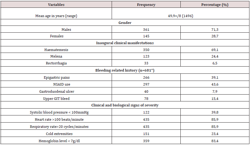
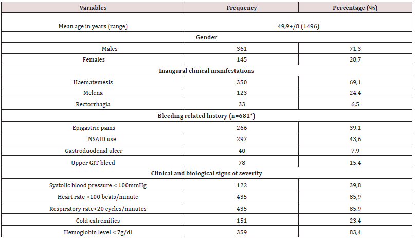
A retrospective collection of data contained in the files of patients admitted for acute UGIB at the Yaounde Central Hospital (Cameroon) between January 1st2010 and December 31st2019, was carried out. Located at the heart of the city of Yaounde, the Yaounde Central Hospital (YCH) was created in 1930. It is the largest public referral hospital in Cameroon with capacity of …. Beds that also acts as a teaching hospital. It houses several services including the Hepato-gastroenterology service with a capacity of 34 beds, three university specialists, four general practitioners and several permanent workers and residents. This service has an upper and lower gastrointestinal endoscopy room and endoscopy equipment. The variables recorded included: demographics (age, sex); clinical and biological characteristics (onset of UGIB, history of bleeding, physical parameters of bleeding severity, and haemoglobin levels on admission); Esogastroduodenal endoscopy (EGDE) findings; specific management of the cause of the bleeding and outcomes (Table1).

Table 1: Clinical and biological parameters of 506 patients admitted at the Yaounde Central hospital for a recognized acute UGIB.

Lupinepublishers-openaccess-gastroenterology-hepatology-journal

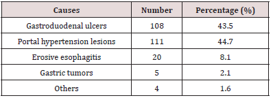
Table 2: Main causes of acute UGIB at the Yaounde Central Hospital amongst 203 patients who had an EGD.

Lupinepublishers-openaccess-gastroenterology-hepatology-journal

The second component, cross-sectional and observational blinded experiments, conducted from December 1st 2017 to May 31st 2018, assessed the diagnostic approach and actual management of patients admitted for acute UGIB in five hospitals of different categories (1st to 3rd) of the Cameroonian health pyramid, in the city of Yaounde. Twenty (20) doctors and 39 nurses in the emergency service who managed 74 patients admitted for acute UGIB were followed. The elements to take care of acute UGIB included recognition of acute UGIB by placing a nasogastric tube or performing a digital rectal exam; assessment of severity by clinical criteria (taking blood pressure (BP), pulse rate (PR), respiratory rate (RR) and room oxygen saturation (SPO2), Glasgow- Blatchford score; fluid resuscitation and transfusions (double venous line, crystalloids filling, proton pump inhibitors (PPI) or vasoactive treatment, oxygenation, restrictive blood transfusion (haemoglobin (Hb) <7 g/dl), hourly monitoring of vital signs and neurological status) and finally, the performance of EGDE and the Rockall and Forrest scores.

Statistical Analysis

Data was analysed using Statistical Package for Social Sciences (SSPS Inc, Chicago, Illinois, USA) version 23.0. Means ± standard deviation was used for quantitative variables; Categorical data was expressed as numbers and proportions. A p value of less than 0.05 was considered statistically significant.

Results

During the retrospective period, 506 patients (prevalence of acute UGIB in the services 5.6%) were included of which 361 men (71.3% and 145 women (28.7 %), given a 2.5 sex ratio. The mean age was 49.9+/- 8 years (maximum 14-96 years) and the peak of bleeding was in the 55-65-year age group. Haematemesis was the initial complaint in 350 patients (69.1%), non-steroidal antiinflammatory drugs (NSAIDs) constituted the most important risk factor in 297 patients (43.6%), in 78 (15.4%). This was a second episode. On admission, clinical parameters relevant to severity were systolic BP <100 mmHg in 122 patients (39.8%); HR >100 beats/ minutes in 435 (85.9%) and RR >20 cycles/minute in 435 (85.9%). Hb was <7g/dl in 359 patients (83.4%). EGDE was performed in 203 patients (40.2%), the major causes of bleeding were: portal hypertension lesions in 111 patients (44.7%) followed by peptic ulcers in 108 (43.5%). Treatment was mainly medical. However, 94 patients (84.7%) with portal hypertension lesions received endoscopic treatment, mainly by injection of sclerosing agent (69.1%), as well as 13 patients (1.2%) with peptic ulcers, mainly isolated injection of dilute adrenaline (1: 10,000) in 11 (84.6%). A total of 75 patients (14.8%) died during hospitalization (Table 2).

The second part concerned the assessment of care offered to 74 patients admitted for acute UGIB (mean age 55 years; sex ratio 2.1). For the recognition of bleeding, digital rectal examination was performed in 43 patients (58.1%) and no patient received nasogastric tube. Regarding the assessment of severity by clinical criteria, BP was taken in 73 patients (98.6%), HR in 61 (82.4%), and RR in 17 (23%), no patient had prognostic scores in the record, including the Glasgow-Blatchford score. Only 17 patients (23%) had SPO2 measurements. Regarding intensive care measures, only 10 patients (13.5%) received a double peripheral venous line, the majority of which was a small-bore venous line. Twenty patients (27%) were filled with crystalloids; restrictive blood transfusion was performed in 24 out of 27 patients (88.9%) with Hb < 7 g / dl. Only 9 patients (12.2%) received nasal oxygen therapy. EGDE was performed in 43 patients (60.6%), all beyond 24 hours after admission and none had a prognostic score after endoscopy (Forrest or Rockall).

Discussion

The study showed that acute UGIB is an emergency with exceptional severity in our environment, as mortality is very high at around 15%. In this study, it was found that acute UGIB affected two and a half times more men than women with a mean age of about 50 years. This is the case in studies conducted in Mali (sex ratio 2.78; mean age 47.45 years), and in Côte d’Ivoire (sex ratio 3.38; mean age 47 years) [8,12]. Indeed, in the sub-Saharan African region, the occurrence of acute UGIB often involves relatively young patients. This can be explained by the fact that the causes of bleeding are often dominated by portal hypertension lesions, especially in young patients, as reported in Mali in a rural area [8]. As opposed to sub-Saharan Africa, in the West, the age of onset of acute UGIB is higher than 70 years due to the use of NSAIDs in the elderly population. Of chronic NSAID users, 25% develop an ulcer, of which 2-4% are complicated by bleeding [2,7,13,14]. The male predominance is universal, and reverses in the West after the age of 80 years due to the higher life expectancy in the female population [2,4,6-9, 11,13,14].

As in several studies reported in literature, hematemesis was the most common initial clinical presentation [2,6,7,15,16]. This initial clinical presentation can be explained by the different lesions found on endoscopy. In fact, portal hypertension lesions, led by oesophageal varices, were the most frequent, alongside peptic ulcers. The frequency of the various aetiologies varies from one region to another [5,6,8]. Thus, in the sub-Saharan African region, several studies report very high frequencies of portal hypertension lesions. This is the case of the study by Diarra et al. in Mali in 2007 [8]. The authors reported a frequency of 55.2% in favour of ruptured oesophageal varices, far ahead of peptic ulcers which represented 16%. The frequency of portal hypertension lesions is also high in Burundi (28.2%) [16] and Gabon (29.5%) [17]. These various countries have liver diseases related to chronic HBV infection in common. Indeed, sub-Saharan African countries are located in a zone of high endemicity according to the World Health Organisation (WHO), i.e. the prevalence of hepatitis B is 8-20% of the general population [10]. Cirrhosis, which causes portal hypertension, is often the result of chronic HBV infection acquired at birth or in early childhood [18]. The annual incidence of varicose veins is approximately 5% [19]. UGIB from ruptured esophageal varices accounts for 70% of gastrointestinal bleeding in cirrhosis, with an estimated overall 2-year bleeding risk of 20% [19]. The aetiological approach to acute UGIB in high- and intermediate-endemic areas where the majority (80%) of the world population lives should therefore consider this high frequency of portal hypertension lesions and bleeding complications.

Regarding the severity of the bleeding, more than 85% of patients were initially unstable with clinical and laboratory signs of severity, despite the absence of Glasgow-Blatchford and Rockall scores in their records. This initial haemodynamic instability can be explained, on the one hand, by the causes of bleeding, in particular the rupture of oesogastric varices, which are exceptionally serious, but also, on the other hand, by the late arrival in hospital structures due to distance, cultural considerations or difficulties of mobility in our country.

Treatment was essentially medical. PPI treatment was most often initiated on admission, even if this treatment did not always fully comply with current recommendations in Europe and Asia [2,7,20,21]. Contrarily, in cases of suspected acute UGIB related to rupture oesogastric varices, vasoactive therapy to reduce portal blood flow was never initiated on admission, in line with international recommendations, including those adapted by the European Society of Gastrointestinal Endoscopy to be applicable to resource-limited settings, including some African countries [2,7,22-24]. Patients with bleeding peptic ulcers have rarely benefited from endoscopic haemostasis, unlike those with portal hypertension lesions. Late arrival at hospital would explain why in sub-Saharan Africa Forrest scores IIc and III, i.e. pigmented stain or clean base of the ulcer, are most frequently found [25]. For the Forrest classification guiding the choice of endoscopic treatment modality for ulcers, there is no endoscopic treatment for these scores [2,3,7,26]. The practice of injecting adrenaline alone instead of combined adrenaline and bipolar coagulation or endoclips was inappropriate. This could be explained by factors such as excessive procedure costs, insufficient training of practitioners and limited logistic equipment.

Read More About Lupine Publishers Journal of Gastroenterology and Hepatology Please Click on Below Link: https://currenttrendsingastroenterology.blogspot.com/

Tuesday, 28 March 2023

Lupine Publishers| Improvement of Liver Function by a Short-term Administration of Luseogliflozin in Patients with Type 2 Diabetes: A Single-arm Study and the Mini-literature Review

Lupine Publishers| Journal of Gastroenterology and Hepatology


Abstract

Background: Non-alcoholic fatty liver disease is not simply the hepatic manifestation of obesity and diabetes but also linked to hepatocellular carcinoma. Yet, its effective treatment has not been established. In this study, we evaluated the effect of a short-term administration of luseogliflozin to patients with type 2 diabetes having non-alcohol fatty liver disease. Luseogliflozin is a unique sodium-glucose cotransporter 2 inhibitor which is metabolized in and excreted by the liver in addition to the kidney. Therefore, the drug might possess an additional effect on other agents of the same class which are exclusively metabolized in the kidney.

Methods: Using alanine aminotransferase >20 IU/L as a diagnostic basis for non-alcoholic fatty liver disease, 19 patients, not taking alcohol, with type 2 diabetes (male/female 15/4, the median age 57 years) was treated with 2.5 mg luseogliflozin for 12 weeks.

Results: Pre- and post-treatment median values for alanine aminotransferase were 51 IU/L and 33 IU/L (p = 0.001), and the corresponding values for the fibrosis index based on the four factors [age (years) ∙ alanine aminotransferase (IU/L)] / [platelets (109/L) ∙ alanine aminotransferase (IU/L)1/2)] were 1.669 and 1.314 (p = 0.043). There was no adverse effect of the drug. Our findings were essentially compatible with the results of the previous studies reviewed.

Conclusion: We conclude that sodium-glucose cotransporter 2 inhibitor could be the choice for the pharmacological treatment of non-alcoholic fatty liver disease. Especially, a short-term administration of it is consistently effective in mild cases.

Keywords: SGLT2 inhibitor; NAFLD; Fibrosis-4 index; GPR-index; APRI.

Abbreviations: SGLT2i: sodium-glucose cotransporter 2 inhibitor; NAFLD: non-alcoholic fatty liver disease; NASH: non-alcoholic steatohepatitis; T2DM: type 2 diabetes mellitus; AST: aspartate aminotransferase; ALT: alanine aminotransferase; GGT: gammaglutamyl transpeptidase; Fib-4 index: Fibrosis-4 index; GPR-index: gamma-glutamyl transpeptidase to platelet ratio; APRI: aspartate aminotransferase to platelet ratio

Introduction

Obesity or overweight is the motherland of non-alcoholic fatty liver disease (NAFLD) and non-alcoholic steatohepatitis (NASH) [1]. Accordingly, with increasing trend of body weight worldwide, the number of patients with the liver problem is relentlessly increasing [2]. Recently, NASH has also been attracting the attention as a cause of hepatoma [3,4]. Under such yet, treatment of NAFLD (NASH and NAFLD are collectively called NAFLD hereafter in this communication), irrespective of presence or absence of diabetes, has not been established. Results of treatment of patients with type 2 diabetes (T2DM) having NAFLD with sodium-glucose cotransporter- 2 inhibitor (SGLT2i) appears promising [3-7], the effectiveness of a short-term treatment, such as 12 week-treatment, has not been established.
Here, we evaluated effectiveness of a short-term administration of SGLT2i, luseogliflozin, that is metabolized not only in the kidney but in the liver [8]. A mini literature review on this issue was also performed to resolve the current inconsistency. This study was approved by the Clinical Research Ethics Committee of Aizawa Hospital (No. 2019-094).

Subjects and Methods

Subjects

Consecutive 29 patients with T2DM who took luseogliflozin for 3 months or longer between January 1, 2019 to May 31, 2020 were initially registered. Because the purpose of this study was to investigate the effect of luseogliflozin on NAFLD/NASH, 8 with alanine aminotransferase (ALT) less than 20 IU/L [9], and other 2 with a habitual alcohol drinking of 20 g/day or more were excluded, and the remaining 19 were analyzed.
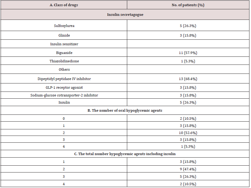
The study was a single-arm, add-on study. Namely, 2.5 mg luseogliflozin was additively prescribed on top of the hypoglycemic agents already taken by the patients, which are shown in Supplemental Table 1. The data before and 12 weeks after luseogliflozin administration was critically compared.

Laboratory measurements

In addition to the routine clinical chemistries, indices of the hepatic fibrosis including Fibrosis-4 index (Fib-4 index), gammaglutamyl trans peptidase (GGT) to platelet ratio (GPR-index), and aspartate aminotransferase (AST) to platelet ratio (APRI) were calculated10: the unit for AST, ALT, GGT as IU/L, platelet counts as 109/L and age as a year. Equations for each index were as follows [10].

Fib-4 = [(AST) ∙ (age)] / [(Platelet counts) ∙ (ALT)1/2]
GPR index = 100 ∙ (GGT) / (Platelet counts)
APRI = 100 ∙ (AST) / (Platelet counts)

Statistical analysis

The data were collected retrospectively and analyzed cross-sectionally and longitudinally. We evaluated the effect of luseogliflozin on the liver function tests and the indices of liver fibrosis. In addition, delta, i.e., the basal value minus 12 week-value for the indices of liver fibrosis was calculated for each study subject, and correlation between the delta values of fibrosis indices and the basal plasma glucose (PG), glycosylated hemoglobin (HbA1c), AST, ALT, GGT, body weight (BW) and body mass index (BMI) were examined. Furthermore, the delta value of the liver fibrosis indices and the delta value of PG, HbA1c, AST, ALT, GGT, BW and BMI were also examined. The statistical analysis was performed using JMP ver.15. Wilcoxon rank-sum test, Wilcoxon signed rank test and Spearman rank correlation were used as needed.

Literaturereview

Representative 10 original reports published in the English language on the treatment of patients with T2DM having NAFLD by SGLT2 inhibitors [6,7,11-18] were summarized to provide a current overview of the issue. In these literature, five kinds of SGLT2i agents were prescribed, with the number of the patients ranging from 9 to 32 and the treatment duration from 12 to 48 weeks.

Results

Baseline characteristics of the patients

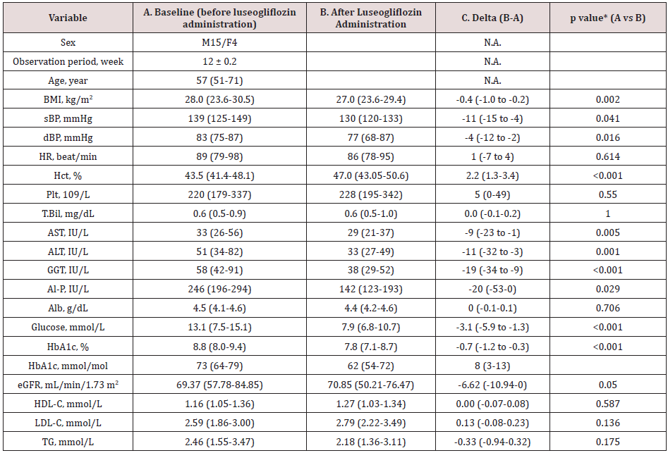
Table 1. Baseline data (A), the data 3 months after the luseogliflozin treatment (B) and the difference (C) between the two (luseogliflozin- basal).

Lupinepublishers-openaccess-gastroenterology-hepatology-journal

Values are median and interquartile ranges except for sex and the observation period: the latter was shown as mean and SD. sBP, systolic blood pressure; dBP, diastolic blood pressure; HR, heart rate; Hct, hematocrit; Plt, platelet count; T.Bil, total bilirubin; AST, aspartate aminotransferase; ALT, alanine aminotransferase; GGT, gamma-glutamyl transpeptidase; Al-P, alkaline phosphatase; Alb, albumin; eGFR, estimated glomerular filtration rate; HDL-C, high density-lipoprotein cholesterol; LDL-C, low density-lipoprotein cholesterol; TG, triglycerides. N.A., not applicable. *Wilcoxon signed-rank test.

The study patients were male dominant (the proportion of males, 79%), middle-aged Japanese adults with the median BMI of 28.0 kg/m2 which was larger than the representative value in the Japanese patients with T2DM in general11(Table 1A). The median HbA1c value of the entire group before luseogliflozin was 8.8% (77 mmol/mol) so that the level of glycemic control was unsatisfactory (Table 1A). Regarding the hypoglycemic agents used before subscribing luseogliflozin, Dipeptidyl Peptidase-4 (DPP-4) inhibitors and biguanide were most frequently employed (Supplemental Table 1), which was typical for the Japanese patients [19].

Supplemental Table 1: Medications before administration of luseogliflozin. 2.5 mg luseogliflozin was added in each patient on top of the medication described above.

Lupinepublishers-openaccess-gastroenterology-hepatology-journal

Values are median and interquartile ranges except for sex and the observation period: the latter was shown as mean and SD. sBP, systolic blood pressure; dBP, diastolic blood pressure; HR, heart rate; Hct, hematocrit; Plt, platelet count; T.Bil, total bilirubin; AST, aspartate aminotransferase; ALT, alanine aminotransferase; GGT, gamma-glutamyl transpeptidase; Al-P, alkaline phosphatase; Alb, albumin; eGFR, estimated glomerular filtration rate; HDL-C, high density-lipoprotein cholesterol; LDL-C, low density-lipoprotein cholesterol; TG, triglycerides. N.A., not applicable. *Wilcoxon signed-rank test.

Liver function and indices of hepatic fibrosis following luseogliflozin administration

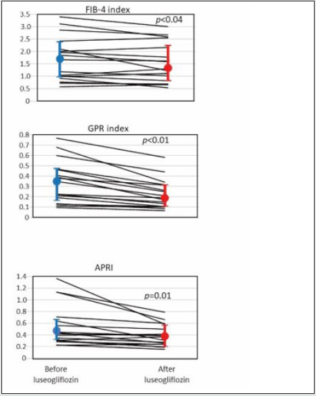
Elevated serum level of ALT was an inclusion criterion, so that the ALT was clearly elevated as a group with the median value, 51 IU/L. As well expected, administration of luseogliflozin significantly decreased PG and HbA1c (Table 1, A and B, before and after luseogliflozin, respectively). In addition, it significantly lowered the serum level of AST, ALT, GGT, and alkaline phosphatase (ALP). The degree of lowering was 12%, 34%, 35 and 42% for the respective enzyme levels. Importantly, the luseogliflozin treatment also significantly lowered the values for Fib-4 index, GPR index, and APRI (Figure 1).

Correlation between delta Fib-4, GPR index, and APRI and baseline and delta values of liver function and the body weight and BMI

Figure 1: Change of indices of liver fibrosis produced by luseogliflozin. Individual lines represent a change of the value in each person and the circles, and the vertical lines indicate the median and interquartile ranges: blue ones for before and red ones for after luseogliflozin. Wilcoxon signed-rank test was used for the statistical analysis.

Lupinepublishers-openaccess-gastroenterology-hepatology-journal

The delta Fib-4 index was significantly and positively correlated associated with higher baseline AST and ALT levels; the delta GPR index was correlated with baseline AST and GGT; the greater delta APRI was associated with higher baseline AST and ALT (Table 2A). Correlation between the delta values and the baseline of liver function was absent for BW and BMI (Table 2A).
On the other hand, there was an inverse correlation between ‘delta Fib-4 and delta AST and delta ALT’, ‘delta GPR index and delta GGT’ and ‘delta APRI with delta AST and delta ALT’ (Table 2B). There was no significant correlation between delta values of the fibrosis indices and the delta of BW and BMI (Table 2B).

Table 2. Correlation between delta Fib-4, GPR index and APRI and baseline value (A) and delta (B) of liver function. Delta means the difference between the value of each test performed on the day of starting luseoglifrozin and 12 weeks later.

Lupinepublishers-openaccess-gastroenterology-hepatology-journal

Values are median and interquartile ranges except for sex and the observation period: the latter was shown as mean and SD. sBP, systolic blood pressure; dBP, diastolic blood pressure; HR, heart rate; Hct, hematocrit; Plt, platelet count; T.Bil, total bilirubin; AST, aspartate aminotransferase; ALT, alanine aminotransferase; GGT, gamma-glutamyl transpeptidase; Al-P, alkaline phosphatase; Alb, albumin; eGFR, estimated glomerular filtration rate; HDL-C, high density-lipoprotein cholesterol; LDL-C, low density-lipoprotein cholesterol; TG, triglycerides. N.A., not applicable. *Wilcoxon signed-rank test.

Read More About Lupine Publishers Journal of Gastroenterology and Hepatology Please Click on Below Link: https://currenttrendsingastroenterology.blogspot.com/

Wednesday, 8 February 2023

Lupine Publishers| Is there a utility of Nordin’s Index in the evaluation of osteoporosis in patients with post viral cirrhosis? Results of a pilot study in Cameroonians

 Lupine Publishers| Journal of Gastroenterology and Hepatology


Abstract

Background: Metabolic bone disorders are frequent in patients with cirrhosis. These two conditions are usually misdiagnosed in Sub-Saharan Africa before the onset of complications. A full evaluation of metabolic bone disorders in patients with liver cirrhosis is difficult in our milieu and osteodensitometry is rarely available. In this preliminary study, we sought to determine the necessity of a cost-effective method, the Nordin’s Index, in the evaluation of osteoporosis in patients with liver cirrhosis.

Methods: This was a prospective cross-sectional study from January to May 2017 in Yaoundé. We compared the data of 19 patients (15 men, 4 women) with post viral liver cirrhosis and 17 controls (13 men, 4 women) paired with age, gender and body mass index (BMI). Data collected included vitamin D levels, serum and urine concentrations of calcium and phosphorus, Nordin’s Index and results of the bone mineral density using an x-ray absorptiometry scan. Statistical analysis was performed using the software SPSS 21. A p value of less than 0.05 was considered to be statistically significant.

Results: The mean age of patients was 38 ±15 years, with a mean BMI of 25 ±8 kg/m². Three of the four women were on menopause. Etiologies of cirrhosis were viral hepatitis B (8 patients), viral hepatitis B and D coinfection (7 patients), and viral hepatitis C (4 patients). The median duration of cirrhosis was 19 [8; 48] months, and 14 patients were classified grade A in Child Pugh classification. There was no statistical difference in the serum and urine concentrations of calcium and phosphorus. Osteoporosis was more frequent in cirrhosis (31.6% versus 11.8%, p<0.05). Nordin’s Index was significantly elevated in patients with cirrhosis compared to controls (0.12 [0.06; 0.13] mg/mg, versus 0.03 [0.01; 0.08] mg/mg, p<0.05), and in patients with cirrhosis associated to osteoporosis compared to those without (0.13 [0.09; 0.13] mg/mg versus 0.07 [0.03; 0.08] mg/mg, p<0.05). Vitamin D deficiency was more observed in controls (13/17 versus 7/19, p<0.05). Factors associated with osteoporosis were disease duration, elevated Nordin’s Index and elevated serum level of transaminases.

Conclusion: The Nordin’s Index, a simple and inexpensive tool for exploration of the phosphocalcic metabolism, could be useful for the evaluation of osteoporosis during viral cirrhosis. However, its performance has to be evaluated in a larger sample.

Keywords: Cirrhosis, chronic viral hepatitis, osteoporosis, Nordin’s Index, Vitamin D.

Abbreviations: ALAT: Alanine Aminotransferase; ALP: Alkaline Phosphate; ASAT: Aspartate Aminotransferase, BMD: Body Mass Density; GGT: Gamma Gluthamyl Transferase

Introduction

Cirrhosis is the outcome of most chronic liver diseases. It remains a major public health problem in Sub-Saharan Africa and particularly in Cameroon, where chronic viral hepatitis is highly endemic [1-3]. In fact, sixty percent of cirrhosis in Africa are attributable to viral hepatitis B and C [4]. In Cameroon, 70.9% of cirrhosis cases are due to chronic viral hepatitis B and 25.5% to chronic viral hepatitis C [5]. The hallmark of cirrhosis is hepatocellular insufficiency and portal hypertension, resulting in impaired liver function and subsequent systemic abnormalities. Bone damage is one of the systemic abnormalities frequently seen in cirrhosis, regardless of etiology, and is known as hepatic osteodystrophy [6].
Hepatic osteodystrophy is a combination of osteoporosis and osteomalacia, the latter being rare during cirrhosis [6]. According to the World Health Organization (WHO), osteoporosis is a diffuse disease of the skeleton, characterized by a decrease in bone mass and an alteration of the micro-architecture of bone tissue, leading to increased bone fragility and an increased risk of fractures [7]. Its pathogenesis during cirrhosis is complex and leads to an increase in bone resorption by osteoclasts to the detriment of its formation by osteoblasts [8]. The prevalence of osteoporosis during cirrhosis is between 12% and 55% [9]. In Africa, more precisely in the Maghreb region, it has been estimated to 28.26% [10]. Osteoporosis is almost asymptomatic until complications. It is a real public health problem as it causes low energy fractures in more than 40% of cases, affecting the morbidity and quality of life of patients with cirrhosis [8]. Several factors are associated with the occurrence of osteoporosis during cirrhosis, including vitamin D deficiency with a prevalence of 32% in Europe and 32.6% in Africa during cirrhosis [10,11]; Elevated Child and Pugh Score, duration of disease, etiology of cirrhosis, low body mass index, with some controversy in the literature [12].
Considering the risk of osteoporosis being high during cirrhosis, it is necessary to have an evaluation of bone mineral density in these patients [13,14]. However, this is rare in our context because of the cost and accessibility of this examination. With the absence of hepatic transplantation in our environment to cure cirrhosis, there is a long duration of cirrhosis and, therefore, an increased risk of complications including osteoporosis. It is therefore important to use simple and inexpensive tools to make the diagnosis or at least to detect high-risk subjects. Nordin’s Index is calculated by the calciuria/creatinuria ratio. It is an old marker of bone resorption, which correlates with post-menopausal osteoporosis and its risk of fracture [15,16]. However, its usefulness in cirrhosis especially post viral has been little studied to the best of our knowledge. In this study, we sought to determine the utility of Nordin’s Index as a cost-effective method in the evaluation of osteoporosis in people suffering from post-viral cirrhosis in a highly endemic area for chronic viral hepatitis and limited access to bone mineral density assessment.

Patients and Methods

Study design

The study used a cross sectional design with a prospective data collection.

Study setting

This study took place from January to May 2017 at the Yaoundé Central Hospital and Cathedral Medical Centre (CMC) at Yaoundé, where the participants were recruited. Laboratory analysis were conducted at the University Hospital Centre of Yaoundé. Bone mineral density measurements were carried out at the Autonomous Centre for Radiology and Medical Imaging (CARIM) at Yaoundé.

Participants

The sample was made up of 19 adult volunteers (15 men and 4 women) followed for a cirrhosis of viral cause (chronic viral hepatitis B, C, and D). They were matched with 17 adult volunteers (13 men and 4 women) without any chronic or acute disease/ condition, paired with age (±2 years), gender and body mass index (BMI; ±2 kg/m²). The diagnosis of liver cirrhosis was based on clinical and biological signs of portal hypertension and chronic liver failure, ultrasonographic signs of chronic liver disease and endoscopic signs of portal hypertension. Ultrasonographic signs included irregular liver outline, heterogeneous echo structure, dysmorphic liver, enlarged portal vein and presence of collateral venous circulations.
The controls were recruited from among the patients’ caregivers to ensure equal exposure to environmental and nutritional factors that may contribute to osteoporosis. We did not include any participants who presented pathologies with known repercussions on bone and/or phosphocalcic metabolism, such as: chronic renal disease, thyroid and parathyroid disorders, Cushing Syndrome, diabetes, HIV infection and cancer. We did not include participants receiving treatment with known effects on bone and/ or calcium phosphorus metabolism such as: hormone replacement therapy, biphosphonates, calcium, vitamin D, corticosteroids, antimetabolites, anticoagulants, anticonvulsants, thyroxine. Pregnant women instead of women with amenorrhea, participants with diabetes, tobacco and alcohol consumers were not included.

Sample size calculation

The sample size was estimated using the formula in Whitley et al. to compare proportions between groups [17]. The parameters used for the calculation of the standardized difference were derived from the study of Goral et al [18]. They found a mean T-score of -1.6 SD in patients with cirrhosis and -0.25 SD in controls, with a standard deviation of 1.3 SD. In our study, the level of significance was set at p 0.05 and the power at 80%. The minimum sample per group was 15.

Procedure

Ethical considerations

We obtained research authorizations in the selected health care facilities and the approval of the Institutional Review Board of the Faculty of Medicine and Biomedical Sciences. All the participants read and signed an informed consent form.

Clinical data

Sociodemographic (age, gender) and clinical data were collected through a questionnaire. Clinical variables were anthropometric parameters (weight and height) for BMI calculation, the history of cirrhosis (duration, etiology, complications, current Child and Pugh’s Score, treatment), suggestive signs of osteoporosis and its complications (history of pathological and or low energy fracture), comorbidities, menopause for women and its duration, and family history of osteoporosis.

Laboratory Analysis

For laboratory analysis, 10 mL of peripheral venous blood sample was collected from each participant after eight hours of fasting. The blood samples were centrifuged for 5 minutes at 3000 rpm using the Humax 14K Germany centrifuge. 500μl of serum was then extracted for the determination of calcium (mg/L), phosphorus (mg/L), albumin (g/L) by colorimetric method and for the determination of creatinine (mg/l), total alkaline phosphatase (U/L), transaminase (Aspartate amino transferase (ASAT), Alanine amino transferase (ALAT), in U/L), gamma gluthamyl transferase (GGT, U/L) by kinetic method, using the Mindray model BS 120 spectrophotometer. The remaining serum was stored at -20° Celsius and used to determine 25(OH) vitamin D (ng/mL) levels by ELISA using the BioTek EL×800 ELISA chain. Serum creatinine was used to estimate the glomerular filtration rate using four parameters’ MDRD formula in mL/min for 1.72m². The measured calcemia was corrected using albuminemia. Hypercalcemia was defined for a corrected Calcemia > 104 mg/L, while Hypocalcemia was defined below 81 mg/L [19]. Hyperphosphatemia was defined as blood phosphorus > 50 mg/L in adults while hypophosphatemia was defined below 25 mg/L [19]. Vitamin D Status was assessed according to the classification of Holick et al [20].

Laboratory Analysis

After consumption of 200 ml of low calcium water (Volvic® type: Ca<10 mg) in a fasting individual with a previously empty bladder, calciuria and creatininuria was measured on the urine collected 2 hours later. Nordin’s Index was calculated: calciuria/ creatininuria (mg/mg). The normal value of the Nordin’s Index is lower than 0.11 mg/mg. An increase in the index indicates hyperresorption of bone. Calcium, phosphorus and creatinine in urine were determined using the same methods as in the plasma.

Bone Mineral Density (BMD)

Bone mineral density was measured by biphotonic X-ray absorptiometry scan using the Hitachi® lunar prodigy DXA system, with 37 Micro SieVert (μSV) dose per measurement site. Bone mineral density was measured at the lumbar spine, the neck of the left femur and the distal end of the left radius. Data on bone mineral density were expressed in g/cm², T-score and Z-score. Osteoporosis was defined as T-score <-2.5 for patients aged 50 years and older or a Z-score <-2 for patients under 50 years of age for measurement of the lumbar spine, femoral neck or distal end of the radius [21].

Statistical analysis

Statistical analysis was performed using SPSS version 21.0. Quantitative variables were expressed as mean ± standard deviation or median and interquartile range [25th and 75th quartiles]. Qualitative variables were expressed as effectives and proportions. The Mann-Whitney test was used to compare the means, while medians were compare using the Median’s test. Fisher Exact Test was used to compare proportions. We searched for associations between continuous variables and bone mineral density on various sites by calculating the Pearson’s Correlation Coefficient to determine the factors associated with the decrease in BMD. For all the tests, the statistical significance level was set at 0.05.

Results

Clinical characteristics of the study samples

The mean age of patients was 38.6 ±15.9 years, while that of controls was 37.8 ±15.9 years (p>0.05). Participants’ ages ranged from 23 to 73 years. There were respectively 2/4 and 2/4 women on menopause in the patients and control groups. Between cases and controls, there was no statistical difference in body mass index (p>0.05) (Table 1). The median duration of cirrhosis was 19 months, range from one to 120 months. The causes of liver cirrhosis were chronic viral hepatitis B (42%), hepatitis C (31%), and B-delta (37%). There was no coinfection with viral hepatitis B and C. Most of the cases were classified on stage A (n=14), while 3 and 2 cases were respectively on-stage B and C in Child and Pugh classification.

Read More About Lupine Publishers Journal of Gastroenterology and Hepatology Please Click on Below Link: https://currenttrendsingastroenterology.blogspot.com/

Friday, 6 January 2023

Lupine Publishers | Efficacy and Safety of Prolonged Alfuzosin Treatment in Patients with Lower Urinary Tract Symptoms Associated with Benign Prostatic Hyperplasia: 7 Years of Observation

 Lupine Publishers | Journal of Urology & Nephrology


Abstract

Introduction: Amongst all of the medications prescribed to BPH patients undergoing conservative therapy, α1-blockers were used in 80% of cases. The question of optimal treatment length is one that has been constantly asked during the last years. The current study will encompass the data we have collected of a small study group of BPH patients treated with 10 mg of Alfuzosin daily, over a period of more than 7 years.

Material and Methods: From 2009 to 2011, 41 patients with mean age 67.7 years began medical therapy taking 10 mg of Alfuzosin per day. The mean treatment length was 7.6 years as of today. 16 patients (39%) with prostates exceeding 60 cm3 were additionally prescribed 5-ARIs.

Results: Overall, a positive dynamic was found in 85.4% of patients. Not a single patient chose to discontinue treatment. A very high level of satisfaction was reported by 88% of our patients. A statistically significant (P < 0.05) decrease in IPSS scores by 8.5 ± 6.1 (47.5%) was found. Mean QoL indices decreased from 3.7 ± 1.1 to 2.3 ± 1.1 over the period of observation. Mean Qmax values increased from 9.7 ± 0.52 mL/s to 14 ± 0.60 mL/s (an increase of 44.3%).

Conclusions: This study has demonstrated a high level of safety and efficacy when using Alfuzosin to treat voiding dysfunction in patients suffering from LUTS/BPH. Our study resolves the issue surrounding a prolonged course of α1-blockers, which, due to its widespread use, remains the gold standard for medical treatment of lower urinary tract symptoms associated with benign prostatic hyperplasia.

Keywords: LUTS, BPH, Alfuzosin, BOO

Introduction

It is well known that the prevalence of voiding dysfunction and patient age are in direct proportion and that prevalence is particularly high in patients over 50 years of age. Typical complaints associated with voiding dysfunction are termed lower urinary tract symptoms (LUTS) and have a wide variety of underlying causes. The most common aetiologies of LUTS are prostatitis, urethral strictures, cystitis, bladder stones, prostate cancer, and benign prostatic hyperplasia (BPH).

BPH is usually managed with surgical intervention, pharmacotherapy, or a strategy of watchful waiting, and numerous factors affect the decision of which method should be employed. Symptom severity is one of the most important subjective factors and is measured using the international prostate symptom score (IPSS) questionnaire. The nature of the symptoms (whether they fall under the category of obstructive or irritative) also influences the final decision. The primary objective factor can be considered to be the quality of voiding as measured by uroflowmetry, the interpretation of which should consist not only of the peak flow rate (Qmax), but also flow time, time to Qmax and many other indicators. Postvoid residual urine is an equally important value because it helps identify poor bladder contraction ability (detrusor underactivity) in the case of progressive intravesical obstruction. There are also those factors, no less important than both uroflowmetry and IPSS, which relate to the potential risk of BPH progression, and thus make their impact on the choice of treatment. Whilst the size of the prostate itself cannot be ignored, it is by no means a marker for surgical intervention when considered by itself, but rather a hallmark for potential future complications such as acute urinary retention (AUR) amongst other things. In recent years, prostate-specific antigen (PSA) levels have also become associated with BPH progression. Today, PSA levels are not only used to screen for prostate cancer, but also to reflect cellular proliferation rates of the prostate. Thus, high PSA levels increase the risk of rapid BPH progression. [1] The sum of the factors mentioned above influences the ultimate decision of which type of treatment should be employed in a specific patient.

Modern standards of medical practice dictate that pharmacotherapy is recommended in BPH patients with moderate LUTS without upper urinary tract complications, patients without obvious surgical indications, and patients who either decline surgical intervention, or are unable to commit to surgery in the nearby future. Watchful waiting is acceptable if a patient exhibits mild LUTS (IPSS ≤ 8), or if the patient’s symptoms do not significantly impact their quality of life (QoL). Patients who fall under this category should be made aware of the necessity to maintain an appropriate lifestyle as well as the importance of regular blood, urine, PSA, uroflowmetry and ultrasound testing. Surgical intervention is needed if a patient exhibits severe symptoms with upper urinary tract complications, or if there are sufficient reasons to suspect that medication will be ineffective[2,8].

In order to better illustrate modern tendencies in the treatment of BPH, we turn our attention to the TRIUMPH study. The TRIUMPH study recorded the treatment and outcomes of 2351 newly presenting LUTS/BPH patients in 6 European countries over a 1-year follow-up period. Out of the entire study population, 23.8% were managed with watchful waiting, 72.5% were prescribed medication, and only 2.7% underwent surgical treatment. These statistics accurately reflect the rise in medical management of LUTS as well as the decreasing need for surgical intervention during the past decade. It should be interesting to note that amongst all of the medications prescribed to BPH patients undergoing conservative therapy, α1-adrenergic receptor antagonists (α1-blockers) were used in 80% of cases [3].

The efficacy of α1-blockers in patients with BPH has been well documented in a large quantity of double-blind, placebo-controlled studies.[4,5,6,7] If it is possible to consider transurethral resection of the prostate (TURP) the method of choice for surgical intervention in BPH patients, then α1-blockers will fall under the same criterion for the medical management of BPH. It is important to note that the European Association of Urology considers α1-blockers the first line of therapy for the management of voiding dysfunction in patients suffering from BPH[8].

Whereas the rationale behind α1-blockers does not arouse suspicion in the majority of specialists, the question of optimal treatment length is one that has been constantly asked during the last 6 years. Solutions offered to address this problem range from a life-long course of medication, to one that only lasts a few months. There is no one correct way to approach this matter, but it is the duty of every qualified specialist to incorporate certain principles when making such a decision. Personal experience and knowledge gained from a wide variety of international studies should encompass the core of such precepts. It is arguable that the lack of a formulated strategy that applies to such clinical trials is hindering the formation of a unified point of view. If we turn to medical literature, we find that despite an abundance of studies indicating the effectiveness of α1-blockers in BPH patients, the studies lack the duration necessary to examine long-term effects and are mostly comprised of 3-6 month trials with only a few mentioning 1-3 year-long results. This has led many to assume that a 3-month course (being the most prominent study length) of α1-blockers is sufficient, whilst the actual reasoning behind the short length of most studies lies in the knowledge that the results achieved during that period of time will continue over the remaining course of treatment, however long it may be. Confirmation of this can only be found in a few currently available publications that demonstrate long-term results. Most articles cite results that were obtained using studies based on principles of Good Clinical Practice (GCP) in which the categories of patient inclusion/exclusion were so rigid, they created unrealistic expectations for the wide variety of patients that are actually treated. Only a few trials provide us with results that are based on a realistic study population. Such trials include those of Roehrborn C. et al., Elhilali M. et al. 2006 and Vallancien G. with Emberton M. et al. 2008[9,10,11].

During the early 21st century, the suitability of α1-blockers was no longer debated in the Russian Federation, whilst the question of long-term use was left unanswered even for us. Even now, considering the ageing population and the necessity of prolonged medical treatment, many specialists are hesitant to prescribe α1-blockers for more than a few years without consulting medical literature about the safety and efficacy of such a treatment.

Materialand Methods

The current study will encompass the data we have collected of a small study group of BPH patients treated with 10 mg of Alfuzosin daily, over a period of more than 7 years. We must mention that the data provided isn’t final, in the sense that patients are still undergoing treatment to this day. We had not originally planned for this to be a study with a specific design, but retrospective aspects make it similar to various non-comparative, real-life surveillance studies. Our goal is to introduce the reader to the results of a longterm course of α1-blockers that, in our opinion, are a safe and effective method of long-term medical therapy in patients suffering from BPH.
From 2000 to 2002, 41 patients with mean age 67.7 years (range 51– 83) began medical therapy. The mean treatment length was 7.6 years as of today. We would like to mention that this group of patients wasn’t specifically chosen as a study group at the time, nor were they the only patients that had been prescribed Alfuzosin. Our detailed observation of the isolated group in question is partially due to chance, and partially due to loss to follow-up. Patients began treatment taking 5 mg of Alfuzosin twice daily. From January 2005 onwards, all patients take 10 mg of Alfuzosin once daily at night due to numerous complaints of increased voiding problems during this time. Prostate volumes fluctuated between 25 cm3 and 100 cm3, and 16 patients (39%) with prostates exceeding 60 cm3 were additionally prescribed 5-alpha-reductase inhibitors (5-ARIs). During the early stages of treatment these patients were prescribed Finasteride, but during the last 2 years many have switched to Dutasteride in accordance with our recommendations.

Indications

Patients with LUTS/BPH

Contraindications

Patients with surgical indications
Patients known to have insufficient results from taking α1- blockers in the past
Patients with postural hypotension
Patients taking alternative medication for LUTS with good results
Patients with unstable angina (pectoris)
Patients with life-threatening comorbidities

Table 1: Summary of initial patient data.
lupinepublishers-openaccess-journal-urology-nephrology

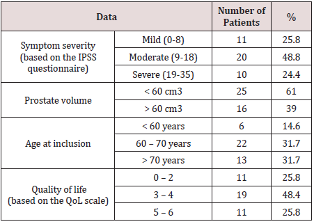
The mean duration of morbidity at the time of patient inclusion in the trial was 1.9±1.1 years. During ultrasound testing, 31.2% of patients had postvoid residual urine measuring 82 ± 41.7 mL. Mean PSA levels of all patients before the trial were 2.9 ± 2.8 ng/mL. The initial data of the patients included in this trial are summarized in Table 1.
Out of the 41 patients, 14 (34.1%) had hypertension 11 of which (26.8%) were receiving some form of hypotensive therapy, and 5 patients had suffered a myocardial infarction before inclusion into the trial.
The efficacy of the treatment was analysed using IPSS scores as well as QoL indices. Additional factors included Qmax and prostate volume in patients who were receiving combination therapy with 5-ARIs. The safety of the treatment was analyzed based on recorded incidents of adverse cardiac side effects (mainly hypotensive) during the trial. Vital-sign dynamics were also analyzed. Follow-ups were conducted biannually during the first 2 years, and no less than annually thereafter.
Two-sided hypothesis tests were conducted with a significance P-value cut-off of 0.05. Dynamics of safety and efficacy variables were analysed before and after the trial (absolute and relative change was considered). A paired Student’s t-test was used if statistics followed a normal distribution; a Wilkinson’s test was used if they did not.

Results

Not a single patient chose to discontinue treatment, notwithstanding the long period of observation. We must mention, however, that several patients stopped being prescribed Alfuzosin during the early stages of the trial but were obviously not considered in the study group. Subjective assessments of patient satisfaction were carried out using a questionnaire specifically tailored to this publication and not used in our routine clinical practice. A very high level of satisfaction was reported by 88% of our patients, whilst the remaining 12% consisted of those actively taking a large quantity of medication for various comorbidities, and patients with diabetes. Many of the aforementioned patients were prescribed medication after they began treatment with Alfuzosin. Therefore, these facts could be taken into consideration by general practitioners who have patients undergoing treatment for LUTS.

Table 2:IPSS scores after 7.6 years of Alfuzosin treatment.
lupinepublishers-openaccess-journal-urology-nephrology

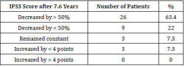
Mean IPSS scores were 17.9 ± 5.3 before the treatment, and 9.4 ± 5.2 after 7.6 years. A statistically significant (P < 0.05) decrease in IPSS scores by 8.5 ± 6.1 (47.5%) was found. A decrease in IPSS scores by > 50% was noted in 28 patients (68.3%). Overall, a positive dynamic was found in 85.4% of patients. IPSS score dynamics are summarized in Table 2.
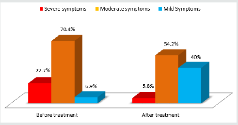
Dynamics of irritative (questions 1, 3, 5 and 6 on the IPSS questionnaire) and obstructive (questions 2, 4 and 7 on the IPSS questionnaire) symptoms were also analyzed. Mean irritative IPSS scores decreased from 6.3 ± 2.9 to 3.7 ± 2.4 (33.7% lower) whilst mean obstructive IPSS scores decreased from 9.6 ± 4 to 5.5 ± 3.7 (35.7% lower). Incidents of nocturia decreased from 2.4 ± 1.1 to 1.5 ± 1.1. Diagram 1 illustrates a major decrease in the percentage of patients with moderate to severe voiding dysfunction symptoms as well as a six-fold increase of patients with mild symptoms. All of the statistics mentioned above were statistically significant (Figure 1).

Figure 1: Severity of LUTS before and after 7.6 years of Alfuzosin treatment.
lupinepublishers-openaccess-journal-urology-nephrology

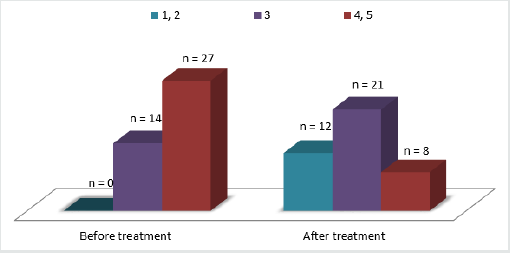
QoL dynamics are presented in diagram 2. Mean QoL indices decreased from 3.7 ± 1.1 to 2.3 ± 1.1 over the period of observation. The mean decrease in QoL indices was 32.3% (reflecting an increase in QoL). All changes were statistically significant. Overall, 72.7% of patients had an increase in their QoL (Figure 2).

Figure 2: QoL indices before and after 7.6 years of Alfuzosin treatment.
lupinepublishers-openaccess-journal-urology-nephrology

It is important to note that the minority of patients who did not demonstrate clear improvements in symptom severity either underwent surgery or continued to take Alfuzosin. The latter group either demonstrated a minimal but satisfactory improvement or was unable to undergo surgery for various reasons. We believe that it is of utmost importance to understand that the current publication presents a comparative analysis before and after Alfuzosin treatment, and whilst all of the patients had demonstrated an improvement during various periods in the trial, the dynamics of IPSS scores and changes in QoL were not registered at those intervals. It is also important to highlight the role of the detrusor in BPH symptoms, something that was originally demonstrated by Russian authors O.B.Loran and E.L.Vishnevskiy in 1998. The contraction capability, level of energy metabolism, and nature of the biochemical events of the detrusor influence voiding quality no less than the level of intravesical obstruction.[13] This could explain treatment ineffectiveness in certain patients.

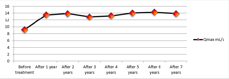
Changes in Qmax over the course of the study were considered as a secondary measurement of treatment effectiveness and were analysed during every follow-up, making this particular variable very intriguing. Mean Qmax values increased from 9.7 ± 0.52 mL/s to 14 ± 0.60 mL/s (an increase of 44.3%). Diagram 3 illustrates Qmax dynamics throughout the entire length of the study. One can conclude that early treatment results remained constant throughout the observation period (Figure 3).

Figure 3: Peak flow rate (Qmax)dynamics during 7.6 years of Alfuzosin treatment.
lupinepublishers-openaccess-journal-urology-nephrology

During the period of observation, 5 patients underwent TURP procedures. The rationale behind TURP in one patient was due to an episode of AUR provoked by alcohol ingestion. After 3 days of catheterisation, the patient did not show any signs of improvement in voiding ability and was subsequently operated. The other 4 patients exhibited worsening LUTS as shown by an increase in their IPSS scores. During ultrasound and transrectal ultrasound testing, a median lobe was detected in all 4 patients. All operations performed did not deviate from a standard TURP procedure.
The median prostate volume of the 16 patients receiving combination therapy had decreased. The value fell from 74.1 cm3 before treatment to 50.1 cm3 after 7.6 years of therapy (a decrease of 32.4%).
Throughout the trial, PSA levels of all patients were measured annually. Median PSA levels were 2.9 ng/mL before the trial and 3.2 ng/mL afterwards, demonstrating no significant variance. PSA levels of patients undergoing combination therapy were considered after the second year of treatment and were doubled. PSA levels of over 4 ng/mL requiring transrectal ultrasound-guided multifocal prostatic biopsies were noted in 3 patients aged 68–74 years. None of the subsequent histological analyses revealed the presence of cancerous cells.
The safety of this treatment was assessed based on the frequency and type of adverse side effects recorded during the trial. In total, 9 patients (22%) exhibited adverse effects. The most common findings were dizziness (2 patients/4.9%) and asthenia (2 patients/4.9%). Another 3 patients experienced dyspepsia, shortness of breath and headaches. Out of the 16-patient subgroup receiving combination therapy, 2 patients complained of decreased libido (possibly attributed to 5-ARIs). One of these patients had their sexual drive normalise after 1 year of therapy, whilst the other retained a low libido throughout the treatment. Considering the relatively small study population involved, a true understanding of the statistical significance of these adverse effects as well as their immediate connection to the treatment in question is not possible. However, the fact that most of the aforementioned adverse effects were present as isolated episodes in patients during the first week of therapy demonstrates a high level of treatment safety.

Discussion

The current study has demonstrated a high level of efficacy with minimal adverse side effects in the treatment of lower urinary tract symptoms (LUTS) arising from benign prostatic hyperplasia (BPH) with α1-adrenergic receptor antagonist (α1-blocker) Alfuzosin. The effectiveness of the treatment was determined using subjective and objective criteria. A 45.7% decrease in LUTS severity as measured by the International Prostate Symptom Score (IPSS) questionnaire was found in 85.4% of the entire study population. Mean Quality of Life (QoL) indices measured using the QoL scale increased by 32.2% and mean peak flow rate (Qmax) values increased by 44.3%. The fact that the positive results achieved during the early months of therapy had stabilised and showed no signs of regression during the entire study period is quite remarkable. Such results verify the notion that a prolonged course of α1-blockers provides consistent long-term results. The high level of patient satisfaction with treatment results and the information that patients themselves have provided us with, mark not only the fact that patients are not bothered by the need to take medication daily, but also (and most importantly) the great sense of confidence patients feel regarding the stability of the results they have achieved, leading to a positive outlook on their future treatment.

Several observational studies indicate results very similar to ours. In the ALFORTI study, 311 patients on Alfuzosin were surveyed over a period of 9 months. A decrease was shown in mean IPSS scores and QoL indices measuring 45.6% and 36.4%, respectively. Adverse effects (most likely due to the medication in question) occurred in 4.4% of the study population.[12] A similar study published by the same authors analyzed the safety and efficacy of Alfuzosin. A decrease in mean IPSS scores and QoL indices was shown and measured 32% and 27.8%, respectively[14].
Combination therapy in the form of α1-blockers and 5-alphareductase inhibitors (5-ARIs) is currently the recommended form of treatment in patients with LUTS and an increased prostate volume according to the European Association of Urology.[12] The safety and efficacy of this treatment has been confirmed by a considerable decrease in LUTS severity and prostate volume (a mean decrease of 32.4%) in our 16 patient subgroup. Similar results have also been demonstrated by such well-known trials as MTOPS, ALTESS and COMBAT[1,2,8,9].
When the information provided by the various authors is analysed, it becomes apparent that Alfuzosin attains a similar degree of effectiveness in every single case. The fact that the results gained from a continuous course of therapy remain consistent throughout prolonged treatment is extremely important, but perhaps the most interesting observation made during our trial is that the considerable results are not only long-lasting, but are achieved and made stable during the first month of treatment.
A study conducted in 2008 by Vallancien G. et al. is one of the few studies (similar to ours in design, but not in duration) that analysed the data collected in a real-life surveillance study format regarding the effectiveness of Alfuzosin during a 3-year course of therapy. The results were as follows: mean IPSS scores decreased by 33.4%, mean QoL indices by 40.7%, and nocturia severity by 25.5%. Adverse side effects (likely related to vascular dilation) were registered in 4.5% of cases, and surgical intervention was required in 5.7% of cases. These results are clearly similar to ours. [10] Similar findings were also recorded earlier during the ALFONE trial published by Elhilali M. et al. in 2006[11].
Whilst several adverse cardiac effects were noted, the analysis of adverse side effects in our trial is far from thorough, which is explained by the lack of the patients enrolled in the trial leading to insufficient statistical integrity. A study involving a multidisciplinary approach could aid the understanding of various guarantees that we may be able to give our patients in the future. These days, patients mostly consist of elderly men whose age and comorbidities make the use of pharmacotherapy ideal for managing the symptoms associated with BPH. Perhaps tomorrow, the necessity of prescribing Alfuzosin for over 10 years will increase considering the continually regressing number of patients who choose surgical intervention for BPH management; those that wish to retain a good quality of life whilst remaining ‘real men’ for many years to come. Today, when patients inquire about the potential length of their treatment, they are told that they may take Alfuzosin for as long as it is required.

Conclusions

This study has demonstrated a high level of safety and efficacy when using Alfuzosin to treat voiding dysfunction in patients suffering from LUTS/BPH. Furthermore, it is important to mention that many researchers have come to the same conclusion. Our study, whilst potentially lacking in statistical magnitude, resolves the issue surrounding a prolonged course of Alfuzosin, which, due to its widespread use, remains the gold standard for medical treatment of lower urinary tract symptoms associated with benign prostatic hyperplasia.

Read More About Lupine Publishers Journal of Urology & Nephrology Please Click on Below Link:
https://lupine-publishers-urology-nephrology.blogspot.com/


Monday, 21 November 2022

Lupine Publishers| Case of Application of Syrolimus in a Patient with Progressing Pultifocal Pepatic Nodular Hyperplasia

 Lupine Publishers| Journal of Gastroenterology and Hepatology


Abstract

Introduction: A case presented to the public demonstrates the possibility of treatment with Sirolimus in a patient with progressive multifocal liver Focal Nodular Hyperplasia (FNH) with progressive growth and a tendency to formation of liver function insufficiency after splenorenal shunt for portal hypertension.

Material and Methods: Patient E. 08.03.2001 year of birth c Diagnosed with multifocal bilobar nodular liver hyperplasia after splenorenal bypass surgery for portal hypertension, established in 2018/ Sirolimus treatment was prescribed from 06.22.18 to the present.

Conclusion: As a result of the treatment with Sirolimus, a normalization of the level of liver enzymes, bilirubin in the blood is noted. According to radiation methods, a reduction in liver size and FNH size is up to 30%.

Keywords:Multifocal liver Focal Nodular Hyperplasia (FNH); Sirolimus

Introduction

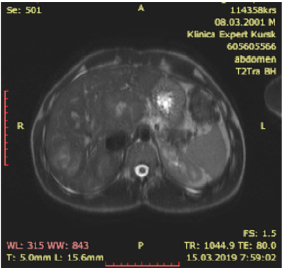
Today, FNH is regarded as a benign vascular formation of the liver. VNG is the second most common benign liver neoplasm [1]. Edmonton first described it in 1958 [2]. FNH most often occurs as a monofocal lesion, in more rare cases there are two or more nodes [3]. FNH is believed to be a hyperplastic response of liver tissue to arterial malformation, and not a true tumor. When conducting radiation diagnostics, the central scar is determined in 44% of cases. The central scar with T2-weighted MRI is hyperintensive [4]. It is possible to determine the filling of blood formation from the center to the periphery, which distinguishes it from monofocal liver hemangiomas. As a rule, in the case of PF, the risk of complications is low; there is no risk of malignancy. Therefore, a patient with VNF rarely requires their surgical removal. The most common indication for removal is pain, which usually occurs with FNH greater than 7 cm in distance [5]. The presence of more than five nodes are characterized as multiple FNH. This is very rare and only a few cases are described in the literature [6]. With progressive multiple FNH, we did not find any treatment recommendations in the literature other than liver transplantation. Therefore, we decided to present a case of treating multiple liver FNHs in a patient using Sirolimus therapy. We also did not find such articles in the literature and therefore we bring this case to the public.

Materials and Methods

Patient E. 08.03.2001 year of birth. Born from the first pregnancy, proceeding against the background of chronic intrauterine hypoxia of the fetus. Childbirth - emergency cesarean section. Height at birth 51 cm, weight 3118 grams, Apgar score of 8 points. In the neonatal period: pneumonia, urinary tract infection, perinatal hypoxic encephalopathy of the fetus, intraventricular hemorrhage. Grew and developed according to age.
In August 2008, he suffered bleeding from the dilated veins of the esophagus and stomach. Bleeding is stopped conservatively. The diagnosis of portal hypertension syndrome, cavernous portal vein transformation. In October 2008, surgical treatment was carried out: the formation of spleno-renal anastomosis “side-byside” (Figure 1). No more bleeding was noted.

Figure 1.

Lupinepublishers-openaccess-gastroenterology-hepatology-journal

In February 2018, during a control study, multiple FNHs were identified in C1, C4, C8 with dimensions of 87x61 mm, in C7 with dimensions of 51 by 43 mm, 50 by 40 mm (Figure 2). Diagnosed with multifocal bilobar nodular liver hyperplasia after splenorenal bypass surgery for portal hypertension. Blood test for alphafetoprotein 1.66 IU / ml. During the control examination in July 2018, an increase in FNH sizes to 90 by 70 mm, an increase in blood transaminases (Table 1), alkaline phosphatase up to 218.00 IU / L (N 42 - 110), a moderate increase in bilirubin were noted. The patient complained of chronic fatigue, weakness, moderate pain in the liver. lack of appetite.

Figure 2.

Lupinepublishers-openaccess-gastroenterology-hepatology-journal

Table 1.

Lupinepublishers-openaccess-gastroenterology-hepatology-journal

After examining the child, we came to the conclusion that the observed progression of the disease can lead to the replacement of the liver parenchyma with the subsequent occurrence of organ failure. This could endanger the patient’s life, but there is no possibility of surgery due to the prevalence of the lesion. Sirolimus (Pfizer, USA) from 06.02.2018 was prescribed orally daily at a dose of 3 mg / day until a therapeutic concentration of the drug in blood serum of 6-15 ng / ml was achieved. Due to the excess of the therapeutic interval, the dose of Sirolimus was consistently reduced from 11/06/2018 to 1 mg / day, 02/05/2019 the concentration of Sirolimus was 7.8 ng / ml.

Résultats

As a result of the treatment with Sirolimus, a normalization of the level of liver enzymes, bilirubin in the blood is noted. According to radiation methods, a reduction in liver size and FNH size is up to 30%. Now the patient is feeling well. There is no pain in the liver. Playing sports. Reception of sirolimus continues to date.

Discussion

Multifocal FNH is a rare form of this disease. An even rarer situation arose in our patient, there was a progression of the disease with signs of emerging hepatic cell failure. There was a high risk of severe disease with subsequent liver transplantation. There is an interesting fact that the physiological characteristics of the patient appeared after application of a splenorenal shunt for portal hypertension. Apparently, in the growth stimulation of the nodes, the FNH played the role of impaired portal blood flow. In the available literature, we found only recommendations for liver transplantation in such patients. Teaching ability of Sirolimus to suppress vascular growth through inhibition of M-TOP, we suggested the possible effectiveness of this therapy. After an explanation with the patient and his legal representatives, we started therapy with Sirolimus at a dose of 3 mg / day, then we reduced it to 1 mg / day taking into account the concentration in the blood. The patient had no complications associated with Sirolimus therapy. After a year of treatment, we noted a clear positive trend. The patient’s condition improved, there was no pain, blood counts returned to normal (Table 1).

Conclusion

Of course, this question still requires research in other patients with multiple progressive FNH, in order to draw conclusions about the effectiveness and safety of Sirolimus. But with this patient, we got encouraging results.

Read More About Lupine Publishers Journal of Gastroenterology and Hepatology Please Click on Below Link: https://currenttrendsingastroenterology.blogspot.com/