Showing posts with label Journal of Nephrology. Show all posts

Thursday, 28 September 2023

Lupine Publishers | The TUR Syndrome Re-Incarnating as ARDS after Saline use as Irrigating Fluid in Endoscopic Surgery

 Lupine Publishers | Journal of Urology & Nephrology


Abstract

Objective: To demonstrate the TUR syndrome characterized with hyponatraemia (HN) will no longer be seen after using saline as irrigating fluid in urology, but it has re-incarnated as the acute respiratory distress syndrome (ARDS) presenting with the same clinical picture of the multiple organ dysfunction syndrome (MODS).
Material and Methods: A focused objective and relevant narrative review of other eminent authors’ work and mine are used here.
Results: The TUR syndrome characterized with HN will no longer occur in urology after the use of saline as irrigating fluid in endoscopic surgery. It has reincarnated as ARDS presenting with the same MODS clinical picture. It is induced by VO caused by iv fluid infusions. This induces cardiovascular shock (VOS) that cause ARDS. The latter is already common in clinical practice due to the excessive us of iv fluids in the management of shock, acutely ill patients, and prolonged major surgery as iatrogenic complication of fluid therapy. The wrong Starling’s law dictates the current faulty rules on fluid management of shock that mislead physicians into giving too much fluid. The correct replacement is the hydrodynamics of the porous orifice (G) tube which should be the new scientific basis for fluid therapy in shock management. The currently available hypertonic sodium therapy of 5%NaCl and/ or 8.4%NaCo3 is lifesaving therapy for HN, the TUR syndrome and ARDS.
Conclusion: The TUR syndrome may seem to have been eradicated in urology with the use of saline as irrigating fluid in endoscopic surgery. However, it has reincarnated as ARDS with the same clinical picture of MODS. It is an iatrogenic complication of fluid therapy dictated by the wrong Starling’s law for which the hydrodynamic of the G tube is the correct replacement that should be the new scientific basis for a new policy on fluid management of shock.

Keywords: The TUR syndrome; Endoscopic Surgery; ARDS; Shock; Fluid Therapy; Starling’s law, Capillary-ISF transfer

Introduction

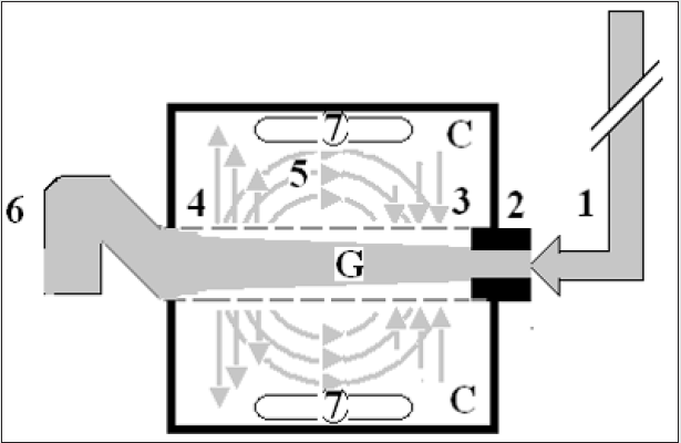
My beginning with the transurethral resection of the prostate (TUR) syndrome started in 1981 after I attended post-mortem (PM) examinations on 3 patients who died after the TURP surgery. I was only an SHO in urology working for the late Mr. KC Perry and JP Ward at DGH in Eastbourne. At the PM examination it was clear and obvious to me that these patients died of internal drowning as result of massive volumetric overload (VO) of fluids used for resuscitation of a cardiovascular shock they suffered, and the fluid was retained in their bodies. When I asked the pathologist why doesn’t he mention that retained VO in his report? He replied: “because it offends treating physicians”? The word offends hit me right hard on my head like a hammer. My next question to myself was if it offends them why do physicians do it? This had led me to immediately replace the term fluid overload with the new and original Volumetric Overload (VO) after adding the cardiovascular hypotension Shock to it to become (VOS) that was introduced to avoid the word offends but it has proved to be a new scientific medical discovery. Another few questions such as: “What is misleading physicians into giving too much fluid during the resuscitation of shock? What shock is it? I communicated with Richard Harrison III (who may be late now) who is the originator of the hyponatraemic shock of the TUR syndrome and the use of 5%NaCl therapy in clinical practice for years during his retirement [1]. I reported later the true pioneer originators of this shock and the hypertonic sodium therapy (HST) were Danowski et al who induced it experimentally in dogs by massive 5%Glucose infusion [2]. Harrison advised me to “put the poison in the honey” that I could not accept. After the PM examination I suspected and incriminated Starling’s law being the scientific basis of fluid therapy in shock that dictates the wrong rules on fluid therapy for shock management documented in articles and books [3-7], for which the hydrodynamics of the porous orifice (G) tube is the correct replacement (Figures 1a&b) [8,9]. I felt so strongly about it that I wrote a letter to the late great professor of physiology Eric Neil and author of Sampson Wright Textbook of Physiology later in 1983 [10,11]. He nicely replied in handwritten letter as he was in retirement asking: Why and how may Starling’s law cause death of patients? The answer is there now after 40 years of hard scientific research and investigations [12].

Figure 1a: shows a diagrammatic representation of the hydrodynamic of G tube based on G tubes and chamber C. This 37-years old diagrammatic representation of the hydrodynamic of G tube in chamber C is based on several photographs. The G tube is the plastic tube with narrow inlet and pores in its wall built on a scale to capillary ultra-structure of pre-capillary sphincter and wide inter cellular cleft pores, and the chamber C around it is another bigger plastic tube to form the G-C apparatus. The chamber C represents the ISF space. The diagram represents a capillary-ISF unit that should replace Starling’s law in every future physiology, medical and surgical textbooks, and added to chapters on hydrodynamics in physics textbooks. The numbers should read as follows:
The inflow pressure pushes fluid through the orifice.
Creating fluid jet in the lumen of the G tube**.
The fluid jet creates negative side pressure gradient causing suction maximal over the proximal part of the G tube near the inlet that sucks fluid into lumen.
The side pressure gradient turns positive pushing fluid out of lumen over the distal part maximally near the outlet.
Thus, the fluid around G tube inside C moves in magnetic field-like circulation (5) taking an opposite direction to lumen flow of G tube.
The inflow pressure 1 and orifice 2 induce the negative side pressure creating the dynamic G-C circulation phenomenon that is rapid, autonomous, and efficient in moving fluid and particles out from the G tube lumen at 4, irrigating C at 5, then sucking it back again at 3,
Maintaining net negative energy pressure inside chamber C.
**Note the shape of the fluid jet inside the G tube (Cone shaped), having a diameter of the inlet on right hand side and the diameter of the exit at left hand side (G tube diameter). I lost the photo on which the fluid jet was drawn, using tea leaves of fine and coarse sizes that run in the centre of G tube leaving the outer zone near the wall of G tube clear. This may explain the finding in real capillary of the protein-free (and erythrocyte-free) sub-endothelial zone in the Glycocalyx paradigm. It was also noted that fine tea leaves exit the distal pores in small amount maintaining a higher concentration in the circulatory system than that in the C chamber- akin to plasma proteins.
lupinepublishers-openaccess-journal-urology-nephrology

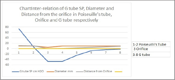
Figure 1b: shows the relationship between SP to the Diameter and length of the G tube which demonstrate a negative SP starting at the orifice (Point 2) (akin to precapillary sphincter) and extends as high negative pressure gradient over the proximal part of the G tube (Point 2-6) to cross 0 line at point 8 and then turn positive of 7 cm water at Point 9. This SP gradient from orifice at Point 2 to G tube lumen (Points 2-6) is negative to become positive DP at point 9 of 7 cm H20 water along the G tube. The wide section diameter of G tube is 7 mm all along the entire tube. The orifice is 5 mm while the distance from orifice to exit represent the tube’ length in which the Fluid jet diameter change with increasing gradient (Figure 1a). Neither Poiseuille’s law nor Bernoulli’s equation can predict SP neither at orifice of Venturi’s effect nor at the G tube proximal part know as Bernoulli’s effect. Thus, the RBCs speed or CBS depend on the dynamic fluid jet diameter not the G tube diameter. Hence the equation in Figure 2g (Figure 2) and graph are wrong giving low RBCs speed or CBS over the capillary length but is correct only at point of the G tube where the jet diameter equals the tube diameter.
lupinepublishers-openaccess-journal-urology-nephrology

What is the TUR Syndrome? And what is causing the “Understanding Gap”? Our prospective cohort study on the TUR syndrome was conducted in 1987-8, a letter to the editor of BJU was reported in 1988 [13], MD Thesis was accepted November 1988 [14], and the article reported in 1990 [15]. The TURP syndrome is a condition induced by gaining large volume of sodium-free fluid overloading the cardiovascular system and spelling over into the interstitial fluid (ISF) space of vital organs and subcutaneous. The fluid of 1.5%Glycine used as irrigating fluid gets absorbed, or rather infused through peri-prostatic veins, during the TURP surgery as well as all endoscopic surgeries performed under sodium-free fluid irrigation of any type such as Mannitol, Sorbitol, Glucose and Cytal. Also, intravenous (iv) infusion of 5% Glucose considerably and significantly contributes to it- as well as saline. What is more, excessive infusion of saline or any sodium-based fluid such as Saline, Hartman, Ringer, plasma, and plasma substitutes, and blood worsens it transferring the shock being treated from VOS 1 into VOS 2 [16] and causing ARDS 1 and 2 [17,18] with apparent correction of HN, and has high morbidity and mortality later.
The TUR syndrome has a characteristic severe drop of serum sodium level causing acute dilutional hyponatraemia (HN) induced by VO 1 (Figures 2 & 3) with severe clinical symptoms affecting all vital organs causing the multiple organ dysfunction syndromes (MODS) (Table 1) or ARDS [17,18] with recognizable clinical picture but one system may predominate such as acute kidney injury (AKI). The HN of <120 mmol/l has 2 paradoxes and 2 nadirs that have eluded authorities and physicians on HN, and that has made the TUR syndrome most elusive and invisible making it though obvious it has remained invisible even to authorities on HN. Professors and consultant urologists who are such swift good resection experts have testified that the TUR syndrome does not exist as no fluid absorption occurs, with a negative prospective study of 100 patients [19]. Off course no such hyponatraemia occurs when the irrigating fluid is saline whatever the volume absorbed and infused. Another important reason that prevents massive 1.5% glycine absorption and the TUR syndrome is for the Urologist not to breach the prostate capsule and not to open the venous sinuses where the irrigating fluid is directly injected intravenously (iv) into the periprostatic veins. There was also another good swift urologist who reported >1000 consecutive TURP surgeries without seeing the TUR syndrome. The risk of VO during endoscopic surgery will continue to occur as long as there are registrars in training and even with the experienced consultants who occasionally and inadvertently breach the prostatic capsule and open the venous sinuses. However, the TUR syndrome due to 1.5% Glycine VO with its characteristic HN has an undoubted reality [13-15] and [20-22]. Our study reported 10% incidence of the TUR syndrome with one near death case that was saved [14] and a similar study done a year earlier in the same department reported 7% incidence of morbidity with 1% mortality [22]. Before the TUR syndrome disappears into oblivion and is totally replaced by ARDS a most comprehensive literature review on the subject was reported in 2018 after the wide use of saline as irrigating fluid in the TURP surgery [23]. Here a distinction between a physiological VO of <2 L infused in less than one hour that is extensively studied by Hahn in volunteers and patients is known as Volume Kinetic (VK) (20) and the pathological VO of 3.5-5 L gained in < 1 h that causes the TUR syndrome [15] is highlighted. This has been a cause of serious misunderstanding gap in the pathogenesis of the TUR syndrome. The physiological response of VK is remarkably different from the pathological response of VO which is paradoxical: VK elevates blood pressure and induces diuresis while VO causes hypotension with bradycardia and causes acute renal failure.

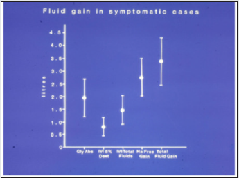
Figure 2: It shows the means and standard deviations of volumetric overload in 10 symptomatic patients presenting with shock and hyponatraemia among 100 consecutive patients during a prospective study on transurethral resection of the prostate. The fluids were of Glycine absorbed (Gly abs), intravenously infused 5% Dextrose (IVI Dext) Total IVI fluids, Total Sodium-free fluid gained (Na Free Gain) and total fluid gain in litres.
lupinepublishers-openaccess-journal-urology-nephrology

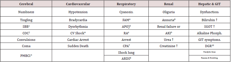
Table 1: Shows the manifestations of VOS 1 of the TURP syndrome for comparison with ARDS manifestations induced by VOS2. The manifestations are the same but one vital organ-system may predominate.
lupinepublishers-openaccess-journal-urology-nephrology

Table Abbreviation
SBB1: Sudden Bilateral Blindness
COC2: Clouding of Consciousness
MBCI3: Paralysis mimicking bizarre cerebral infarctions, but is recoverable on instant use of HST of 5%NaCl and/or NaCO3, and so is coma and AKI
FAM4: Frothing Around the Mouth
APO5: Acute Pulmonary Oedema.
RA6: Respiratory Arrest.
CPA7: Cardiopulmonary Arrest; ARDS$: Occurs on ICU later.
Annuria8: That is unresponsive to diuretics but responds to HST of 5%Ncl and/or 8.4%NaCO3; AKI8: Acute Kidney Injury. Also occurs the excessive bleeding at
AKI9: Acute Kidney Injury
DGR10: Delayed Gut Recovery; CV Shock*:
Excessive bleeding may occur at the surgical site and leucocytosis occurred in the absence of sepsis and septic shock.

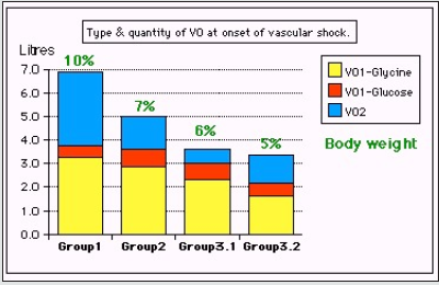
Figure 3: Shows volumetric overload (VO) quantity (in liters and as percent of body weight) and types of fluids. Group 1 was the 3 patients who died in the case series as they were misdiagnosed as one of the previously known shocks and treated with further volume expansion. Group 2 were 10 patients from the series who were correctly diagnosed as volumetric overload shock and treated with hypertonic sodium therapy (HST). Group 3 were 10 patients who were seen in the prospective study and subdivided into 2 groups; Group 3.1 of 5 patients treated with HST and Group 3.2 of 5 patients who were treated with guarded volume.
lupinepublishers-openaccess-journal-urology-nephrology

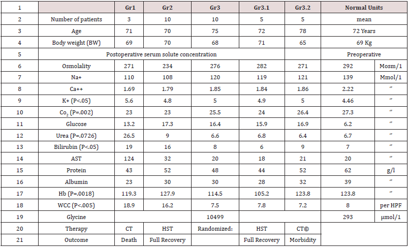
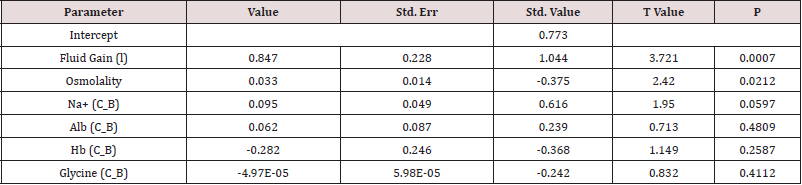
The TURP syndrome starts by presenting with cardiovascular hypotension shock to anaesthetists and surgeons in theatre [24,25] and at times by cardiac or cardiopulmonary arrest [26] and sudden death. By next morning the surviving patients present with coma, convulsion and bizarre paralysis to physicians, neurologists, and ICU specialists [15]. It has the characteristic serum hypo-osmolality. BUT other solute contents dilutions seem to be apparently spontaneously improving due to water shift into cells [Table 2, Figures 1 and 2]. The HN of <120 mmol/l causes cardiovascular hypotension shock. Volumetric overload (VO) is the most highly significant factor causing its patho-aetiology with a (p=0.0007). Osmolality was also significantly low (p=0.02) while all other serum solute changes including the most remarkable drop in serum sodium and huge elevation in serum glycine did not reach statistical significance in the multiple regression analysis, yet it did alone when pre- and post-operative levels are compared!? [Table 2 and 3]. This cardiovascular shock of VOS is easily confused with and mistaken for haemorrhagic or septicaemia shock and is wrongly treated with further massive volume expansion that usually kills the patient as happened in the 3 patients mentioned above!?

Table 2: Shows the mean summary of data, therapy and outcome comparing the 3 groups of 23 case series patients who’s (whose) VO is shown in Figure 3. Groip-1 was the 3 patients who died and had post-mortem examination, Group-2 were a series of severe TURP syndrome cases successful ly treated with hypertonic sodium therapy (HST), and Group-3 were 10 patients encountered in the prospective study who were randomized between HST (3.1) and conservative treatment (CT) (3.2). The significant changes of serum solute contents are shown in bald font with the corresponding p- value. Most of the patients showed manifestation of ARDS of which the cerebral manifestation predominated, being on initial presentation (Regional Anaesthesia) and representation of VOS 1 (General Anaesthesia). However, most patients were given large volume of saline that elevated serum sodium to near normal while clinical picture became worse. They suffered VOS2 that caused ARDS. The VO of patients to whom these data belong is shown.
lupinepublishers-openaccess-journal-urology-nephrology

Table 3: Shows the multiple regression analysis of total per-operative fluid gain, drop in measured serum osmolality (OsmM), sodium, albumin, Hb and increase in serum glycine occurring immediately post-operatively in relation to signs of the TURP syndrome. Volumetric gain and hypo-osmolality are the only significant factors.
lupinepublishers-openaccess-journal-urology-nephrology

The toxic theory of the TUR syndrome and septic theory of ARDS.

Sepsis and septic shock in the pathogenesis of ARDS is as innocent as the wolf in Josef story [18], so is glycine in the aetiology of the TUR syndrome [15], particularly as correctly mentioned that the TUR syndrome occurs with Mannitol, Sorbitol, and Glucose. Professor Alan Arieff has clearly reported the morbidity and mortality of hyponatraemia (HN) of the TUR syndrome induced by 1.5%Glycine as well as the excessive 5%Glucose infused intravenously during prolonged surgery in healthy women [27]. That does not mean that I deny the toxicity of glycine and the seriousness of sepsis. I am just saying they are misleading like a mirage to someone thirsty and lost in the desert. While thinking about it please, try to attend the PM examination of some patients who died from the TUR syndrome and ARDS. Every anaesthetist should examine own practice when he embarks on Bolus Fluid Therapy (BFT) during anaesthetic induction and watch out how much fluid is given during prolonged major surgery. Review the scientific basis of fluid therapy in the management of septic and all other types of shock on which bases the current practice is implemented.

Fluid therapy Regimen and Iatrogenic complications

The TUR syndrome occurs because of combination of fluid absorption and direct iv infusion of the irrigating fluid when the prostatic capsule is breached, and venous sinuses are open. In clinical practice all ARDS cases occur as result of iv infusion of fluids. In our study 7 cases of capsule breaching occurred among the 10 TUR syndrome cases as observed by the surgeon. The iv infusion occurs with both the liberal regimen of Early Goal-Directed Therapy (EGDT) and Bolus fluid therapy (BFT) of the conservative regimen. Hahn is a professor and consultant of anaesthesia and intensive care. He is also a leader and world authority on fluid therapy and the editor of a book on the same subject. I would and have recommended him as the head of a committee to write the new guidelines on fluid therapy in shock management. He has my new book that will help him for >8 months now, please read it if you’ve not done so already. Like all anaesthetists, Intensive care therapists, surgeons, and physicians of the whole world who remain to practice the liberal fluid therapy regimen also well known as EGDT in the management of shock, don’t you? Go to any ICU near you and observe the swollen-up ARDS patients mostly with trunk oedema comparing their body weight on hospital admission with their current weight while suffering from ARDS. Try to attend the PM examination of the TURP patients and ARDS patients. Allow me to reproduce this section from my article later that is most recommended reading to all physicians interested in the subject of fluid therapy, the TUR syndrome, HN, VOS and ARDS [18].

The role of Starling’s law

Starling’s law [28,29] dictates the current faulty rules on fluid therapy in the management of shock. It thus misleads physicians into giving too much fluid during shock resuscitation [30]. More than 21 reasons were reported to show that Starling’s law is wrong [31], none of it can be denied or refuted. The correct replacement is the hydrodynamic of the porous orifice (G) tube [8,9] (Figure 1 a & b) that was built on capillary ultrastructure anatomy of having precapillary sphincter [32] and a porous wall [33] that allow the passage of plasma proteins-hence nullify the oncotic pressure. It follows that the extended Starling Principle is wrong and a misnomer [34,35] and all the equations are also wrong.

Two types of VO inducing VOS and causing ARDS of type 1 and 2

There are two types of VO: Type 1 induced by sodium-free fluid and Type 2 induced by sodium-based fluid. These in turn induce VOS 1 and VOS 2 which cause ARDS 1 and ARDS 2, respectively. The clinical picture is the same for both types (Table 1). Type 1 is characterized with HN of the TUR syndrome with which the cerebral neurological manifestations of coma, convulsions, and bizarre paralysis predominate while type 2 may have moderate hypoproteinemia if induced by crystalloids and none when plasma, plasma substitutes and blood are used. Type 2 may complicate Type 1 or may occur do novo. Manifestations of the multiple organ dysfunction syndrome (MODS) are the same and appear in every case, but one system may predominate. When Hahn sent me his article on Revised Starling Principle calling for revalidation [34] I immediately responded with an article: Revised Starling’s Principle (RSP): a misnomer as Starling’s law is proved wrong. I considered research on validating RSP is a total waste of money, time, and efforts.

Proof by eminent authors on the VO role in the aetiology of the TUR syndrome and ARDS

Professor Robert Hahn from Sweden has done lots of research infusing various types of fluid used in clinical practice to normal adult volunteers and patients, as well as animal research and clinical studies and reported >340 articles on the TURP syndrome alone (PubMed 2017) and 532 articles in total (PubMed search 2021): Here is what Robert Hahn said: in the abstract of an article reported in 2017 [36]:

Abstract [36]:

“Adverse effects of crystalloid fluids are related to their preferential distribution to the interstitium of the subcutis, the gut, and the lungs. The gastrointestinal recovery time is prolonged by 2 days when more than 2 liters is administered. Infusion of 6-7 liters during open abdominal surgery results in poor wound healing, pulmonary oedema, and pneumonia. There is also a risk of fatal postoperative pulmonary oedema that might develop several days after the surgery. Even larger amounts cause organ dysfunction by breaking up the interstitial matrix and allowing the formation of lacunae of fluid in the skin and central organs, such as the heart.” Thank you, Professor Hahn for a most impressive work indeed. New guidelines based on currently available evidence on fluid therapy for resuscitation of sepsis, septic shock, trauma patients, critically ill patients, ARDS and patients undergoing prolonged major surgery are badly needed. Professor Hahn is the expert witness on fluid therapy.
Why does not Hahn believe his own results? Why doesn’t he make the most obvious conclusion based on what he said in the abstract above? What and how much more evidence and years that he needs to believe that the pathological VO of massive fluid infusions induces cardiovascular shock that is VOS of both types and causes ARDS? If my articles referenced here and the books [3- 7] particularly the one Hahn has now for 8 months and being held in the press awaiting his introduction, then allow me most sincerely and humbly to give you a helping hand to lift you up to where I stand and clearly see the picture on the real issues discussed here. Hahn does not need to do any more research studies. Just report a re-analysis of data from previously reported articles he has done and reported before, based on his previous published articles on the TUR syndrome and saline-based fluid infusions. Please, reexamine and re-analyse your own research work in a manner and method identical to your article reported here [20]. Please, Hahn don’t bother with equations that are hard to understand and are meaningless and perhaps misleading or even wrong. Do not use fancy sophisticated graphs that does not impress me. I would love, most sincerely and humbly, to give you a hand to get you out of the huge maze you have been lost inside it for >3 decades. All you need to do my friend now is to liberate yourself from the illusive and misleading concepts of the toxic/septic hypotheses of glycine and sepsis!? One must unlearn old bad habits to be able to receive and acquire the new correct ones.

Evidence for the VO Theory causing VOS and ARDS

“The prevalence of “liberal fluid infusion” in resuscitation of all types of shocks not only septic shock in clinical practice all over the world is attributed to an impactful article by Rivers et al, reported at The N Engl J Med 2001 [37]. Dr Rivers’ investigation reported EGDT in the treatment of severe sepsis and septic shock. In this singlecenter study published more than 20 years ago involving patients presenting to the emergency department with severe sepsis and septic shock, the conclusion was: “mortality was markedly lower among those who were treated according to a 6-hour protocol of EGDT, in which intravenous fluids, vasopressors, inotropes, and blood transfusions were adjusted to reach central hemodynamic targets, than among those receiving usual care” Usual care means conservative fluid regime. There is something grossly wrong with this conclusion, but I cannot tell what is it? Not yet. Let us see what other author investigators have said first. The EGDT of liberal fluid infusion has been termed “aggressive” by some authors. However, it has been adopted all over the world not only for the therapy of septic shock but also whenever fluid therapy is required for the management of all types of shocks.
“In another article by Dr Rivers 11 years later in 2012 [38] he compared the liberal to the conservative approach concluding in his last statement: “In contrast to what is true in politics, in fluid management of acute lung injury, it is OK to be both liberal and conservative.” So, Dr Rivers says it is OK to have it both ways: “one for the ebb and one for the flow”! Sorry, sir, I disagree. It is not OK. It is not politics either. No, you cannot have it both ways. The right way is only one. The issue here is how much fluid should be infused during the ebb phase of shock and does it have a maximum limit? Replace the loss but do not overdo it. Since the cardiovascular system (CVS)’ maximum capacity of an adult is 7 L and the normal blood volume is 5 L, the maximum infused volume of fluid should be limited by the maximum capacitance of the CVS. What do you expect when you try to fit 10-15 L of fluid into a 7 L capacity container? Simple physics and common sense indicate that it must spell over if it is open system or burst if closed! The cardiovascular system is no exception. Dr Rivers should re-examine his own data and tell us where and why he went so grossly wrong.” The EGDT has spread like fire in a haystack, and it remains operative in current clinical practice all over the world that is why ARDS is so common yet remains under recognized and underestimated affecting and killing hundreds of thousands of patients per year.” Other authors have confirmed the significant role of VO of crystalloids in causing the morbidity and mortality of ARDS both in adult and children of trauma patients [39,40]. All authors have stopped short of recognizing VOS as Cause of ARDS or MODS morbidity and mortality. Quoting also from this article [18] I mention here the remarkable multicenter study by Rowan et al. [41] Like Hahn they reported results that demonstrate the massive VO retained in the body of surviving ARDS patients. After sending 3 emails to Rowan commending the authors on their results and asking about the dead patients retained fluid VO, none of the 40+ authors replied. “The PRISM Investigators reported its Trial by Rowan et al at NEJM 2017 [41] concluded: “In this meta-analysis of individual patient data, EGDT did not result in better outcomes than usual care and was associated with higher hospitalization costs across a broad range of patient and hospital characteristics.” Thank you, Dr Rowan and colleagues for the excellent research and report. This is good evidence-based medicine, but more is needed, from you, and you have the data to provide it. Based on this conclusion that agrees with other multicenter trials I wonder is time to say goodbye Dr Rivers? The aggressive and deleterious liberal approach of EGDT is no longer wanted. It should be abandoned immediately. Even when the nasty liberal approach goes away, hopefully soon, it remains bad enough with the conservative regime as it is now that must be sorted out! I wonder what Dr Rivers has to say about this, particularly as authors of 3 other huge prospective multicenter trials of The ProCESS/ARISE/ProMISe reported similar conclusion by Huang et al. [42]. So, Rowan gave the results of: The cumulative VO was -136 ml in the conservative-strategy group, as compared with 6992 ml in the liberal-strategy group (P<0.001). For patients who were in shock at baseline, the cumulative seven-day VO was 2904 ml in the conservative-strategy group and 10,138 ml in the liberal strategy group (P<0.001). For patients who were not in shock at baseline, the cumulative VO was −1576 ml in the conservative-strategy group and 5287 ml in the liberal-strategy group (P<0.001)”. “First, the negative sign (-) indicating negative fluid balance has appeared in the data above and is very important. It characterizes the nonsymptomatic patients among the conservative-strategy group.
These patients should be used as the controls for the statistical analysis of the data. I have been waiting for 40 years to see these VO results. I am still waiting to see VO data with statistical significance in mortality patients. I plead with and urge the respected authors of major randomized Trials of FACCT, PRISM, ProCESS, ARISE, and ProMISe to come forward with these data, please,

Clinical picture of (VOS, The TUR syndrome, ARDS and MODS)

The clinical picture of ARDS is that of the multiple organ dysfunction syndrome (MODS) (Table 1) reported previously by Khadarow and Marshal in 2002 [43]. Another remarkable article was reported by Schrier in 2010 [44]. Demonstrating the link of the TUR syndrome with ARDS by having identical clinical picture with minor variations was reported by Ghanem as complications of VO covering the cardiovascular/hematological that appear first under general Anaesthesia with bradycardia [45], the cerebral/ neurological with coma appear first under spinal/epidural Anaesthesia and convulsions and bizarre paralysis predominate in the TUR syndrome, not in ARDS [46], the respiratory of ARDS and hepatic/gastrointestinal manifestations [47] and AKI predominate later were documented recently in individual specific reports. Excessive bleeding and leukocytosis in the absence of sepsis also occur.

Therapy of VOS, the TUR syndrome and ARDS [17] Prevention

Based on the above discussion, ARDS is an iatrogenic complication of fluid therapy in hospital, never in community, that is overlooked and underestimated. Being iatrogenic; means it is preventable. In order to prevent VOS and ARDS a limit to the maximum amount of fluid used during shock resuscitation or major surgery must be agreed upon. Professor Hahn [36] found that infusing 2 L of saline to human volunteers produces symptoms. Infusing >3 L is pathological. More than 5 L is associated with deleterious morbidity [38,39]. So, the maximum volume of fluids that can be infused safely to an adult patient is 3 L which is the daily fluid requirement, and no more fluid of any kind is given for 24 hours except replacing the actual loss that does not include urine loss. The patient should be put on a weighing scale every day from hospital admission till discharge or death. Any retained volume of fluid above his body weight on admission is pathological. On using CVP for monitoring fluid therapy, please refrain from persisting to elevate CVP to levels above 12 and up to 18-22 cm saline [48]. This is a major cause for inducing VO and VOS and ARDS during shock resuscitation, particularly septic shock [37]. Look up any physiology textbook to find out that the normal CVP is 0 and it swings between -7 and +7 cm saline which is the level that should be aimed at in monitoring fluid replacement in shock of sepsis, trauma, and bleeding, acutely ill and during major surgery. Elevating CVP is not synonymous with elevating arterial pressure. If hypotension develops later during ICU stay, inotropic drugs, hydrocortisone 200 mg and HST should be used. The latter restores the pre-capillary sphincter tone (peripheral resistance) so that the capillary works as normal G tube again [9], but no isotonic crystalloids or colloids infusions of above the daily fluid requirement should be given. If persistence with the current liberal regimen of Early Goal-Directed Therapy (EGDT) and conservative Bolus Fluid Therapy regime continues, then more reports on ARDS will continue. Future authors will be hopefully taking into consideration the mentioned above data concerning VO/Time, or the retained fluid VO at the time of inducing ARDS or death on reporting new trials or case reports.

Treatment of ARDS [6]

Hypertonic sodium therapy (HST) of 5%NaCl and/or 8.4%NaCo3 has truly proved lifesaving therapy for the TUR syndrome and acute dilution HN [17,18] as well as Secondary VOS 2 that complicates fluid therapy of VOS 1 causing ARDS. It works by inducing massive diuresis; being a potent suppressor of antidiuretic hormone. My experience in using it for treating established ARDS with sepsis and primary VOS 2 that causes ARDS is limited. However, evidence on HST suggests it will prove successful if given early, promptly, and adequately to ARDS patients while refraining from any further isotonic crystalloid or colloid fluid infusions using saline, Hydroxyethyle starch and/or plasma therapy- just give the normal daily fluid requirement and no more. After giving HST over one hour using the CVP catheter already inserted, the patient recovers from AKI and produces through a urinary catheter massive amount of urine of 4-5 L as you watch. This urine output should not be replaced. Just observe the patient recovering from his AKI, coma and ARDS and asks for a drink. This is done in addition to the cardiovascular, respiratory, and renal support on ICU. Patients with AKI on dialysis, the treating nephrologist should aim at and set the machine for inducing negative fluid balance. The HST of 5%NaCl and/or 8.4%NaCo3 is given in 200 ml doses over 10 minutes and repeated. I did not have to use more than 1000 ml during the successful treatment of 16 patients. Any other hypertonic sodium concentration is not recommended- I know Hahn tried 1.8%NaCl and it does not work. A dose of intravenous diuretic may be given but it does not work in a double or triple the normal dose. A dose of 200 mg of hydrocortisone is most useful. Antibiotic prophylactic therapy is given in appropriate and adequate doses to prevent sepsis and septic shock. No further fluid infusions of any kind of crystalloids, colloids and blood is given. The urinary loss should not be replaced as this represent a surplus in the body and must be discarded otherwise defeats the objective of treatment.

Addendum: Relevant articles on the history of the TUR syndrome and ARDS

This addendum is dedicated to important landmark articles on the history of the TUR syndrome and ARDS that could not be fitted directly on the above focused narrative review on how the TUR syndrome has been reincarnated into ARDS. It is optional reading for the interested reader, but it completes this review. The first part is dedicated to eminent authors on the TUR syndrome and ARDS whether directly or indirectly. The second part is a section on selfreferences by the author that report important issues that highlight aspects of the presentation.

A. Other Eminent Authors

Creevy was the first author to report the TUR syndrome as acute water Intoxication [49]. Ashbaugh et al were the first to report ARDS in the Lancet in 1967 [50]. Lessels et al. reported in a letter to the editor as the only article on death during prostatectomy [51]. Hendry was first to report that the osmotic pressure of various body fluid is the same as plasma [52]. Guyton and Coleman reported the negative pressure of the subcutaneous space of -7 cm water, a fact that cannot be explained by Starling’s law [53]. Calnan et al reported the negative pressure in lymphatic vessels [54]. Renkin was the first to call for reconsideration of Starling’s law [55]. The Coshran injuries Group, Finfer, Vincent and futier et al demonstrated that oncotic pressure does not work and the argument on albumin versus saline is obsolete [56-59].

B. Self-references

Articles 60 and 61 have educational and entertainment value. Articles 62 and 63 shows the relevance of my work on ARDS to Covid-19 pandemic ARDS. Article 64- 66 corrects other received misconceptions on capillary physiology to augment the discovery of the G tube hydrodynamics and its impact on the capillary- ISF transfer. Articles 67 and 68 report the two clinical studies on which the above article is based. Article 68 corrects some errors and misconceptions on fluid therapy. Article 70 is on preventing renal failure in the critically ill patients. Article 71 reports my Experience with cystoprostadenectomy with “prostatic capsule sparing” for orthotopic bladder replacement. Article 72 is on Features and Complications of Nephroptosis Causing the Loin Pain and Haematuria Syndrome. Article 73 reports “New Discoveries in Medicine and Physiology Originated in Urology”. Article 74 is on an Update on Ghanem’s new scientific discoveries in physics, Physiology, and Medicine, Article 75 is on Goodbye Starling’s law, hello G tube.

Conclusion

The TUR syndrome as defined and characterized with acute dilutional hyponatraemia will no longer be seen in urology after the use of saline as irrigating solution in endoscopic surgery. However, the ARDS will replace it with identical clinical picture of MODS that continue to occur with high morbidity and mortality that is underrecognized and underestimated. The ARDS is common in clinical practice and is induced by excessive sodium-based fluid infusion and is likely to occur in urology due to the added risk of irrigating fluid absorption and infusion through periprostatic veins. Neither the toxic theory nor the septic theory plays the significant assumed rule in the pathogenesis of the TUR syndrome and ARDS. Both are iatrogenic complications of fluid therapy, induced by VO of > 3 L in <1 h time and is severe at 7-10 L of retained fluid VO in surviving ARDS patients wile mortality occur with 12 L, and both have preventative and curative therapy of HST of 5%NaCl and/or 8.4%NaCo3.

Conflict of Interest

Conflict of Interest: None

Funds received

Funds received: None

Read More About Lupine Publishers Journal of Urology Please Click on Below Link:
https://lupine-publishers-urology-nephrology.blogspot.com/

Saturday, 5 August 2023

Lupine Publishers | Swathed Ureter, an Enigma in Diagnosis- A Pictorial Essay

 Lupine Publishers | Journal of Urology & Nephrology


Abstract

This pictorial essay is an educational article aiming to provide both textual and visual portrayals consisting of a collection of images and texts on an important issue by reviewing and extrinsic encasing pathology of the ureter to provide a guide to those who are involved in diagnostic intervention. While considering diseases of the urinary system, physicians mainly focus on the kidneys and the bladder. Only scant attention is paid to the ureters. Most of the UTO due to calculi are readily identifiable whereas many cases of ureteric exterior encasement are frequently missed from early detection even by experienced clinicians and radiologists. Failure in recognition of the encasement of ureters and its causes may lead to mistaken diagnosis with resultant inappropriate management. However, problems with the ureters can adversely affect the functioning of the kidneys and could even be lethal. In this article we focus only ‘encasement of ureters’ with a few common examples and salient signs that help in the diagnosis.

Keywords: Encasement of Ureters; Endometriosis; MRI; RPF; Obstructive Uropathy

Background

Urinary tract Obstruction (UTO) an alarming but common clinical condition affecting more women than men at any age, though common in 20 to 60 years of age having an overall incidence of hydronephrosis in 3.1% of autopsy. Over time, UTO results in irreversible loss of numerous nephrons leading to obstructive nephropathy and end-stage renal failure. If the obstruction of the ureter is partial and brief and if intervention is done at correct time, after relief of obstruction. complete recovery of renal function is possible. One has to be aware that UTO lasting more than 24 hours may cause irreversible loss of renal function. Radiology investigations may show UTO without ureteric dilation and dilation of ureter without UTO creating potential pitfall in radiologic diagnosis of UTO. Most of the UTO due to calculi are readily identifiable whereas many cases of ureteric exterior encasement are frequently missed from early detection even by experienced clinicians and radiologists. Failure in recognition of the encasement of ureters and causes may lead to mistaken diagnosis with resultant inappropriate management [1]. The most common benign cause of encasement of ureters is retroperitoneal fibrosis and the most frequent malignant causes are extension from an adjacent primary tumour such as sarcoma, lymphoma, (E.g.: sarcomas and lymphomas of uterus, ovaries, urinary bladder and prostate). Among benign conditions of swathed ureters, Extrinsic benign tumours, Retroperitoneal lymphadenopathy, Retroperitoneal abscess, Retroperitoneal fibrosis, Inflammatory abdominal aortic aneurysm or iliac artery aneurysm, and Endometriosis are significant. Chronic fibrosing conditions of the abdomen may involve multiple systems by their proliferative deep fibromatoses which form pseudotumor which cannot be differentiated from neoplastic conditions at imaging. Peri-ureteral inflammation (E.g.: peritonitis, salpingitis, and diverticulitis), multifocal idiopathic fibrosclerosis and schistosomiasis are some other known causes. Encasement may also be associated with blocked ureter [2-4]. Besides the inherent features of the disease causing the encasement of the ureters, in general, clinical features include recurrent fever, pain abdomen, oliguria, frequency, dysuria, haematuria nocturia, and hypertension. Patients may also present with fatigue, anorexia, weight loss, fever, hydroceles, scrotal pain, lower extremity oedema, and pulmonary embolism. Since ureters may be affected, various degrees of ureteral obstruction, hydronephrosis, and renal failure are also considered early and common clinical manifestations. In this article, we will review the four important diseases that cause ureteric encasement with some key imaging features for the diagnosis [5,6].

Imaging

On imaging ureteral pathology, plain abdominal radiography does not play a major role in imaging. Intravenous excretory urography (EUG or IVU) was once the study of choice for evaluating the renal collecting system and ureters; although now replaced by Computed Tomography urography (CTU), IVU is still performed in some centres. IVU has limited utility in patients with impaired renal function. In patients with renal insufficiency the risk for contrast-induced nephropathy from iodinated contrast media is high [7-10]. Systemic IV contrast should be avoided, and direct injection of a contrast medium can be performed with antegrade or retrograde pyelography. This allows evaluation of the collecting system and ureters and the opportunity for interventions such as stent placement. In cases where a detectable mass is not present, computed tomography and ultrasound, while helpful, are probably less sensitive and less specific than the retrograde ureterogram. Ultrasonography is not at all used to evaluate mid ureteral stricture or encasement; but is useful to diagnose if ureters are well distended in urinary obstruction due to a retroperitoneal mass and retroperitoneal fibrosis. However, overlying bowel often obscures visualization of the mid-ureter.

CT scan is the first choice to demonstrate ureters CT urography (CTU) is the major imaging modality for evaluating the ureters and secondary findings that help to narrow the differential diagnosis of the cause of the ureteral stricture and allows visualization of adjacent structures to differentiate an extrinsic pathology from an intrinsic process. For CTU, a three-scan CT protocol is used. After voiding completely all patients are asked to drink 800-1000 mL of water immediately prior to the examination [11]. The urinary system is imaged in both unenhanced and contrast enhanced CT (CECT) scan, by using multidetector helical CT scanner. On CECT, 100 mL of Non-ionic Low-osmolar contrast media (LOCM) is injected(2.5ml/sec); the kidneys are scanned 25-80 seconds after intravenous administration of the contrast for cortico-medullary phase, after 100 seconds for nephrographic phase, 8-10 minutes after contrasting medium injection for pyelographic phase, by using a maximum collimation of 1.0 mm. All three scans are performed with a tube voltage of 120 kVp and a tube current of 200 mAs.

Magnetic resonance urography (MRU) is very useful in patients with renal failure and or with Iodine contrast allergy. Absence of ionizing radiation, evaluation of paediatric patients and pregnant women, the high T2 signal intensity of urine in the dilated collecting system, are the advantages. Disadvantages of MRU are higher cost, motion artifacts from respiration and ureteral peristalsis, small field of view because of coil size nephrogenic systemic fibrosis secondary to gadolinium and bowel artifacts, However, when compared with CT, MRI offers better contrast resolution, but CT has higher spatial resolution [12-14]. Among Nuclear Medicine imaging in the setting of ureteral stricture and obstruction, diuretic renograms can be used to differentiate collecting system dilation from urinary obstruction. Radionuclide studies are not used for the detection of encasement of the ureters although positron emission tomography (PET) scan may show increased uptake. The role of PET in evaluating urothelial lesions is limited. Occasionally, ureteral obstruction may be identified in patients undergoing PET for other processes [15,16]. In general, on imaging, fibrosis displays hypoechogenic with acoustic shadowing at ultrasonography (US), hypovascularity at Doppler imaging, isoattenuating to muscle at computed tomography (CT), and isointensity at T1W1 and markedly low signal intensity at T2-weighted imaging. Due to reduced cellularity, the fibrous tissue typically does not show significant restriction at diffusion-weighted imaging. Contrastenhanced CT and MR will not be informative although delayed phase enhancement may be present. Other diseases mimicking Chronic fibrosing conditions such as mesenteric panniculitis, retractile mesenteritis, mesenteric carcinoid, Crohn disease with fibrofatty proliferation, and desmoid (mesenteric fibromatosis), carcinoid and carcinomatosis, mesenteric lipogenic liposarcoma will be the primary differential diagnoses [17].

IgG4-related sclerosing disorders

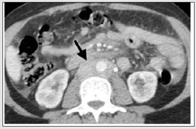
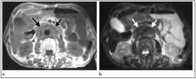
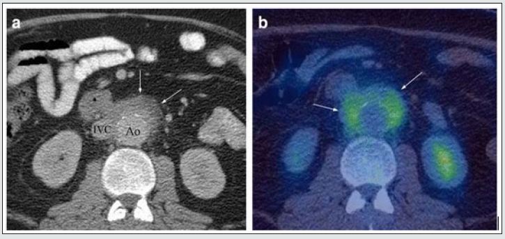
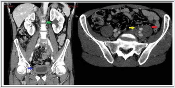
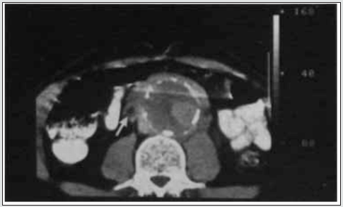
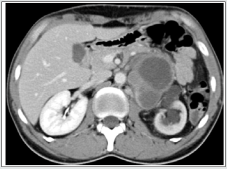
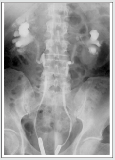
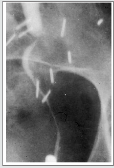
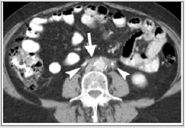
Retroperitoneal Fibrosis (RPF) is now considered an important abdominal component of the group of IgG4-related sclerosing disorders. It is an aggressive, rare fibro- proliferative process causing deposition of unencapsulated fibrous tissue masses in the retroperitoneal space usually centered at the lumbosacral junction. it is typically seen in men (2-3 times commoner than in women) between the 5th and 7th decades of life. Up to 15% of RPF patients have additional fibrotic processes in the mediastinum, thyroid (Riedel thyroiditis), biliary tree. sclerosing mesentery, and orbital tissue(pseudotumor). It leads to progressive encasement of the retroperitoneal structures, especially the ureters. and may cause hydronephrosis secondary to the extrinsic compression of the ureters [18]. RPF plaque may involve both ureters and may draw them medially towards the spine. This appearance on retrograde pyelogram resembles a narrow-waisted maiden (Figure 1a). Encasement of the ureter cause abrupt tapered narrowing of the ureteral lumen The dilated proximal ureter (the bullet) and the nondilated, encased distal ureter may appear as ‘The bullet and bodkin’ and this appearance can be caused by malignancy such as lymphoma, or rarely by RPF (Figure 1b). However, on the above imaging pictures, the ureteric encasement is not visually demonstrable. On CT, the area of fibrosis and encasement appears as a soft-tissue mass with variable contrast enhancement. The active and inactive stages of the disease can be differentiated on CT and MRI. In the retroperitoneum, RPF tissue surrounds the aorta (arrow) sparing posterior aspect (Figure 2a) indicating a benign stage. In contrast to the figure above, in malignant RPF, the RPF has mass effect with adjacent structures (Figure 2b below). Another noteworthy finding is the intensity difference of RPF mass on MRI imaging (Figure 3). A high intensity signal of the mass on T2WI is suggestive of early active RPF whereas a low intensity signal on T2WI is highly suggestive of benign RPF in a late inactive stage. On FDG PET/CT imaging, the fibrous tissue in the affected areas exhibit avid FDG uptake (Figure 4b). When the infiltrating tissue encompass both ureters and compromise the lumen, bilateral hydronephrosis will ensue regardless of the location of the mass in the abdomen or pelvis (Figure 5a-b). Abdominal pain and obstructive uropathy should, therefore, raise suspicion for retroperitoneal fibrosis and imaging should be obtained. On the other hand, new onset of hypertension, flank pain radiating to groin in a known case of RPF or malignancy should arise the suspicion of ureteral encasement and appropriate imaging should confirm [19]. Failure in recognition of the encasement of ureters and causes may lead to mistaken diagnosis with resultant inappropriate management.

Figure 1: Retrograde ureteropyelogram 1a. Maiden waist deformity.

lupinepublishers-openaccess-journal-urology-nephrology

Figure 1b: Bullet and Bodkin Sign (Courtesy: Classic Signs in Uroradiology; Raymond B. Dyer, Michael, Ronald)

lupinepublishers-openaccess-journal-urology-nephrology

Figure 2a: Plaque-like Soft tissue surrounding the aorta (arrow) sparing posterior aspect without elevating the aorta from the spine; encases both ureters (arrow heads) findings indicative of benign RPF. (courtesy: Ali Jiwani https://www.slideshare.net/AliJiwani/retroperitoneal-fibrosis- radiology)

lupinepublishers-openaccess-journal-urology-nephrology

Figure 2b: Axial CECT demonstrates well defined mass surrounding Aorta and IVC pushing the aorta anterior to the spine, a finding suggestive of malignant RPF. (courtesy: Ali Jiwani https://www.slideshare.net/AliJiwani/retroperitoneal-fibrosis-radiology)

lupinepublishers-openaccess-journal-urology-nephrology

Figure 3a: Histologically confirmed idiopathic RPF mass surrounding the abdominal aorta and IVC, showing medium intensity on T1WI (3a); and low signal intensity on T2WI (3b) a finding suggestive of late stage. (courtesy: Ali Jiwani https://www. slideshare.net/AliJiwani/retroperitoneal-fibrosis- radiology)

lupinepublishers-openaccess-journal-urology-nephrology

Figure 4: Example of IgG4 retroperitoneal fibrosis. 4a Axial CECT showing enhancing soft tissue deposit partly encasing the IVC and atherosclerotic aorta (arrows). b Axial FDG PET/CT showing increased FDG uptake in the abnormal retroperitoneal soft tissue (arrows). (Courtesy: Christopher Siew Wai Tanghttps://insightsimaging.springeropen.com/articles/10.1007/ s13244- 018-0618-1).

lupinepublishers-openaccess-journal-urology-nephrology

Figure 5a: Axial and 5b: coronal computerized tomography images demonstrating left-sided pelvic mass causing hydroureteronephrosis (green arrow) with diffuse bladder wall thickening (blue arrow). Asymmetric pelvic mass extending to posterior presacral region and psoas (red arrow), with ureter encased by pelvic mass (yellow arrow). Courtesy: R Bechtold, M I Shaff; PMID: 6879267; DOI: 10.1097/00007611-198308000-00023.

lupinepublishers-openaccess-journal-urology-nephrology

Perianeurysmal Fibrosis

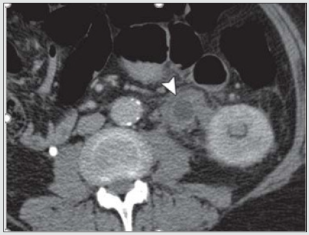
Reports of 56 previous cases showed striking male predominance. An abdominal mass may be palpable in 45% of cases. Fibrosis is often seen in association with atherosclerosis of the aorta (Figure 6), possibly due to fibrosis as an immune response to the leakage of ‘ceroid’ from the atheromatous plaque into the perivascular tissues. When perianeurysmal fibrosis occurs in association with an abdominal aortic aneurysm it may encase and produce ureteric obstruction and renal function impairment. The computerized tomography scan is able to provide accurate details of aortic aneurysm, intraluminal thrombosis, calcification, periaortic inflammation, entrapped ureters and hydronephrosis (Figure 6).

Figure 6: At L3/4 DISC SPACE level – Aorta enlarged with calcifications in the wall. The left ureter (black arrow) is encased by the periaortic aneurysmal fibrosis and the right ureter (white arrow) lies adjacent to fibrosis (partly encased). Both ureters are small, the possibility of Ureteric obstruction secondary to perianeurysmal fibrosis should be in the pre-operative evaluation of abdominal aortic aneurysms. (Courtesy: https://www.researchgate.net/ R N Gibson Alison Halliday A Mansfield).

lupinepublishers-openaccess-journal-urology-nephrology

Neoplasm

Locally invasive neoplasm in the retroperitoneum and pelvis can involve ureters by three ways: 1. by malignant infiltration of their walls causing narrowing of their lumen (stricture) and affecting the peristalsis; 2. by external compression by the mass itself that may encroach, encompass and compress and displace the kidneys and ureters (Figure 7a) or by the adjacent aorto-caval and common iliac lymphadenopathy around abdominal ureter and internal and external iliac nodes around the pelvic ureter (carcinoma of prostate, uterus, cervix and or colon cause lymph node enlargement or organ enlargement); 3. by encircling and encasing the ureter as a whole. True hematogenous or lymphatic metastases to the ureter may occur from primary somewhere (Figure 7b). The primary may be in stomach, pancreas, lung, breast, neuroendocrine tumour) But it is exceedingly uncommon to find ureteral obstruction due to metastases to the ureter from distant primary tumors. Until now, only about 400 cases confirmed by post-mortem have been reported [20,21].

Figure 7: Retroperitoneal soft tissue sarcoma Encased and dilated left ureter (https://radiopaedia.org/cases/retroperitoneal).

lupinepublishers-openaccess-journal-urology-nephrology

Figure 7b: Ureteral metastasis of lung cancer Encased and dilated left ureter https://cdn.amegroups.cn/journals/amepc/fil.

lupinepublishers-openaccess-journal-urology-nephrology

Endometriosis

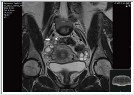
Currently endometriosis is considered between benign and malignant status and is known as a “malignant” benign disease by experts. Its histopathologic, and molecular data suggest that endometriosis has malignant potential and is associated with ovarian cancer [22,24]. Although urinary tract endometriosis occurs in ~1% of women mainly in women of child-bearing age with pelvic endometriosis it may cause hydronephrosis by involving the ureters secondarily by encompassing and or compressing them. Any lapse or delayed diagnosis can lead to renal failure. Ultrasound, CT and MRI (Figure 8) may be revealed and or laparoscopy may reveal the encasement of ureters.

Figure 8: MRI scan of the pelvis showing abnormal tissue (arrowhead) which encases the right distal ureter (arrow). (Courtesy: Apostolos Vrettos, Maria Prasinou, Rob Frymann doi: 10.21037/qims.2016.01.02)

lupinepublishers-openaccess-journal-urology-nephrology

Conclusion

Both ureters are retroperitoneal throughout their course in the abdomen. An encompassing inflammation and or infiltrative malignant process usually cause circumferential wall thickening and an abrupt change in the diameter of the ureter resulting in dilated proximal ureter with a narrow or normal-calibred distal ureter. RPF is usually diagnosed through clinical presentation and imaging studies. The severity of RPF is emphasised by the fact that, in about 56%–100% of patients with idiopathic RPF, the fibroinflammatory tissue entraps the ureters and causes obstructive uropathy and subsequent renal failure. However, in any case biopsy should be considered to exclude malignancy. Any of the above discussed pathologies may involve the renal vessels and contribute to renal insufficiency or cause renovascular hypertension. New onset of hypertension, flank pain radiating to groin in a known case of RPF or malignancy should arise the suspicion of ureteral encasement and appropriate imaging should confirm. Failure in recognition of the encasement of ureters and its causes may lead to flawed diagnosis and irrecoverable damage to the kidneys. Early detection, corticosteroids, Radiation therapy, Retrograde ureteral stent and PCN placement, bilateral ureterolysis and resection of the aneurysm and other surgical interventions are some in the management algorithm to be chosen to broaden the treatment arena and provide encouraging results.

Acknowledgement and Disclaimer

The author would like to thank Taylor’s University, Malaysia for the time granted for reviewing work. I am also grateful for the insightful comments offered by the anonymous peer reviewers of the Journal of Urology & Nephrology studies. This paper would not have been possible without the exceptional support of my amazing partner A. Riaz Ahmed. His enthusiasm, contributions and the responses kept my work on track.

Read More About Lupine Publishers Journal of Urology & Nephrology Please Click on Below Link:
https://lupine-publishers-urology-nephrology.blogspot.com/
Thursday, 22 June 2023

Lupine Publishers | Lasers in Urology – what has Survived of our Research Starting 1970*

 Lupine Publishers | Journal of Urology & Nephrology 


Short Communication

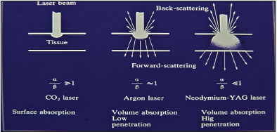
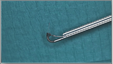
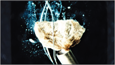
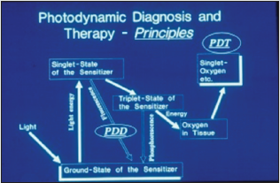
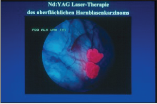
In 1970 I started to investigate the Laser-Technology for urologic surgery together with H. Müßiggang. First of all, it was to check the interacting of the different lasers with biological tissue. The result you can gather from the Figure 1 Decisive are the two factors: absorption and scattering of light into the tissue. The strong light absorption of the CO2 – laser leads to an excellent incision effect with low edema reaction. The light of the is mainly absorbed in the tissue by hemoglobin and pigment colorings and therefore suitable for the destruction of highly vascularized tumors or malformations. For achieving greater volume effects, - necessary for destruction of solid tumors, bilharzial bladder-lesions and inflamed areas in interstitial cystitis, - the Nd:YAG – laser was used by us since 1976 (Figure 2). Presupposed for the clinical application of lasers was the developing of a quartz glass fiber transmission system by Nath, a physicist from the Neuherberg Laser Labor (1973) and the developing of a special cystoscope insert, designed by my working-group (Staehler, Frank et al.) and constructed by the Storz Compagny/Tuttlingen/Germany (Figure 3). The next steps were the laser induced shock wave lithotripsy, developed between 1978 to 1986 (Munich/Lübeck) and the photodynamic procedures for early tumor diagnosis (Figures 5a & 5b).

Figure 1: Effects of different lasers on human tissue.

lupinepublishers-openaccess-journal-urology-nephrology

Figure 2: Bladder Tumor Destruction in 1976.

lupinepublishers-openaccess-journal-urology-nephrology

Figure 3: Insert for Laser-Endoscope.

lupinepublishers-openaccess-journal-urology-nephrology

Figure 4: Shockwave application on a stone using an optomechanical coppler.

lupinepublishers-openaccess-journal-urology-nephrology

Figure 5a: Principles of PDD/PDT.

lupinepublishers-openaccess-journal-urology-nephrology

Figure 5b: Fluorescending tumor under ALA-Fluorescence.

lupinepublishers-openaccess-journal-urology-nephrology

Now is to ask – what survived?

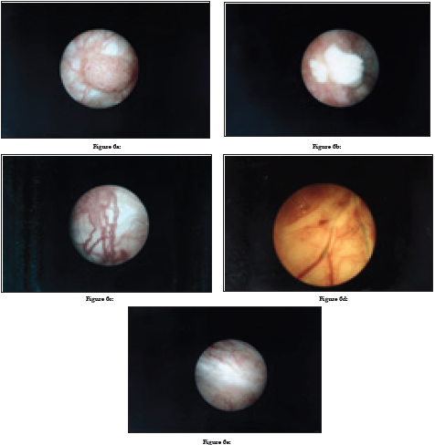
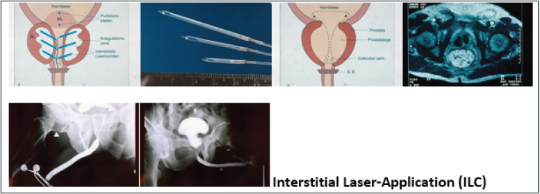
The laser-lithotripsy, especially in ureter, and after a long fight, the photodynamic diagnosis for bladder tumors and Cis, are on routine. Also in tumors of external genitalia, - e. c. condylomata acuminate, hemangiomas, penile cancer, - the ND:YAG- and Diodelasers are well established. Established are also the vaporization and enucleation of prostate tumors by laser. In contrast to these indications, the Nd: YAG-laser-application in bladder-tumors seems to be forgotten. I think this method is head and shoulders above the common TUR-resection because it generates a total homogenous necrosis with closing the blood- and lymphatic vessels simultaneously, it means, no bleeding, no tumor-cellspreading. You can perform the operation under excellent viewing, so that it`s possible to destroy also any satellite-tumors and the tumor vessels (Figures 6a, b, c, d, e). Histological examinations of the tumor tissue after Nd:YAG-laser-application have never been problematic according to our pathologist Dr. Keiditsch. It depends on the fact, that during the coagulation the temperature didn`t exceed 100 C°, it means, the cell-structure remains unchanged. I observed 5 patients, who refused the radical cystectomy, only treated by ND:YAG –laser, between 10 and 40 years. They all remain tumor free. Our photodynamic therapy of localized prostate cancer, published 1993, is still in the early stages. It seems, nobody is interested on this procedure. Forgotten seems to be also our interstitial laser coagulation (ILC) of prostate adenomas and bladder-neck-stenoses, although by this procedure the retrograde ejaculation can be avoided in the most cases (Figures 7a-f).

Figure 6 a-e: a/b: Tumor before and after Nd:YAG-Irradiation, c/d: Destruction of tumor-vessels, e: six weeks later.

lupinepublishers-openaccess-journal-urology-nephrology

Figure : 7a= scheme, b= glasfibers with lightdistributors, c= scheme after laser application, d= CT after ILC, e = big adenoma of the prostate, f= state after ILC.

lupinepublishers-openaccess-journal-urology-nephrology

Read More About Lupine Publishers Journal of Urology & Nephrology Please Click on Below Link:
https://lupine-publishers-urology-nephrology.blogspot.com/

Friday, 28 April 2023

Lupine Publishers | Chronic Kidney Disease, Data from MIKD

 Lupine Publishers | Journal of Urology & Nephrology


Abstract

Chronic kidney disease (CKD) is a leaping up public health matter. It has an augmented effect on cardiovascular diseases and affects every system in body. In developed countries the extent of prevalence is available due to renal registries. In middle or low socio economic countries the proportion of this evolving health issue is not known. CKD is rising and a major contributor is soaring number of diabetes mellitus worldwide. Other significant addition in this cohort of CKD is by rising numbers of obese and hypertensive patients. In order to curtail this rising health matter immediate and intense measures both at preventive and curative levels are required.

Aim: Due to lack of formal renal registry we wanted to see spectrum of chronic kidney disease (CKD) in our catchment area and at what CKD stage they present to tertiary care hospital.

Methods: All patients who presented to Emergency department of Multan Institute of kidney disease from 01 Sep 2017 till Sep 2019 data were evaluated. Record of Emergency department patients were taken from electronic system of our hospital. Some patients had multiple visits and we took first visits kidney function in our analysis. eGFR was calculated from serum creatinine with help of CKD-EPI equation.

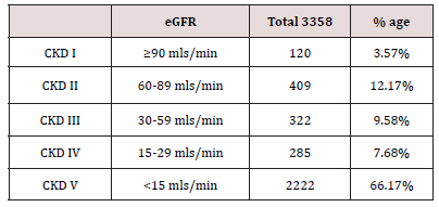
Results: Total 4303 patients were included in study. Males were 60% and females 40%. Age range from 13 years to 96 year old. 945 patients were excluded as they were not falling in chronic kidney disease category. Remaining 3358 patients had chronic kidney disease. Sub-analysis according to CKD stage showed 66.17% patients presented at CKD stage V.

Conclusion: Kidney disease is rising globally. Countries where renal registries are established provide incidence of chronic kidney disease ranging from 10 to 15%. Still a lot of countries worldwide do not have established system of data collection so true incidence is not established. Our work is first of its kind reported from this area. Drastic preventive strategies are need of time from health budget planners.

Introduction

Multan Institute of kidney diseases is a tertiary care nephrology and urology hospital located in Multan (Largest city) in South Punjab, Pakistan. Catchment area include Multan division, Dera ghazi khan division and Bahawalpur division. According to last census the total population of these 3 divisions is just over 34 million. All patients who presented to Emergency department of our institute from 01 September 2017 till 01 September 2019 were taken from electronic medical record of hospital.

Inclusion & Exclusion Criteria

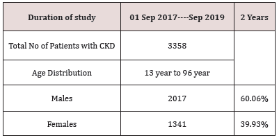
Total number of patients in our data are 4303. Patients from Age 13 year and above were taken in study. Less than age 13 were not included. Maximum age recorded was 96 year old. In case of multiple visits to Emergency department by same patient, only first presentation kidney functions were included in study and subsequent visits kidney function were excluded. 467 patients had normal kidney functions, normal urinalysis and no structural abnormality on renal imaging and were excluded from interpretation. 478 patients were found to have acute kidney injury as per acute kidney injury network (AKIN) classification. These patients were also excluded from analysis. Rest of the patients (n=3358) had chronic kidney disease [Table 1].

Table 1:

lupinepublishers-openaccess-journal-urology-nephrology

Kidney Function Measurement (eGFR)

Serum Creatinine was taken to measure estimated glomerular filtration rate (eGFR). Various methods of eGFR calculation are available, including the Cockcroft-Gault equation, MDRD (Modification of Diet in Renal Disease) Study equation, and the CKD-EPI (Chronic Kidney Disease Epidemiology Collaboration) equation. We took CKD-EPI for measuring eGFR. Patients were divided in CKD stages from 1 to 5.

CKD Stages and Prevalence

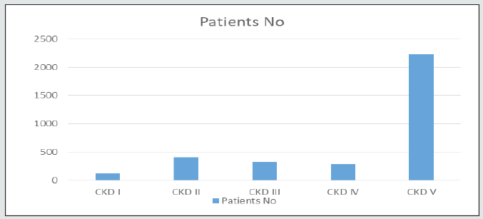
Total 3358 patients were identified with chronic kidney disease. Majority (n=2222) of patients presented when eGFR was <15mls/min for the first time to tertiary care facility. CKD stages with number of patients in each group are in following Table 2 [Figure 1].

Table 2:

lupinepublishers-openaccess-journal-urology-nephrology

Figure 1:

lupinepublishers-openaccess-journal-urology-nephrology

Causes of CKD

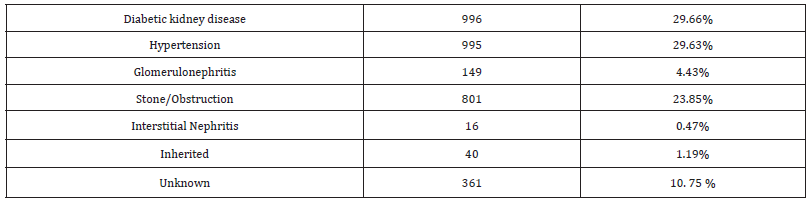
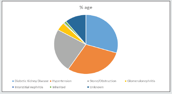
We analyzed our data to look into etiologies of chronic kidney disease. It showed 30 % patients had diabetic kidney disease and same proportion were hypertensive nephropathy patients. Next in line was obstructive nephropathy with 23.85%. Glomerulonephritis was about 4.5% and roughly 10% patients cause was not established as they presented late. Inherited diseases were just over 1% and interstitial nephritis was just 0.47% [Table 3]. Among the patient cohort diabetic kidney disease was separated based on presence of diabetes mellitus and one of the criteria (proteinuria, normal size kidneys, diabetic retinopathy on fundoscopy or >5 years duration of diabetes mellitus). Nearly 30% of total patients had diabetic kidney disease and more than 2/3rd of them had CKD-5 on presentation. A detailed analysis of CKD stages of diabetic group is in following table [Table 4] [Figure 2].

Table 3:

lupinepublishers-openaccess-journal-urology-nephrology

Table 4:

lupinepublishers-openaccess-journal-urology-nephrology

Figure 2:

lupinepublishers-openaccess-journal-urology-nephrology

Discussion

Chronic kidney disease is rising worldwide. It has big impact on health economics [1,2]. Developed countries have registries showing its prevalence which helps in health budget planning. Unfortunately under developed countries lack such planning. In Pakistan recently a national renal registry has started working and will give informative statistics regarding renal disease spectrum. This will help in health management planning and focusing on areas where rampant growing renal issues are focused.

Reviewing published data spectrum of renal disease is variable in different geographical areas [3, 4]. Ethnicity, dietary habits, use of herbal medicines, low birth weight & climate all have been postulated as contributing factors in prevalence of chronic kidney disease. According to a Meta-analysis, chronic kidney disease has prevalence of 11% to 13% globally [5]. In Pakistan studies published on chronic kidney disease prevalence are usually from tertiary hospital or region based [6]. Tertiary care centers are in urban cities so there is no good data from rural areas. South Punjab of Pakistan majority comprise of rural area. According to last census the population of south Punjab 3 divisions (Multan, Bahawalpur and Dera Ghazi Khan) is 34 million [7]. This is the catchment area served by Multan institute of kidney disease (MIKD). MIKD is an emerging kidney institute in this area. We analyzed all patients who presented to Emergency department of our institute. We analyzed presentation creatinine and used CKD-EPI Creatinine Equation for eGFR calculation as recommended by the National Kidney Foundation [8]. Chronic kidney disease in Pakistan is on rise and presentation to specialist center is late. Majority of patients present with CKD-stage 5 requiring dialysis. Our hospital’s 2 year record tells us that 66.17% patients who attended Emergency room were at CKD-5 and majority required renal replacement therapy. Diabetic kidney disease was alarmingly on 29.66 %. Further sub analysis of diabetic kidney disease patients revealed that majority patients (72%) were at CKD stage 5 when presented to hospital emergency department. We labelled diabetic kidney disease based on presence of one of the following criteria’s (established diabetic retinopathy and/or micro albuminuria and/or long duration (>5 years) of diabetes mellitus). Further stage of CKD was established based on CKD-EPI calculation. Diabetes mellitus is at tremendous rate in Pakistan. International diabetes federation data reveals that 17.1% rising adult population of Pakistan is now living with diabetes mellitus [9]. With such enormous rise in diabetics in last decade, the burden of diabetic nephropathy is on rise too. In rural areas a lot of people are undiagnosed and usually present to health facility with end organ damage. According to systematic analytic study done in 2016 prevalence of diabetes mellitus in Pakistan is more in males as compared to females and more common in urban areas [10]. The rising burden of diabetes is imputed to environmental and emotional changes. The main contributors are sedentary lifestyle including internet and TV usage, caloric rich diets leading to increasing obesity. Pakistan with very high numbers of prevalent diabetes needs a cost-effective population based approach for screening [11]. Obstruction leading to chronic kidney disease was immensely at 23.85% in our studied population. Obstructive nephropathy due to stone is highly prevalent in this area and reviewing published data the number is almost 2-3 times in comparison to other parts of world. Nephrolithiasis is a highly prevalent disease worldwide with rates ranging from 7 to 13% in North America, 5–9% in Europe, and 1–5% in Asia. Due to high rates of new and recurrent stones, management of stones is expensive and the disease has a high level of acute and chronic morbidity [12]. Epidemiologic studies have demonstrated that the stone risk incidence increases with Body Mass Index, through multiple pathways. Metabolic syndrome and diabetes are associated with an increased renal stones disease incidence [13, 14]. There is a known high incidence of stone disease in Pakistan as this country belongs to the so-called stone belt [15]. In summing up our chronic kidney disease burden we have identified Diabetic kidney disease, Hypertension and Kidney stones as our bulk. This helps in health budget planning and preventive strategies to cut down disease load. Lack of renal registry in Pakistan has been emphasized [16]. Recently at national level Pakistan renal data system (PKDRS) is established and will provide data from all parts of country in coming years [17]. Curtailing diabetes will have significant reduction in diabetic kidney disease and strategies have been proposed. Creating multidisciplinary team, primary preventive strategies and nationwide diabetic care programme are proposed steps to deal this impending pandemic [18].

Read More About Lupine Publishers Journal of Urology & Nephrology Please Click on Below Link:
https://lupine-publishers-urology-nephrology.blogspot.com/
Friday, 6 January 2023

Lupine Publishers | Efficacy and Safety of Prolonged Alfuzosin Treatment in Patients with Lower Urinary Tract Symptoms Associated with Benign Prostatic Hyperplasia: 7 Years of Observation

 Lupine Publishers | Journal of Urology & Nephrology


Abstract

Introduction: Amongst all of the medications prescribed to BPH patients undergoing conservative therapy, α1-blockers were used in 80% of cases. The question of optimal treatment length is one that has been constantly asked during the last years. The current study will encompass the data we have collected of a small study group of BPH patients treated with 10 mg of Alfuzosin daily, over a period of more than 7 years.

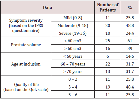
Material and Methods: From 2009 to 2011, 41 patients with mean age 67.7 years began medical therapy taking 10 mg of Alfuzosin per day. The mean treatment length was 7.6 years as of today. 16 patients (39%) with prostates exceeding 60 cm3 were additionally prescribed 5-ARIs.

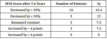
Results: Overall, a positive dynamic was found in 85.4% of patients. Not a single patient chose to discontinue treatment. A very high level of satisfaction was reported by 88% of our patients. A statistically significant (P < 0.05) decrease in IPSS scores by 8.5 ± 6.1 (47.5%) was found. Mean QoL indices decreased from 3.7 ± 1.1 to 2.3 ± 1.1 over the period of observation. Mean Qmax values increased from 9.7 ± 0.52 mL/s to 14 ± 0.60 mL/s (an increase of 44.3%).

Conclusions: This study has demonstrated a high level of safety and efficacy when using Alfuzosin to treat voiding dysfunction in patients suffering from LUTS/BPH. Our study resolves the issue surrounding a prolonged course of α1-blockers, which, due to its widespread use, remains the gold standard for medical treatment of lower urinary tract symptoms associated with benign prostatic hyperplasia.

Keywords: LUTS, BPH, Alfuzosin, BOO

Introduction

It is well known that the prevalence of voiding dysfunction and patient age are in direct proportion and that prevalence is particularly high in patients over 50 years of age. Typical complaints associated with voiding dysfunction are termed lower urinary tract symptoms (LUTS) and have a wide variety of underlying causes. The most common aetiologies of LUTS are prostatitis, urethral strictures, cystitis, bladder stones, prostate cancer, and benign prostatic hyperplasia (BPH).

BPH is usually managed with surgical intervention, pharmacotherapy, or a strategy of watchful waiting, and numerous factors affect the decision of which method should be employed. Symptom severity is one of the most important subjective factors and is measured using the international prostate symptom score (IPSS) questionnaire. The nature of the symptoms (whether they fall under the category of obstructive or irritative) also influences the final decision. The primary objective factor can be considered to be the quality of voiding as measured by uroflowmetry, the interpretation of which should consist not only of the peak flow rate (Qmax), but also flow time, time to Qmax and many other indicators. Postvoid residual urine is an equally important value because it helps identify poor bladder contraction ability (detrusor underactivity) in the case of progressive intravesical obstruction. There are also those factors, no less important than both uroflowmetry and IPSS, which relate to the potential risk of BPH progression, and thus make their impact on the choice of treatment. Whilst the size of the prostate itself cannot be ignored, it is by no means a marker for surgical intervention when considered by itself, but rather a hallmark for potential future complications such as acute urinary retention (AUR) amongst other things. In recent years, prostate-specific antigen (PSA) levels have also become associated with BPH progression. Today, PSA levels are not only used to screen for prostate cancer, but also to reflect cellular proliferation rates of the prostate. Thus, high PSA levels increase the risk of rapid BPH progression. [1] The sum of the factors mentioned above influences the ultimate decision of which type of treatment should be employed in a specific patient.

Modern standards of medical practice dictate that pharmacotherapy is recommended in BPH patients with moderate LUTS without upper urinary tract complications, patients without obvious surgical indications, and patients who either decline surgical intervention, or are unable to commit to surgery in the nearby future. Watchful waiting is acceptable if a patient exhibits mild LUTS (IPSS ≤ 8), or if the patient’s symptoms do not significantly impact their quality of life (QoL). Patients who fall under this category should be made aware of the necessity to maintain an appropriate lifestyle as well as the importance of regular blood, urine, PSA, uroflowmetry and ultrasound testing. Surgical intervention is needed if a patient exhibits severe symptoms with upper urinary tract complications, or if there are sufficient reasons to suspect that medication will be ineffective[2,8].

In order to better illustrate modern tendencies in the treatment of BPH, we turn our attention to the TRIUMPH study. The TRIUMPH study recorded the treatment and outcomes of 2351 newly presenting LUTS/BPH patients in 6 European countries over a 1-year follow-up period. Out of the entire study population, 23.8% were managed with watchful waiting, 72.5% were prescribed medication, and only 2.7% underwent surgical treatment. These statistics accurately reflect the rise in medical management of LUTS as well as the decreasing need for surgical intervention during the past decade. It should be interesting to note that amongst all of the medications prescribed to BPH patients undergoing conservative therapy, α1-adrenergic receptor antagonists (α1-blockers) were used in 80% of cases [3].

The efficacy of α1-blockers in patients with BPH has been well documented in a large quantity of double-blind, placebo-controlled studies.[4,5,6,7] If it is possible to consider transurethral resection of the prostate (TURP) the method of choice for surgical intervention in BPH patients, then α1-blockers will fall under the same criterion for the medical management of BPH. It is important to note that the European Association of Urology considers α1-blockers the first line of therapy for the management of voiding dysfunction in patients suffering from BPH[8].

Whereas the rationale behind α1-blockers does not arouse suspicion in the majority of specialists, the question of optimal treatment length is one that has been constantly asked during the last 6 years. Solutions offered to address this problem range from a life-long course of medication, to one that only lasts a few months. There is no one correct way to approach this matter, but it is the duty of every qualified specialist to incorporate certain principles when making such a decision. Personal experience and knowledge gained from a wide variety of international studies should encompass the core of such precepts. It is arguable that the lack of a formulated strategy that applies to such clinical trials is hindering the formation of a unified point of view. If we turn to medical literature, we find that despite an abundance of studies indicating the effectiveness of α1-blockers in BPH patients, the studies lack the duration necessary to examine long-term effects and are mostly comprised of 3-6 month trials with only a few mentioning 1-3 year-long results. This has led many to assume that a 3-month course (being the most prominent study length) of α1-blockers is sufficient, whilst the actual reasoning behind the short length of most studies lies in the knowledge that the results achieved during that period of time will continue over the remaining course of treatment, however long it may be. Confirmation of this can only be found in a few currently available publications that demonstrate long-term results. Most articles cite results that were obtained using studies based on principles of Good Clinical Practice (GCP) in which the categories of patient inclusion/exclusion were so rigid, they created unrealistic expectations for the wide variety of patients that are actually treated. Only a few trials provide us with results that are based on a realistic study population. Such trials include those of Roehrborn C. et al., Elhilali M. et al. 2006 and Vallancien G. with Emberton M. et al. 2008[9,10,11].

During the early 21st century, the suitability of α1-blockers was no longer debated in the Russian Federation, whilst the question of long-term use was left unanswered even for us. Even now, considering the ageing population and the necessity of prolonged medical treatment, many specialists are hesitant to prescribe α1-blockers for more than a few years without consulting medical literature about the safety and efficacy of such a treatment.

Materialand Methods

The current study will encompass the data we have collected of a small study group of BPH patients treated with 10 mg of Alfuzosin daily, over a period of more than 7 years. We must mention that the data provided isn’t final, in the sense that patients are still undergoing treatment to this day. We had not originally planned for this to be a study with a specific design, but retrospective aspects make it similar to various non-comparative, real-life surveillance studies. Our goal is to introduce the reader to the results of a longterm course of α1-blockers that, in our opinion, are a safe and effective method of long-term medical therapy in patients suffering from BPH.
From 2000 to 2002, 41 patients with mean age 67.7 years (range 51– 83) began medical therapy. The mean treatment length was 7.6 years as of today. We would like to mention that this group of patients wasn’t specifically chosen as a study group at the time, nor were they the only patients that had been prescribed Alfuzosin. Our detailed observation of the isolated group in question is partially due to chance, and partially due to loss to follow-up. Patients began treatment taking 5 mg of Alfuzosin twice daily. From January 2005 onwards, all patients take 10 mg of Alfuzosin once daily at night due to numerous complaints of increased voiding problems during this time. Prostate volumes fluctuated between 25 cm3 and 100 cm3, and 16 patients (39%) with prostates exceeding 60 cm3 were additionally prescribed 5-alpha-reductase inhibitors (5-ARIs). During the early stages of treatment these patients were prescribed Finasteride, but during the last 2 years many have switched to Dutasteride in accordance with our recommendations.

Indications

Patients with LUTS/BPH

Contraindications

Patients with surgical indications
Patients known to have insufficient results from taking α1- blockers in the past
Patients with postural hypotension
Patients taking alternative medication for LUTS with good results
Patients with unstable angina (pectoris)
Patients with life-threatening comorbidities

Table 1: Summary of initial patient data.

lupinepublishers-openaccess-journal-urology-nephrology

The mean duration of morbidity at the time of patient inclusion in the trial was 1.9±1.1 years. During ultrasound testing, 31.2% of patients had postvoid residual urine measuring 82 ± 41.7 mL. Mean PSA levels of all patients before the trial were 2.9 ± 2.8 ng/mL. The initial data of the patients included in this trial are summarized in Table 1.
Out of the 41 patients, 14 (34.1%) had hypertension 11 of which (26.8%) were receiving some form of hypotensive therapy, and 5 patients had suffered a myocardial infarction before inclusion into the trial.
The efficacy of the treatment was analysed using IPSS scores as well as QoL indices. Additional factors included Qmax and prostate volume in patients who were receiving combination therapy with 5-ARIs. The safety of the treatment was analyzed based on recorded incidents of adverse cardiac side effects (mainly hypotensive) during the trial. Vital-sign dynamics were also analyzed. Follow-ups were conducted biannually during the first 2 years, and no less than annually thereafter.
Two-sided hypothesis tests were conducted with a significance P-value cut-off of 0.05. Dynamics of safety and efficacy variables were analysed before and after the trial (absolute and relative change was considered). A paired Student’s t-test was used if statistics followed a normal distribution; a Wilkinson’s test was used if they did not.

Results

Not a single patient chose to discontinue treatment, notwithstanding the long period of observation. We must mention, however, that several patients stopped being prescribed Alfuzosin during the early stages of the trial but were obviously not considered in the study group. Subjective assessments of patient satisfaction were carried out using a questionnaire specifically tailored to this publication and not used in our routine clinical practice. A very high level of satisfaction was reported by 88% of our patients, whilst the remaining 12% consisted of those actively taking a large quantity of medication for various comorbidities, and patients with diabetes. Many of the aforementioned patients were prescribed medication after they began treatment with Alfuzosin. Therefore, these facts could be taken into consideration by general practitioners who have patients undergoing treatment for LUTS.

Table 2:IPSS scores after 7.6 years of Alfuzosin treatment.

lupinepublishers-openaccess-journal-urology-nephrology

Mean IPSS scores were 17.9 ± 5.3 before the treatment, and 9.4 ± 5.2 after 7.6 years. A statistically significant (P < 0.05) decrease in IPSS scores by 8.5 ± 6.1 (47.5%) was found. A decrease in IPSS scores by > 50% was noted in 28 patients (68.3%). Overall, a positive dynamic was found in 85.4% of patients. IPSS score dynamics are summarized in Table 2.
Dynamics of irritative (questions 1, 3, 5 and 6 on the IPSS questionnaire) and obstructive (questions 2, 4 and 7 on the IPSS questionnaire) symptoms were also analyzed. Mean irritative IPSS scores decreased from 6.3 ± 2.9 to 3.7 ± 2.4 (33.7% lower) whilst mean obstructive IPSS scores decreased from 9.6 ± 4 to 5.5 ± 3.7 (35.7% lower). Incidents of nocturia decreased from 2.4 ± 1.1 to 1.5 ± 1.1. Diagram 1 illustrates a major decrease in the percentage of patients with moderate to severe voiding dysfunction symptoms as well as a six-fold increase of patients with mild symptoms. All of the statistics mentioned above were statistically significant (Figure 1).

Figure 1: Severity of LUTS before and after 7.6 years of Alfuzosin treatment.

lupinepublishers-openaccess-journal-urology-nephrology

QoL dynamics are presented in diagram 2. Mean QoL indices decreased from 3.7 ± 1.1 to 2.3 ± 1.1 over the period of observation. The mean decrease in QoL indices was 32.3% (reflecting an increase in QoL). All changes were statistically significant. Overall, 72.7% of patients had an increase in their QoL (Figure 2).

Figure 2: QoL indices before and after 7.6 years of Alfuzosin treatment.

lupinepublishers-openaccess-journal-urology-nephrology

It is important to note that the minority of patients who did not demonstrate clear improvements in symptom severity either underwent surgery or continued to take Alfuzosin. The latter group either demonstrated a minimal but satisfactory improvement or was unable to undergo surgery for various reasons. We believe that it is of utmost importance to understand that the current publication presents a comparative analysis before and after Alfuzosin treatment, and whilst all of the patients had demonstrated an improvement during various periods in the trial, the dynamics of IPSS scores and changes in QoL were not registered at those intervals. It is also important to highlight the role of the detrusor in BPH symptoms, something that was originally demonstrated by Russian authors O.B.Loran and E.L.Vishnevskiy in 1998. The contraction capability, level of energy metabolism, and nature of the biochemical events of the detrusor influence voiding quality no less than the level of intravesical obstruction.[13] This could explain treatment ineffectiveness in certain patients.

Changes in Qmax over the course of the study were considered as a secondary measurement of treatment effectiveness and were analysed during every follow-up, making this particular variable very intriguing. Mean Qmax values increased from 9.7 ± 0.52 mL/s to 14 ± 0.60 mL/s (an increase of 44.3%). Diagram 3 illustrates Qmax dynamics throughout the entire length of the study. One can conclude that early treatment results remained constant throughout the observation period (Figure 3).

Figure 3: Peak flow rate (Qmax)dynamics during 7.6 years of Alfuzosin treatment.

lupinepublishers-openaccess-journal-urology-nephrology

During the period of observation, 5 patients underwent TURP procedures. The rationale behind TURP in one patient was due to an episode of AUR provoked by alcohol ingestion. After 3 days of catheterisation, the patient did not show any signs of improvement in voiding ability and was subsequently operated. The other 4 patients exhibited worsening LUTS as shown by an increase in their IPSS scores. During ultrasound and transrectal ultrasound testing, a median lobe was detected in all 4 patients. All operations performed did not deviate from a standard TURP procedure.
The median prostate volume of the 16 patients receiving combination therapy had decreased. The value fell from 74.1 cm3 before treatment to 50.1 cm3 after 7.6 years of therapy (a decrease of 32.4%).
Throughout the trial, PSA levels of all patients were measured annually. Median PSA levels were 2.9 ng/mL before the trial and 3.2 ng/mL afterwards, demonstrating no significant variance. PSA levels of patients undergoing combination therapy were considered after the second year of treatment and were doubled. PSA levels of over 4 ng/mL requiring transrectal ultrasound-guided multifocal prostatic biopsies were noted in 3 patients aged 68–74 years. None of the subsequent histological analyses revealed the presence of cancerous cells.
The safety of this treatment was assessed based on the frequency and type of adverse side effects recorded during the trial. In total, 9 patients (22%) exhibited adverse effects. The most common findings were dizziness (2 patients/4.9%) and asthenia (2 patients/4.9%). Another 3 patients experienced dyspepsia, shortness of breath and headaches. Out of the 16-patient subgroup receiving combination therapy, 2 patients complained of decreased libido (possibly attributed to 5-ARIs). One of these patients had their sexual drive normalise after 1 year of therapy, whilst the other retained a low libido throughout the treatment. Considering the relatively small study population involved, a true understanding of the statistical significance of these adverse effects as well as their immediate connection to the treatment in question is not possible. However, the fact that most of the aforementioned adverse effects were present as isolated episodes in patients during the first week of therapy demonstrates a high level of treatment safety.

Discussion

The current study has demonstrated a high level of efficacy with minimal adverse side effects in the treatment of lower urinary tract symptoms (LUTS) arising from benign prostatic hyperplasia (BPH) with α1-adrenergic receptor antagonist (α1-blocker) Alfuzosin. The effectiveness of the treatment was determined using subjective and objective criteria. A 45.7% decrease in LUTS severity as measured by the International Prostate Symptom Score (IPSS) questionnaire was found in 85.4% of the entire study population. Mean Quality of Life (QoL) indices measured using the QoL scale increased by 32.2% and mean peak flow rate (Qmax) values increased by 44.3%. The fact that the positive results achieved during the early months of therapy had stabilised and showed no signs of regression during the entire study period is quite remarkable. Such results verify the notion that a prolonged course of α1-blockers provides consistent long-term results. The high level of patient satisfaction with treatment results and the information that patients themselves have provided us with, mark not only the fact that patients are not bothered by the need to take medication daily, but also (and most importantly) the great sense of confidence patients feel regarding the stability of the results they have achieved, leading to a positive outlook on their future treatment.

Several observational studies indicate results very similar to ours. In the ALFORTI study, 311 patients on Alfuzosin were surveyed over a period of 9 months. A decrease was shown in mean IPSS scores and QoL indices measuring 45.6% and 36.4%, respectively. Adverse effects (most likely due to the medication in question) occurred in 4.4% of the study population.[12] A similar study published by the same authors analyzed the safety and efficacy of Alfuzosin. A decrease in mean IPSS scores and QoL indices was shown and measured 32% and 27.8%, respectively[14].
Combination therapy in the form of α1-blockers and 5-alphareductase inhibitors (5-ARIs) is currently the recommended form of treatment in patients with LUTS and an increased prostate volume according to the European Association of Urology.[12] The safety and efficacy of this treatment has been confirmed by a considerable decrease in LUTS severity and prostate volume (a mean decrease of 32.4%) in our 16 patient subgroup. Similar results have also been demonstrated by such well-known trials as MTOPS, ALTESS and COMBAT[1,2,8,9].
When the information provided by the various authors is analysed, it becomes apparent that Alfuzosin attains a similar degree of effectiveness in every single case. The fact that the results gained from a continuous course of therapy remain consistent throughout prolonged treatment is extremely important, but perhaps the most interesting observation made during our trial is that the considerable results are not only long-lasting, but are achieved and made stable during the first month of treatment.
A study conducted in 2008 by Vallancien G. et al. is one of the few studies (similar to ours in design, but not in duration) that analysed the data collected in a real-life surveillance study format regarding the effectiveness of Alfuzosin during a 3-year course of therapy. The results were as follows: mean IPSS scores decreased by 33.4%, mean QoL indices by 40.7%, and nocturia severity by 25.5%. Adverse side effects (likely related to vascular dilation) were registered in 4.5% of cases, and surgical intervention was required in 5.7% of cases. These results are clearly similar to ours. [10] Similar findings were also recorded earlier during the ALFONE trial published by Elhilali M. et al. in 2006[11].
Whilst several adverse cardiac effects were noted, the analysis of adverse side effects in our trial is far from thorough, which is explained by the lack of the patients enrolled in the trial leading to insufficient statistical integrity. A study involving a multidisciplinary approach could aid the understanding of various guarantees that we may be able to give our patients in the future. These days, patients mostly consist of elderly men whose age and comorbidities make the use of pharmacotherapy ideal for managing the symptoms associated with BPH. Perhaps tomorrow, the necessity of prescribing Alfuzosin for over 10 years will increase considering the continually regressing number of patients who choose surgical intervention for BPH management; those that wish to retain a good quality of life whilst remaining ‘real men’ for many years to come. Today, when patients inquire about the potential length of their treatment, they are told that they may take Alfuzosin for as long as it is required.

Conclusions

This study has demonstrated a high level of safety and efficacy when using Alfuzosin to treat voiding dysfunction in patients suffering from LUTS/BPH. Furthermore, it is important to mention that many researchers have come to the same conclusion. Our study, whilst potentially lacking in statistical magnitude, resolves the issue surrounding a prolonged course of Alfuzosin, which, due to its widespread use, remains the gold standard for medical treatment of lower urinary tract symptoms associated with benign prostatic hyperplasia.

Read More About Lupine Publishers Journal of Urology & Nephrology Please Click on Below Link:
https://lupine-publishers-urology-nephrology.blogspot.com/