Showing posts with label IGWHC. Show all posts

Saturday, 7 October 2023

Lupine Publishers | Laparoscopic Ovarian Cystectomy Hemostatic Maneuvers Impact and Influence on Ovarian Reserve Parameters and Indices

 Lupine Publishers | Journal of Genecology and Women's Healthcare 


Abstract

Background: Electrocoagulation during performing laparoscopic ovarian cystectomy as a method used for hemostasis is one of the most commonly and preferred methods due to simplicity and speedy applicability. Fertility outcomes after ovarian cystectomy is a global concern that should be respected and investigated by further research efforts trying to elucidate the best method and protocol in surgical practice.

Aim: To investigate the impact and influence of hemostatic methods implemented in laparoscopic ovarian cystectomy on ovarian reservoir function.

Methodology: A prospective research clinical trial conducted on 72 research study subjects from January 2017 till April 2018. Research study subjects were recruited for the study having unilateral or bilateral ovarian cysts they were equally categorized into research groups according to the hemostatic method implemented during the surgical procedure.

Results: There was no statistical significant difference as regards the ovarian reserve parameters observed before surgery between both electrocoagulation and suture research groups as regards FSH, LH, E2, AMH, AFC, PSV (P value =0.229, 0.063, 0.475, 0.068, 0.609, 0.187 consecutively) after 1 month follow up there was statistically significant difference between suture and electro surgical research groups as regards the FSH, E2, AMH (P values =0.002,<0.001,0.028 consecutively) being more favorable within suture research group ,finally at 6 months follow up there was statistically significant difference between suture and electro coagulation research groups as regards FSH, E2, AMH, AFC, PSV (P values =<0.001, 0.013, 0.023, 0.027, 0.012 consecutively).

Conclusion: Further research efforts are required to evaluate if the difference is clinically significant concerning fertility potential and premature ovarian failure by implementing larger study samples putting in consideration the age and racial differences among the observed cases.

Keywords: Laparoscopic; Ovarian cystectomy; Anti mullerian hormone

Introduction

Ovarian cysts are a frequent form of ovarian lesions that could carry concerns to gynecologists according to their sonographic and histopathological characteristics [1,2].

One of the cornerstone issues and chief factors particularly in child bearing age is the impact of ovarian cystectomy on ovarian reserve function. The hemostatic methods that are implemented during the surgical procedure could influence the functional performance of the ovary at physiological, cellular and molecular levels due to the healing process that follows performance of the hemostatic method [3,4].

Electrocoagulation during performing laparoscopic ovarian cystectomy as a method used for hemostasis is one of the most commonly and preferred methods due to simplicity and speedy applicability. Research debate and many unanswered questions around the clinical and reproductive outcome that various hemostatic methods on ovarian reservoir function such as suturing and electrocoagulation [5,6].

 Fertility outcomes after ovarian cystectomy is a global concern that should be respected and investigated by further research efforts trying to elucidate the best method and protocol in surgical practice that would preserve the best ovarian functional reservoir after surgery [7,8].

Aim

To investigate the impact and influence of hemostatic methods implemented in laparoscopic ovarian cystectomy on ovarian reservoir function.

Methodology

A prospective research clinical trial conducted on 72 research study subjects from January 2017 till April 2018. Egyptian research study subjects from Zagazig Governate were recruited for the study having unilateral or bilateral ovarian cysts with good clinical general condition they were equally categorized into research groups according to the hemostatic method implemented during the surgical procedure 36 cases within suture research group and 36 cases within electrocoagulation research group. Inclusive research criteria were as follows in which cases recruited for the research study had an age range 21 to 36 years old, 28-35 days menstrual cycle length; ovarian cyst diagnosis by B- mode sonography; cyst diameter was 5 to 8 cm , no past history of gynecological surgery; no coexisting endocrinological diseases e.g. DM ,hyperthyroidism, and no hormone usage within the previous 6 months before the surgical intervention.

Surgical intervention

After anesthetic induction, the cases have been positioned in supine position, head down and hip up. After classic laparoscopic preliminary steps Pneumoperitoneum have been accomplished observed ovarian cortex covering the cyst wall have been incised in which the ovarian tissue have been observed to be characterized by being thin with few blood vessels. The surgical incision was performed in a manner to be located away from the ovarian hilum anatomical zone. The cyst wall has been stripped off from the normal ovarian tissue using blunt dissection and exteriorized using an endobag. Ovarian tissue hemostasis has been accomplished using bipolar electrocoagulation within the electrocoagulation research group. The probe was in contact with the bleeding points no more than 3 sec, causing the heat to achieve hemostasis. After performing electrocoagulation hemostatic procedure, no suturing was performed for the residual ovarian tissue left, and the ovarian edges have been left to heal by the process of secondary intention. The suture research group ovarian tissue bleeding has been managed by usage of 4-0 absorbable sutures. The surgical incision has been sutured in a manner perpendicular in direction to the ovarian longitudinal axis using a continuous suturing needle was running close to but not via the ovarian cortical tissue.

Ovarian reserve parameters observed

Biochemical 3rd menstrual cycle FSH, LH, E2, AMH were assayed preoperatively,1 month and 6 months after the surgical procedure by using radioimmune assay. Sonographic vaginal color Doppler sonographic assessment after 6 months from performing the surgical intervention the following was obtained as research data to be statistically analyzed involving the following basal antral follicle count in both ovaries (number of antral follicles within early menstrual cycle measuring less than 10 mm in diameter), peak systolic velocity of ovarian stromal blood flow was observed, ovarian volume.

Statistical analysis

Research data was collected, revised, coded and entered to the Statistical Package for Social Science (IBM SPSS) version 23. The quantitative data were presented as mean, standard deviations and ranges when parametric and compared between two independent groups using Independent t-test. Also, qualitative variables were presented as number and percentages and compared between groups using Chi-square test and/or Fisher exact test when the expected count in any cell found less than 5. The confidence interval was set to 95% and the margin of error accepted was set to 5%. So, the p-value was considered significant at the level of < 0.05.

Results

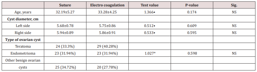
(Table 1) reveals and displays the age, cysts diameters mean +/-SD on left and right sides type of ovarian cysts in which there was no statistically significant differences between both research groups (p values=0.174, 0.609, 0.595, 0.598 consecutively).

Table 1: Cases age, cyst diameter and type.
lupinepublishers-openaccess-journal-gynaecology-women-health-care

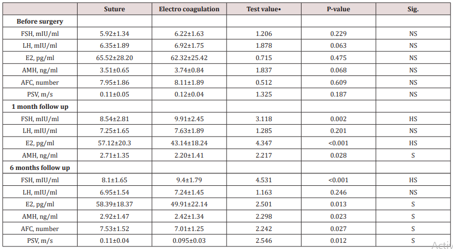
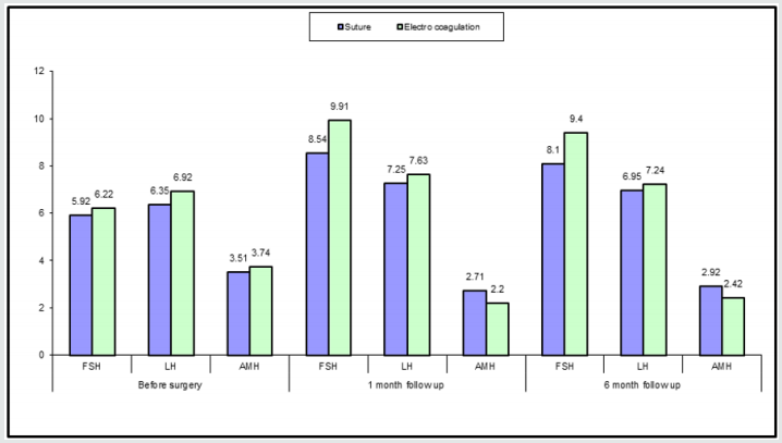
(Table 2) and (Figure 1) reveals and displays the that there was no statistical significant difference as regards the ovarian reserve parameters observed before surgery between both electrocoagulation and suture research groups as regards FSH, LH, E2, AMH, AFC, PSV (P value =0.229, 0.063, 0.475, 0.068, 0.609, 0.187 consecutively) after 1 month follow up there was statistically significant difference between suture and electro surgical research groups as regards the FSH, E2, AMH (P values =0.002,<0.001, 0.028 consecutively) being more favorable within suture research group, finally at 6 months follow up there was statistically significant difference between suture and electro coagulation research groups as regards FSH, E2, AMH, AFC, PSV (P values =<0.001, 0.013, 0.023, 0.027, 0.012 consecutively).

Table 2: Ovarian reserve parameters and indices in suture and electrocoagulation research groups before surgery, 1 month, 6 months after surgery.
lupinepublishers-openaccess-journal-gynaecology-women-health-care

•: Independent t-test

Figure 1: Ovarian reserve parameters in both research groups (FSH, LH, AMH) before ,1 month,6 months after surgery.
lupinepublishers-openaccess-journal-gynaecology-women-health-care

(Table 3) reveals and displays that there was no statistically significant difference between both suture and electrocoagulation research groups as regards cyst diameter and pregnancy occurrence within 1 year (p values =0.073,0.729 consecutively).

Table 3: Ovarian cysts diameter and occurrence of pregnancy within 1 year after surgery among both suture and electro coagulation research groups.

lupinepublishers-openaccess-journal-gynaecology-women-health-care

*: Chi-square test

Discussion

Ovarian reserve function after ovarian cystectomy remains the cornerstone research debate issue among infertility practitioners all over the globe, requiring the balance between the best management protocol that preserves the residual ovarian function for infertility management and occurrence of conception after the procedure [9,10].

Hemostatic maneuvers implemented are necessarily required during performance of ovarian cystectomy that needs to have minimal negative impact on ovarian reserve. Ovarian follicular damage and release of inflammatory mediators besides the affection of ovarian vasculature integrity are all proposed negative impacts that could affect the ovarian function particularly after ovarian cystectomy [11,12].

Reproductive potential of cases is measured by various indices and parameters that reflect the ovarian function such as Anti mullerian hormone, antral follicular count, peak systolic velocity among others are affected by age and surgical interventions affecting the ovarian structural integrity [13,14].

There is no single assay that could reflect ovarian reservoir function with 100% precision. Integrated assessment of various parameters and indices have shown by various prior research groups of investigators to be more precise for ovarian reserve predictability [15,16].

Interestingly a prior research systematic review compared and contrasted the impact of suturing and surgical energy hemostasis implemented during ovarian cystectomies on ovarian reserve function statistical analysis of the pooled research data powerfully supports the usage of suturing rather than surgical energy (e.g. bipolar coagulation) for hemostasis since it offers enhanced ovarian function preservation during ovarian cystectomy. In which ovarian reserve markers AMH, AFC, PSV, ovarian volume have been revealed and displayed to have a positive correlation to suturing usage the research team of investigators in harmony and similarity to the current research study findings have revealed that suturing for ovarian cystectomy hemostasis is superior to electrocoagulation for ovarian function preservation [1,3,7].

A previous similar research study to the current study investigated, suturing and electrocoagulation as hemostatic methods, to control ovarian wound bleeding during laparoscopic ovarian cystectomy performance, and assessment of ovarian reserve after intervention was conducted by the research team of investigators. The serum levels of E2, FSH and AMH have statistically significant at 1- and 6-months post procedure between both research groups (P values <0.05). Further more it was observed that the blood flow of the ovarian stroma, antral follicular count and peak systolic velocity post- procedural indices among the electrocoagulation research group was lower in comparison and contrast to the suture research group, in a statistically significant fashion (P values <0.05). The research team to the conclusion that hemostatic suturing during laparoscopic ovarian cystectomy is superior in maintenance of the follicles and cortical blood supply within the residual ovarian tissue. on the other hand, electrocoagulation hemostasis caused a reduction in of ovarian functional performance in a more considerable manner than hemostatic suturing. those research findings in addition show great harmony and similarity to the current research study findings [4,6,15].

Conclusion and Future Research Recommendations

Further research efforts are required to evaluate if the difference is clinically significant concerning fertility potential and premature ovarian failure by implementing larger study samples putting in consideration the age and racial differences among the observed cases.

Read More About Lupine Publishers Journal of Gynaecology Please Click on Below Link:
https://lupinepublishers-gynecology.blogspot.com/

Saturday, 26 August 2023

Lupine Publishers | Laparoscopic Colpopexy is it an Effective Intervention for Urinary Incontinence Associated with Female Pelvic Organ Prolapse?

 Lupine Publishers | Journal of Gynaecology and Women's Healthcare


Abstract

Background: Pelvic organ prolapse is a distortion in normal anatomical correlations and integrity resulting in chronic distressing symptoms. Laparoscopic sacrocolpopexy is a frequently performed mesh-based surgical procedure for severe forms of pelvic organ prolapse having a high cure rate. Functional clinical outcomes evaluation after repair of pelvic prolapse is one of the cornerstone issues that determines the quality of health care service implemented. Aim: To investigate and evaluate the functional clinical outcomes of conducting laparoscopic sacrocolpopexy by comparing and contrasting preoperative and postoperative urodynamic testing in cases clinically classified to have stages II to IV pelvic organ prolapse.

Methodology: A prospective research study conducted on 48 research study subjects from January 2016 till February 2018 undergoing laparoscopic sacrocolpopexy due to stage II–IV pelvic organ prolapse.

Results: urodynamic indices pre and post-operative in which as regards there was statistically significant difference regarding uroflowmetric indices, Qmax (maximum urinary flow), Elevated PVR (p value<0.001,0.001 consecutively ) as regards cystometric indices there was statistically significant difference between pre and post-operative findings concerning, low bladder compliance , involuntary detrusor contractions, detrusor muscle pressure, involuntary detrusor contractions, positive Valsalva (vesical) leakpoint pressure, (p values =0.016,0.001, <0.001 and 0.016 consecutively) although there was no statistical significant difference concerning maximum cystometric capacity, Abdominal VLPP positive(p value=0.098,0.617 consecutively. As regards pressure flow study indices there was statistical significant difference between pre and post-operative readings as regards Qmax, PVR, mean +/- SD Opening pressure , mean +/-SD Pdet Qmax (detrusor pressure at maximum flow), Median (IQR) of Time to maximum flow, Voiding time, Urogenital Distress Inventory, BOO bladder outlet obstruction, (p values<0.001 ,0.001) Whereas projected isovolumetric pressure <35 wasn’t statistically significant pre and post-operative (p value=0.063).

Conclusion and recommendations: Laparoscopic sacrocolopexy when properly performed on required cases results in marked improvement of most urodynamic indices. Future research efforts should be conducted in a multicentric manner to evaluate the effectiveness of this mode of management on long term.

Keywords: Pelvic organ; Laparoscopic sacrocolpopexy; Urogynecological; Peritoneum

Introduction

Pelvic organ prolapse is a distortion in normal anatomical correlations and integrity resulting in chronic distressing symptoms that could be severe to affect the quality of life particularly in advanced stages [1-3].

Irritative or storage voiding symptoms or urinary incontinence could result from this anatomically altered clinical scenario frequently presented in every day gynecological practice particularly among post-menopausal women sometimes urinary incontinence is masked due to change within the urethral angle resulting from obstructive impact of pelvic organs prolapsed [4-6].

The chief presenting symptom for pelvic organ prolapse is urinary stress incontinence and various issue correlated to distortion of normal anatomical support of the urinary system crucial for normal functioning resulting in repeated urinary tract infections and overactive bladder symptoms that cause social embarrassment of cases affected [7-9].

Prolapse surgical repair procedures result in improvement of voiding functions however sometimes new symptoms arise from the procedure itself or recurrence that is common particularly when predisposing factors are not corrected [10,11].

Functional clinical outcomes evaluation after repair of pelvic prolapse is one of the cornerstone issues that determines the quality of health care service implemented in urogynecological practice [12,13].

Researchers recently have increased interest in enhancing quality of life in that category of cases not only by proper choice and technique of operative intervention but in addition by close follow up of those patients that would improve the long-term outcomes [14-16].

Laparoscopic sacrocolpopexy is a frequently performed mesh-based surgical procedure for severe forms of pelvic organ prolapse having a high cure rate with limited number of operative complications reported [17-19].

Aim of the work

To investigate and evaluate the functional clinical outcomes of conducting laparoscopic sacrocolpopexy by comparing and contrasting pre-operative and post-operative urodynamic testing in cases clinically classified to have stages II to IV pelvic organ prolapse.

Methodology

A prospective research study conducted on 48 research study subjects from January 2016 till February 2018 undergoing laparoscopic sacrocolpopexy inclusive research criteria stage II–IV pelvic organ prolapse classified according to the Pelvic Organ Prolapse Quantification system, cases preferring to preserving their uteri after exclusion of any uterine pathology clinically by sonographic examination and not requiring concomitant incontinence procedures. Exclusive research criteria are cases that require incontinence procedures e.g. TVT, TOT, cases that have undergone hysterectomy. All recruited subjects have undergone full assessment for clinical functional outcomes by pre- and postoperative urodynamic investigations.

Full relevant clinical medical history, examination, multichannel urodynamics, was conducted for all recruited cases.

Cases had clinical follow up at 1, 3, 6, and 12 months consecutively after performance of the operative procedure. After surgery by 6 months, all cases performed a urodynamic re- assessment and filled an assessment questionnaire.

Surgical intervention

Four trocars were used for the laparoscopic procedure as follows: a sub umbilical 12-mm trocar for the 0° scope, a 10-mm trocar medial to the superior–anterior iliac spine, another 5-mm trocar medial to the superior–anterior iliac spine on the other side, and a 5-mm trocar midway between the umbilicus and symphysis. A polypropylene mesh rectangular in shape have been fixed to the anterior wall of the vagina using four sutures (polyglycolic 1-0) after bladder dissection till the bladder neck. An additional rectangular polypropylene mesh has been fixed to the posterior aspect of the vagina by using four sutures (polyglycolic 1–0) after surgical dissection downwards till reaching the levator ani tissue plane the retroperitoneum was opened using monopolar diathermy from sacral promontory to vault. The mesh size has been accustomed to suit the patient. Both meshes have been fixed by one or two non-absorbable sutures (2.0 prolene) to presacral ligament in the promontory with an undue tension. Another option is to use self-tailored H shaped mesh. The peritoneum is closed over the mesh to prevent any bowel entrapment. No associated surgical intervention for anti-incontinence was conducted.

Urodynamic assessment

Cystometry, the bladder was filled with room-temperature saline solution at 50ml/min with the patient in a supine position. No prolapse reduction was performed during evaluation. Detrusor over activity have been clinically defined as involuntary detrusor contractions During cystometry filling phase, spontaneous, provoked, phasic or terminal, revealing a cystometrogram wave form variable in both duration and amplitude. Bladder outlet obstruction was observed according Defreitas Nomogram [maximum flow at uroflowmetry ≤12ml/s and a detrusor pressure at maximum flow phase throughout pressure-flow study ≥25cm H2O]. All urodynamic investigations were conducted by a consultant specialized uro- gynecologist. Bladder compliance calculated by division of the change in volume by detrusor pressure changes. Two typical indices have been used the beginning of filling and cystometric capacity.

Statistical analysis

Data were collected, revised, coded and entered to the Statistical Package for Social Science (IBM SPSS) version 23. The quantitative data were presented as mean, standard deviations and ranges when parametric and median with inter-quartile range (IQR) when non-parametric and percentiles was used to assess the distribution of some parameters. Also paired groups regarding qualitative variables were done by using McNemar test while with quantitative parameters were done using paired t-test when parametric and Wilcoxon rank test when non-parametric. The confidence interval was set to 95% and the margin of error accepted was set to 5%. So, the p-value was considered significant at the level of < 0.05.

Results

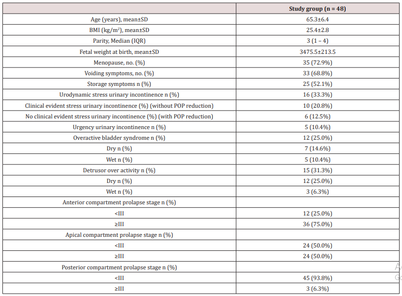
(Table 1) reveals and displays the demographic and basic clinical features of the research study cohort in which mean /SD of age, BMI, fetal weight at birth =65.3±6.4 years , 25.4±2.8kg/m2, , 3475.5 ± 213.5 grams, whereas parity median (IQR)= 3 (1 – 4),35 cases were menopausal representing 72.9% of the research study cohort ,33 cases had voiding symptoms representing 68.8% of the research study cohort ,storage symptoms was present in 25 cases(52.1%),16 cases had Urodynamic stress urinary incontinence (33.3%), Clinical evident stress urinary incontinence without POP reduction was present in 10 cases (20.8%), No clinical evident stress urinary incontinence with POP reduction was present in 6 cases (12.5%),5 cases had Urgency urinary incontinence (10.4%),12 cases had Overactive bladder syndrome (25.0%) 7 of them were dry and 5 were wet ,15 cases had detrusor over activity (31.3%) 12 cases were dry and 3 cases were wet , Anterior compartment prolapse stage <III was present in 12 cases(25.0%) whereas stage ≥III was present in 36 cases (75%), Apical compartment prolapse stage <III was present in 24 cases (50%),whereas stage ≥III was present in 24 cases also(50%), Posterior compartment prolapse stage <III was present in 45 cases (93.3%),whilst stage ≥III existed in 3 cases only (6.3%).

Table 1: Demographic and basic clinical features.

lupinepublishers-openaccess-journal-gynaecology-women-health-care

(Table 2) reveals and displays the urodynamic indices pre and post-operative in which as regards there was statistically significant difference concerning Uroflowmetry indices, Qmax, Elevated PVR (p value<0.001,0.001 consecutively ) as regards Cystometry indices there was statistically significant difference between pre and post-operative findings concerning, Low bladder compliance, involuntary detrusor contractions, Detrusor pressure, involuntary detrusor contractions, Positive Valsalva (vesical) leak-point pressure, (p values =0.016,0.001,<0.001 and 0.016 consecutively )Whereas there was no statistical significant difference as regards Maximum cystometric capacity , Abdominal VLPP positive(p value=0.098,0.617 consecutively. As regards pressure flow study indices there was statistical significant difference between pre and post-operative readings as regards Qmax (maximum urinary flow rate, PVR (post voidal residual urine), mean +/-SD Opening pressure, mean +/-SD Pdet Qmax detrusor pressure at maximum flow, Median (IQR) of Time to maximum flow, Voiding time, Urogenital Distress Inventory, BOO bladder outlet obstruction,(p values <0.001, 0.001) Whereas projected isovolumetric pressure <35 wasn’t statistically significant pre and post-operative (p value=0.063).

Table 2: Urodynamic findings pre and post-operative among research study cohort.

lupinepublishers-openaccess-journal-gynaecology-women-health-care

Q max maximum flow, PVR post void residual, PVR >30% of voided volume, IDC involuntary detrusor contractions, VLPP Valsalva (vesical) leak-point pressure, P det max detrusor maximum pressure, Pdet Qmax detrusor pressure at maximum flow, PIP projected isovolumetric pressure, BOO bladder outlet obstruction, UDI Urogenital Distress Inventory

*McNemar test

*: Paired t-test

Discussion

Urodynamic evaluation in uro-gynecologic corrective procedures is a challenging issue due to complexity of functional components of voiding processes. Anatomical correlations regained by surgical interventions is the cornerstone aim of all urogynecologists trying to achieve the best short and long term functional out comes however some components of micturition function could be still affected and clinical follow up in a systematic manner would enhance the quality of life of those cases [20-22].

Complexities arise also due to frequent comorbid clinical scenarios such as DM and hypertension making management protocols for those cases best achieved by multidisciplinary approach [23,24].

The lack appropriate cutoff values for uroflowmetry indices is considered a research challenge when Investigating voiding functions in females having pelvic organ prolapse issues [25,26].

Prior research groups of investigators have revealed that the voiding dysfunction within prolapse patients have a statistically estimated range of 12.8% to 39% in addition a prior research study similar to the current research in approach and methodology have shown that voiding dysfunction rate among cases having pelvic organ prolapse was about 39% whereas another prior research team of investigators mentioned a rate of 29.25%, 67 of 229 cases having clinical symptoms of lower urinary tract. Functional various facts and issues could elucidate the cause for the high incidence observed in some prior research studies such as postmenopausal state severe forms pelvic organ prolapse , besides it was interestingly observed by experienced uro-gynecologists that anterior vaginal wall prolapse cases are more liable to suffer voiding functional disorders due to urethral compression by prolapsed pelvic organs [27,28].

Prior researchers have observed and displayed among their research study findings that around 78% of preoperative voiding dysfunction could be resolved after abdominal sacrocolpopexy, and newly developed voiding disorders developed in 3% of patients another group of investigators have revealed and displayed that around 28% of cases suffering pelvic organ prolapse had preoperative voiding dysfunction, and 10% had persistent voiding dysfunction issues postoperatively [1,3,9].

Additionally, in an interesting fashion a prior research team of investigators have revealed and observed among their research study findings that around 89% of cases with severe forms of Pelvic organ prolapse and raised post voiding residual urine indices had normalization of PVR after POP surgical corrective procedures. Similar to the current research study consecutive urodynamic evaluation before and after laparoscopic sacrocolpopexy research findings and results revealed that bladder volume indices at first sense of desire to void was statistically significantly raised and that maximal detrusor pressure during voiding have been statistically significantly reduced. Besides, it was observed that there were no unfavorable urodynamic results after laparoscopic sacrocolopopexy. Interestingly those research findings show great similarity and harmony with the current research study findings [4,10,15].

A prior prospective research study recruited Forty-nine cases that have undergone a modified laparoscopic sacrocolpopexy/hysteropexy procedure. The investigators aimed to investigate the impact of surgical intervention on pelvic anatomy, urodynamic observations, cases satisfaction and symptom scoring levels. The research team of investigators revealed and displayed the following results in which they observed that Laparoscopic sacrocolpopexy corrected successfully vaginal vault prolapse. In all cases assessed by urodynamics workup six months after the procedure in which the bladder volume during start of voiding desire had statistically significantly increased and the maximal detrusor pressure at voiding phase have been statistically significantly reduced. Furthermore, the irritative and storage voiding complains have been reduced in a considerable manner showing statistical significance [17-22].

Another priorly conducted research studies assessed and evaluated the voiding functional changes at 3 months after performance of laparoscopic sacrocolpopexy. They came to the conclusion that preoperative voiding dysfunction is powerfully correlated and linked to pelvic organ prolapses and considerably improves in a statistically significant fashion at 3 months after performance of laparoscopic sacro- colopopexy [19,24,28].

The current research study findings that have shown great improvement in urodynamic findings could be justified by the fact that the sacrocolpexy procedure improves the anatomical pelvic structural positioning and support therefore enhances the urinary continence function.

Conclusion and Recommendations

Laparoscopic sacrocolopexy when properly performed on required cases results in marked improvement of most urodynamic indices and parameters ,however future research efforts should be conducted in a multicentric manner to evaluate the effectiveness of this mode of management on long term .Racial ,ethnic and anatomical variations in the presentation of the pelvic organ prolapse should be put in consideration in efforts of future research to aid in enhancement and improvement of urodynamic practice and health care requirement for those category of cases.

Read More about Lupine Publishers Journal of Gynecology Please Click on Below Link
https://lupinepublishers-gynecology.blogspot.com/

Friday, 7 July 2023

Lupine Publishers | Sonographic 3 D Features Correlated to Uterine Pathological Issues

 Lupine Publishers | Journal of Gynaecology and Women's Healthcare


Abstract

Background Adenomyosis is a frequent gynecological disease of unknown etiology causing menstrual pain disorders and pelvic congestion, that definite histopathological based diagnosis of adenomyosis relies on the presence of endometrial glands and stroma within the myometrial tissue, junctional zone assessed sonographically could elucidate the nature of disease progressive pathological changes.

Aim: To investigate the value and usefulness of Junctional Zone indices in suspicion and diagnosis of adenomyosis disease in correlation to histopathological findings

Methodology: A clinical research trial conducted on 200 research study subjects scheduled for hysterectomy procedure due to abnormal uterine bleeding and/or dysmenorrhea unresponsive to medical treatment performed at Ain Shams University maternity hospital from January 2018 till March 2019, all research study subjects have undergone two‐ and three‐dimensional transvaginal sonography before the day of surgery.

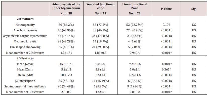
Results: There was statistically significant difference between research groups (Adenomyosis of the inner myometrium, Serrated junctional zone, Linear junctional zone,) as regards 2 D features anechoic lacunae, asymmetric corpus myometrium ,myometrial cysts, fan shaped shadowing, mean number of 2D features (p values <0.001), concerning 3 D features Mean JZ max, Mean JZ diff, JZ interruption, Sub endometrial lines and buds, mean number of 3D features (p values <0.001).

Conclusion: Junctional zone changes could be denoting early phases of adenomyosis disease development furthermore the 3D sonographic features implemented were considered and shown to be more valuable in elucidating the pathological changes confirmed by histopathological examination.

Keywords: Adenomyosis; Sonographic; Endometrial lining; Myometrium

Introduction

Adenomyosis is a frequent gynecological disease of unknown etiology causing menstrual pain disorders and pelvic congestion ,being heterogeneous in nature from being focal or simple to severe or diffuse affecting the whole myometrial tissue mass .Sonographic features revealed by 2D and 3D sonography are considered elucidating tools aiding in the diagnosis of the clinical condition however research interests are rising to correlate the sonographic criteria to histopathological and clinical criteria of adenomyosis [1,2]. Junctional zone representing an interface between the endometrial lining and myometrium is considered a cornerstone sonographic tool when properly applied in 3D and 2D sonography technologies to elucidate the severity and distribution patterns of adenomyosis [3,4].

Interestingly prior research studies have revealed and displayed that the pathophysiological origin of adenomyosis starts by repeated traumas to the junctional zone, making the anatomical and histological boundaries between the endometrium and myometrial tissues weaker causing subsequent progression of the disease by successive menstrual cycles [5,6]. On the other hand, it is crucial to mention that definite histopathological based diagnosis of adenomyosis relies on the presence of endometrial glands and stroma within the myometrial tissue. on the contrary other researchers have mentioned that endometrial‐sub endometrial myometrial zone disruptive issues observed by imaging techniques e.g. as junctional zone thickening, disruption or infiltration, should be considered a separate category than adenomyosis. Interestingly pathologists were not capable to clearly discriminate between the junctional zone and the outer myometrial layer further more by light microscopic examination, the muscle fibers directions of the inner and outer myometrial zones could show variability [7,8].

Aim

To elucidate and investigate the value and usefulness of Junctional Zone indices in suspicion and diagnosis of adenomyosis disease in correlation to histopathological findings using 2D and 3D transvaginal sonographic imaging

Methodology

A clinical research trial conducted on 200 research study subjects scheduled for hysterectomy procedure due to abnormal uterine bleeding and/or dysmenorrhea unresponsive to medical treatment performed at Ain Shams University maternity hospital from January 2018 till March 2019, all research study subjects have undergone two‐ and three‐dimensional transvaginal sonography before the day of surgery. Junctional zone maximum thickness, junctional zone maximum irregularity and sonographic features of adenomyosis have been compared with the junctional zone histopathology described as follows

a) Adenomyosis within the inner myometrium, ≥2mm myometrial invasion without contact to the basal endometrial layer,

b) Serrated junctional zone, >3mm myometrial invasion with contact to the basal endometrial layer or

c) Linear junctional zone, no or marginal myometrial invasion ≤3mm with contact to the basal endometrial layer.

Exclusive research criteria was as follows Uterine volume above 300mL due to fibroids, the existence of four or more fibroids, malignant disease, sonographic examinations involved the usage of Voluson E8 Expert machine using a multifrequency trans vaginal probe (6‐12MHz) sonographic maximum thickness measurement of the anterior and posterior uterine walls and existence of any myometrial lesions (fibroids and adenomyosis characteristics) have been recorded defined and measured. Adenomyosis features implemented were as follows. Heterogeneity, myometrial cystic lesions (>1mm), anechoic lacunae (<1mm), shadowing being fan shaped and asymmetry of myometrial corpus. JZ was evaluated in all three planes (sagittal, transversal and coronal) and measured as the maximum (JZmax) and minimum (JZmin) JZ in each wall. Anterior and posterior uterine walls have been measured using the sagittal or transverse planes and the fundal and two lateral walls were measured using the coronal plane. The walls with the largest junctional zone maximum thickness and junctional zone difference measurements have been implemented to determine peak JZ irregularities.

Uterine specimen histopathological examination

The uterus was divided into two halves immediately after performance of hysterectomy procedure fixed using formalin before histopathological examination with a slice thickness of 1‐1.5cm all over the uterus. Slices were examined in the beginning macroscopically. Followed by detailed by microscopic examination comparison with normal myometrial tissue was performed with macro and microscopic examination.

Statistical analysis

Research data were collected, revised, coded and entered to the Statistical Package for Social Science (IBM SPSS) version 23. The quantitative research data were presented as mean, standard deviations and ranges while qualitative variables were presented as number and percentages. The comparison between groups regarding qualitative data was done by using Chi-square test. Also the comparison between more than two independent groups with quantitative data and parametric distribution was done by using One Way ANOVA. The confidence interval was set to 95% and the margin of error accepted was set to 5%. So, the p-value was considered significant at the level of < 0.05.

Results

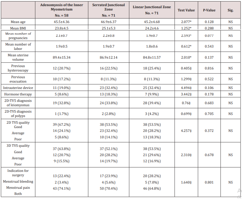
Table 1 reveals and displays demographic research data of the three research groups categorized according to presence of adenomyosis within inner myometrium (n=58), serrated junctional zone (n=71), linear junctional zone (n=71)in which there was no statistical significant difference as regards mean age, BMI, number of pregnancies, number of labor, mean uterine volume ,previous hysteroscopy ,previous evacuation ,intrauterine device ,hormone therapy, 2D-TV5 diagnosis of leiomyoma’s, 2D-TVS diagnosis of polyps, 2D TVS quality, 3D TVS quality, Indication for surgery (p values=0.128,0.288, 0.077, 0.543, 0.137, 0.816, 0.522, 0.106, 0.178, 0.683, 0.705, 0.372, 0.678, 0.801 consecutively).

Table 1: Demographic data of the studied research groups.

lupinepublishers-openaccess-journal-gynaecology-women-health-care

*: One Way ANOVA; ‡: Chi-square test

Table 2 reveals and displays that there was statistically significant difference between research groups (Adenomyosis of the inner myometrium, Serrated junctional zone, Linear junctional zone,) as regards 2 D features anechoic lacunae, asymmetric corpus myometrium ,myometrial cysts, fan shaped shadowing, mean number of 2D features(p values <0.001),concerning 3 D features Mean JZ max, mean JZ diff, JZ interruption, sub endometrial lines and buds, mean number of 3D features (p values<0.001).

Table 2: Demographic data of the studied research groups.

lupinepublishers-openaccess-journal-gynaecology-women-health-care

Discussion

Adenomyosis a frequent uterine pathology that is usually diagnosed in a definitive manner after hysterectomy reveal the requirement and necessity to understand its imaging features from early to late stages of pathological development that could aid in enhancement of management protocols for those category of cases permitting early definitive management that could prevent progression of the pathological disease process [9] Adenomyosis is a pathological insult that could represent in focal or diffuse manner leading to pelvic congestion issues and uterine bleeding disorders affecting the woman’s quality of life. Implementing advanced 3D sonographic technology with junctional Zone measurements could enhance the clinical diagnosis however still that requires extensive research efforts to elucidate the normal anatomical and histological changes of junctional zone in correlation to adenomyosis disease [10,11].

The current research study recruited 200 research study subjects undergoing hysterectomy procedures due to uterine bleeding issues unresponsive to conservative measures and all research study subjects had gone through extensive and meticulous 2D and 3d sonographic assessment before the hysterectomy procedure the research team of investigators have revealed an displayed the following research results in which there was statistically significant difference between research groups (Adenomyosis of the inner myometrium, Serrated junctional zone, Linear junctional zone) as regards 2D features anechoic lacunae, Asymmetric corpus myometrium ,Myometrial cysts, fan shaped shadowing ,mean number of 2D features(p values <0.001),concerning 3D features Mean JZ max, Mean JZ diff ,JZ interruption, Sub endometrial lines and buds, mean number of 3D features (p values<0.001.

In a prior research study have revealed and displayed that, a high percentage of cases having severe menstrual Bleeding issues and/or dysmenorrhea scheduled for a hysterectomy or had considerable junctional zone changes [12].Another research team of investigators have shown that around one‐third of cases having adenomyosis within the inner myometrial zone have junctional zone serration [13,14]. Interestingly in harmony with the current research study findings a prior research team of investigators revealed a strong statistical correlation between junctional zone changes and abnormal uterine bleeding disorders [1,4,12]. Even though it was displayed by other research studies contradicting to the current research findings that junctional zone serration can be a normal histological finding due to physiological changes in multigravida cases [3,9,10].

Furthermore, another research study similar to the current research study in approach and methodology have shown that 3D transvaginal sonography observed a statistically significant correlation between junctional zone increased thickness and irregularities and adenomyosis disease within the inner myometrial zone [8,14]. Another interesting issue observed and mentioned from previous research study findings that there is raised rates of false‐positive sonographic diagnosis of adenomyosis disease using 2D than with 3D‐ trans vaginal sonography, particularly within cases showing junctional zone serration those research findings could be justified by the fact that 3D‐ trans vaginal sonography is superior in elucidating junctional zone measurements precisely than 2D trans vaginal sonography. 7 However prior research groups have revealed that there should be a discrimination between junctional zone diseases and abnormalities and adenomyosis however junctional zone changes could reflect adenomyosis at early phases of pathological course of development [11].

Another prior research study have implemented histopathological based categorization using 3mm endometrial invasion depth as a cutoff value to denote junctional zone abnormalities furthermore junctional zone thickening could be a pathological responsiveness to the endometrial invasiveness consequently thickening of junctional zone usually co-occurs with interruption of junctional zone integrity, sub endometrial sonographic lines and budding [5,9,13].

Conclusion and Recommendations

The current research study reveals and displays that the junctional zone changes could be denoting early phases of adenomyosis disease development furthermore the 3D sonographic features implemented were considered and shown to be more valuable in elucidating the pathological changes confirmed by histopathological examination. However, the future research studies are required to be multicentric in fashion putting in consideration racial and ethnic differences and the presence of various uterine volumes to aid in future development of clinical imaging guidelines to suspect and diagnose adenomyosis at early stages enhancing the level of clinical care offered for those categories of cases.

Read More about Lupine Publishers Journal of Gynecology Please Click on Below Link:
https://lupinepublishers-gynecology.blogspot.com/

Saturday, 27 May 2023

Lupine Publishers | Laparoscopic Surgery in Pregnancy and Consequences According to Gestational Age

 Lupine Publishers | Journal of Gynaecology and Women's Healthcare


Abstract

Introduction: Laparoscopic surgery has been revealed and displayed by various research teams to reduce operative time, postoperative pain, hospital length of stay, recovery time, and wound complications in comparison to laparotomy during pregnancy. However, research efforts and analytical methods should explore the feasibility and safety to perform laparoscopic surgery at different gestational ages. Surgical and anesthetic challenges arise due to anatomical and physiological changes that normally appear as a normal adaptation to pregnancy status.

Aim of the Work: The main aim of this research study was to evaluate the clinical and obstetric consequences of interventional laparoscopic procedures conducted during late gestation in comparison to cases conducted in early gestation.

Methodology: The research was conducted in a retrospective manner on 18 cases of laparoscopic surgeries performed during pregnancy from January 2011 till March 2017 comparing cases performed early in pregnancy to those in late pregnancy.

Result: The research data was collected and analyzed from 18 pregnant study subjects that had undergone laparoscopic procedures during the research study period. The mean operative time was significantly lower among women of early pregnancy research group [45.0±7.7 min vs. 57.9±3.9min, p value=0.001]. None of women of early pregnancy group had postoperative complications, in contrast to 3 cases in the late pregnancy group [1 (14.3%) had port-site infection after laparoscopic appendectomy and 2 (28.6%) had threatened preterm labor that was managed by rectal indomethacin for 48 hours; this difference was close to be statistically significant (p value =0.059) there were no conversions to laparotomy.

Conclusion: According to the current research findings, laparoscopic interventional procedures in late and early gestation have a high safety profile and with appropriate preparation are considered feasible without any hazardous impact on pregnancy as regards maternal and fetal wellbeing.

Introduction

The requirement of abdominal surgery during pregnancy is a common concern and issue that integrates the efforts of various subspecialties to perform a multidisciplinary management for those cases scenarios in favor of the maternal and fetal wellbeing. The rates and incidences of abdominal surgery requirement varies widely according to various prior research studies however the cornerstone issue of concern is safety and capability to perform laparoscopic procedures with the great challenge of anatomical and physiological changes that normally exist during pregnancy, that requires laparoscopic surgeon that have the experience and skill to conduct the surgery taking into account the anatomical changes that vary greatly according to the gestational trimester. Research efforts in this aspect are still inadequate and scarce and requires various investigations as regards the type and safety profile of laparoscopic surgery during pregnancy [1,2]. The most frequent etiology in non-gynecological surgical interventions is appendicitis and cholecystitis and the most common gynecological interventional challenges is ovarian torsion and symptomatic adnexal masses requiring both surgical and clinical skills and expertise to have a proper and safe management handling of those clinical scenarios to avoid unnecessary surgical intervention or misdiagnosis [3,4]. Laparoscopy in pregnancy is a growing area of interest in research in the last two decades due to the advantages of minimally invasive surgeries although it carries concerns around possible injuries or complications that could affect the maternal and fetal health [5,2]. The presence of a viable intrauterine gestation with requirement of surgical intervention is a challenge that needs proper counselling and preparation of patients due to the potential risks [6].

Methodology

This study was carried out in Saudi Arabia, in Jeddah at a private hospital (Bugshan Hospital) for a period starting from January 2011 to March 2017. The study protocol was approved by the institutional ethics review board. The patients were provided with an informed consent after receiving a full explanation of the nature and protocol of the study. This study was conducted in a retrospective way the medical records of 18 pregnant cases that performed laparoscopic procedures were reviewed as regards demographic and clinical features and indication of laparoscopic procedure performance and clinical outcomes of conducted cases in from January 2011 till March 2017. Usage of tocolytic Therapy preoperatively or postoperatively was conducted on a case-by-case basis, and not in a routine manner. Cases scheduled for laparoscopic surgical intervention have been positioned in the dorsal supine position with slight a left sided tilt to avoid aorto-caval compression by the gravid uterus. Positional changes have been adjusted in harmony and conjugated efforts with the anesthesia team and according to the cases hemodynamic monitoring parameters. General anesthesia has been implemented in all cases. The laparoscopic and operative trocars placement was adjusted according to uterine size and corresponding gestational age. The following was generally performed as a rule in all cases performed and recruited in the research study in which first gestational trimester cases, Initial placement of the trocar for the laparoscopic procedure was in the umbilicus by positioning a Veress-Palmer needle followed by a 10mm trocar. The other regions for trocar placement were decided according to the surgical intervention performed.

Surgical laparoscopic interventions during the second and third gestational trimesters, the preliminary trocar was positioned within the umbilicus or supra umbilical zone, within midline by implementing Hasson open technique, being cephalic a few centimeters to the uterine fundus to avoid entry injuries to the uterus. Laparoscopic procedure insufflation has been conducted using Co2 pressure sustained under 12mmHg for sufficient venous return, to reduce aorto-caval pressure and avoid subsequent fetal acidosis.

Abdominal structures were intraoperatively manipulated according to the site, symptoms, and features of the pelvic or abdominal pathology using laparoscopic graspers and manipulators to permit adequate and optimal performance of the required interventional surgery, facial layer was closed when above 10mm trocars were implemented in the laparoscopic surgical intervention to avoid incisional abdominal hernia. While performing the procedures minimized manipulations of the gravid uterus as much as possible. Routine preprocedural performance of sonographic assessment of fetal wellbeing was performed for all cases and Cardio tocography for gestations above 24 gestational weeks. Post-operative follows up for premature contractions was conducted and tocolytic therapy was administered when required. Sonographic and CTG assessment for fetal wellbeing was repeated after one day from performing the surgical procedure according to the gestational age.

Statistical analysis

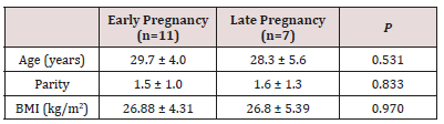
Inferential analyses were done for quantitative variables using independent t-test in cases of two independent groups, ANOVA test for more than two independent groups with post hoc Tuky’s test. In qualitative data, inferential analyses for independent variables were done using Chi square test for differences between proportions and Fisher’s Exact test for variables with small expected numbers. Logistic regression was done for factors affecting clinical and completed first trimester pregnancy among the studied cases. The level of significance was taken at P value < 0.050 is significant, otherwise is non-significant. Table 1 reveal and display that 11 study subjects (61.1%) were in early pregnancy (before 14 weeks of gestation) while 7 cases (38.9%) were in late pregnancy. The mean (± SD) gestational age of women in the early pregnancy group was 9.39±1.92 weeks (range: 7-12.57 weeks), while that in the late pregnancy group was 32.63±1.05 weeks (range: 30.43 - 33.86 weeks). There were no statistically significant differences between women of both research groups regarding the initial characteristics (age, parity and BMI) (p values=0.531,0.833,0.970, consecutively) (Table 1).

Table 1: Initial Characteristics in Included Women.

lupinepublishers-openaccess-journal-gynaecology-women-health-care

Data presented as mean ± standard deviation

BMI body mass index (calculated as weight in kilograms divided by squared height in meters)

Analysis using independent student’s t-test

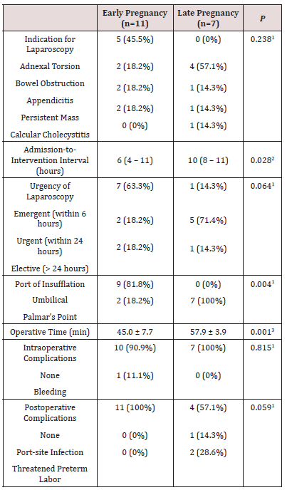
Table 2 reveals and displays that 11 women of early pregnancy research group, 5 (45.5%) underwent laparoscopy for suspected adnexal torsion, while 2 (18.2%) for bowel obstruction, 2 (18.2%) for appendicitis, and 2 (18.2%) for persistent adnexal mass. On the contrary, of the 7 women of late pregnancy group, 4 (57.1%) were for appendicitis, 1 (14.3%) for persistent adnexal mass, while 1 (14.3%) for persistent pain and vomiting due to Calcular Cholecystitis. These differences were not statistically significant (Table-2). The median admission-to-intervention interval was significantly lower among women of early pregnancy group (p=0.028), owing to the close-to-be-significant higher rate of emergent (within 6 hours after admission) laparoscopy among women of early pregnancy group (p=0.064) (Table 2).

Table 2: Laparoscopy Procedure Characteristics in Included Women.

lupinepublishers-openaccess-journal-gynaecology-women-health-care

Data presented as number (percentage); median (interquartile range); or mean ± standard deviation

1 Analysis using chi-squared test

2 Analysis using Mann-Whitney’s U-test

3 Analysis using independent student’s t-test

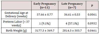
Among women of early pregnancy group, only 2 (18.2%) women had the insufflation needle inserted through the Palmar’s point (left midclavicular line, subcostal), in contrast to all women [7 (100%)] of the late pregnancy group; this difference was statistically significant (p=0.004) (Table 2). The mean operative time was significantly lower among women of early pregnancy group [45.0±7.7 min vs. 57.9±3.9 min, p=0.001] (table-2). Only 1 (11.1%) case had significant intraoperative bleeding that required blood transfusion (a case of adnexal torsion who underwent detorsion and ovarian cystectomy). None of women of early pregnancy group had postoperative complications, in contrast to 3 cases in the late pregnancy group [1 (14.3%) had port-site infection after laparoscopic appendectomy and 2 (28.6%) had threatened preterm labor that was managed by rectal indomethacin for 48 hours; this difference was close to be statistically significant (p=0.059) (table-2). Table 3 reveals and displays that the mean gestational age at delivery and mean birth weight were both significantly lower in women of the late pregnancy group [p=0.006 and p=0.046, respectively]. Only 1 (9.1%) woman of the early pregnancy group versus 4 (57.1%) women of the late pregnancy group had preterm labor (< 37 weeks of gestation); this latter difference was statistically insignificant (p=0.093) (Table 3).

Table 3: Obstetric Outcome in Included Women.

lupinepublishers-openaccess-journal-gynaecology-women-health-care

Data presented as mean ± standard deviation; or number (percentage)

1 Analysis using independent student’s t-test

2 Analysis using chi-squared test

Discussion

Recently laparoscopy have shown great advances in practice and became increasingly popular and more frequently conducted during Pregnancy. Laparoscopy as minimally invasive procedure permits early patient ambulation that avoids serious clinical issues such as deep venous thrombosis. As pregnancy is a hypercoagulable state and thromboembolic events are more frequent in pregnant women consequently early ambulation is considered a desired privilege obtained by laparoscopic approach in surgical management of cases requiring intervention in pregnancy. Even though laparoscopic procedures are feasible in all gestational trimester’s injuries are greater issues of concerns as gestation advances since the gravid uterus is at increased risk of injury in late gestational ages raising the risks of prematurity [4,7]. Besides the gravid uterus could affect the adequate and proper visualization. The growing uterus may make the traditional umbilical site less desirable. To decrease the risk of laparoscopic entry injuries, it is crucial to consider alternative sites for peritoneal injury, in advanced pregnancy gestation other than umbilical zone [8,9].

In the current research study, a cohort of 18 gravid women undergone laparoscopy during the interval between January 2011 and March 2017; 11 cases (61.1%) were in early gestation (before 14 weeks of gestation) whereas 7 study subjects (38.9%) were in late gestation. The mean (±SD) gestational age of women in the early pregnancy research group was 9.39±1.92 weeks (range: 7-12.57 weeks), whereas that in the late pregnancy research group was 32.63±1.05 weeks (range: 30.43-33.86 weeks). There were no statistically significant differences between women of both research groups regarding the initial characteristics (age, parity and BMI) Of the included 11 women of early pregnancy research group, 5 (45.5%) undergone laparoscopy for suspected adnexal torsion, whereas 2 (18.2%) for intestinal obstruction, 2 (18.2%) for appendicitis, and 2 (18.2%) for persistent adnexal mass. On the contrary, of the 7 women of late pregnancy research group, 4 (57.1%) were for appendicitis, 1 (14.3%) for persistent adnexal mass, whereas 1 (14.3%) for persistent pain and vomiting due to Calcular Cholecystitis. Those differences were not statistically significant. The median admission-to-intervention interval was statistically significantly lower among women of early pregnancy research group (p value =0.028), owing to the close-to-besignificant higher rate of emergent (within 6 hours after admission) laparoscopy among women of early pregnancy group (p value =0.064). Among women of early pregnancy research group, only 2 (18.2%) women had the insufflation needle inserted through the Palmar’s point (left mid clavicular line, subcostal), in contrast to all women [7 (100%)] of the late pregnancy group; this difference was statistically significant (p value =0.004).

The mean operative time was significantly lower among women of early pregnancy research group [45.0±7.7 min vs. 57.9±3.9 min, p value =0.001]. Only 1 (11.1%) case had considerable intraoperative bleeding that required blood transfusion (a case of adnexal torsion who underwent de-torsion and ovarian cystectomy). None of cases recruited from early pregnancy research group had postoperative complications, in contrast to 3 cases in the late pregnancy research group [1 (14.3%) had port-site infection after laparoscopic appendectomy and 2 (28.6%) had threatened preterm labor that was managed by rectal indomethacin for 48 hours; this difference was close to be statistically significant (p value =0.059). The mean gestational age at delivery and mean birth weight were both statistically significantly lower in women of the late pregnancy group [p value =0.006 and p value =0.046, consecutively]. Only 1 (9.1%) woman of the early pregnancy group versus 4 (57.1%) women of the late pregnancy group had preterm labor (< 37 weeks of gestation); this latter difference was statistically insignificant (p=0.093). A prior similar research study similar to the current research study revealed and displayed that laparoscopic surgeries, could be performed with adequate safety profile despite the indication for surgical intervention in advanced gestations up to 34 weeks [10,11].

As in the non-pregnant cases, laparoscopic surgical approaches have been revealed by various research teams and cases series to reduce operative time, postoperative pain, hospital admission time, period of recovery, and wound complications in comparison to laparotomy during gestation. Various research issues and concerns of grate debate and controversies such as risk of uterine injury and pneumoperitoneum during pregnancy that had make draw backs for performing laparoscopic surgeries in the past [12,13]. Prior research groups have revealed and displayed that CO2 insufflation triggered maternal hypercapnia, which caused fetal hypercapnia, tachycardia, and hypertension. On the other hand, that contradicts with recent research based, evidence that have emerged to show that laparoscopy, even in late gestational ages, could be undertaken safely and is the preferred management modality for many various clinical and surgical scenarios faced in every day practice in pregnant cases [1,3,14]. Laparoscopy could permit better abdominal exploration with less uterine manipulation in comparison to laparotomy that favors laparoscopic approach. the open, closed or optical trocar initial entry techniques are implemented, depending on the fundal height and experience of the surgeon. Left subcostal entry or entry under sonographic guidance have been described to prevent uterine injury. Interestingly CO2 insufflation pressure levels of 10-15mm Hg are recommended by the Society of American Gastrointestinal and Endoscopic Surgeons for the pregnant cases [2,4].

When first trimester ends the organogenesis phase of development is completed, subsequently gestational loss rate falls. Laparoscopic procedures require great skill and experience by the operator to avoid any complications possible due to physiological and anatomical changes in pregnancy which is considered a surgical and anesthetic challenge. in various clinical and surgical scenarios, the surgeon is faced by a restricted by the reduced peritoneal space available to conduct the surgery. Interestingly pregnant cases must be positioned in the left lateral decubitus position to reduce aorto- caval compression, therefore improving venous return and cardiac output. Carbon dioxide insufflation of 10-15mmHg could be implemented with adequate and acceptable safety levels for laparoscopic surgical procedures in pregnancy [5,4]. Some research studies have revealed and displayed the issue of concern that the intra-abdominal insufflation pressure should be kept under 12mmHg to prevent deterioration of pulmonary physiological status in gravid cases on the other hand other research teams have claimed that less than 12mmHg insufflation does not provide enough visualization of the intra-abdominal cavity. Additionally, 15mmHg pressure have been implemented during laparoscopic surgeries in pregnant cases without any adverse clinical outcome, whether at maternal or fetal levels [6,11]. Research groups didn’t resolve the concern and continuous debate about prophylactic tocolysis therefore all clinicians consider this issue pre or post operatively according to clinical experience and clinical scenarios tailored according to the cases scenario and requirements [3,6,13].

Conclusions and Recommendations for Future Research

Laparoscopic procedures in pregnancy appears to have a high safety profile levels however future research is recommended to be conducted in multicentric fashion with larger sample sizes and to put in consideration various indications for surgical intervention and different gestational ages .Future research should also consider ethnic and racial differences in anatomical detailed changes that could influence surgical laparoscopic practice approach to aid in future guideline implementation in practice.

Read More About Lupine Publishers Journal of Gynaecology Please Click on Below Link:
https://lupinepublishers-gynecology.blogspot.com/
Saturday, 18 March 2023

Lupine Publishers | Two Consecutive Successful Pregnancy Outcomes in a woman with Eisenmenger’s Syndrome: A Case Report

 Lupine Publishers | Journal of Gynaecology and Women's Healthcare


Abstract

Eisenmenger’s syndrome is a very rare condition in pregnant women. The incidence of ES is about 3% of the pregnant patients with congenital heart defects; however, it can be accompanied with high incidence of maternal and neonatal morbidity and mortality. Therefore, these patients should have efficient contraception or termination of pregnancy in the first trimester. We present two poorly-controlled consecutive pregnancies with good outcomes in a woman with ES to clarify the appropriate function of teamwork in the management of emergency situations in similar cases. Alongside teamwork, good prenatal care is also important because it can result in elective termination in higher gestational age of pregnancy.

A 21-year old woman, Repeat II cesarean belonging to a very low socioeconomic class, with ES was admitted to the emergency ward of Imam Hossein teaching hospital affiliated to Shahid Beheshti University of Medical Sciences with labor pain and severe dyspnea in 28 weeks of pregnancy. Echocardiography indicated a PAP of 120 mmHg. The patient had supportive treatment in intensive care unit until she was discharged. Despite previous reports of poor pregnancy outcomes in women with ES, high quality and significant treatment through labor and postpartum period lead to good outcomes in both mother and neonate.

Keywords:Eisenmenger complex; outcome; pregnancy; pulmonary hypertension

Introduction

Several congenital heart defects may result in Eisenmenger’s syndrome (ES) [1]. The progress of ES in patients with congenital heart defects depends on the heart defect size and location [2]. Signs and symptoms of ES include right ventricular (RV) failure due to right ventricular hypertrophy, nail clubbing, cyanosis, dyspnea, edema, fatigue, dizziness, and arrhythmia [1]. For the first time in 1897, Victor Eisenmenger described a 23-year-old man with a large ventricular septal defect and pulmonary arterial hypertension and termed the condition as ES [3]. The major causes of death in ES are right ventricular failure, pulmonary hypertension crisis, arrhythmia and stroke [1]. ES is a very rare condition in pregnant women. The incidence of ES is about 3% of the pregnant patients with congenital heart defects [4]. Although ES progresses slowly in non-pregnant women, the increased blood volume during pregnancy may advance the disease during a relatively short time [5]. When pulmonary hypertension exceeds 70% of systemic blood pressure, pregnancy may be associated with complications and cause maternal death [6]. In a review, the maternal mortality rate from 1978 through 1996 due to ES was 36% and the risk of maternal death remained unchanged over the period [5]. Neonatal outcome of pregnancy with ES is also poor. ES is a strong risk factor for spontaneous abortion, preterm birth, and intrauterine growth retardation (IUGR) [7].

As a rule, pregnancy in women with ES must be prevented or terminated in the first trimester [1,4,8,9]. In women who choose to continue pregnancy, a team consists of an obstetrician, perinatologist, cardiologist and an anesthesiologist is needed to care the pregnancy and labor [4,9]. Women with ES should be hospitalized in the second trimester of pregnancy [10]. Pulmonary vasodilator agonists have been used for lowering pulmonary hypertension with good results [9,11,12]. The best mode of delivery is a non-instrumental vaginal delivery using a labor pain relief through epidural block. Spinal analgesia is also preferred for cesarean in these women [5,9]. In general, treatment of ES is supportive [13] and includes oxygen therapy, the use of digitalis, diuretics, vasodilators and anticoagulants [4]. Usually anticoagulant therapy is prescribed to prevent thrombotic events; however, it may increase the risk of hemorrhage in the postpartum. Kahn reported a pregnancy in a 23-year old woman G4P3 with ES who was transferred to the hospital at 38 weeks with a two-month history of dyspnea and edema. After vaginal birth, she was treated with heparin to prevent thromboembolism. In the next day, severe vaginal bleeding developed and finally she expired [8]. We present two consecutive pregnancies with ES and good outcomes in a 21- year old woman, with poor prenatal care. If she had regular visits during her pregnancy by specialists in obstetric, perinatology and cardiology she would have a higher chance of elective termination in later gestational age of her pregnancies; however, in emergent situations, a good teamwork can lead to saving both the patient and her neonate as it happened in both pregnancies in this patient. Therefore, women with ES may have a chance to experience motherhood.

Case Presentation

A 21-year old mother G1P0 in 34 weeks of pregnancy was admitted in the emergency ward of Imam Hossein teaching hospital affiliated to Shahid Beheshti University of Medical Sciences with labor pain and dyspnea. She was a known case of ES since 25 weeks of her pregnancy. The most important findings were pulse rate (PR) of 100beat/min, respiratory rate (RR) of 24/min, blood pressure (BP) of 100/60 mmHg, O2 SAT of 92% and no cyanosis was seen. The echocardiography revealed a mild right ventricular enlargement, a mild left atrial enlargement, a mild reduced right ventricular function, a mild left ventricular hypertrophy (LVH), ejection fraction (EF) of 50%, Pulmonary artery systolic pressure (PAPs) of 50mmHg, and a large ventricular septal defect (VSD) progressing to ES. She continued her pregnancy until presented dyspnea and cyanosis in the 34th week of pregnancy. The vital signs were as follows: PR=100 beat/min, RR=34/min and BP=100/70mmHg. The second echocardiography reported EF of 50% and PAP of 98 mmHg. O2 SAT was 86%. Due to severe pulmonary hypertension, viability of fetus and low Bishop score, cesarean was performed, and a neonate was born with Apgar score 9/10 and birth weight of 2kg. She was admitted to the intensive care unit (ICU) and after four days, she was discharged from the hospital in good condition.

Again, in her second pregnancy, she was admitted for an elective therapeutic abortion in the 16th week of pregnancy but she did not accept and left the hospital. In physical examination she had PR of 110 beat/min, RR of 32/min, BP of 110/70mmHg and O2 SAT of 85%. The patient also presented acrocyanosis. Echocardiography in the 16th week of pregnancy detected EF of 50%, PAP of 80mmHg, mild systolic dysfunction and mild right ventricle enlargement. In electrocardiography, a sinus tachycardia was seen. After 12 weeks, she hospitalized in emergency ward with labor pain and dyspnea. Blood pressure, pulse rate, and respiratory rate were 100/70 mm Hg, 112 beat/min, and 30/min, respectively. Finding in arterial blood gas analysis (ABG) were as follows: PH = 7/52, partial pressure of carbon dioxide (PCO2) = 18mm Hg, bicarbonate (HCO3) = 27mEq\L and O2 SAT = 90%. In the electrocardiogram, a sinus tachycardia was detected. Echocardiography indicated a PAP of 120mmHg. Immediately oxygen was administered, emergency consultation with a cardiologist was done According to consultation with an anesthesiologist, pethidine was injected to reduce the labor pain. Supportive management such as oxygen and pain control continued. While stabilizing the patient, magnesium sulfate for neuroprophylaxis of the baby and betamethasone for fetal lung maturity were administered. The patient was admitted to the ICU. After 8 hours, because of intensified labor pain, cesarean was done under general anesthesia and a preterm baby girl with Apgar score of 7/8 and birth weight of 1800gm was born. Tubal ligation was performed and again she was transferred to the ICU ward. Enoxaparin was administered to prevent thromboembolic events in the postpartum period. The result of the echocardiography after cesarean showed PAPs of 110mm Hg and EF of 48%. Four days later, she transferred to the post-cesarean ward and after one week, she discharged with good general condition.

Ethics

We obtained patient’s informed consent for publishing this report.

Discussion

It is strongly recommended that women with ES be discouraged having pregnancy or be advised to terminate in the first trimester of pregnancy [4,9]; however, few reviews and case reports indicated that outcomes for women with ES have been improved [11-16]. Geohas and McLaughlin reported a 21-year old woman G3P2 in the 34th week of pregnancy with ES who suffered from dyspnea and edema in the third trimester. She was treated with epoprostenol and was terminated by cesarean. The outcome was good and a newborn with good Apgar score was born [13]. Our patient was belonging to an underprivileged social group and did not have appropriate prenatal care during her pregnancies. She was a known case of ES since second trimester of her first pregnancy which was continued until gestational age of 34 weeks. In her second pregnancy, she maintained her pregnancy and finally was terminated in 28 weeks of pregnancy. The outcome of both pregnancies was good similar to the results reported in previous studies [10,13,17] while in Duan’s report, the perinatal outcome of pregnant women with ES were poor [1]. In a systematic review from 1978 through 1996 on 73 women with ES, patient`s age was a risk factor of maternal death [5]. Our patient was young, and it can be one of the reasons leading to good outcome. In addition, O2 sat and hemoglobin did not show any relationship to the outcome in ES [5].

Although vaginal delivery is preferred in these patients [4] other factors such as Bishop score and maternal and fetal condition are important to determine delivery route. In our case cesarean was preferred. In three reviews, 65% to 100% of patients gave birth by cesarean due to deteriorating maternal condition during the third trimester of pregnancy [4,12,18]. In a review study by Wang, and colleagues on 13 pregnancies, no pregnancy continued to term [19]. In our study, both pregnancies were preterm and terminated at the gestational age of 34 and 28 weeks of pregnancy, respectively. Despite poorly controlled prenatal care, the mother saved because she was young and received proper care in a tertiary center during labor and postpartum period. Besides, preterm labor at the 34th and 28th week of pregnancy may contribute to the successful outcomes of the patient due to stopping the progress of hemodynamic changes and worsening the condition. On the other hand, the patient was referred to the same hospital and we had access to her past medical history. Because she was not supervised between her pregnancies, we were not aware of the medications and the cares she received and whether she was adherent to the treatment.

Conclusion

Although prevention of pregnancy or termination in the first trimester is usually recommended in patients with ES, in this case, two consecutive pregnancies developed with good outcomes. Although the patient received no proper prenatal care including visiting a cardiologist before pregnancy and having timely care by a perinatologist or even an expert obstetrician in managing high risk patients, receiving a significant treatment in a tertiary center during labor and postpartum period resulted in good outcomes in this patient. Women with ES may have a chance to experience motherhood.

Acknowledgment

Authors thank the patient for agreeing to publish this report

Ethical Approval

We obtained patient consent for publishing this report.

Read More About Lupine Publishers Journal of Gynaecology and Women's Healthcare Please Click on Below Link:
https://lupinepublishers-gynecology.blogspot.com/

Friday, 10 February 2023

Lupine Publishers | Attitudes of Married Women with Advanced Maternal Age and their Spouses Towards Family Planning and Evaluation of the Effects of Trainings related to This Issue

 Lupine Publishers | Journal of Gynaecology and Women's Healthcare


Abstract

Purpose: In this study, we aimed to evaluate the attitudes of married women with advanced maternal age and their spouses towards family planning and to assess the effects of training related to this issue.

Design: The sample group of the study was composed of 170 women and their spouses who were registered to the three health clinics located in Kiziltepe district of Mardin province. Participants were selected according to the family planning attitude scale results.

Results: When we evaluate the mean attitude scale scores of both women and their spouses before and after the trainings, it can be concluded that trainings positively affected the attitudes of them towards the family planning.

Implications: Trainings are effective in changing attitudes and behaviors of individuals who are strongly are engaged in traditional cultural characteristics.

Keywords: Family planning; Attitude scale; Nursing, Education; Culture, Mardin

Introduction

Family planning is defined as that all couples and individuals have right to have desired number of children, to decide the duration between the births freely and responsibly, and to have knowledge, tools and education in this regard. In other words, Family Planning is a protective service which allows married couples to have desired number of children according to their economical possibilities and personal wishes, and it ensures the couples to have births at appropriate intervals in accordance with the maternal and child health William et al. [1], Yıldırım [2]. Population growth has been the determinative factor in Family planning service requirements of countries. According to the World Bank reports between 2004 and 2020, the average population growth rates of come countries are estimated as follows; India (1.3), Ireland (1.2), Mexico (1.1), USA (0.9), Canada (0.8), China (0.6), Norway (0.5), Netherlands and the UK (0.3), Greece (0.1), Germany and Italy (0), Japan (-0.1). On the other hand, the population growth rate in Turkey in the same time interval will be higher than the world average (1.2 and 1.1). This population growth rate of Turkey will be one of the highest population growth rates in the world and Turkey will be one of the three countries in this regard (India and Ireland) with the highest rates Ulusoy [3], Population and the Environment [4].

In each minute, 380 women conceive, 190 women are faced with an unplanned or undesired pregnancy, 100 women experience complications related to pregnancy, 40 women experience unsafe abortion, and 1 woman die. Among all these deaths, 1% of them happen in developed countries and 99% of them happen in developing countries. In developing countries, 1 woman dies in each minute due to complications of the pregnancy. Almost 90% of these deaths can be preventable. In Turkey, there are approximately 1,8 million pregnancies in a year. Among all, 500.000 of them are abortions, 23.000 of them are stillbirths, and 1.350.000 of them are live births. Besides, 39.000 babies die before the age of one Altıparmak [5]. It has been reported by the World Health Organization (WHO) that annually 12% of 500.000 women die due to complications of abortion and 99% of these women are from developing countries. Studies conducted particularly in developing countries show that almost half of the abortions are performed as a family planning method due to the undesired pregnancies and these findings indicate the importance of the family planning services after these abortions Kabalcıoglu et al. [6], Erol et al. [7]. In Turkey, mother-child health indicators and high population growth rates require to give priority to family planning services among other health services Gemalmaz [8]. It has been shown that the rate of the desired abortion is 24% and spontaneous abortion rate is 20% in Turkey. The rate of 25 year-old or younger women who experience induced abortion is lower than 10%. However, this rate is higher than 40% in women between the ages of 45 and 49. Yigitler and Donmez conducted a study in Antalya and they stated that women who have an induced abortion are mostly in the 40-49 age group Okçay & Öztürk [9]. In a study conducted in England, the 35-40 age group of women had high risks in gestational diabetes, placenta previa, breech presentation, operative vaginal delivery, elective cesarean delivery, emergency cesarean delivery, postpartum hemorrhage, birth before 32 weeks, and birth weight and stillbirth under 5. percentile Jolly & Sebire [10].

According to the 2013 Turkey Demographic and Health Survey data, the total fertility rate is 3.41 children per woman and the highest rate is detected in the Eastern Region of Turkey. In line with this information, Southeastern Anatolia Project shows that the fertility rate is even higher in Southeastern part of the Turkey (3.46 births per woman) compared to other East regions. The fertility rate in Mardin (3.52 births per woman) is higher than the average values according to the 2014 data. The fertility rates of regions other than the East regions are similar or lower than the average of Turkey. This finding shows that the fertility rates change between the rural and urban areas and also between the east and other regions of Turkey TNSA [11]. The use of contraceptive methods alters according to the age of the woman. The frequency of contraceptive method use is the lowest among 15-19 year-old married women (44%) and this frequency increases up to 81% in 30-34 year-old women and decreases to 50% in 45-49 year-old women. The death risk due to the pregnancy is 2-3 times more in 35-39 year-old women compared to women at the age of 20s and this risk increases in women older than 40 years old Eserdağ [12]. As the age of the mother increases, the complications and death rates during the birth or after the delivery also increase Çetinoğlu et al. [13].

In Turkey, the family structure contains strong cultural elements which prevents the use and disseminates the family planning methods Cüceloğlu [14]. Authoritarian and patriarchal structures of the Turkish family relationships require the approval of man in family planning management as in other issues Depe & Ayten [15]. Uskun et al. [16], Turkistanli et al. [17], Kitis et al. [18] conducted studies and they detected that the use of family planning methods by women is affected by the educational status of the women and their spouses, the family structure, the number of children, and the view of men towards the family planning. More active involvement of men in family planning services and the use of male-specific methods will facilitate the reduction of excessive fertility Uskun et al. [19], Türkistanlı et al. [17], Kitiş et al. [18]. International and national studies indicate that the choice of method related to reproduction and fertility control depends on patriarchal traditions and this affects reproductive behaviors of women. The decision of man is prioritized about which protection method to be used and the number of family members. Men have a right to decide in family planning in various societies. It is believed that inappropriate family planning management and unhealthy attitudes due to the insufficient knowledge can be decreased in case men actively participate in the family planning Depe &Ayten, [15]. Certain practices, beliefs, attitudes, myths, customs and habits of each society depend on cultures of societies Babadağlı & şahin [20]. For instance, ceremonies and use of herbs for women’s fertility are very common in Nigerian culture. In African culture, individuals used to apply hot water, salt or vinegar/lemon juice inside the vagina after the sexual activity in order to prevent the pregnancy. There are some other contraceptive methods such as eating arsenic and castor oil seeds and drinking the water used to wash corpses Akın & senlet [21], Keller [22]. In Turkey, the number of female children increases 5-6 times due to the recurrent deliveries till the couple has a male child. In our region, there are still various wrong beliefs and applications related to the family planning. For instance, it is believed that the oral contraceptive drugs will lead to cancer, the intrauterine tool can go to the stomach, vasectomy may cause impotence, and connecting pieces of the fallopian tubes can lead to infertility. These are only some of these wrong beliefs Örsal & Kubilay [23].

Materials and Methods

Participants

The universe of the study was composed of 35-49 year-old married women who did not enter menopause, who did not use any of the effective family planning methods and their spouses. Totally 68860 participants were selected from individuals who were registered to the 1., 2. and 3. Health Clinic located in the center of Mardin province.

Data Collection

In order to measure the attitudes of individuals towards family planning in Turkey, we used the unique scale “Family Planning Attitude Scale” which was developed by Orsal. The scale was composed of 34 items and the answers to questions were ranked from 1 to 5. The minimum score was 34 and the maximum score was 180.The increased scores were considered as a positive output.

Data Analysis

The study was conducted for almost two years. The survey form and attitude scale were applied to women and their spouses by using face-to-face method via home visits. During home visits, researchers asked all questions of the survey and attitude scale to women and answers were also written by researchers. Men filled the survey and the attitude forms by themselves. The family planning attitude scale was applied before and after the trainings.

Trustworthiness

Statistical Package for The Social Sciences (SPSS) for Windows 11,5 was used to code the data and the data assessments. The reliability of the scale was tested and Chronbach’s alpha internal consistency coefficients for sub scales were calculated. The statistical analyses were done by using SPSS 11.5 program. The significance was accepted for p values either equal or lower than 0.05 (p≤0.05).

Results

Table 1: Distribution of Characteristics of Women related to Fertility and Family Planning.

lupinepublishers-openaccess-journal-gynaecology-women-health-care

In this part, the socio-demographic features of women and their spouses, fertility and family planning characteristics, the mean scores of attitude scale before and after trainings, and comparison of some variables can be seen. The highest age range when the women gave birth 19 and 22. More than half of the women had 4 to 6 children, 81.8% of them stated that they desired to have 4 to 6 children. Women stated that they did not use any contraceptive methods because more than half of them were using withdrawal method and 29.7% of them wanted to have a child (Table 1). There was a significant difference between the attitude scale scores of women and their spouses before and after trainings. Trainings led to behavioral change in both women and men (Table 2). According to our results, we detected significant differences between society, method and pregnancy subscale scores. When we evaluate the differences between the scores before and after trainings, we can conclude that the lowest differences were obtained in case of society sub scale scores and highest differences were detected in case of pregnancy sub scale scores (Tables 3 & 4). There was a significant difference between society and method sub scale scores of men before and after trainings. However, we did not detect significant difference between pregnancy sub scale scores of men before and after trainings. The lowest differences between scores before and after trainings were found in case of pregnancy subscale scores (0,505) and the highest differences were detected between the scores of society sub scale scores (7,52).

Table 2: Distribution of Attitude Scale Scores of Women and their Spouses before and after the Trainings.

lupinepublishers-openaccess-journal-gynaecology-women-health-care

Table 3: Subscale scores of women before and after the trainings.

lupinepublishers-openaccess-journal-gynaecology-women-health-care

Table 4: Distribution of the sub scale scores of men before and after trainings.

lupinepublishers-openaccess-journal-gynaecology-women-health-care

Discussion

In this part, we discussed the results of our study which was conducted to evaluate the effect of attitudes of women and their spouses towards family planning and trainings related to this issue.

In our study, we found that more than half of the women had 4 to 6 children (54.1%). In a study conducted in Isparta province, it was shown that the mean number of children was 1.95 and this number was lower compared to findings of other studies (2.5, 2, 2.7, 1.92, and 2.07) Kişioğlu et al. [16]. In Diyarbakir, according to the findings of the study conducted in Woman Health Institution, the mean number of living children was 4.3. This similarity can be because of that these studies were conducted in the same province Saka et al. [24]. It was shown that women desired to have at least four children (81.8%) because the study was conducted in East Anatolian Region of Turkey, the mean number of children was 6, it was not the preference of women to have high number of children, and women had to behave according to traditions. It was detected that women were affected by various factors while applying different family planning methods. In a study conducted in Van province, 57.9% of the women living in the city and 55.2% of the women living in villages or towns stated that they did not use any family planning method because their older family members did not allow them to use the contraceptive methods. In the same study, it was shown that 21% of the women living in the city and 30% of the women living in villages or towns did not prefer to use contraceptive methods because they believed that it is sin and 3.4% and 2.6% of them respectively thought that family planning methods led to infertility Şahin et al. [20].

In our study, more than half of the women did not use the contraceptive methods because they were using withdrawal method. Furthermore, 29.7% of them did not use these methods because they wanted to have a child. In Ankara, it was determined that the most common contraceptive method was the withdrawal method (33.7%). According to the results of Soylu, 54.4% of the women wanted to have a child and thus they did not use these family planning methods Yıldırım et al. [25]. Our findings are similar to these results. According to TNSA (2003), withdrawal method was the most common one among family planning methods TNSA [26]. Another study conducted in Mardin reported that using the withdrawal method was very common because other modern methods were not well known Ertem et al. [27]. In Turkey, it was shown in a study conducted with two different groups that individuals did not use family planning methods mostly because their spouses or families did not allow them to use or they believed that using contraceptive methods is a sin. It was shown in this study that older family members and men primarily decided on the choice and use of contraceptives, individuals had lower educational level, they had wrong religious beliefs, families were under the effect of feudal and closed society structure. These factors led to unsuccessful family planning services Tunç [28].

According to our findings, there was a significant difference between mean attitude scale scores of women and their spouses before and after trainings. The differences of mean scores of women before and after trainings were lower compared to men. We can state that trainings were more effective on women than men. For instance, women were asked whether or not they agree with the statement “The main task of woman to give birth” and 33.5% of the women answered “Yes, I agree.” before trainings. However, 17% accepted that this statement was right after trainings. Furthermore, 25.8% of the women agreed that “Contraceptive methods are impositions of western countries” before trainings. After trainings, only 13.5% of the women participated in this statement. Similarly, 75.3% and 37% of the women accepted the idea that “I can tell that I have a child in case I have a son” respectively before and after trainings. According to answers of the spouses of women, 28.3% of them supported the idea that “it is true that the main task of woman is giving birth.”. However, this rate was decreased to 7.1% after trainings. Similarly, 31.2% of the men answered as “I agree” to the statement “Contraceptive methods are impositions of western countries. After trainings, this rate was 9.5%. There was more difference between the scores of women who did not use previously any contraceptive methods before and after trainings. These women had a greater interest in trainings. This can be because that these women had an advanced maternal age and they did not use any family planning methods and they received trainings in their own home. The lowest scores were obtained from women who did not use contraceptive methods due to their wrong religious beliefs. We observed that the scores of this group mostly increased after trainings. It is possible due to their scores before trainings were prominently lower compared to scores of others.

In our study, there was a statistically significant difference between the society, method and pregnancy subscale scores of women before and after trainings. We found that the difference between scores was maximum for the society subscale scores and minimum for the pregnancy sub scale scores before and after trainings. Regarding the society subscale items, 57% of the women supported the idea of “higher number of children provides a stronger social environment” before trainings. This rate decreased to 28.2% after trainings. Furthermore, 29% of the women participated in the statement “Higher numbers of children show the power of men”. However, only 12.9% of them agreed this statement after trainings. 51.1% and 18.8% of the women agreed the statement “Men have a boy child” respectively before and after trainings. There was a significant difference between the society and method subscale scores of men before and after trainings. However, there was no significant difference between the pregnancy subscale scores of men before and after trainings. It is most probably that men perceive that the pregnancy is only the task of women. Society sub scale scores before and after trainings were the highest scores. According to answers of men, 45.8% of them agreed the statement “higher number of children provides a stronger social environment” before trainings. This rate decreased to 28.4% after trainings. Furthermore, 25.9% of the men participated in the statement “Higher numbers of children show the power of men”. However, only 8.8% of them agreed this statement after trainings. 52.5% and 29% of them agreed the statement “Men have a boy child” respectively before and after trainings.

In our study, the society, method and pregnancy subscale scores of women according to their educational statuses increased as the educational level of them increased. We observed that this difference was even higher for women particularly graduated from secondary school or higher level of educational institution. For instance, 37.1% of the women agreed the statement “Contraceptive coil leads to headache”. However, this rate decreased to 11.2% after trainings. 47% and 8% of them supported the idea that “Contraceptive drugs can lead to cancer” respectively before and after trainings. Regarding pregnancy subscale items, the statement “pregnancy makes the women more attractive” was accepted by 35.9% of the women before trainings. However, this rate decreased to 14.1% after trainings. The statement “men should marry women who do not have knowledge about family planning methods” was accepted by 28.2% of the women before trainings. However, this rate decreased to 6% after trainings. In Japan, a study related to oral contraceptive use was conducted in 1999 and it was emphasized that women should be informed about the contraceptive methods, their contribution in the health of women, and particularly about their side effects in order to reach the success in new family planning strategies Goto et al. [29].

In Turkey, the family structure contains strong cultural elements which prevent the use and disseminate the family planning methods. Authoritarian and patriarchal structures of the Turkish family relationships require the approval of man in family planning management as in other issues Depe & Ayten [15]. The use of family planning methods by women depends on various factors such as the education statuses of women and their spouses, the number of children, the family structure, and the perspective of men on family planning (DepeAyten,2006). Furthermore, according to the results of TNSA [26], it was detected that the modern contraceptive use rates increased in case women approved the attitudes of their spouses towards the family planning. However, there are limited numbers of studies conducted on the use of family planning methods and perspectives of men on family planning TNSA [26]. In Turkey, withdrawal method is one of the methods which have limited impacts (97.5%). The failure rate of these types of methods such as withdrawal method is known as 30%. In a study conducted in Turkey, it was shown that individuals who use the withdrawal method considered that it is a reliable and safe method. Therefore, family planning trainings and consultancy services should be given to men and they should be informed about the methods with limited impacts Yurdakul [30]. It has been shown in one of the studies that men can have wrong knowledge about various contraceptive methods in Izmir even though they are living in one of the well-developed cities in Turkey. According to studies, it has been detected that the education status of men does not affect their attitudes towards using contraceptive methods Gönener & Altay [31]. According to our results, we found that the mean attitude scale scores were significantly difference from each other before and after trainings. These findings can show us that trainings effective because a nurse who gave these trainings was aware of the structure of the society, she was living in the same society, and she was a well-known sand a reputable person.

Read More About Journal of Gynecology Please Click on Below Link:
https://lupinepublishers-gynecology.blogspot.com/