Showing posts with label Journal of Gynecology and Women's Health. Show all posts

Thursday, 8 December 2022

Lupine Publishers | Undetectable = Untransmissible: Unpacking HIV Risk and Transmission Concerns for Women Living With HIV

 Lupine Publishers | Journal of Gynecology and Women's Healthcare


Short Communication

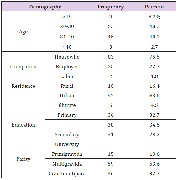
Figure 1:

lupinepublishers-openaccess-journal-gynaecology-women-health-care

The notion that Undetectable = Untransmissible (U=U) is revolutionizing the way people living with HIV are seen and treated around the world. No longer the harbingers of disease and death, those living with HIV who take HIV anti-retroviral therapy (ART) daily as prescribed and achieve and maintain an undetectable viral load (< 50 copies/ml) have effectively no risk of sexually transmitting the virus to an HIV-negative partner [1]. The global U=U campaign has been credited with beginning to change public perception of HIV transmissibility [2]. However, sexual transmission of HIV is only one way in which HIV is transmitted and it is women living with HIV who have to shoulder the additional burden of the risk of possibly transmitting HIV to their child either pre or post nattily. And even though the basis of the rationale behind U=U was proven almost two decades ago by proving that a woman prescribed ART and is virally suppressed prior to contraception, and remains undetectable throughout their pregnancy, there is virtually no vertical transmission of HIV from a mother to her infant [3] (Figure 1).

To explore some of the questions and concerns of women living with HIV regarding the broad application of U=U to their lives, ICASO commissioned a Community Brief on U=U for women living with HIV. Launched in September 2018 at the National AIDS Conference in Australia, this brief was written by a team of women living with HIV and was guided by a global community advisory committee, also made up by women living with HIV. Together they collected stories from over 65 women living with HIV from around the world and held two community consultations at the International AIDS Conference in Amsterdam in July 2018 to refine and test the content and construction of the brief. In particular, this global brief focused upon capturing the voices of women living with HIV and contains quotes of over 20 women from every region in the world. Their voices illuminate the intensely personal experiences of pregnancy, motherhood and infant feeding as well as some of the nuances around sexuality, access, equity and gender which exist in the lives of women living with HIV. The widespread adoption of the U=U discourse has provided an important opportunity to ensure that the sexual and reproductive rights of women living with HIV are recognized in the context of U=U and importantly, the profile of the U=U message has the potential to be a catalyst for the much-needed conversations about gender inequalities and violence, women’s*1 self-determination, access to treatment, women’s* involvement in research, body autonomy and informed choice as essential aspects in the lives of women* regardless of whether or not a woman* living with HIV is virally undetectable.

Perhaps the most contentious issue arising from the brief is the absence of quality research and evidence regarding the risk of HIV transmission via breastfeeding. There is an urgent need to recognize the social, emotional, practical and cultural challenges that women experience when it comes to the pressures, they are under to breastfeed or not with differing advice provided to women around the world often associated with access to clean water to provide infant formula in preference to breastfeeding. What the women expressed throughout the construction of the brief is a disconnect between medical evidence, breastfeeding guidelines, cultural practices and public health approaches to the risks and harms in relation to breastfeeding. What is most concerning is the fact that there is a fear on the part of women* living with HIV that they will not receive all the information on breastfeeding, nor will they receive appropriate social or medical support if they express their desire to breastfeed. This is both troubling and concerning. This prevents women* living with HIV from having the information they require to make their own informed decisions and choices that may affect the future health of their children and families.

This brief concludes with four actions that were ubiquitous across the communities of women* living with HIV that require urgent attention including;

I. The right of women* living with HIV to make informed choices about their sexual and reproductive rights, including the right to the birth control options of their choice as well as access to safe and legal abortion,

II. Better research into vertical transmission in the context of U=U,

III. Supporting and respecting women* living with HIV to make informed choices and the best decisions about infant feeding options for themselves and their children

IV. Increasing, improving and guaranteeing access to the range of HIV treatments that work best for women* living with HIV around the globe.

Optional Quote

“The U=U movement is transformative . . . so profound in its implication and impact but so simple in its concept. And women - all women everywhere - too must benefit from this concept. More than ever before, we now have within our hands the opportunity to counter both perceived and experienced stigma and really embrace the fact that when individuals know their status, link to antiretroviral therapy and become virally suppressed then life can return to normal in the fullest sense.”

Foot note

1 This community brief acknowledges the diversity of women living with HIV, which includes transgender men, gender queer people and others identified female at birth. The writing team felt it was important to indicate this inclusivity in this brief by using an asterisk * where our diversity is included.

Read More About Lupine Publishers Journal of Gynaecology Please Click on Below Link:
https://lupinepublishers-gynecology.blogspot.com/

Thursday, 15 September 2022

Lupine Publishers | Associated Factors with Sexual Assaults in A Gynaecology Emergency Department

 Lupine Publishers | Journal of Gynaecology and Women's Healthcare


Abstract

Objective: To study associated factors with sexual assaults suffered by patients attended in the Gynaecology Emergency Department in Miguel Servet University Maternity Hospital.

Methods: Descriptive cross-sectional study of sexual assaults from October 2016 to April 2018 in Miguel Servet University Maternity Hospital.

Results: 52 cases of sexual assault were attended from 50 different women. The patient profile was: young woman, with an average age of 28 years old (24% were minors), attacked by penetration (57.7%), by a single assailant (80.8%), with no objective lesions in genitals (86.5%) nor other parts of the body (65.4%). 50% of the assaults took place during the week-end and 63.5% of the victims came to the Gynaecology Emergency Department in less than 24 hours from the incident. 75% of women reported the assault to the police.

Conclusion: Patients were mostly young, without secondary physical lesions and assaulted with penetration by a single assailant. An appropriate coordination between health workers, the police, and forensic experts is fundamental to handle the situation properly.

Keywords: Sexual assault; Associated factors; Epidemiology; Emergency Department; Patient profile.

Introduction

Violence against women constitutes an infringement of fundamental rights, not only affecting the victims but the whole society itself. According to a survey published in 2014 by the FRA (European Union Agency for Fundamental Rights), of 42,000 women from the 28 European Union Member States, 1 out of 10 women (10%) has suffered any kind of sexual violence since they were 15 years old; 1 out of 20 women (5%) has been raped after the age of 15; and 1 out of 5 women surveyed (20%) had suffered indecent touching against their will. In addition, 1 out of 4 women (25%) did not report the incident of sexual violence to the police [1]. The objective of our study consists in compiling data from patients who came to the maternity emergency room of Miguel Servet University Hospital (a tertiary-level hospital, reference in Aragón) for sexual assault and to describe the victim and assailant’s characteristics as well as the context of the incident to identify possible risk factors associated.

Materials and Methods

Descriptive cross-sectional study including all women attending the Gynaecology Emergency Department of Miguel Servet University Maternity Hospital in Zaragoza from October 2016 to April 2018. A total of 50 women were included, one of them was attended in 3 different occasions. Data was collected by the patient’s hospital discharge report and electronic clinical history. Women arrived either accompanied by the UFAM (in Spanish, Family and Women Unit of the Judicial Police) or by themselves. In this last case, police were warned from the Emergency Department and came to the hospital to take testimony from the victim. Likewise, a forensic expert was informed. The testimony, as well as the sample gathering, were always taken in presence of the forensic expert together with two specialists in Gynaecology and Obstetrics.

For the data collecting, a specific worksheet was design using the IBM Statistics Process Social Sciences 22.0 for Windows application, that allowed the statistical analysis at a later stage. This database did not use personal details, guaranteeing the patient’s anonymity and hence, respecting the principle of confidentiality. This study was carried out respecting the current laws and regulations including the ethical principles contained in the Declaration of Helsinki. For the descriptive analysis of our sample, arithmetic means were used for quantitative variables and percentages for categorical variables.

Results

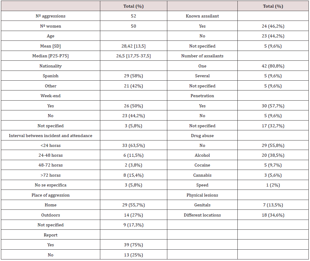
Table 1: Description of the characteristics of patients attended, assailant and context of incident. Data is shown as relative frequencies and percentages. Age is shown as arithmetic mean and median.

lupinepublishers-openaccess-journal-gynaecology-women-health-care

SD: standard deviation.
P25-75: percentile 25-75.

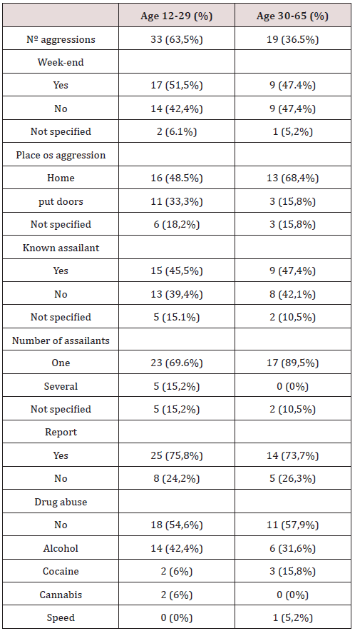
During the study period, 50 women were attended for sexual assault, one of them came in 3 different occasions for the same reason. The main characteristics are exposed in Table 1. Women’s average age was 28.42 years old. Nearly half of them (48%) were under 25, and 12 (24%) were minors. Half of the assaults occurred during the week-end. Most women attended to the Gynecology Emergency Department in the first 24 hours from the incident. 55.7% of aggressions took place in a place of residence (15.4% in the victim’s house, 28.8% in the assailant’s house and 11.5% in the house the victim and assailant shared). 30 women (57.7%) referred some kind of penetration (vaginal, anal or oral). In 46.2% of cases, the assailant was someone known by the victim (partner, friend or colleague), being in these cases the aggressor a single person. From the 23 women who reported the aggression by an unknown assailant, in 5 cases (21.7%) there were 2 or more aggressors. A sub-analysis has been performed based on the age of the patients. Results are shown in Table 2. 44.2% of women recognized having being under the effects of alcohol or any other drugs at the time of the assault. 20 women (38.5%) had drank alcohol in moderatehigh quantities, 5 women (9.7%) had used cocaine and 3 women (5.6%) cannabis. 13 urine samples were collected for toxicological analysis. 30% of samples were positive in toxics (1 in cocaine, 1 in cannabis, 1 in cocaine and cannabis and 1 in benzodiazepines and cocaine). During the physical examination, lesions in external genitals (erythema and erosions) were found in 7 women (13.5%). Neither vaginal or anal tears nor vulvar or perineal bleeding lesions were found. In 18 women (34.6%) skin injuries were found (bruisings, grazes, erosions and ecchymosis).

Table 2: Sub-analysis of the characteristics of patients attended, assailant and context of the incident according to the age of the victim. Data is shown as relative frequencies and percentages.

lupinepublishers-openaccess-journal-gynaecology-women-health-care

Discussion

In our study, victims of sexual assault were mostly young women (with an average age of 25 years old [2]), and the mechanism of aggression was penetration by a single assailant. The majority did not suffered objective physical lesions. In 2013, the US Department of Justice published a report collecting data on women suffering sexual violence including 146570 women. Statistics showed that 78% of sexual violence involved an offender known by the victim; 58% of women suffered some type of physical injury during the incident; 55% of aggressions occurred at or near the victim’s home; 90% of events was carried out by one single aggressor; and 64% of women had not reported the assault to the police [3]. These data are slightly far from those published by Larsen et al. in a descriptive case study carried out in Denmark. 2541 women attending the Center for Victims of Sexual Assault in Copenhagen from 2001 to 2010 were surveyed. According to this study, 70% of the victims reported the assault to the police (figure more consistent with data in our study showing 75% of reports to the police); 44% of assaults were carried out by a perpetrator known by the victim (similar to 46% in our statistics); a physical injury was found in 53% of victims (in our study the percentage was lower, 35%); alcohol was involved in 60% of the cases (compared to 38% in our sample); and 57% of the assaults had occurred at a place of residence [4]. A very similar percentage was obtained in our study (56%).

In Spain, Grau Cano et al. published in 2011 an individual cross-sectional study of sexual assault treated in the Emergency Department of a referral hospital, including 712 patients. According to this study, when the perpetrator was known by the victim, the aggression was more frequently carried out during the week, at home, and the mechanism of aggression was penetration by a single assailant. On the contrary, when the perpetrator was not known by the victim, the aggression took place more frequently in a public place and the percentage of assaults carried out by more than one assailant increased. Moreover, when the aggression took place during the week-ends, victims were mainly young women and the assailants unknown [5]. 87.5% of women included in the study reported the assault to the police. The rate of reports to the police by sexual assault victims differs significantly amongst different publications. Jones et al. in 2009, published a study where causes why victims of sexual assault decided not to report the aggression to the police were analyzed. According to this study, 25% of women attending a hospital’s Emergency Department for sexual assault decided not to report the incident. Authors concluded than the main reasons for not reporting the assault were the fear that the assailant went to jail, a prior relationship with the assailant and feeling that the police would blame the victim or be insensitive. Psychological barriers such as shame, anxiety or fear were not significantly associated with reporting the sexual assault [6]. This is the first paper in our reference population the study the patient’s characteristics attended for sexual assault. The sample obtained is still small and the inclusion of more patients in our data-base is necessary to obtain definitive conclusions.

Conclusion

The patient profile of women attending the Emergency Department for sexual assault in our environment was young woman (24% were minors), attacked by penetration, during the week-end, by a single assailant with no objective physical lesions. An appropriate coordination between health workers, police and forensic experts is fundamental to manage these situations and guarantee an accurate intervention for the victims.

Read More About Lupine Publishers Journal of Gynaecology Please Click on Below Link:
https://lupinepublishers-gynecology.blogspot.com/

Friday, 29 July 2022

Lupine Publishers | Effect of Diet on Evolutionary Obstetrics

 Lupine Publishers | Journal of Gynaecology and Women's Healthcare


Opinion

Diet and health are both related directly to the reproductive functions of a female in that nutrient intake effects exactly what hormones are produced and at what amounts. According literature women of Western societies produce more progesterone and estradiol from their ovaries leading to high rates of breast cancer in the West [1]. Another facet to the Western diet is foods that are rich in sugar thus contributing to the blood sugar level and ultimately affecting resistance to insulin. This resistance to insulin, which is called diabetes, may lead to many problems of the reproductive function of women. Obesity, insulin resistance, and hyperinsulinemia are all common of women in the West and lead to oligomenorrhea, amenorrhea, and chronic anovulation [2]. Regions pay big role into nutrition in the sense that different areas have abundance of different types of foods, a bulk of North America simply gets absorbed into the “western” diet, meaning increased intake of unhealthy food due to increased availability of fast food. Since these female populations are adopting nutritious lifestyles leading to obesity, insulin resistance, and hyperinsulinemia, these newly developing populations have women who are unable to reproduce as well, thus making this “developing society” not “develop.” The current research is supported with case studies demonstrating insulin levels, SHBG, and testosterone levels of women who are of south Asian descent living in affluent societies such as the United Kingdom [3]. The evidence provided portrayed that though there were two type of women living in the same society, women from south Asia that were adopting the new Western lifestyles were more susceptible to having high levels of insulin and insulin resistance, thus proving that the societies that are westernizing is leading to reproductive failure of the women. What this portrays is that high levels of obesity, insulin resistance, and insulin itself leads populations in transition to have reduced fecundity in women and based on the examples provided, it is a logical conclusion [4].

Another, “purposeful” way of preventing pregnancy is to take oral contraceptives or using contraceptives altogether before having sexual intercourse. There are many different types of ways a woman who is sexually active can prevent pregnancy. The most recent type of contraception is the menstrual-suppressing oral contraceptives which lead to virtually no menstrual cycle or a very consistent menstrual cycle like one every 3 months [5]. The basis of the menstrual cycle is avoided because of the menstrualsuppressing oral contraceptives. The main reason this happens is to procure sexual development and gain levels in estrogen and progesterone. There are two general standpoints on whether these contraceptives should be taken: the fact that the menstrual cycle is physiologically tolling, or menstruation is energetically less costly than maintain the endometrium between ovulations [6]. There are many different hypotheses present, after these standpoints are mentioned, which talk about what menstruation can do and why it is there. These hypotheses are that menstruation was an evolutionary byproduct, it aids in pathogen removal, it weeds out defective embryos, and it advertises fertility. How women feel about menstruation also would aid in decisions on whether to use contraception or not. According to the surveys, women are inadvertently split close to even in that some women feel they need the period and some feel that they should take oral contraceptives to ease the pain and reduce bleeding that occurs [7]. There are various pros and cons when considering oral contraceptives, so it is completely a woman’s choice on to take one or not. The menstrual cycle is neither beneficial nor deleterious when compared with women taking contraception, thus leaving the argument open ended on whether taking these contraceptives are viable. Poor diet garners an overall unwanted experience while contraception is used for the sole purpose of deterring pregnancy; these are two of the few ways that women are preventing pregnancies [8].

Read More About Lupine Publishers Journal of Gynaecology Please Click on Below Link:
https://lupinepublishers-gynecology.blogspot.com/

Thursday, 10 February 2022

Lupine Publishers | A Critical Review on Artavavaha Srotas as Concept of Female Reproductive System in Ayurveda

 Lupine Publishers | Journal of Gynaecology and Women's Healthcare


Abstract

According to Ayurveda, health of a female starts in the fetal stage itself which describes the measures to yield a good female child. The distinctive anatomical and physiological features of women in each age group were well studied by the ancient sages of Ayurveda. Just as the river is cleansed by its flow the women are purified by the menstrual flow. That is the reason for the non-susceptibility of women to many diseases. The regimens to be followed during menstrual and post-menstrual periods are well advocated in Ayurveda. Disregard to these regimens is the leading cause for many of the gynecological and systemic diseases in women. In addition to the systemic diseases, 20 gynecological diseases are described, which are explained in the classics under the entity of Yonivyapad and all the Yoni Roga are the disease of anatomical components of Artavavaha Srotas (Reproductive system).

In Ayurveda there is vast description of Artavavaha Srotas in terms of its moolsthana, Patho-physiology, clinical conditions and their ayurvedic management. Artavavaha Srotas shows quite similarities with female reproductive system explained in modern medical science. Anatomical defects of the reproductive system could be one of the commonest causes of bad obstetric history. Approximately 12 - 15% of women with recurrent abortion have uterine malformation. So there is need of time to elaborate the physio- anatomical and applied ayurvedic knowledge regarding female reproductive system for better female health.

Keywords: Ayurveda; Artavavaha Srotas; Female Reproductive System; Yonivyapad; Moolsthan

Abbrevations: ART: Assisted Reproductive Technology; HSG: Hystero Salpingo Grams; MRI: Magnetic Resonance Imaging

Introduction

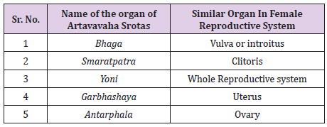
Ayurveda explained different structures or parts of the Artavavaha Srotas which are similar to the structures of the female reproductive system. The term Bhaga is in Ayurveda refers to the Smaramandira and Yoni which shows similarity with Vulva. It is of 12 Angular in length. It seems to be the description of circumference of entire vulva instead of introitus of vagina. Thus the word Bhaga gives the meaning of Yoni, which denotes the external genital organs of female i.e. vulva or the introitus of vagina. Smaratpatra is situated in upper portion of the vagina similar to the clitoris which is highly stimulated/erectile structure during sexual act. The word Yoni in Ayurvedic classics refers to entire reproductive system and also as individual organs. The word Yoni is used in different contexts to denote different organs of female reproductive system [1-10].

The structure of the Yoni is like a conch shell, it is broader at start, kinked at middle and again broader at end. It is described to be composed of three Avarta. Prathamavarta comprises vagina and accompanying structures, Dwitiyavarta comprises cervix and accompanying structures and Tritiyavarta includes uterus along with its appendages. Garbhashaya consists of two words Garbha+Ashaya means the organ that holds the Garbha (the foetus) called Garbhashaya. It is placed between Pittashaya (small intestine) and Pakvashaya (large intestine). Yoni looks like a conch shell and it is composed of three Avarta and Garbhashaya is situated in the third Avarta of Yoni. Garbhashaya means uterine cavity which resemble to shape of fish named Rohit.

It is triangular in shape, apex being at the mouth, says that its mouth is small and mean while internal cavity is a big. Rajovahi Sira (uterine vessels) is the blood supply of the organ. While considering the concept of Artavavaha Srotas it can be understood by its functions, Moolsthan or retrogression method which means symptoms produced due to injury (Viddha Lakshana), or Dushti Lakshana (pathological changes) occurs in Artavavaha Srotas. Aratvavaha Srotas is physio-anatomical structure present in female pelvic cavity having Garbhashaya and Artavavahi Dhamani of its Moolsthan i.e., most important parts. It is quite related with female reproductive system of modern science in which uterus along with fallopian tube and ovary are the most important parts for its structural and functional essentiality. Concept of Artavavaha Srotas has been resolute in two ways Macroscopic and microscopic. Macroscopically it is considered as reproductive tract where menstruation, conception, and foetal development take place. While microscopically we must understand physiological aspect as Artavavaha Srotas is physio-anatomical concept [11-15].

Functions of reproductive system like ovulation, menstruation, conception, endometrial changes etc. all these are controlled by various hormones under HPO Axis while some functions happen due to its proper blood supply and nerve supply. Capillary network present in reproductive system also play significant role in nutrition, development and proper functioning of the whole system. Both Ayurveda and modern science accept that Fertilization, Implantation, nutrition and development of foetus take place in Garbhashaya (uterus). Any injury to the Artavavaha Srotas or its Moolsthan causes symptoms like menstrual disturbances, dyspareunia or even infertility which is also accepted and well explained by modern science. By all the above argument concerning Artavavaha Srotas shows reasonably similarities with reproductive system.

Moolsthan of Artavavaha Srotas

While considering the Moolsthan of any Srotas, following points are taken in consideration like Utpattisthan (origin point of view), Sangrahasthan (storage) and Vahanasthan (conduction). The Moolsthan or source is considered that without which the origin, maintenance and destruction of that specific carrier of body nutrient cannot be possible and the place which controls the entire functional dealings and processes of the specific carrier. In females, additional Srotas is explained named as Artavavaha Srotas. Ayurveda mentions the Garbhasaya and Artavavahi Dhamani as a Moolsthan of Artavavaha Srotas. Garbhashaya is mainly responsible for conception, production and expulsion Artava, from origin point of view. It shows same structural and functional characteristics of uterus along with ovaries and both have same pathological and clinical conditions after injury. Artavavahi Dhamani can be taken as fallopian tube which conducts Artava (ovum) towards uterus during menstrual phase and ovarian and uterine vessels, Dhamana word refers to contraction, hence in this study as by contracting fallopian tube conduct the ovum so it works like an Artavavahi Dhamani [16-25].

In Garbhashaya, the word Ashaya refers as cavity or space in that particular organ which are prime functional areas or cavities of the body. Garbhashaya is a space which helps in implantation and development of Garbha (foetus) here it doesn’t refers to only a single organ but it is the hollow space in the body where various bio physiological activities happens. Yoni is a very important structure of female body, shaped as “Shankha Nabhi Akriti”, contains three Avarta (circular folds) in its structure. Garbhashaya or Garbhashayya is an important structure situated in third Avarta of Yoni. Here the term Yoni has ample meaning represent the whole reproductive system of female in which shows three circular folds or subdivision yoni(vagina), Garbhashayamukha (cervical canal) and Garbhashayya (uterus) from external to internal respectively (Table 1).

Table 1: Shows the structure mentioned in Ayurveda having similarity with the organs of female reproductive system.
lupinepublishers-openaccess-journal-gynaecology-women-health-care

There are number of important things to remember about “anatomical problems” that cause abnormality of menstrual disorders. First, uterine cancer accounts for less than 1% of these. Although cancer is among the causes of menstrual disorders, it is rare in women under the age of 50 and, if caught early, is a very curable disease. Though, majority of women in their 30’s and 40’s who have periods that are heavy because of anatomic reasons, do not have cancer. For most women it is at least comforting to know that however inconvenient or disabling their symptoms may be, it is the rare woman with heavy periods that has uterine cancer. The uterus contains two types of tissue one is inner lining (endometrium) that sheds each month with menstrual cycle another muscular portion (myometrium). Both of these tissue types can develop growths. When the lining tissue develops growth, it called polyps. When the overgrowth of muscle tissue causes tumors, they are referred to as myomas (or fibroids) [26-34].

Fibroids mostly produce two types of symptoms like abnormal uterine bleeding and infertility (although other symptoms occur less frequently). Many women with fibroids don’t have symptoms and the majority of them can simply choose to live with these benign tumors. Fibroids, as you can see from the drawing, can grow anywhere in the uterus. If a fibroid is closer to the center of the uterus (the inner cavity) it is more troublesome causing heavy menstrual bleeding and infertility (or miscarriages). Ayurveda is an open to discussion science. At every place, wherever it was concerned, Ayurvedic Seers have given the freedom to the thinkers and researchers to open a new window for the elaboration of the science. Because this work was based upon the literary review so it was a mere effort to understand Ayurvedic literature and its different prospective with relation to the modern day available sciences. This work is important in sense of exploration of Ayurveda within scientific background. Gynecology is a matter of concern for all of the medicinal sciences because- start of life begins with a baby only and health of the mother is the most important factor in this process. Today in totality, problems of gynecology are increasing day by day and Ayurveda has lot to contribute in this process. All the Ayurvedic Samhita have emphasized on this topic and this is the reason, why in each and every Samhita one can found some elaborated material about the gynecological problems.

In the same context all the Samhita and scholars of Ayurveda have a unified thought that Aartava represents the main and important activities of the females. This is a cyclic activity which can be understood in different ways. This activity not only represents the cyclic monthly blood flow from the vagina on gross level but also represents the hormonal changes related with the female reproductive system. Aartava makes women very specialized in sense of physiological activities. This physiological specialization is not possible without specialized anatomical structures. Clinical importance of the Aartava is very clear because in case of any female reproductive system related examination and questioning first question with everyone will be the pattern of the monthly menstrual flow. Srotas represents the “physiological entities” of the body on the gross level as well as on the subtle level both. Generally scholars have described the Srotas as some part of a particular system. The main concept of the Srotas seems very much clear after the review of the literature deeply- this is a concept of Physiological Anatomy and Pathological Anatomy

As Ayurveda is a completely practical science and all the concepts described in Ayurveda are all about either for the maintenance of the health of a healthy individual or these are related with the cure of some diseases. In the same both physiology and pathology are most important to understand for any medical science. Medicinal sciences or health sciences can only be applied in every sense; these cannot be plain theoretical in any way. This is the reason perhaps nowadays modern medical sciences are also returning to the Ayurvedic way of understanding the Anatomy and are emphasizing on the functional anatomy more than the plain theoretical anatomy. Description of Srotas is mainly with the sense of functional anatomy suggests that a Srotas can represent the whole of the system easily. When it comes to defining the pair nature of the female genital system- this is made up of two parts- embryological view clears this. Second is the confusion over the number and counting of the Srotas. When text was seen properly, it is very clear that approach of Sushruta and Charaka Samhitas are entirely different, one thinks like a physician and other one’s view is totally surgical.

Aartava and Srotas collaboratively are Aartava Vaha Srotas as stated above that this represents the whole female reproductive system, because all normal activities are quite impossible if one of these parts won’t work properly. All minor and major parts of the system maintain its normal activities. These are the anatomical specifications of a part which decide the role and activity of that particular part and same is applicable on the Aartva Vaha Srotas or female reproductive system. Right from the positioning of a system up to the mucosal lining and blood and nervous supply, all parts together decides the activities of any system. There are different types of the anatomical anomalies in the female reproductive system. These can be classified into three parts on a whole

a) Structural Deformities

b) Embryological Deformities

c) Histological Deformities

a) Structural Deformities: The structural deformities there is deformities like- Antramukhi, Phallini, Mahayoni, Vatiki Yoni, Prasransini along with Udvrtta and Apavrtta phala Yoni Vyapada. These all structural deformities mainly represent the “dislocation”, “displacement” and “disarrangement” of the female genital organs. Today modern medical sciences also agree that when there is some problem with the positioning of the uterus it may hamper the feminine characters and activities, very easily!

b) Embryological deformity: Under this heading we can summarize- Suchi Mukhi, Shandi and Bandhya Yoni Vyapadas. Beside this Kashypa has elaborated this very clearly and has given some other correlations by giving some examples of the different type of the vaginal openings on the basis of embryological developments. So all this can be used for the exploration of the embryological development and different anomalies,

c) Histological deformities: According to Ayurveda in this category Karnini and Yoni Kanda can be included because in these two deformities will be on histological levels.

There is no direct correlation possible in different conditions between Ayurveda with modern sciences because the way to acquire knowledge is totally different in both the sciences. Still there are some of the analogues based on the description available in the text. The table below is for the same “analogue” in different conditions of Ayurveda and Modern Science

Discussion

When we look at the Female genital system, this whole system is developed by two mullerian ducts during the development. These two fuse together and develops a complete system and this system as whole is one and same! This defies the “Copulate” nature of the Aartava Vaha Srotas as a whole and is perfect even in terms of the analogue of different thoughts from Ayurveda and Modern Anatomical Sciences. Secondly, any system works properly, when all of its components are working well, even small tiny cells are important in well functioning of the system. When we are looking at the human body, our approach should be subjective not objective. Anatomy in the text is not discussed to find out what and where alone in Ayurveda. The main purpose of the Anatomy in Ayurveda is to understand the structural importance to treat a patient and this should be taken in the same way. If the whole female genital system is Aartava Vaha Srotas then what is the Ayurvedic concept about that? This was the next quest in clearing the questions. There is an ample description of the Yoni and Garbhashya in the text, which clearly covers the whole genital system of the females.

According to Sushruta Samhita there are four “whorls” of the Yoni and these are situated in shape similar to a conch shell and every whorl represents a part of the female genital system. Coming to the anatomical deformities Women’s health is a topic of concern in the medical field, as women are an important factor in the reproduction of healthy progeny. The environmental factors, fast-changing lifestyles, and various addictions (drug abuse) as well as excess use of drugs (like steroids) have endangered their health. Congenital or acquired anatomic defects remain important considerations in the investigation of recurrent pregnancy loss. When repeated first or second trimester losses, preterm delivery, or abnormal fetal presentations are documented, the suspicion of a structural abnormality should be high. Multitudes of female anatomical deformities are uncommon. However, their impact on reproduction can be profound. The aim of this review is to remind the practicing physician of the clinically relevant embryology and summarize the studies that look at the impact of such various anomalies on a woman’s fecundity.

This retrospective longitudinal study was undertaken in order to determine the incidence and reproductive impact of uterine malformations on women desiring to conceive during their reproductive years. More couples where the female partner has a uterine anomaly are seeking care in the current clinical practice of reproductive medicine. This apparent increase is not due to a change in the prevalence of uterine anomalies in the population but is due the availability of better imaging techniques of the uterus itself and the practice of assisted reproductive technology (ART). In this era of ART, there has been more attention paid to the impact of Mullerian anomalies and their potential therapies on the outcomes of these assisted pregnancies.

The true prevalence of uterine anomalies in the population is unknown. It is insufficient to consult the older medical literature because of inconsistent diagnostic techniques utilized in the past and the heterogeneity of the subject populations that were studied. With the general wide spread use of transvaginal ultrasound and hysterosalpingograms (HSG) in reproductive-age women, increased detection of uterine anomalies in the general population can be expected, especially in the infertile and recurrent miscarriage subgroups. Following detection of uterine anomalies by ultrasound and HSG, the availability of magnetic resonance imaging (MRI) and three-dimensional ultrasound (3D.com) should increase the accurate diagnosis of these anomalies as diagnostic criteria are applied more consistently. In 1998, the American Society for Reproductive Medicine (international organization of fertility experts) classified mullerian anomalies in an attempt to provide clinicians with a tool to better document the actual anomaly and subsequently follow their patients in regards to both conception and pregnancy outcome. In general, uterine anomalies present some difficulty in pregnancy retention and overall pregnancy outcome with natural conception and ART. Arcuate uterus probably has no impact on reproductive capacity.

The uterine septum is more definitively associated with recurrent miscarriage and, unlike the bicornuate uterus; surgical correction is technically easier and less morb the bicornuate uterus appears to suffer from an increased miscarriage rate and preterm delivery and the surgical repair is more extensive. The didelphic uterus was originally thought to have no impact on reproductive outcome. Re-evaluation of the literature shows that it does increase preterm deliveries and miscarriage rates. Like the unicornuate uterus, the didelphic uterus has an increased risk of malpresentation and cesarean section for dystocia. Patients with a unicornuate uterus have the poorest outcome: higher miscarriage rates, higher ectopic rates, higher preterm delivery rates and lower live-birth rates. In Ayurvedic classics majority of the gynecological disorders have been described under the heading of Yoni-vyapad. Some people perceive Yoni as vagina, but that is just the literal meaning. The real meaning comprises the whole female reproductive system. All the Yoni Roga is the disease of anatomic components of reproductive organs like vagina, cervix, and uterus. No work is complete in terms of studies and research. This work needs more exploration with help of some clinical surveys and studies so that we can elaborate the clinical anatomy in Ayurveda. Clinical contribution is more important for any health science and Ayurvedic Anatomy has a wide space for that

Conclusion

A. Artavavaha Srotas shows quite similarity with the female reproductive system of modern medical science. It is determined in two ways macroscopic and microscopic. Macroscopically it is reproductive tract as conducting point of view and microscopically it is capillary network of uterus in context of nutrition point of view.

B. Moolsthan of Artavavaha Srotos are the regions or structures in the body from where Artava originates, store for small duration and carry for ejection outside body. Here Garbhashaya (uterus) and Artavavahi Dhamani (blood vessels and capillaries of uterus) are the Moolsthan of Artavavaha Srotas.

C. The clinical conditions related to Rituchakra (Menstrual cycle) are resulting from a range of factors such as hormone levels, anatomical deformities, functioning of the central nervous system, health of the reproductive organs.

D. Finally we conclude that, the Concept of female reproductive system is well explained in Ayurveda in terms of Artavavaha Srotas along with its basic units, physio-anatomy, clinical.

Read More About Lupine Publishers Journal of Gynecology Please Click on Below Link:
https://lupinepublishers-gynecology.blogspot.com/

Friday, 10 December 2021

Lupine Publishers | Laparoscopic Management of Early Post operative Small Bowel Adhesive Obstruction following Hysterectomy: a Series of 2 Cases

 Lupine Publishers | Journal of Gynaecology


Abstract

Objective: To describe laparoscopic management of two cases of early post operative small bowel obstruction. One case that occurred after a total laparoscopic hysterectomy with uterosacral colpopexy and the second case following vaginal hysterectomy.

Design: Case series

Setting: Academic Medical and Research Centre.

Patient: We report a 50 year old woman who developed small bowel obstruction five days after total laparoscopic hysterectomy with bilateral salpingo oophorectomy and uterosacral colpopexy due to adhesion of an ileal loop to the vaginal vault. The second patient presented ten days after vaginal hysterectomy with symptoms of bowel obstruction

Intervention: Both patients were managed conservatively for 48 hours with bowel rest and intravenous hydration. Due to unsatisfactory recovery of clinical symptoms and supportive radiologic findings, diagnostic laparoscopy was performed. The adherent ileal loop was released by blunt dissection in both the cases. Immediate clinical improvement was seen during post operative period.

Main Outcome MeasureClinical resolution of small bowel obstruction symptoms.

Result:No bowel resection was needed.

Conclusion: Adhesive small bowel obstruction can occur in the immediate post operative period and can be managed laparoscopic ally. It can occur following laparoscopic and vaginal hysterectomy in low risk patients and should be borne in mind when evaluating symptoms of small bowel obstruction in these patients

Keywords: Laparoscopy; Hysterectomy; Post Operative Adhesions; Small Bowel Obstruction

Case Presentation

Background

Peritoneal adhesions following pelvic and abdominal surgery are a significant cause of post operative morbidity. Adhesions are the cause of approximately 75% of all cases of small bowel obstruction [1]. In gynecological surgery; adhesion formation tends to occur at the vaginal vault and pelvic sidewall. These adhesions frequently involve the bowel, omentum and adnexa. Minimal invasive surgery has been increasingly performed in the recent years. Laparoscopic procedures result in fewer adhesions in comparison with laparotomy. However, early small bowel obstruction (ESBO) though common after open surgery can occur following laparoscopy as well [2]. In the management of ESBO, there is a role for conservative approach which if fails needs exploration by laparoscopy or laparotomy. Conservative management with gastric decompression may be effective in well selected cases [3]. Tissue injury is inevitable with every surgery. Conventionally it was thought that achieving adequate hemostasis could possibly prevent adhesions. Practically it is not feasible to predict the occurrence of adhesions. Several studies involving the use of adhesion prevention barriers like Interceed, Seprafilm suggest that these agents are safe for using in gynecological surgery, but there is limited evidence regarding long term benefits.

Case 1

A 50 year old woman who underwent Total laparoscopic hysterectomy with bilateral salpingo oophorectomy and uterosacral colpopexy in our hospital presented on fifth post operative day to the outpatient department with complaints of recurrent vomiting, altered bowel habits, abdominal distension and colicky pain abdomen for two days. She had two normal vaginal deliveries; her previous surgical history was only that of an open sterilization. Her initial presentation prior to hysterectomy was with severe anemia and menorrhagia. On examination, the uterus was 24 weeks size, irregularly enlarged, mobile with hypertrophied cervix and first degree uterovaginal descent. Ultrasound reported multiple intramural fibroids. After correction of anemia pre operatively with parenteral iron, blood transfusion; she was planned for total laparoscopic hysterectomy with bilateral salpingo oophorectomy. On laparoscopy, uterus was 24 weeks size; specimen was retrieved vaginally by cold knife morcellation. Uterosacral colpopexy was done with no.2¯0 PDS. Her post operative period was uneventful. She was started on clear liquids the next day, discharged after she was comfortable with soft diet.

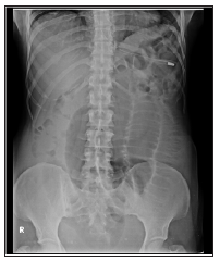
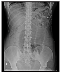
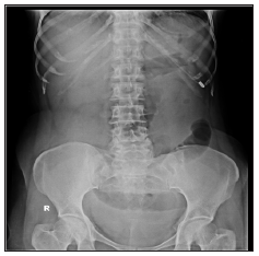
After discharge from the hospital, she had history of obstipation for three days following surgery. She consulted a local doctor for the same and was given laxative and intravenous fluids. She had one episode of loose stools on the day of admission. There is no history of fever or other symptoms suggestive of genitourinary infection. On examination, patient was hemodynamic ally stable, hydration air, abdomen was mildly distended and there was mild diffuse tenderness. There were no signs of peritonitis and bowel sounds were sluggish. Vault appeared healthy on examination. Rectum was not ballooned and there was no fullness in the pouch of douglason per rectal examination. Initial management of the patient was with intravenous fluid administration, nasogastric tube insertion, baseline blood investigations, erect and supine abdominal x rays, ultrasound abdomen and pelvis. Her blood reports were normal. There were dilated jejunal loops and collapsed distal ileal loops with no free air under the diaphragm on x ray (Figure 1). Ultrasound examination was suggestive of sub acute intestinal obstruction with dilated small bowel loops and minimal interloop fluid in abdomen.

Figure 1: CASE1- dilated jejunal loops and collapsed distal ileal loops with no free air under the diaphragm on x ray.

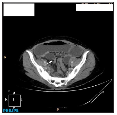
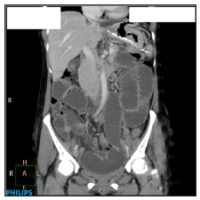
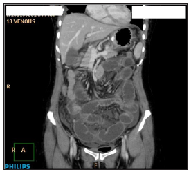
The decision was for a conservative management and the patient was monitored accordingly for 48 hours. During the course of conservative therapy, patient was nil per oral with good intravenous fluid hydration and her electrolytes were normal. Abdominal girth was noted to have increased, bowel sounds were sluggish and ryles tube aspirate was thin bilious with an average aspirate of 1000ml for 24 hours. Since x ray repeated after 48 hours showed similar findings (Figure 2), computed tomography of the abdomen with oral contrast was performed which reported multiple dilated small bowel loops with zone of transition in the proximal ileum and collapsed distal ileal loops (Figures 3 & 4). The patient was planned for a diagnostic laparoscopy as there was delayed recovery with the conservative approach.

Figure 2: CASE 1- Repeat X Ray after 48 hours.

Figure 3: CECT Abdomen- multiple dilated small bowel loops.

Figure 4: CASE 1-CECT Abdomen with zone of transition in the proximal ileum and collapsed distal ileal loops.

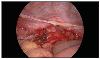
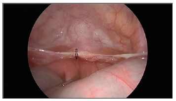
On laparoscopy, minimal peritoneal fluid along with dilated small bowel loops was noted. A small segment of the ileal loop of approximately 6 cm length was adherent to the vault more towards the left side and the same was released easily by blunt dissection (Figures 5 & 6). There was no evidence of fibrous bands/ other pathologies. Bowel walk was normal. Appendix and the vault were healthy. Interceed was placed over the vault prior to port closure.

Post operatively, the patient was administered three doses of Inj Ornidazole 500 mg and oral Erythromycin 250 mg thrice daily until discharge. There was immediate improvement with respect to abdominal pain, abdominal distension and gradual resumption of normal bowel activity in the post operative period. Ryle’s tube aspirate was nil. After 24 hours of surgery, nasogastric tube was removed and the patient was started on clear liquids. She was discharged on fourth postoperative day once she was comfortable with soft diet and bowel movements were regular.

Figure 5: site of ileal loop of approximately 6 cm length adherent to the vault more towards the left side.

Figure 6: Site of small bowel adhesion after separation by blunt dissection.

Case 2

Figure 7: CASE 2-dilated ileal loops and collapsed distal ileal loops with no free air under the diaphragm on x ray.

A 45 year old post operative patient was referred with complaints of sudden onset of abdominal pain and vomiting from one day. She underwent vaginal hysterectomy elsewhere for pelvic organ prolapse and presented with the above symptoms ten days after surgery. On examination, patient was hemodynamic ally stable, hydration fair, abdomen was mildly distended and there was mild diffuse tenderness. There were no signs of peritonitis and bowel sounds were sluggish. Vault appeared healthy on examination. There was no history of constipation, loss of appetite. Initial management of the patient was conservative with intravenous fluid administration, nasogastric tube insertion, baseline blood investigations, erect and supine abdominal x rays, ultrasound abdomen and pelvis. Her investigations were normal. There were dilated ileal loops and collapsed distal ileal loops with no free air under the diaphragm on x-ray (Figure 7). Ultrasound examination was suggestive of sub acute intestinal obstruction.

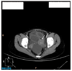
The decision was observation and monitoring the patient for 48.Abdominal girth was noted to have increased, bowel sounds were sluggish. As there was no satisfactory resolution of the symptoms, computed tomography of the abdomen with oral contrast was performed which reported minimally dilated small bowel loops, Zone of transition in the left lateral pelvic wall, adhesions of distal small bowel loops to the left lateral pelvic wall and collapsed distal ileal loops (Figures 8 & 9). Large bowel loops were normal. The patient was planned for a diagnostic laparoscopy as there was no clinical improvement with the conservative approach. On laparoscopy, minimal peritoneal fluid noted. The distal part of the ileum was adherent to left lateral pelvic wall and the raw area of the sutured vault. Proximal small bowel distended and terminal ileum distal to adhesions collapsed. Adhesiolysis done and adherent ileum freed from lateral pelvic wall. Bowel walk done. Raw area over lateral pelvic wall covered with interceed. Patient recovered well post operatively. All symptoms of bowel obstruction were resolved and she was discharged on the third day.

Figure 8: CECT Abdomen depicting adhesions of distal small bowel loops to the left lateral pelvic wall and collapsed distal ileal loops.

Figure 9: CECT Abdomen depicting collapsed distal ileal loops.

Discussion

Laparoscopic surgery and vaginal approach have been associated with fewer adhesions postoperatively than conventional open surgery. Hysterectomy is the most commonly performed gynecological surgery worldwide. The formation of post operative adhesions following abdominal surgery is well known. Adhesions may develop in 90 % after major abdominal surgery and 55 to 100 % after pelvic surgery [4]. These adhesions can be entirely asymptomatic; can cause acute symptoms or long term morbidity in the form of chronic pelvic pain, infertility, adhesive bowel obstruction etc. The pathogenesis of adhesion formation is complex. Normal healing results in the breakdown of fibrinous exudates within a period of 72 hours. Any factor interrupting this breakdown can cause persistence of fibrous matrix which is replaced by granulation tissue, formation of adhesive band. The main contributing factors are tissue injury and ischemia [5-7]. A study on the impact of adhesions on readmissions following gynecological surgeries by Lower et al has reported a 34.5% admissions at some time after surgery for an adhesion related problem [8]. As of now, there is no study that has compared the time interval for development of adhesions based on the type of surgery.

The definition of early post operative small bowel obstruction in literature has been variably considered from the time of surgery to six weeks in different studies. George et al in their study had arbitrarily defined a period of 50 days for ESBO as most of their patients had presented between 35 and 50 days after laparotomy. The earliest readmission in their study was on eight post operative day [9]. This report presents two cases with a similar presentation. Our first case presented with an ESBO within five days after laparoscopic surgery and the second case was admitted with ESBO ten days after a vaginal hysterectomy. There are no previous reports of an adhesion related ESBO that had presented in such a short time after laparoscopic hysterectomy. However, an adhesion causing ESBO within 36 hours after surgery was reported after total abdominal hysterectomy and a bilateral salpingo oophorectomy [10]. Ridgeway et al have reported three cases with ESBO following a vaginal reconstructive surgery; all cases underwent uterosacral vaginal vault suspension and total vaginal hysterectomy [11].

Thus, when a concomitant vault suspension procedure is done with a vaginal hysterectomy, it might be a predisposing factor for post operative bowel obstruction. Thus, adhesion formation causing ESBO has to be borne in mind as a possible diagnosis when the patient presents with vomiting, pain abdomen in the immediate post operative period. Adhesions can cause a range of complications and therefore measures should be taken intraoperatively to minimize their formation. There are no risks factors identified that have been definitely associated with post operative adhesion formation. Our patient did not have any risk factors in particular that could have caused adhesion formation. She was anemic that was corrected preoperatively that might have interfered with wound healing. However there is no evidence to support the same. The size of the uterus was relatively large in our patient and the specimen was retrieved vaginally by cold knife morcellation. The other possible contributing factors in this patient could have been a lengthy vault after closure or the suture material following uterosacral colpopexy. None of the above causes have been proven to cause adhesion formation.

Measures to decrease adhesion formation include minimal tissue handling, securing good hemostasis, thorough irrigation and following a sterile technique. Avoiding bowel injury, prevention of post operative infection, use of non reactive suture material has been suggested as preventive measures. Role of adhesion barriers in the prevention of adhesions is unclear. Studies have not shown concrete evidence of adhesion prevention after the use of adhesion barriers such as interceed, seprafilm. There is no clear role in preventing ESBO after their use. There are numerous studies comparing use of intercede versus no adhesion barrier. The incidence of pelvic adhesion formation was reported to be lesser after using interceed both after laparotomy and laparoscopy.

It was noted that the formation of new adhesions as well as reformation of adhesions was minimized with the use of interceed [12]. There is no data regarding the reduced incidence of small bowel obstruction, chronic pelvic pain. Proper hemostasis should be ensured before placing interceed as fibrin deposition and adhesion formation may increase if interceed is mixed with blood. However, no adverse effects after the use of adhesion barriers have been reported. Use of adhesion barriers has been recommended in high risk patients (with endometriosis, pelvic inflammatory disease) by the Royal college of Obstetricians and gynecologists.

Conclusion

Adhesion formation though less frequent after laparoscopic and vaginal hysterectomy than abdominal hysterectomy can present in the immediate post operative period. The possibility of ESBO should be evaluated for and excluded. It can be managed safely by laparoscopy. Sub acute intestinal obstruction may be more common after vaginal hysterectomy than total laparoscopic hysterectomy .This is more so if a vaginal reconstructive procedure is done probably due to the increased amount of suture material. However, there is limited evidence and further studies are required. Adhesion barriers may be used in high risk patients.

Read More About Lupine Publishers Journal of Gynecology and Women's Health Care Please go through Below Link:
https://lupinepublishers-gynecology.blogspot.com/

Saturday, 11 September 2021

Lupine Publishers | The Fetal Alcohol Spectrum Disorders: A Mini-Review

 Lupine Publishers | Journal of Gynaecology


Abstract

Since the first description of the fetal alcohol syndrome in 1973 the fetal alcohol spectrum disorders (FASDs) are still the leading preventable cause of birth defects,intellectual and neurodevelopmental disabilities. A recent WHO study estimates the global prevalence 7,7 per 1000 population,with large differences between countries.Renewed awareness results in new prevention and screening campaigning for this completely preventable global public health problem. In this mini-review, the various parts of FASD, as FAS, ARND and ARBD are discussed.

Introduction

The term fetal alcohol syndrome was first used in 1973 [1,2]. Fetal Alcohol spectrum disorders are caused by drinking alcoho during pregnancy [3,4]. Prenatal exposure to alcohol can damage the developing fetus and is the leading preventable cause of birth defects and intellectual and neurodevelopmental disabilities [1,3,4]. It presented as a cluster of birth defects[1]. Fetal alcohol spectrum disorders (FASDs) encompasses a range of possible diagnoses, including fetal alcohol syndrome (FAS), partial fetal alcohol syndrome (pFAS), alcohol-related neurodevelopmental disorder (ARND), alcohol related birth defects (ARBD) and neurobehavioral disorder associated with prenatal alcohol exposure (ND-PAE). FAS is a distinct clinical entity refering to a specific constellation of physical behavioral and cognitive abnormalities, resulting from prenatal alcohol exposure (PAE)-[1].

The lack of uniformly accepted diagnostic criteria for FAS and other related disorders has limited efforts to determine accurate prevalence figures. The World Health Organization (WHO) estimates the global prevalence to be 7,7 per 1000 population (95% CI; 4,9-11,7 per 1000 population). The WHO European Region had the highest prevalence (19,8 per 1000 population) and the WHO Eastern Meditarranean Region had the lowest (0,1 per 1000 population).South Africa was estimated to have the highest prevalence of FASD at 111,1 per 1000 population, followed by Croatia (53,3/1000 population)-[5].

Alcohol-related birth defects and developmental disabilities are completely preventable,when pregnant women abstain from alcohol use.Neurocognitive and behavioral problems resulting from prenatal exposure are lifelong.Early recognition,diagnosis, and therapy along the FASD spectrum can result in improved outcomes. There is no amount of alcohol intake to consider safe.There is no safe trimester to drink alcohol. All forms of alcohol pose similar risk and binge drinking poses dose-related related risk to the developing fetus [6]. There is a global need to establish an universal public health message about the potential harm of prenatal alcohol exposure and a routine screening protocol,according to the WHO [5]. Globally, nearly 10 percent of women drink alcohol during pregnancy [7]. In this mini-review the various parts of the fetal alcohol spectrum disorders will be discussed. The pathogenesis,pathophysiology and treatment are beyond the scope of this mini-review.

Fetal Alcohol Syndrome (FAS)

For the diagnosis FAS, patients must have at least one growth abnormality eg short stature,as well as all three characterisic facial abnormalities;short palpebral fissure length,a thin upper lip and a smooth philtrum.They must also have at least one diagnosed structural or functional abnormality of the central nervous system, eg microcephaly or impaired executive function. Confirmation of intrauterine exposure to alcohol is not obligatory for the diagnosis [8].

Growth abnormalities in FAS: The percentiles for weight are significantly lower in children with FAS at birth and in following outpatient consultations,,compared with children without FAS in a retrospective study of 322 FAS children. Moreover 22% had a body mass index below the third percentile compared with 3% of those withoit FAS[9]. Day et al. (cohort study,n=580) found, that 14 year old children,whose mothers had drunk alcohol in the first and second trimester of pregnancy showed reduced body weight, and maternal alcohol consumption in the first trimester led to smaller body length [10]. Explanation of the growth disturbance by other causes as eg prenatal deficiency states, hormonal disorders, malabsorption, malnutrition, neglect, and genetic syndromes should be excluded [11].

Facial anomalies in FAS: Several studies described the characteristic facial abnormalities in children with FAS [12-16]. Regardless of ethnicity and sex, the most powerful discriminating features for FAS proved to be smoothing of the philtrum, a thin upper lip and short palpabral length. These facial screening criteria for FAS showed a sensitivity of 100% and a specificity of 89,4%. To add quantitative assessment of upper lip thickness and philtrum smoothness Astley and Clarren developed a lip-philtrum guide with five photographs comparable to a five-point Likert scale. Upper lip and philtrum scores of 4 or 5 are considered pathological in the context of suspected FAS [17,18].

Central nervous system (CNS) abnormalities in FAS: Early injury of the brain,due to alcohol toxicity may be manifested by microcephaly. Affected children and adolescents show behavioral phenotypes of toxic damage to brain structures. As most studies are exploratory case-control studies no specific neuropsychological profile of children with FAS can currently be defined. Most of these children show below-average performance [19-27]. Functional abnormalities of the CNS should be evaluated by means of standardized neuropsychological tests together with behavioral assessment by a psychologist or physician [27]. Bell et al. found in a cohort from 2 FAS centers, that 5,9% of children with FASD showed epilepsy. THhs is much higher than the 0,6% prevalence found in the normal population [28,29]. THere is no agreement in the literature regarding a recorded cut-off value for the presence of microcephaly in children with FAS.

Studies about the head circumference in children with FAS yielded conflicting results. Therefore,a head circumference

Importance of confirmation of maternal alcohol consumption in FAS: Burd et al. [37] investigated the importance of confirmation of alcohol consumption of the mother for the certainty of the diagnosis of FAS. In cases where maternal alcohol consumption could not be confirmed sensitivity for the diagnosis FAS was higher (unconfirmed 89%,confirmed 85%), while specificity was lower (71,1% versus 82,4%). In other words,more children with FAS actually have FAS diagnosed,when alcohol consumption by their mother is not confirmed. Documentation of maternal alcohol intake is difficult. Many mothers are not questioned about their alcohol consumption during pregnancy, because carers are worried about loss of trust in the caregiving relationship. Otherwise mothers frequently deny alcohol use during pregnancy for reasons of social acceptability. The diagnosis of FAS remains difficult, because the characteristic abnormalities in children with FAS change with age. Facial abnormalities and growth deficiencies are obvious in childhood,but less distinct in adolescence and adulthood [38].

ARND

Unfortunately children with FAS represent only the tip of the iceberg of affected children, as numerous children exposed to alcohol in utero have significant physical or neuro developmental abnormalities, without all the features of FAS [39]. Alcohol related neurobehavior disorder (ANRD) refers to a constellation of neurobehavioral and central nervous system effects, occuring in the absence of the characteristic facial and growth abnormalities associated with FAS. These abnormalities include; head circumference< or equal to the 10th percentile,learning disabilities, poor impulse control, seizures, deficits in higher level receptive and expressive language, and problems with mathematical skills, memory, attention and judgement [40].

ARBD

Individuals that exhibit the typical FAS facies along with specific structural anomalies, that are known to be associated with alcohol exposure, such as low set ears,micrognathia, epicanthal folds, low nasal bridge, short upturned nose,strabismus, clinodactyly, ”hockey stick” palmar crease, radioulnar synostosis, renal anomalies and cardiac defects, but have normal growth and development, are classified as having alcohol-related birth defects ( ARBD)-[41]. The prevalence of ARND and ARBD is estimated to be at least 4 times more common than FAS [42,43]. Combining prevalence rates for FAS, ARND And ARBD indicates that 1% to 3% of all children born in the United States are affected by alcohol. This is probably an underestimation,because primary care providers and others, who care for children, do not routinely screen for FASD [44].

Conclusion

FASDs are still the leading cause of birth defects,intellectual and neurodevelopmental disabilities. There is a global need to establish an universal public health message about the potential harm of prenatal alcohol exposure and a routine screening protocol, according to the WHO [8,10]. Ten percent of pregnant women use alcohol and 1% to3% of all children born in the United States are affected by alcohol.

Read More About Lupine Publishers Journal of Gynecology Please Click on Below Link:
https://lupinepublishers-gynecology.blogspot.com/

Thursday, 29 July 2021

Lupine Publishers | Correlation between Ultrasound Findings of Ectopic Pregnancy And Operative Findings in Sudanese Women

 Lupine Publishers | Journal of Gynaecology and Women's Healthcare


Abstract

Designs and objectives This study is a prospective, descriptive, hospital-based study, done in Omdurman Maternity Hospital and Omdurman New Hospital, to assess correlation between ultrasound findings of ectopic pregnancy and operative findings in Sudanese women.

Methods: The study was conducted on patients attended to gynecological Casualty with vaginal bleeding and abdominal pain in early pregnancy diagnosed as Ectopic pregnancy, ultrasound done to all the patients, with direct interview using predesigned questionnaire, and all of these is after consent obtained, ultrasonic findings was correlated to the intraoperative findings, sensitivity of the ultrasound was calculated using SPSS.

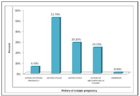
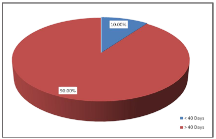
Results: out of 110 women, the most common age was 20-30 years (48.2%), multiparous were predominated by (53.6%). The commonest risk factors included; history of IUCD usage found in (52.7%), history of PID in (29.1%), history of abdomino-pelvic surgery in (24.5%), and history of ectopic pregnancy in (6.4%). On examination, there were (71.8%) women were stable, abdominal tenderness found in (54.5%), pallor in (57.3%), and (10%) of women were shocked. Sensitivity of U/S in diagnosis of ectopic pregnancy was 97.3%.

Conclusion: It was concluded that, U/S is reliable tool for diagnosing ectopic pregnancy

Keywords: Ectopic Pregnancy; Ultrasound Intraoperative; Sudan

Abbrevations: TVUS: Transvaginal Ultrasound; TAUS: Transabdominal Ultrasound; SPPS: Statistical Package for Social Science

Introduction

Implantation of pregnancy outside the normal endometrial cavity called an ectopic pregnancy, which accounts for about 10% of all pregnancy-related deaths, despite improved diagnostic methods leading to earlier detection and effective treatment [1]. Also, it increases the chances of secondary infertility as well as incidence of the subsequent ectopic pregnancy [2]. The percentages of the women with ectopic pregnancy go to an emergency department with first trimester bleeding, pain, or both about (6 to 16%) [3]. Which is strongly associated with an increased incidence of pelvic inflammatory disease [4]. Symptoms may present in both ruptured and enraptured cases [5]. Ectopic pregnancy should be suspected in any woman of reproductive age with these symptoms, especially those who have risk factors for an extrauterine pregnancy [6]. The diagnosis is usually made clinically, based upon results of the imaging studies (ultrasound) and laboratory tests (hCG). The diagnosis can also be made by observation of the ectopic gestation at surgery or histopathological examination [7]. Transvaginal ultrasound (TVUS) is the principle approach used for sonographic evaluation of pregnancy of unknown location. TVUS allows for earlier and more reliable detection of an intrauterine or ectopic pregnancy (abdominal pregnancies are an exception) and for more reliable detection of a fetal heartbeat compared with transabdominal ultrasound (TAUS) [7]. A pseudosac can be seen in up to 20 percent of women with an ectopic pregnancy [8]. An extraovarian adnexal mass, seen in 89 to 100 percent of women with ectopic pregnancy, is the most common.com finding in tubal pregnancy [8-10].

Methodology

This is a descriptive, prospective hospital-based study .It was conducted in Omdurman Maternity Hospital and Omdurman New Hospital. Sudan, during the period from August 2014 to Jan 2015 all women diagnosed clinically and by ultrasound with ectopic pregnancy attended study area were included in the study. Time-frame sample size was be taken in a period of six months from August 2014 to Jan 2015. Variables assessed were: sociodemographic data, previous ectopic pregnancy, contraceptive usage, tubal pathology and surgery, PID, clinical presentation, ultrasound findings and intra-operative findings. Intra-operative findings were correlated with clinical presentation, examination, and ultrasound findings. Sensitivity and specificity was calculated. The data was collected by direct interview using predesigned questionnaire. Abdominal ultrasound, and/or vaginal ultrasound were done to all the patients according to clinical presentation. Ultrasound was done by senior obstetrician and radiologist. The data was analyzed by computer program; statistical package for social science (SPPS), results presented in tables and graphs. The test of significance was be calculated by P value (0.05: 95% confidence). Written consent was obtained from SMSB provided to the hospital administration. Women consent was obtained verbally. Privacy of data collected was considered.

Results

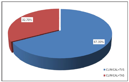
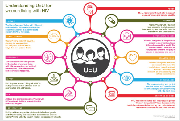
The study included 110 women diagnosed as ectopic pregnancy by ultrasound and clinically and they attended to OMH and ONH during the period from Aug. 2014 to Jan. 2015. The study aimed to correlate clinical presentation, and ultrasound findings versus intraoperative findings. Age distribution showed that, 53(48.2%) of women in the age group of 20-30 years, 45(40.9%) in the age group of 31-40 years, 9(8.2%) in the age group of 19 years or less, other socio demographic distribution of women in the study shown in Table 1. Obstetric history showed that, 58(52.7%) of women found to have history of IUCD usage, 32(29.1%) had history of PID, 27(24.5%) had history of abdominal-pelvic surgery, 7(6.4%) had history of ectopic pregnancy and 40 one woman had subfertility. No woman found to have IVF as shown in Figure 1. In addition to clinical examination, transvaginal scan for confirmation of the diagnosis was used in 74(67.3%) of women, and transabdominal scan was used in (32.7%) of women. Operators were distributed to 51(46.4%) radiologists and 59(53.6%) consultants as shown in Figures 2 & 3.

Figure 1: Risk factors distribution among Sudanese women diagnosed with ectopic pregnancy at OMH and ONH, Aug. 2014 - Jan. 2015

Figure 2: Amenorrhea in Sudanese women diagnosed with ectopic pregnancy at OMH and ONH, Aug. 2014 - Jan. 2015.

Figure 3: Method of diagnosing Sudanese women with ectopic pregnancy at OMH and ONH, Aug. 2014 - Jan. 2015.

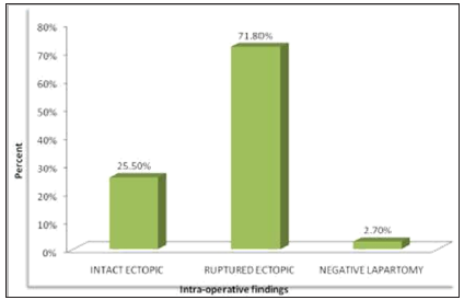
Figure 4: Intra-operative findings in Sudanese women diagnosed with ectopic pregnancy at OMH and ONH, Aug. 2014 - Jan. 2015

Table 1:

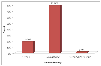
Intra-operative findings showed intact ectopic pregnancy in 28(25.5%), ruptured ectopic was found in 79(71.8%) and negative laparotomy found in 3(2.7%) as shown in Figures 4 & 5. Specific findings showed that gestational with positive fetal cardiac activity in 10(9.1%) of women, gestational sac containing fetal pole and/or yolk sac in 13(11.8%). Non-specific findings of ultrasound imaging showed complex mass in 3(2.7%), complex mass with free fluid in 54(49.1%), free abdominal fluid in 0(9.1%) and pre-pelvic + complex mass with free fluid in 20(18.2%). All participants found managed surgically by laparotomy. Correlation between intraoperative and ultrasound findings showed significant association (P value = 0.002), 19(67.9% out of 28 specific findings) of women with intact ectopic showed specific finings in U/S, among women with ruptured ectopic (79), 76(96.2%) had non-specific U/S findings, while all women with negative laparotomy had non-specific U/S findings. Correlating intra-operative findings with tender abdomen and shocked patients showed no significant association (P value > 0.05).

Figure 5: Ultrasound findings in Sudanese women diagnosed with ectopic pregnancy at OMH and ONH, Aug. 2014 - Jan. 2015

Discussion

This study carried out in OMH and ONH to assess correlation between ultrasound findings of ectopic pregnancy and operative findings in Sudanese women total number of patients was 110, age distribution showed that, 53(48.2%) of women had age of 20- 30 years, 45(40.9%) had age of 31-40 years, 9(8.2%) had age 19 years and less. Multiparous women predominated among women in the current study, they represented by (53.6%), on the other hand findings indicated considerable percentage of women who were grand multiparous (32.7%) versus few primigravida (13.6%). Multi-parity revealed by Kopani F and colleagues as risk factors for ectopic pregnancy 56. The most frequent criteria considered as risk factors for ectopic pregnancy in the current study were history of using intra-uterine contraceptive device, history of pelvic inflammatory disease and history of abdomino-pelvic surgery (previous caesarean section) and they were represented by 52.7%, 29.1% and 24.5% respectively. Other risk factors with less incidence were history of ectopic pregnancy (6.4%) and infertility. Similar findings were reported by Parashi S and colleagues in 2014, who revealed significant association between prior ectopic pregnancy, prior tubal ligation, use of intrauterine device, and prior abdominal/pelvic surgery with ectopic pregnancy (p<0.05) 57 (12).

Of the risk factors reported by Kopani F and colleagues, there was previous surgery was registered in 26% of patients and history of previous ectopic pregnancy in 7-8% of patients reported high incidence of ectopic pregnancy [5-7]. While other studies vast majority of women in the current study presented with vaginal bleeding and abdominal pain (86.4%), while abdominal pain was found in 13.6% women. These findings are compatible with what reported by Parashi S and colleagues [8] in 2014 who found that, initial symptoms were nonspecific, consisting of a period of amenorrhea and abdominal pain or tenderness, with or without unexpected vaginal bleeding. New onset pain was reported, dull or sharp in nature, which was generalized or localized to one area. Furthermore, they complained about spotty or irregular vaginal bleeding. Other study reported that, abdominal pain was a presenting symptom in 99 percent, amenorrhea in 74 percent, and vaginal bleeding in 56 percent. On examination at presentation, most patients found stable, pale and have abdominal tenderness which represented by 71.8%, 57.3% and 54.5% respectively. Shock was found in 10%, and no soft abdomen noticed. In absence of ultrasound findings, HCG test, clinical manifestation of ectopic pregnancy complicates the diagnosis because of their broad spectrum that run from asymptomatic until acute abdomen and hemodynamic shock.

Most women diagnosed through transvaginal scan (67.3%), less women diagnosed by transabdominal scan. Kopani F and colleagues revealed that, trans-vaginal ultrasound has a sensitivity of 97% and specificity of 95%. The most frequent intra-operative findings of the current study was ruptured ectopic (71.8%), followed by intact ectopic which found in 25.55% and negative laparotomy in 2.7%. Intra-operative findings were reported by Tulandi T showed that, in one representative series of 147 patients with ectopic pregnancy (78 percent were ruptured) [8]. On the other hand, most findings by ultrasound diagnosis were non-specific (79.1%) (E.g. Complex mass with free fluid and free abdominal fluid), while specific findings reported in 19.1% (e.g. Gestational sac containing fetal pole and/or yolk sac). An extra ovarian adnexal mass was reported in previous study as most common non-specific sonographic findings; they reported that, it was seen in 89 to 100 percent of women with ectopic pregnancy, is the most common.com finding in tubal pregnancy (20,26). The sensitivity of U/S method in the current study was found to be 97.3%. Findings of the study revealed good accuracy of ultrasound method to diagnose ectopic pregnancy, because imaging was mostly done by seniors and any suspicious result repeated several times in other place.

Out of 110 cases suspected with ectopic pregnancy with U/S method, only 3 cases found negative. Of the limitations faced by the current study, is scarcity of published literature comparing U/S imaging and intra- operative diagnosis in ectopic pregnancy patients, the only study available conducted in London by Condous G and colleagues in 2005, it assessed accuracy of TVS accuracy, and it was concluded that, the sensitivity of TVS found to be 90.9% . All women in the current study managed surgically by laparotomy, and they were all ectopic, salpingectomy was done. Intra-operative findings indicated that, intact ectopic were 28, out of which 67.9% showed specific findings in U/S. out of 79 ruptured ectopic, most cases showed non-specific findings in U/S. while all negative laparotomy showed non-Specific findings. Intra-operative findings indicated that, all shocked women in the study were found ruptured ectopic, except one case which was found intact. Correlating intra-operative findings with shocked cases and with abdomen tenderness found statistically not significant (P value was 0.326 and 0.230 respectively).

Conclusion

According to the current findings, we can conclude that, assessing the role of ultrasound in diagnosing ectopic pregnancy in comparison to intra-operative findings, high diagnostic accuracy was revealed with sensitivity of 97.3%. The study obtained good results because imaging was mostly done by seniors and any suspicious result repeated several times in other place. With regard to clinical presentation, most patients were stable, pale and have abdominal tenderness in addition to that some patients were in shocked. From the study the risk factors for ectopic pregnancy were history of using intra-uterine contraceptive device, history of pelvic inflammatory disease and history of abdominal-pelvic surgery. The most common intra-operative findings of the current study was ruptured ectopic, followed by intact ectopic and negative laparotomy.

Recommendations

a) Since ectopic pregnancy is a top emergency situation, and it is a leading cause of maternal death, clinical assessment should be done as accurate as possible, and cases should be scanned by highly skilled cadres.

b) Establishment of early pregnancy assessment unit in every maternity hospital.

c) Organization of IUCD workshops including proper treatment of associated infection.

d) All hospitals, especially obstetrical department should be supplied by well-established U/S unit and well trained operators.

e) Availability of laparoscopic unit with experienced personnel for managing intact ectopic and stable patients laparoscopically.

f) Ultrasound and intra-operative findings can help establishing a reported program to monitor, evaluate and manage ectopic pregnancy by routine studies.

To Know More About Lupine Publishers Journal of Gynaecology Please Click on Below Link:
https://lupinepublishers-gynecology.blogspot.com/