Showing posts with label Journal of Surgery. Show all posts

Friday, 29 September 2023

Lupine Publishers | Covered Perforativnye Ulcers Gastroduodenal Zone

 Lupine Publishers | Journal of Surgery & Case Studies


Abstract

Of the 215 patients with gastroduodenal ulcer probodnymi zone 17 (7.9%) She wore covered nature. The author notes some difficulties in diagnosing this type of pathology. If you open probodenijah symptomatology are manifestnyj nature and errors in diagnosis is usually not observed, then covered the clinical picture had ulcers was wiped out in nature, requiring additional research methods, including x-ray, to recognize this pathology. An inexperienced clinician may resort to using gastrografii contrast barium dredge. As a result of the suspension enters the free abdominal cavity, which further accompanied by the development of severe adhesive disease. This cause of this serious pathology he watched from 2 patients operated in various hospitals of Kuban. The author describes a diagnostic algorithm of this pathology in 17 patients admitted in the hospital, or with acute cholecystitis, or other pathology, and only a few hours when they are accidentally fibro gastroduodenoscopy the true cause has been identified the emergence of pain in epigastralna area is covered with a perforated gastroduodenal ulcer zone.

Keywords:Gastroduodenal Ulcer Zone Covered Perforation; Diagnostic

The aim of the study was the definition of diagnostic methods to identify covered perforating ulcers of the stomach and duodenum (PPJaZhD).

Introduction

Identification of covered probodnyh ulcers gastroduodenal zone represents some diagnostic difficulties. If you open perforations of ulcers leading clinical symptom is triad-Mond or (ulcerative anamnesias, “pain in the abdomen, doskoobraznoe tension of the abdominal wall) and x-ray-presence of free gas in the abdominal cavity, when covered probodnoj ulcer all they are missing [1- 3]. More-over, and other clinical symptoms are not defined, i.e. dullness, no liver kept blunting in sloping are-as of the abdomen and other [4,5,6]. Patients usually complain of mild pain in the area epigastralna, accompanied by diarrheal events malaise [7,8]. As their doctors have had the impression that the patient has occurred, or the aggravation of gastric ulcers or chronic cholecystitis [9,10]. Such patients are hospitalized in a therapeutic institution, and begin to be a typical survey, sometimes with the use of barium dredge that is blunder-its particles embedded in the peritoneum, and remain there, despite intensive abdominal lavage varying fluid. This leads to the development of a total of commissural process. To avoid this error, in the middle of the 20 centuries, Weber resorted to inflation of the stomach using gastric probe. This was accompanied by a delaminating of agglomerated tissues in area of ulcers and gastric cavity air rushed to free abdominal cavity (usually under the left or right of the dome of the diaphragm). This manipulation it produced during rentgeno gastroscopy, which al-lowed him to clearly observe the screen air outlet outside the cavity of the body [1,5]. Something similar can be observed in fibro gastroduodenoscopy-as soon as the endoscopies begins to inflate the stomach for inspection of its walls, the patient occur severe abdominal pain that is associated with the forced penetration of air from the stomach cavity in free abdominal cavity. In such a situation urgently carry out a repeated review x-rays of the abdomen, and if, in the first survey of the air in the abdominal cavity is not detected, then now he’s clearly defined [1,2,10]. So, the diagnosis is established. With this same purpose, some radiologists have resorted to the use of water-soluble contrast media.

Material and Methods

For 3 years in 2-ohm ECHO KGCSMP were treating patients with 215 probodnymi ulcers stomach and duodenal ulcers, of whom 17 (7.9%) There were PPJaZhD. All of these patients, who were all men aged 31 to 67 years, was on the front wall, or duodenal ulcers (12), or pyloric stomach Division (5). At 9 (4.18%) these patients the disease began with the emergence of moderate pain in epigastralna area, which was accompanied by vomiting. District therapists they suspected food poisoning and within days conducted appropriate outpatient treatment effect. By the end of the specified term of treatment in patients pain intensified, and at the same time appeared the strain in the right podreberie. With suspected acute cholecystitis, they were sent to hospital treatment. The remaining 8 (3.72%) disease patients developed similarly but when seeking medical help, through 6-12 hours from onset of symptoms, they immediately were suspected of acute cholecystitis. With this diagnosis were hospitalized in the surgery department. The first survey of all 17 patients was missing symptoms of irritation of the peritoneum. ULTRASOUND of abdomen radiography review pathology. For x-ray study of stomach using barium dredge, not steel, as in earlier Office underwent surgical treatment of patient with 2 adhesive ileus, which developed due to the falling mist abdomen (this error allowed doctors in other hospitals). In this situation, esophagogastroduodenoscopy was shown, but when I try to run it in all patients immediately arose strong abdominal pain. The procedure was discontinued. Within one hour from all 17 patients developed a picture of acute peritonitis. Repeat-ed survey radiography of abdominal cavity revealed the presence of free gas in it. Patient’s emergency laparotomy was performed.

The Result of The

During surgery in the abdominal cavity found muddy effusion. Around probodnoj the holes had inflammatory infiltrate, and it was partially obscured the top adjacent organs and tissues. All 17 patients carried out organ-preserving operations the results of the study. Of the 17 patients with PPJaZhD (5.9%) and 1 died the patient (from cardiovascular insufficiency). The overall mortality in probodnyh ulcers was 4.65% (of 215 patients died 10).

Discussion

When examining the data received on time execution of operations on the stomach and duodenum from these 17 patients, it can be concluded that cover probodnoj ulcers occurred due to the adhesive around the process expressed ulcers, with the rapid development of inflammatory infiltration. This can happen only when expressed protective immunity when around the ulcer occurs hearth with positive electric potential, to which attracted surrounding organs, having all the negative potential, and fabric to stick together. So in vivo survival occurs. When stoking the stomach this protective barrier is destroyed. That’s why even from ancient times people with abdominal pain otljozhivalis and ate almost nothing for a few days. Range that they at that time carried naked stomach ulcers. Apparently, positive role plays and reflex vomiting, during which adopted on the eve of food almost in full erupts outward. Body cavity decompression promotes podsasyvaniju surrounding organs to probodnymi.

Discussion

As you know, the most informative diagnostic process ulcers gastroduodenal zones possess esophagus gastroduodenoscopy and x-ray studies. However, we observed patients with PPJaZhD these methods initially did not give the expected result. And this was due to the fact that when abdominal radiography review absent such Cardinal diagnostic test, as the detection of free gas in it. Increased pain in the abdomen that occurred during the execution of fibro gastroduodenoscopy can be attributed to the violent disintegration of infiltration air, which at this time vduvalsja in the stomach cavity. As a result of its contents from falling into the free abdomen, that led to the development of a peritonitis. Repeated survey radiography of abdominal cavity allowed recognizing the true cause of the critical deterioration of patients, i.e. the development of widespread peritonitis, which is when the hollow organ perforation took only locally limited form. Thus, the leading value in the diagnosis of PPJaZhD has repeated panoramic radiography of abdominal cavity, which is performed after the failed fibro gastroduodenoscopy. The use of barium dredge to diagnose ulcers of stomach stones leads to the development of heavy adhesive disease of abdomen (due to the introduction of particles of barium in thickness of the peritoneum).

Read More About Lupine Publishers Journal of Surgery Please Click on Below Link:
https://surgery-casestudies-lupine-publishers.blogspot.com/

Friday, 11 August 2023

Lupine Publishers | Covered Perforativnye Ulcers Gastroduodenal Zone

 Lupine Publishers | Journal of Surgery


Abstract

Of the 215 patients with gastroduodenal ulcer probodnymi zone 17 (7.9%) She wore covered nature. The author notes some difficulties in diagnosing this type of pathology. If you open probodenijah symptomatology are manifestnyj nature and errors in diagnosis is usually not observed, then covered the clinical picture had ulcers was wiped out in nature, requiring additional research methods, including x-ray, to recognize this pathology. An inexperienced clinician may resort to using gastrografii contrast barium dredge. As a result of the suspension enters the free abdominal cavity, which further accompanied by the development of severe adhesive disease. This cause of this serious pathology he watched from 2 patients operated in various hospitals of Kuban. The author describes a diagnostic algorithm of this pathology in 17 patients admitted in the hospital, or with acute cholecystitis, or other pathology, and only a few hours when they are accidentally fibro gastroduodenoscopy the true cause has been identified the emergence of pain in epigastralna area is covered with a perforated gastroduodenal ulcer zone.

Keywords:Gastroduodenal Ulcer Zone Covered Perforation; Diagnostic

The aim of the study was the definition of diagnostic methods to identify covered perforating ulcers of the stomach and duodenum (PPJaZhD).

Introduction

Identification of covered probodnyh ulcers gastroduodenal zone represents some diagnostic difficulties. If you open perforations of ulcers leading clinical symptom is triad-Mond or (ulcerative anamnesias, “pain in the abdomen, doskoobraznoe tension of the abdominal wall) and x-ray-presence of free gas in the abdominal cavity, when covered probodnoj ulcer all they are missing [1- 3]. More-over, and other clinical symptoms are not defined, i.e. dullness, no liver kept blunting in sloping are-as of the abdomen and other [4,5,6]. Patients usually complain of mild pain in the area epigastralna, accompanied by diarrheal events malaise [7,8]. As their doctors have had the impression that the patient has occurred, or the aggravation of gastric ulcers or chronic cholecystitis [9,10]. Such patients are hospitalized in a therapeutic institution, and begin to be a typical survey, sometimes with the use of barium dredge that is blunder-its particles embedded in the peritoneum, and remain there, despite intensive abdominal lavage varying fluid. This leads to the development of a total of commissural process. To avoid this error, in the middle of the 20 centuries, Weber resorted to inflation of the stomach using gastric probe. This was accompanied by a delaminating of agglomerated tissues in area of ulcers and gastric cavity air rushed to free abdominal cavity (usually under the left or right of the dome of the diaphragm). This manipulation it produced during rentgeno gastroscopy, which al-lowed him to clearly observe the screen air outlet outside the cavity of the body [1,5]. Something similar can be observed in fibro gastroduodenoscopy-as soon as the endoscopies begins to inflate the stomach for inspection of its walls, the patient occur severe abdominal pain that is associated with the forced penetration of air from the stomach cavity in free abdominal cavity. In such a situation urgently carry out a repeated review x-rays of the abdomen, and if, in the first survey of the air in the abdominal cavity is not detected, then now he’s clearly defined [1,2,10]. So, the diagnosis is established. With this same purpose, some radiologists have resorted to the use of water-soluble contrast media.

Material and Methods

For 3 years in 2-ohm ECHO KGCSMP were treating patients with 215 probodnymi ulcers stomach and duodenal ulcers, of whom 17 (7.9%) There were PPJaZhD. All of these patients, who were all men aged 31 to 67 years, was on the front wall, or duodenal ulcers (12), or pyloric stomach Division (5). At 9 (4.18%) these patients the disease began with the emergence of moderate pain in epigastralna area, which was accompanied by vomiting. District therapists they suspected food poisoning and within days conducted appropriate outpatient treatment effect. By the end of the specified term of treatment in patients pain intensified, and at the same time appeared the strain in the right podreberie. With suspected acute cholecystitis, they were sent to hospital treatment. The remaining 8 (3.72%) disease patients developed similarly but when seeking medical help, through 6-12 hours from onset of symptoms, they immediately were suspected of acute cholecystitis. With this diagnosis were hospitalized in the surgery department. The first survey of all 17 patients was missing symptoms of irritation of the peritoneum. ULTRASOUND of abdomen radiography review pathology. For x-ray study of stomach using barium dredge, not steel, as in earlier Office underwent surgical treatment of patient with 2 adhesive ileus, which developed due to the falling mist abdomen (this error allowed doctors in other hospitals). In this situation, esophagogastroduodenoscopy was shown, but when I try to run it in all patients immediately arose strong abdominal pain. The procedure was discontinued. Within one hour from all 17 patients developed a picture of acute peritonitis. Repeat-ed survey radiography of abdominal cavity revealed the presence of free gas in it. Patient’s emergency laparotomy was performed.

The Result of The

During surgery in the abdominal cavity found muddy effusion. Around probodnoj the holes had inflammatory infiltrate, and it was partially obscured the top adjacent organs and tissues. All 17 patients carried out organ-preserving operations the results of the study. Of the 17 patients with PPJaZhD (5.9%) and 1 died the patient (from cardiovascular insufficiency). The overall mortality in probodnyh ulcers was 4.65% (of 215 patients died 10).

Discussion

When examining the data received on time execution of operations on the stomach and duodenum from these 17 patients, it can be concluded that cover probodnoj ulcers occurred due to the adhesive around the process expressed ulcers, with the rapid development of inflammatory infiltration. This can happen only when expressed protective immunity when around the ulcer occurs hearth with positive electric potential, to which attracted surrounding organs, having all the negative potential, and fabric to stick together. So in vivo survival occurs. When stoking the stomach this protective barrier is destroyed. That’s why even from ancient times people with abdominal pain otljozhivalis and ate almost nothing for a few days. Range that they at that time carried naked stomach ulcers. Apparently, positive role plays and reflex vomiting, during which adopted on the eve of food almost in full erupts outward. Body cavity decompression promotes podsasyvaniju surrounding organs to probodnymi.

Discussion

As you know, the most informative diagnostic process ulcers gastroduodenal zones possess esophagus gastroduodenoscopy and x-ray studies. However, we observed patients with PPJaZhD these methods initially did not give the expected result. And this was due to the fact that when abdominal radiography review absent such Cardinal diagnostic test, as the detection of free gas in it. Increased pain in the abdomen that occurred during the execution of fibro gastroduodenoscopy can be attributed to the violent disintegration of infiltration air, which at this time vduvalsja in the stomach cavity. As a result of its contents from falling into the free abdomen, that led to the development of a peritonitis. Repeated survey radiography of abdominal cavity allowed recognizing the true cause of the critical deterioration of patients, i.e. the development of widespread peritonitis, which is when the hollow organ perforation took only locally limited form. Thus, the leading value in the diagnosis of PPJaZhD has repeated panoramic radiography of abdominal cavity, which is performed after the failed fibro gastroduodenoscopy. The use of barium dredge to diagnose ulcers of stomach stones leads to the development of heavy adhesive disease of abdomen (due to the introduction of particles of barium in thickness of the peritoneum).

Read More About Lupine Publishers Journal of Surgery Please Click on Below Link:
https://surgery-casestudies-lupine-publishers.blogspot.com/

Friday, 23 June 2023

Lupine Publishers | The Case Number 130 of Townes Brocks Syndrome

 Lupine Publishers | Journal of Surgery & Case Studies


Abstract

Townes Brocks syndrome is a very rare genetic syndrome with 129 well-documented patients reported in the medical literature. Townes Brocks syndrome has not been reported before in Iraq. The main aim of this book is to describe the first case of this syndrome in Iraq which seems to be the case number 130.

Introduction

Townes Brocks syndrome is a rare autosomal dominant hereditary disorder which was probably first described in 1972 by Dr Philip L. Townes and Dr Eric Brocks. Dr Philip was professor of pediatrics at the University of Rochester, and Eric Brocks was a medical student. The syndrome is characterized by a triad of imperforate anus, limb defects, and ear abnormalities [1,2].

Main features of the disorder include [1,2]:

a. Anorectal malformations including imperforate anus (absence of an anal opening), recto-vaginal fistula, anal stenosis, unusually placed anus.

b. Hand and foot abnormalities including hypoplastic thumbs, fingerlike thumbs, syndactyly (webbed fingers/toes), fusion of the wrist bones, overlapping foot and/or toe bones.

c. Abnormalities of the ears with sensori-neural or conductive hearing impairment or loss or deafness.

d. Other organ abnormalities including hypoplastic kidneys, multi-cystic kidneys, dysplastic kidneys, and congenital heart defects such as tetralogy of Fallot and defects of the ventricular septum.

The main aim of this book is to describe the first case of this syndrome in Iraq which seems to be the case number 130.

Case report

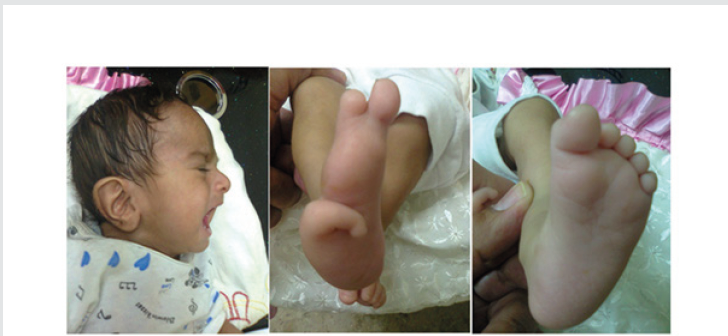
Figure 1: The girl had low set ears and deformity of the right foot with the presence of only three toes. There was no obvious abnormality of left foot, but the big toe was relatively large.

Lupinepublishers-openaccess-Surgery-Casestudies

R.J was first seen at about the age of four months during November 2018 because of poor feeding, failure to thrive, poor response to sounds, and poor head control. The girl also had low set ears, and deformity of the right foot with the presence of only three toes. There was no obvious abnormality of left foot, but the big toe was relatively large (Figure1). She was delivered at 38 weeks by cesarean section. She didn’t pass motion and was found to have imperforated anus. She had colostomy, and the surgeon reported that the sigmoid was not present. The five-centimeter colon ended at the pelvis, and cecum found on the left side. Cloaca treated with diversion colostomy. The parents were relatives and have three normal children. Echocardiography performed during the first month showed normal findings. Brain ultrasound performed on the fifth of August 2018 showed normal findings. Abdominal ultrasound was also performed on the fifth of August 2018 and showed small hypoplastic right kidney (18 x 12 mm) with normal shape. At the age of forty-six days (16, August 2018), a second abdominal ultrasound showed small hypoplastic right kidney. The left kidney had normal size.

Discussion

Authors from Germany, the Netherlands, the UK, the USA, Belgium, Italy , Switzerland and the Czech republic (Jürgen Kohlhase et al ,1998; Jürgen Kohlhase et al ,1999) defined Townes Brocks syndrome as a rare autosomal dominant malformation syndrome with a combination of anal, renal, limb and ear anomalies. Townes Brocks syndrome is a very rare genetic syndrome with 129 welldocumented patients reported in the medical literature [1,2]. In this paper the first case of this syndrome in Iraq is reported which is the case number 130.

Read More About Lupine Publishers Journal of Surgery Please Click on Below Link:
https://surgery-casestudies-lupine-publishers.blogspot.com/

Thursday, 4 May 2023

Lupine Publishers | Multidisciplinary Management of Elderly Cancer Patients: The Radiation Oncologist’s Point of View

 Lupine Publishers | Journal of Surgery & Case Studies


Abstract

Oncological treatments of elderly patients are extremely complex; so far there’s no agreement even on the definition of “geriatric patient”. From the point of view of global health, the problem is of the outmost importance as the number of older patients will increase dramatically in the next years, leading to a change in world epidemiology with a significant increase of chronic-degenerative diseases such as cancer. For this reason, it’s mandatory to provide clinical oncologists with multidisciplinary algorithms aiming at the best treatment of older cancer patients.

Materials and Methods

The complexity of oncological treatments for elderly patients starts from the very beginning, since the definition of “geriatric patient” is not univocal. From the point of view of public health, the problem is of the outmost importance. since nowadays there are 600 million people over 65, in 10 years their number will overcome 1 billion and in 2050 there will be nearly half billion people over 80 [1-3]. Such an increase of longevity will lead to a change in world epidemiology with a significant increase of non-communicable disease such as cancer. In 2030 the annual incidence of new cases of cancer in aged people will be 13,7 million and, which is even more important, nearly half of such cases will be in low-income countries [4]. Very few clinical trials (which are the cornerstone of Oncology) take older patient into account, and usually they are very selected cases [5-8], quite differently from every day’s practice, so there’s paucity of data about care of older patients, which makes clinical oncologist’s task even harder [5].Moreover, at least till few years ago, most guidelines and recommendations only consider chronological age in order to determinate the treatment’s choice, and this policy has led to over- or under-treatments [5]. With the purpose of optimizing the treatment’s choice it is imperative to focus on the concept of biological or functional age, in opposition to merely chronological age [6,7].

We should briefly review the physiological modifications caused by aging, which affects every organ and apparatus. Considering for instance Nervous System, there is a decrease of cortical volume and of synaptic density, which leads to a weaker memory and attention. Cardiovascular system is strongly affected, with a diminished cardiac output, increased arterial stiffness, slower modulation of cardiac frequency etc. [9]. Osteo-muscular apparatus is involved too, with a decrease of bone density leading to an increased risk of fracture and a sarchopenia which causes decreased physical activity with parallel increased fatigue and asthenia [10]. Additionally, for most of oncologic treatments, liver and kidney’s function is crucial; with their reduction, drug toxicity increases. In some aged patients an aforementioned change is plain; in other they can be silent in conditions of balance, becoming evident in stress situations such as a malignant disease and its treatments [1].

Biomolecular Markers of Aging

Aging is an extremely complex phenomenon, showing deep differences among individuals, consequently so far, it’s difficult to identify biological markers which enable us to divide subjects of the same chronological age into different functional ages. Several markers have been suggested, starting with markers of systemic inflammation such as CRP, D-dimer, IL 6 [8]. They are easily quantifiable, and they’ve been associated with functional decline in aged people, but their levels are influenced also by frankly pathological conditions like infections and cancer itself [11,12]. Markers of cellular aging have been considered too, such as telomeres or cell cycle components [8]. Dosing such markers is anyway extremely expensive, and moreover they have a significant interindividual variability. Another marker which could document a link between cancer and aging is P16 INK 4A, which has been showed to increase in aged breast cancer patients receiving chemotherapy [13]. Nevertheless, all of these markers are, so far, not completely validated and reliable.

Geriatric Assessment

As long as validated and reliable biomarkers are not established, the best way to assess a geriatric cancer patient is clinical evaluation [14-17]. Geriatric assessment is a multidisciplinary and multi parametric evaluation which takes into account physical aspect, nutritional status, neurological and cognitive status and even social support [18,19].

Comorbidities

When planning an oncological treatment at any age it’s mandatory to take into account comorbidities. This is mainly true in aged patient, beginning from the commonest pathologies such as cardiovascular diseases, diabetes, chronic renal failure, collagenopathies [20].

Polypharmacy

About 50% of aged patients are on 5 or more different medical therapies before undertaking an oncologic treatment, so it’s mandatory to evaluate all of these therapies and their potential interaction\interference with anticancer therapy [21].

Nutritional Status

Malnutrition and weight loss are deeply connected with cancer and its treatments and they have been shown to be linked to increased risk of toxicity and mortality [22].

Functional Status

All oncologists are familiar with ECOG and KPS scoring systems. In aged cancer patients it’s appropriate to integrate them with other evaluation systems [5,8] such as ADL (Activities of Daily Living) and IADL (instrumental activities of daily living). For instance, an extremely simple and reliable indicator of functional status is the number of falls. They are seldom taken into account, but they seem to be connected with oncological treatments’ toxicity.

Cognitive Status

The risk of cognitive decline increases with age. During anticancer treatment, it can cause for instance a diminished comprehension of its side effects which can be communicated with a delay, increasing the toxicity of the treatment itself, even in a serious way [1].

Psychological Status

Anxiety and depression worsen quality of life and precipitate functional decline, with a lower adherence to therapies [23].

Social Support

Many older people, so even aged cancer patients, live alone. It’s been documented that social isolation is linked to a significant higher mortality in cancer patients [24].

Screening Tools

Many screening tools have been validated with the purpose of identifying aged cancer patients who can take advantage of a multidimensional geriatric assessment [25,26]. Among the commonest ones we mention G8 and Vulnerable Elderly Survey 13 [18]. The final result of these screening and of the subsequent geriatric evaluation is the final decision to perform an oncological treatment (and its intensity) or not [7]. Sometimes it could be appropriate not to perform a treatment with curative purpose in an aged patient because of multiple comorbidities (which could lead to increased toxicities and a reduction in life expectancy). In other circumstances, on the opposite side, it could be an error not to undertake a treatment only because of chronological age. Patient’s preferences must be taken into account too; main international guidelines recommend to including patient in the therapeutic decision [27]. In this setting we must insert geriatric assessment; it’s been documented in literature that multidimensional geriatric evaluation has lead to significant changes in treatment planning, in most of cases with the aim of attenuate it [28]. At the present a multidimensional geriatric assessment is not often performed in the process of decision making regarding oncologic treatment of older patients. It’s been shown anyway that older cancer patients who have been evaluated in such a way have completed their treatment in a significant higher percentage, and with less modifications, compared to those who haven’t received it.

Radiotherapy

radiotherapy is the clinical discipline which aims at curing cancer by means of ionizing radiations; it could be employed as the sole therapeutic modality or in association with surgery and \or systemic therapies. [29,30]. A geriatric evaluation is strongly advisable for older patients who are candidates to radiotherapy, first of all for those treatments which consider its association with a systemic therapy, but also for the exclusive setting. Around 70% of cancer patients will require a radiation treatment, and this is especially true in older patients, as state of art radiotherapy techniques offer higher cure rates with less side effects. Moreover, treatment time can be reduced, and this can help patient with logistic difficulties (e.g. distance from radiotherapy facility) and their family\caregivers. This is true first of all in the palliative setting (e. g. Treatment of pain from bone metastases), but it could be accomplished even in the non-palliative setting, with the adoption of shortest scheduled.

Conclusion

The first dilemma of radiation and medical oncologists treating aged patients is how to decide if a patient is suitable for a given treatment and whether to treat patients with standard protocols or with adapted regimens. So, it’s advisable to include a geriatrician in multidisciplinary oncological teams (Tumour Board).

If it’s not possible, a good result can be achieved even with a conventional geriatric evaluation and a higher cooperation among specialists. Anyway, even after an effective evaluation has declared that an older patient is fit enough to undergo an oncological treatment, it’s mandatory to monitor such a patient in a closer and stricter way compared to a younger one [31].

Read More About Lupine Publishers Journal of Surgery Please Click on Below Link:
https://surgery-casestudies-lupine-publishers.blogspot.com/

Friday, 10 March 2023

Lupine Publishers | Inguinal Hernias Repair by Laparascopy. Repair of Inguinal Hernias by Laparascopy

 Lupine Publishers | Journal of Surgery & Case Studies


Abstract

Introduction: A series of 78 laparoscopic hernioplasties performed in the General Teaching Hospital “Enrique Cabrera”. Objective: To determine perioperative events, surgical complications and the evaluation of the pain referred by the operated patients.

Methods: Between January 2012 and December 2018, 78 hernioplasties were performed laparoscopic in 60 patients; 18 had bilateral inguinales hernias. He collected the variables: age, sex, type of hernia, perioperative events and complications, and a pain scale was applied. A database was filled and processed statistically.

Results: The male sex predominated in a 5:1 ratio, the surgical time average was 53.5 minutes for unilateral hernias and 71.3 minutes for the bilateral ones. The most frequent complication in the transoperative period was bleeding lower in 27.0%, and in the postoperative period the hematoma was in 15.3%, it recurred two hernias (2.5%). At 15 days after surgery, 93.3% of theoperated did not complain of pain, but the social and labor reintegration was of only 34% of patients.

Conclusion: laparoscopic inguinal hernioplasty is a therapeutic option more, mainly in patients with bilateral and reproduced hernias.

Keywords:Laparoscopic Hernioplasty; Inguinal Hernia; Hernia Recurrence

Introduction

Since the concept of endoscopic inguinal hernia repair was first described by Ger R [1] in 1982, the endoscopic techniques are gone modifying, going through a time when failures and complications -united to high cost-exceeded initial enthusiasm [2]. Laparoscopic hernioplasty (HL) has been gaining popularity in the last decade, and numerous controlled studies appear in the literature comparing the laparoscopic techniques with conventional techniques [3-7]. In recent years, HL, despite consolidated as a therapeutic option to consider. The advantages of this have been demonstrated method in bilateral hernias, relapsed and in the active labor subject, that requires a precocious labor reintegration [5-7].

Methods

Between June 2012 and June 2018, a prospective descriptive study of Longitudinal section of 60 patients operated by hernia endoscopy of the region inguinal, in the Department of Surgery of the General Teaching Hospital “Enrique Cabrera”. The inclusion criteria were: - Patients who agreed with the type of surgical intervention and the study, and They gave their informed consent. Patients older than 30 years classified ASA I-III, without contraindications anesthetics for laparoscopic interventions.

a) Patients classified as Nyhus III and IV. Exclusion criteria.

b) Patients with previous surgical wounds in the inguinal region to operate, not dependent on inguinal hernias reproduced.

c) Inguinal hernias complicated, irreducible or slipped.

The surgical techniques were: laparoscopic inguinal hernioplasty completely extraperitoneal (TEP) of total extraperitoneal English and inguinal hernioplasty laparoscopic transabdominal preperitoneum (TAPP) preperitoneal), and one or the other was performed, at the discretion of the main surgeon. The TEP technique was executed with some variants such as: not using the trocar balloon, the preperitoneal space was decoloured by means of the 0º laparoscope, and the insufflation of CO2 at 13 mmHg. In patients with large herniated rings, placed a polypropylene cone in the hernia defect and then a 15 x 12 cm polypropylene prosthesis. There was no need to fix with clips the tights. The TAPP technique was performed on a regular basis [8]. In the immediate postoperative period, the scale of visual pain analog scale was applied (VAS), [9] and a value was assigned to pain through “caritas”, which starts very cheerful (value I) until very sad (value X). The quantification of pain was repeated in consultation at 7, 15 days and one month after surgery.

Results

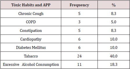
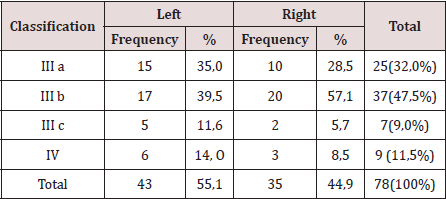
There were operated 78 hernias in 60 patients (18 patients [30.0%] suffered from bilateral hernias, 69 primary hernias and 9 reproduced hernias). The average of age was 55.6 years, the youngest patient was 30 years and the oldest was 77 years, but the majority (12 patients) were in the fifth decade of life. The male sex predominated in 82.9%, which represented a relationship man / woman of 5: 1. 42.9% of patients performed large efforts habitual physicists. The pathological history of the patients (Table 1). It is observed that 24 patients (40.0%) consumed tobacco, and in 11 an excessive consumption of alcohol was collected 18.3%. COPD: Chronic Obstructive Pulmonary Disease (Table 2). The distribution of the series according to the Nyhus classification. Right hernias predominated (55.1%), the indirect variety with large dilation of the ring and destruction of the posterior wall (IIIb) was the most frequent (37 hernias), and 7 femorales hernias and 9 recurrent hernias were operated 73 PET (93.6%) and 5 TAPP (6.4%) were performed. Two of the patients in whom a TEP technique was started were converted to a conventional prosthetic technique by accidental perforation of the peritoneum, passing the CO2 into the peritoneal cavity, and consequently, the loss of the preperitoneal surgical space. Of the 5 TAPP repairs, 3 of them were in the course of a laparoscopic cholecystectomy, and another was the conversion of a failed PET technique. The average surgical time of unilateral hernias was 53.5 min, with a minimum of 25 min and a maximum of 120 min. In bilateral repairs, the average surgical time was 71.3 min, and a minimum of 40 and a maximum was observed. of 110 minutes.

Table 1: Toxicos habits, and personal phalogical history (APP).

lupinepublishers-openaccess-surgery-case-studies-journal

Table 2: Distribution According to the Nyhus Classification.

lupinepublishers-openaccess-surgery-case-studies-journal

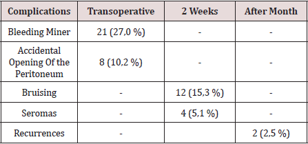
The hospital stay was less than 24 h in 50 patients (71.4%), in 5 it extended from 24 to 48 h, and in 5 to more than 48 hours. The most frequent complication in the transoperative period (Table 3) was minor bleeding in 21 repairs (27.0%) that gave rise to 12 hematomas (15.3%). No complications were observed after the second week, but two patients suffered recurrences (2.5%), more than two months after surgery. The application and evaluation of the VAS scale (Table 4). In the immediate postoperative period, after the patient recovered from anesthesia, 56 individuals (93.3%) were classified as VAS I, and 4 as VAS II. Twenty-four hours after surgery, 14 patients (23.3%) were classified as VAS I, 40 (66.6%) as VASII, 4 patients as VAS III and 2 VAS IV. In the consultation of the first week of postoperatively, 42 patients (70.0%) were classified as VAS I and [10] as VAS II, and two patients with moderate pain (VAS V) appeared in this period. Fifteen days after surgery, 56 individuals (93.3%) were VAS I and a month were 58 (96.6%). The incorporation to the usual activities, including work, was 3 patients a week after surgery, at 15 days they were 19 patients (31.6%) and 54 patients a month (90.0%).

Table 3: Complications.

lupinepublishers-openaccess-surgery-case-studies-journal

Table 4: Evaluation of the Visual Analogy Scale (VAS).

lupinepublishers-openaccess-surgery-case-studies-journal

Discussion

Currently, with the improvement of laparoscopic techniques, these are outlined as safe, reproducible and as a therapeutic option regardless of the age of the patient; nevertheless, the evaluation of the individual must be correct and meticulous in the preoperative period, specifically cardiorespiratory function, since with the TEP method a working space is created between the sheets of the crosssection sheet, richly vascularized, so that absorption and elimination of CO2 is greater than that produced in the peritoneal cavity during the performance of the pneumoperitoneum [10]. Although men predominated, there was a slight increase in women in the series with respect to other authors, [3-5] which could have been due to the inclusion in the study of 3 women who underwent the diagnosis of inguinal hernia, in the course of a laparoscopic cholecystectomy. In laparoscopic practice, it is not uncommon finding of hernia defects diagnosed in the transoperative, in men and women, the latter basically with a history of gynecological disorders. Although the usefulness of hernia repairs in asymptomatic patients is questioned in some articles, [11,12] the authors consider that it would be beneficial for the patient, if conditions permit, the repair of the hernia defect by the TAPP method. The relationship between the hernia disease and the physical efforts, is classic from the Cooper era [1]. In the series, 68% of the patients performed physical activities involving large and medium efforts, and also analyzing the multifactorial character in the pathogenesis of hernia disease, was striking, that approximately half of the patients operated on were smokers, a factor that influences the metabolism of collagen, significantly linked to hernia recurrences [13]. The majority of the repairs were by means of the PET technique, and we consider, as other authors [10,14 -19], that although the TAPP technique brings us closer to the area from a family perspective to the surgeon (peritoneal cavity) and facilitates the so-called “learning curve”, the hernial disease - considering it a parietal defect- should be given solution from this same plane to avoid the likelihood of serious complications of intrabdominal , and to leave the transperitoneal method as a tactical resource when the totally extraperitoneal method is unsuccessful.

The average surgical time was similar to other series [3-6]. It is known that this tends to decrease when the surgical team gains experience [16]. The largest surgical time recorded was in a patient, who was started with a PET technique, but Due to technical difficulties, it was converted to a conventional posterior repair. The fundamental complications were in relation to minor bleeding in the transoperative period and postoperative hematomas. In 3 patients it was necessary to drain the hematoma due to the discomfort caused, however, in the rest of the patients with hematomas and seromas they were treated with conservative measures. In two patients, the recurrence occurred 2 months after surgery, which was interpreted as a technical error. Our results coincide with numerous studies [3-7], that indicate the least postoperative pain of the minimum access techniques, as well as a prompt social and labor reincorporation of the patients. Despite the fact that 70% and 93.3% a week and 15 days postoperatively, respectively, had no pain or discomfort were minimal, only [18] individuals (30%) were incorporated into their usual activities before 15 days. These results contrast with other studies that report a return to work and social activities between 10-15 postoperative days, 4-17- although it is likely that some sociocultural factors are influencing these results. It can be concluded by noting that laparoscopic inguinal hernioplasty is another therapeutic option, mainly in patients with bilateral and reproduced hernias. In the series there were no major transoperative or postoperative complications, only minor bleeding and bruising were present. Most patients were not afflicted by pain 2 weeks postoperatively, however, return to social and labor activities after 15 days was low [18-20].

Read More About Lupine Publishers Journal of Surgery & Case Studies Please Click on Below Link:
https://surgery-casestudies-lupine-publishers.blogspot.com/

Friday, 20 January 2023

Lupine Publishers | Endometriosis Involving Colon and Rectum: A Literature Review and Laparoscopic Management

 Lupine Publishers | Journal of Surgery & Case Studies


Abstract

Introduction: Endometriosis is characterized by the presence of functional endometrial tissue consisting of glands and/ or stroma located outside the uterus [1], although implanted ectopically, this tissue presents histopathological and physiological responses that are similar to the responses of the endometrium [2].

Clinical Features: Endometriosis usually becomes apparent in the reproductive years when the lesions are stimulated by ovarian hormones. Forty percent of the patient’s present symptoms in a cyclic manner, which are usually related with menses Pelvic pain, infertility and dyspareunia are the characteristic symptoms of the disease, but the clinical presentation is often non-specific [1].

Diagnosis and Investigations: A precise diagnosis about the presence, location and extent of rectosigmoid endometriosis is required during the preoperative workup because this information is necessary in the discussion with both the colorectal surgeon and the patient. Furthermore, almost all patients with intestinal endometriosis have lesions in multiple pelvic locations and it is difficult to know what symptoms are caused by the intestinal disease versus the pelvic disease.

Treatment: Treatment must be individualized, taking the clinical problem in its entirety into account, including the impact of the disease and the effect of its treatment on quality of life. Pain symptoms may persist despite seemingly adequate medical and/ or surgical treatment of the disease. In such circumstances, a multi-disciplinary approach involving a pain clinic and counselling should be considered early in the treatment plan.

Endometriosis is characterized by the presence of functional endometrial tissue consisting of glands and/ or stroma located outside the uterus [1], although implanted ectopically, this tissue presents histopathological and physiological responses that are similar to the responses of the endometrium [2].

Prevalence and Epidemiology

The reported prevalence of endometriosis is 1%-20% in asymptomatic women, 10%-25% in infertile patients and 60%- 70% in women with chronic pelvic pain [1]. Endometriosis is a common benign disease among women of reproductive age and affects the intestinal tract in 15%-37% of all patients with pelvic endometriosis [3]. Multiple births and extended intervals of lactation decrease the risk of being diagnosed with endometriosis, whereas nulliparity, early menarche, frequent menses, and prolonged menses increase the risk [4]. Endometriosis also appears to be associated with a taller, thinner body habitus and lower body mass index [5]. The prevalence appears to be lower in blacks and Asians than in Caucasians [6]. Growth and maintenance of endometriotic implants are dependent upon the presence of ovarian steroids. As a result, endometriosis occurs during the active reproductive period: women aged 25 to 35 years [6]. Other factors that appear to play important roles in determining if a woman will develop the clinical condition include [7]:

a) Reproductive lifestyle, especially a delay in childbearing

b) Poorly understood immunological factors

c) Some environmental factors, probably including exposure to a range of environmental toxins

d) Reproductive tract occlusion, such as an imperforate hymen.

Pathogenesis

Endometriosis is a common disease of unknown etiology. Many theories have been proposed to explain this condition: retrograde menstruation theory, metaplastic, transformation, the migration of cells through the lymphatic system or via hematogenous spread, Iatrogenic during CS. However, other factors, immunological, genetic and familial, could be involved in the pathogenesis of this disease [1].

Sampson’s Theory of Retrograde Menstruation

The implantation theory proposes that endometrial tissue passes through the fallopian tubes during menstruation, then attaches and proliferates at ectopic sites in the peritoneal cavity. Recent studies using laparoscopy have demonstrated that retrograde menstruation is a nearly universal phenomenon in women with patent fallopian tubes. Classic studies performed in the 1950s demonstrated viability of sloughed endometrial cells and the capacity to implant at ectopic sites. Patients with mullerian anomalies and obstructed menstrual flow through the vagina may have an increased risk of endometriosis. The anatomic distribution of endometriosis also provides evidence for Sampson’s theory [8].

Coelomic Metaplasia Theory

The theory of coelomic metaplasia proposes that endometriosis may develop from metaplasia of cells lining the pelvic peritoneum. Iwanoff and Meyer are recognized as originators of this theory. A prerequisite of the coelomic metaplasia theory is that mesothelial cells lining the ovary and pelvic peritoneum contain cells capable of differentiating into endometrium. An attractive component of the coelomic metaplasia theory is that it can account for the occurrence of endometriosis anywhere mesothelium is found. This includes reports of endometriosis occurring in the pleural cavity. Pleural endometriosis could result from local metaplasia of pleural mesothelium. On the other hand, it could also result from transdiaphragmatic passage of peritoneal fragments of endometrium as well as vascular metastasis of endometrium. Coelomic metaplasia is thought to account for the rare occurrences of endometriosis reported in males. In these reports of endometriosis, the men were all undergoing estrogen therapy. Although coelomic metaplasia was a possibility, estrogen stimulation of mullerian rests could not be excluded. Likewise, the occurrence of endometrial carcinoma in males is thought to possibly arise from mullerian remnants. Still, further support for the coelomic metaplasia theory may be found in the study of benign and malignant epithelial ovarian tumors. Both are considered to be derivatives of germinal epithelium. The presence of ovarian surface endometriosis could be accounted for by this type of transformation [8].

Induction Theory

The induction theory is an extension of the coelomic metaplasia theory. This theory proposes that menstrual endometrium produces substances that induce peritoneal tissues to form endometriotic lesions [8].

Embryonic Rests Theory

Von Recklinghausen and Russell are credited with the theory that endometriosis results from embryonic cell rests. These embryonic rests, when stimulated, could differentiate into functioning endometrium. As described above, rare cases of endometriosis have been reported in men. Transformation of embryonic rests is a plausible explanation for this phenomenon [8].

Lymphatic and Vascular Metastasis Theories

The lymphatic metastasis theory of endometriosis is often referred to as Hal ban’s theory. He reported that endometriosis could arise in the retroperitoneum and in sites not directly opposed to peritoneum. Sampson had also suggested that endometriosis could result from lymphatic and hematogenous dissemination of endometrial cells. An extensive communication of lymphatics has been demonstrated between the uterus, ovaries, tubes, pelvic and vaginal lymph nodes, kidney, and umbilicus. Metastasis of endometrial cells via the lymphatic system to these areas is therefore anatomically possible. These findings are consistent with a literature review showing a 6.7% incidence of lymph node endometriosis in 178 autopsy cases. Lymphatic and vascular metastasis of endometrium has been offered as an explanation for rare cases of endometriosis occurring in locations remote from the peritoneal cavity. In addition to pleural tissue, endometriosis has been reported in pulmonary parenchyma. Vascular or lymphatic metastasis may also explain cases of endometriosis that have been reported in bone, biceps muscle, peripheral nerves, and the brain [8].

Composite Theory

Javert proposed a composite theory of the histogenesis of endometriosis which combines the implantation, vascular/ lymphatic metastasis, as well as a theory of direct extension of endometrial tissue through the myometrium. Along similar lines, Nisolle and Donnez have recently argued that the histogenesis of endometriosis depends on the location and ‘type’ of the endometriotic implant. For example, peritoneal endometriosis can be explained by the implantation theory. Ovarian endometriomas could be the result of coelomic metaplasia of invaginated ovarian epithelial inclusions. Rectovaginal endometriosis, which often resembles adenomyosis, could result from metaplasia of Mullerian remnants located in the rectovaginal septum. These composite theories are attractive in that they recognize a multifaceted mechanism of histogenesis. It seems logical that a disease with such variable manifestations may originate via several mechanisms [8].

Altered Immunity

Alterations in immunologic response to retrograde menstruation have been implicated in the genesis and maintenance of the endometriotic lesion. This defective immunosurveillance may lead to decreased clearance of menstrual debris from the peritoneal cavity and may allow for attachment of ectopic endometrium to peritoneal surfaces. An abnormal immune response could also promote the persistence and growth of ectopic endometrial tissue [8].

The “Neurologic Hypothesis”

It is a new concept in the pathogenesis of endometriosis: There is a close histological relationship between endometriotic lesions of the large bowel and the nerves of the large bowel wall. Endometriotic lesions seem to infiltrate the large bowel wall preferentially along the nerves, even at distance from the palpated lesion, while the mucosa is rarely and only focally involved [9].

Pathology and Sites of Involvement

Sites

Endometriosis can be divided into intra- and extraperitoneal sites. In decreasing order of frequency, the intra-peritoneal locations are ovaries (30%), uterosacral and large ligaments (18%-24%), fallopian tubes (20%), pelvic peritoneum, pouch of Douglas, and gastrointestinal (GI) tract. Extra-peritoneal locations include cervical portio (0.5%), vagina and rectovaginal septum, round ligament and inguinal hernia sac (0.3%-0.6%), navel (1%), abdominal scars after gynaecological surgery (1.5%) and caesarian section (0.5%). Endometriosis rarely affects extraabdominal organs such as the lungs, urinary system, skin and the central nervous system [1]. Endometriosis affects the intestinal tract in 15% to 37% of patients with pelvic endometriosis [10], involvement have been reported from the small bowel to the anal canal, but more frequently the disease involves the rectum and the sigmoid colon (74%), followed by the rectovaginal septumn (12%), cecum (2%), and appendix (3%) . When the ileum is involved, the most common tract is the distal part. A full-thickness involvement of the colonic wall is infrequent since the mucosa is usually spared. One of the classic locations is the anterior rectal wall in the region of the pouch of Douglas. This can be single nodule or can simulate a cancer. Because of the invasive appearance, the disease can be mistaken for cancer [11].

Gross and Microscopic Pathology of Bowel Endometriosis

The appearance and size of the implants are quite variable. Areas of endometriosis appear as raised flame-like patches, whitish opacifications, yellow-brown discoloration, translucent blebs, or reddish or reddish-blue irregularly shaped islands. The peritoneal surface may be scarred or puckered.

The Microscopic Appearance

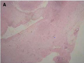
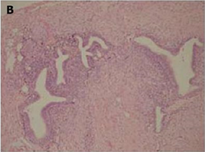
Of endometriotic tissue is similar to that of endometrium in the uterine cavity; the two major components of both are endometrial glands and stroma. Unlike endometrium, however, endometriotic implants often contain fibrous tissue, blood, and cysts (Figure 1(a) & 1(b)).

Figure 1(a): Low-power image of the colonic wall, with a few endometrial glands and stroma embedded in the muscular layer.
Lupinepublishers-openaccess-surgery-case-studies-journal

Figure 1(b): High-power view of the colonic wall, with endometrial glands and stroma embedded in the smooth muscle of the colon [12].
Lupinepublishers-openaccess-surgery-case-studies-journal

Link to Cancer

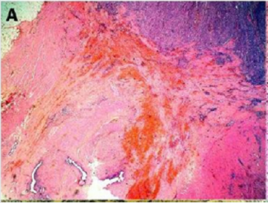
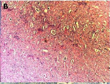
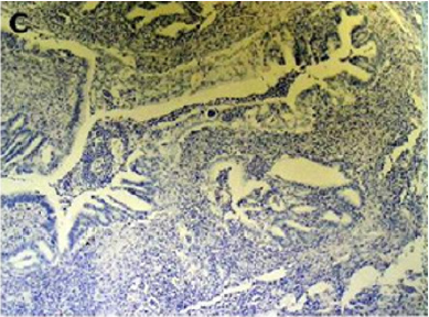
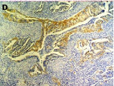
Endometriosis is considered a benign disorder; however, it shares some of the characteristics of malignancy, such as abnormal morphology, deregulated cell growth, cellular invasion, and neoangiogenesis. The glandular epithelium occasionally displays DNA aneuploidy. In vitro evidence suggests that endometriosis may have a monoclonal origin. In addition to being monoclonal, endometriotic deposits showed loss of heterozygosity in 28% of lesions. In 2002, Nezhat et al. with immunohistochemistry, found that alterations in bcl-2 and p53 may be associated with the malignant transformation of endometriotic cysts [12]. The development of a malignancy is a relatively common complication of endometriosis. In fact, several publications have reported malignant neoplasms arising from endometriosis. Most of these publications are case reports or refer to a small series of patients presenting either ovarian carcinomas with associated endometriosis or invasive endometrioid adenocarcinomas involving adjacent pelvic structures. Malignant transformation of extraovarian endometriosis, including the intestinal tract, however, has not been reported as frequently. The largest reported series of neoplastic changes in gastrointestinal endometriosis includes 17 cases [10] (Figure 2(a-d)). Some studies suggest that the development of malignancies may occur in up to 5.5 % of female patients with endometriosis. Only 21.3% of the cases arise from extragonadal pelvic sites, and endometriosis-associated intestinal tumors are even rarer. Malignant transformation of primary gastrointestinal endometriosis without pelvic involvement is uncommon, and its real incidence is unknown. It can mimic a primary gastrointestinal neoplasm. Most of these neoplasms are carcinomas, but sarcomas and müllerian adenosarcomas have also been described. Petersen et al, in a large review of the previously published endometrioid adenocarcinomas arising in colorectal endometriosis, report less than 50 cases of neoplastic transformation, 22 of which were adenocarcinomas. The others included sarcomas and mixed müllerian tumors. The progression to invasive cancer has been related with hyperestrogenism, either of endogenous or of exogenous origin. A possible genetic background favoring the onset of cancer has been reported in some patients without hyperestrogenism and with a family history of cancer. The anatomic distribution and frequency of these cancers parallel the occurrence of which benign endometriosis is found at various sites. In order to classify a malignancy as arising from endometriosis, strict histopathologic criteria need to be fulfilled. Sampson first proposed these criteria in the year 1925. He suggested that the following should be fulfilled:

Figure 2(a): Rectal endometriod adenocarcinoma with adjacent focus of endometriosis (hematoxylin-eosin, 20x).
Lupinepublishers-openaccess-surgery-case-studies-journal

Figure 2(b): Rectal endometriod adenocarcinoma endometriosis (hematoxylin-eosin, 100x).
Lupinepublishers-openaccess-surgery-case-studies-journal

Figure 2(c): Cytokeratin 20 immunostaining negative (100x).
Lupinepublishers-openaccess-surgery-case-studies-journal

Figure 2(d): Cytokeratin 7 immunostaining positive (100x). [10]
Lupinepublishers-openaccess-surgery-case-studies-journal

a) the presence of both malignant and benign endometrial tissue in the same organ.

b) the demonstration of cancer arising in the tissue and not invading it from elsewhere.

c) the finding of tissue resembling endometrial stroma surrounding characteristic glands.

Years later, Scott suggested an additional qualification to complete Sampson’s criteria: the demonstration of microscopic benign endometriosis contiguous with the malignant tissue [10]. Endometriosis and its possible malignant changes should be taken into account in the differential diagnosis of intestinal masses in females. Also, clinical suspicion for malignancy should be aroused in patients with abdominal pain or rectal bleeding and a previous history of quiescent endometriosis. Recognition of these lesions is important because of the different management required by primary gastrointestinal neoplasms and by those arising from endometriosis. These differences may have significant clinical implications [10].

Clinical Features

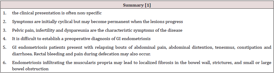
Endometriosis usually becomes apparent in the reproductive years when the lesions are stimulated by ovarian hormones. Forty percent of the patient’s present symptoms in a cyclic manner, which are usually related with menses Pelvic pain, infertility and dyspareunia are the characteristic symptoms of the disease, but the clinical presentation is often non-specific [1]. Symptoms are initially cyclical but may become permanent when the lesions progress. It is difficult to establish a preoperative diagnosis of GI endometriosis, because GI tract symptoms can mimic a wide spectrum of diseases, including irritable bowel syndrome, infectious diseases, ischemic enteritis/colitis, inflammatory bowel disease and neoplasm. GI endometriosis patients present with relapsing bouts of abdominal pain, abdominal distention, tenesmus [1], constipation and diarrhoea. Rectal bleeding and pain during defecation may also occur. Endometriosis infiltrating the muscularis propria may lead to localized fibrosis in the bowel wall, strictures, and small or large bowel obstruction. The true incidence of endometriosis causing bowel obstruction is unknown, although complete obstruction of the bowel lumen occurs in less than 1% of cases. Endometriosis of the distal ileum is an infrequent cause of intestinal obstruction, ranging from 7% to 23% of all cases with intestinal involvement. The incidence of intestinal resection for bowel obstruction is 0.7% among patients undergone surgical treatment for abdominopelvic endometriosis [1]. Rectal bleeding may be caused by mucosal injury during the passage of stools through a stenosed colon with the intramural endometriotic tissue increased at the time of menses if it occurs. Colonic mucosa heals rapidly, and no signs are detectable at endoscopy [1] (Table 1).

Table 1:
lupinepublishers-openaccess-surgery-case-studies-journal

Differential diagnosis [1]

a) irritable bowel syndrome,

b) infectious diseases,

c) ischemic enteritis/colitis,

d) inflammatory bowel disease

e) neoplasm

f) Other causes of intestinal obstruction (Acute/chronic, small/large bowel)

Diagnosis and Investigations

A precise diagnosis about the presence, location and extent of rectosigmoid endometriosis is required during the preoperative workup because this information is necessary in the discussion with both the colorectal surgeon and the patient. Furthermore, almost all patients with intestinal endometriosis have lesions in multiple pelvic locations and it is difficult to know what symptoms are caused by the intestinal disease versus the pelvic disease. In particular, in the case of sigmoid endometriosis, the lesion cannot be suspected at clinical examination, which is why sigmoid endometriosis is often diagnosed only during surgery. Although several radiological techniques have been proposed for the diagnosis of bowel endometriosis, data are inconclusive, and no gold standard is currently available [13].

Colonoscopy

Although endoscopic diagnosis of colonic endometriosis has been reported, the mucosa is usually normal or shows minimal mucosal abnormalities, friability, extrinsic process or fibroses stenoses [1]. Endoscopic biopsies usually yield insufficient tissue for a definitive pathologic diagnosis as endometriosis involves the deep layers of the bowel wall [14]. Endometriosis can induce mucosal changes without any specific pattern, which mimic findings of other diseases such as inflammatory bowel disease, ischemic colitis or neoplasm [1]. Colonoscopy is helpful to rule out colorectal malignancy [11].

Double Contrast Barium Enema

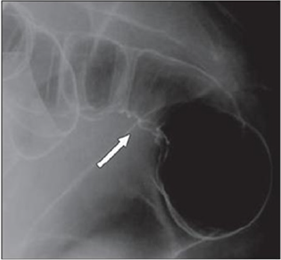
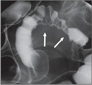
Radiologically, lesions of endometriosis are either of constricting and polypoid type or both. On barium studies, radiographic findings caused by implants in the ileum are similar to those in the colon. Rectosigmoid or cecal endometriosis on double contrast barium enema studies is seen as an extrinsic mass with speculation and tethering of folds [1]. Shortening or flattening of the bowel wall, crenulation of the mucosa, or a combination of these factors [15], Double-contrast barium enema may be effective in determining the precise location of the endometriotic nodules, but it cannot clearly demonstrate the depth of parietal involvement. Furthermore, the experience of the radiologist in the diagnosis of bowel endometriosis remains a critical limit of this technique [13] (Figures 3 & 4(a & b)).

Figure 3: Thirty-four years old woman with suspected intestinal implants of endometriosis. A and B, Lateral A and oblique B spot images show three endometriotic lesions exhibiting extrinsic mass effect with crenulation of contour and speculation that are direct signs of infiltration of bowel wall (arrows). Small polypoid lesion (arrowhead) is benign tubular adenoma confirmed at surgery [15].

Lupinepublishers-openaccess-surgery-case-studies-journal

Figure 4(a): Twenty-eight years old woman with suspected intestinal implants of endometriosis and finding of rectal localization of intestinal endometriosis. DCBE image shows extrinsic mass effect and speculation (arrow) of rectal wall that appears infiltrated [15].
Lupinepublishers-openaccess-surgery-case-studies-journal

Figure 4(b): Twenty-three years old woman with suspected intestinal implants of endometriosis. DCBE examination showing pathologic pelvic process involving bowel serosa at rectosigmoid junction. Finding of extrinsic mass effect and speculation (arrows) owing to poor wall distention after air insufflation suggesting wall infiltration [15].
Lupinepublishers-openaccess-surgery-case-studies-journal

Transvaginal Us

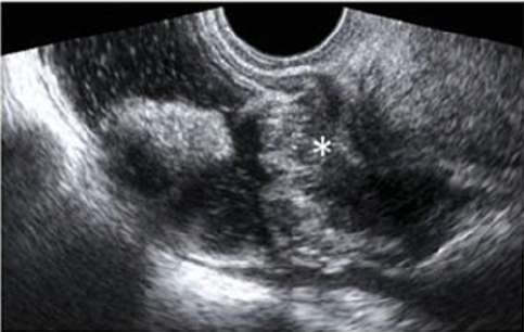
Transvaginal ultrasonography can be useful not only in the first-line exploration of the pelvic cavity, but also in diagnosing rectosigmoid endometriosis. However, relevant limitations of transvaginal ultrasonography consist in the impossibility of determining the exact distance of rectal lesions from the anal margin and of evaluating precisely the depth of rectal wall involvement. In addition, locations above the rectosigmoid junction might be beyond the field of view of a transvaginal approach and limited by the presence of air for a transabdominal approach [15]. Transvaginal us combines with rectal water contrast is more accurate than TVS in diagnosing rectal infiltration reaching at least the muscularis propria in women with rectovaginal endometriosis. However, this exam cannot determine whether the infiltration reaches the rectal submucosa. RWC-TVS may be more painful than TVS, therefore it could be used when TVS cannot exclude the presence of rectal infiltration in women with rectovaginal endometriosis [15] (Figure 5).

Figure 5: A large rectovaginal nodule infiltrating the bowel muscularis (indicated by the asterisk) demonstrated by Rectal Water Contrast- Transvaginal Sonography (RWC-TVS) [16].
Lupinepublishers-openaccess-surgery-case-studies-journal

CT & MSCT

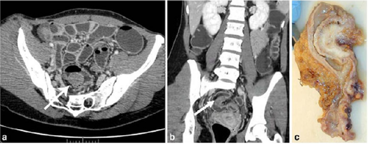
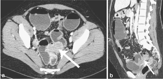
CT is not the primary imaging modality for evaluation of bowel endometriosis, although it can occasionally demonstrate a stenosing rectosigmoid mass. Multislice CT (MSCT) has a great potential for detecting alterations in the intestinal wall, especially if it is combined with enteroclysis (MSCTe). Biscaldi et al carried out a study on 98 women with symptoms suggestive of colorectal endometriosis and MSCTe identified 94.8% of bowel endometriotic nodules [1]. Biscaldi et al reported the usefulness of multislice CT combined with distention of the colon by rectal enteroclysis for bowel endometriosis. The sensitivity was 98.7% and specificity was 100% in identifying women with intestinal endometriosis. This method is thought to be very helpful for diagnosing intestinal endometriosis, but requires bowel preparation, such as the need for a low-residue diet for 3 d, drinking of 4-6 doses of a granular powder dissolved in 500 mL of water per dose and intravenous administration of iodinated contrast medium. This technique is thus inappropriate for patients with obstructive symptoms or allergy to iodinated contrast medium [3] (Figures 6 & 7).

Figure 6: Endometriotic nodule infiltration the muscular layer, A: Axial MSCTe image of the abdomen, the arrow indicates the endometriotic nodule. The lesion is enhanced, and it infiltrates the bowel wall involving the muscular layer. B: Coronal reconstruction demonstrating the extension of the sigmoid endometriotic nodule (indicated by the arrow) C: Formaldehydefixed resected bowel segment, the endometriotic nodule of the sigmoid colon was previously demonstrated by MSCT [14].
Lupinepublishers-openaccess-surgery-case-studies-journal

Figure 7: Endometriotic nodule infiltration the muscular layer, A: Axial MSCTe image of the abdomen, the arrow indicates the endometriotic nodule. The lesion is enhanced, and it infiltrates the bowel wall involving the muscular layer. B: Coronal reconstruction demonstrating the extension of the sigmoid endometriotic nodule (indicated by the arrow) C: Formaldehydefixed resected bowel segment, the endometriotic nodule of the sigmoid colon was previously demonstrated by MSCT [14].
Lupinepublishers-openaccess-surgery-case-studies-journal

Magnetic Resonance Imaging (MRI)

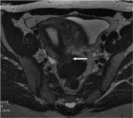
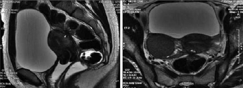
Magnetic resonance imaging (MRI) has a high sensitivity (77%- 93%) in the diagnosis of bowel endometriosis. The depth of rectal wall infiltration by endometriosis is poorly defined by MRI. A combination of MRI and rectal endoscopic ultrasonography (EUS) has recently been proposed. When retroperitoneal infiltration is present, it is mandatory to know if the bowel wall is involved in order to identify patients requiring bowel resection. Both rectal EUS sensitivity and negative predictive value range from 92% to 100%. The specificity and positive predictive value are rather poor, which are 66% and 64%, 83% and 94%, respectively, as reported in two different studies [1]. Imaging examination is thus essential for the preoperative diagnosis of intestinal endometriosis, but some reports have described preoperative confusion between this disease and cancer according to colonoscopy and CT with barium enema, particularly in patients with lesions involving the mucosal surface. In such patients, MRI is helpful for differential diagnosis. In a typical endometrial lesion, MRI showed signal hyperintensity on T1-weighted imaging and signal hypointensity on T2-weighted imaging. However, smooth muscle components are reportedly recognized frequently in endometrial lesions. In such lesions, as seen in the present case, MRI indicates signal hypointensity on both T1- and T2-weighted imaging, and differential diagnosis from other diseases such as cancer and gastrointestinal stromal tumor is thus difficult. In fact, Chapron et al reported that MRI specificity for deeply infiltrating endometriosis was 97.9%, but sensitivity was only 76.5% [3] (Figures 8 & 9).

Figure 8: T2- weighted axial view: fecal matter attached to the rectal wall, simulating thickening of the rectal wall [17].
Lupinepublishers-openaccess-surgery-case-studies-journal

Figure 9: T2- weighted sagittal (a) and axial (b) views.

Nodule of the rectosigmoid junction adhering to the posterior surface of the uterus [17].
Lupinepublishers-openaccess-surgery-case-studies-journal

Transrectal EUS

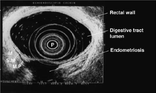
The involvement of the colon is difficult to detect because the implants rarely invade through the intestinal mucosa. For this reason, the rectal ultrasound is of primary importance to assess the rectal involvement [11]. Also, the depth of rectal wall infiltration by endometriosis is poorly defined by MRI. A combination of MRI and rectal endoscopic ultrasonography (EUS) has recently been proposed. When retroperitoneal infiltration is present, it is mandatory to know if the bowel wall is involved in order to identify patients requiring bowel resection [1]. Endoscopic ultrasonography is also a useful and noninvasive examination for the diagnosis of intestinal endometriosis. Sensitivity and specificity are reportedly about 97% for the diagnosis of rectal involvement in patients with known pelvic endometriosis. In addition, EUS-FNAB provides accurate tissue and may be the only procedure for correct preoperative diagnosis of intestinal endometriosis, but the overall specificity, sensitivity and accuracy of EUS-FNA for neoplasms of the gastrointestinal tract are reportedly 88%, 89% and 89%, respectively [3]. Among these examinations, it is considered that MRI and EUS (and/or EUS-FNAB) are the most useful examinations for intestinal endometriosis. However, it is important to perform valuable examinations for diagnosis of intestinal endometriosis, including radiological, histological and etiological examinations, as the condition basically involves a benign lesion requiring minimally invasive treatment [3] (Figure 10).

Figure 10: Rectal endoscopic ultrasonography showing a uterosacral endometriosis nodule (2 cm x 3 cm) with bowel infiltration.

P = probe, M = mucosa, SM = submucosa, MP = muscularis propria [18].
Lupinepublishers-openaccess-surgery-case-studies-journal

7- Serum Markers

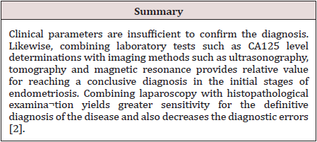
There is a great interest in the use of serum markers to diagnose endometriosis, but they are not sufficiently accurate for use in clinical practice. Cancer antigen CA-125 has been used to monitor the progress of endometriosis [16]. CA19-9 has a lower sensitivity than CA-125, and cytokine interleukin-6 may be more sensitive and specific than CA-125 [1]. Mol et al reported a systematic review of the diagnosis of endometriosis and concluded that serum CA125 level may be elevated in endometriosis, but this measurement had no value as a diagnostic tool compared to laparoscopy [3].

Laparoscopy

Laparoscopy is a primary diagnostic and therapeutic tool providing the opportunity to explore the abdominal cavity and obtain biopsies. The magnified vision enables the surgeon to operate with the best possible exposure. Although it was once believed that intestinal endometriosis was best managed by hormonal regimens or surgical castration, the advent of laparoscopic surgery has dramatically changed this approach [11] (Table 2).

Table 2:
lupinepublishers-openaccess-surgery-case-studies-journal

Treatment

Despite being a gynecologic pathology, deep infiltrating endometriosis is not of exclusive gynecologic concern. A multidisciplinary approach involving urologists and colorectal surgeons therefore is recommended strongly for complete evaluation and correct management. A minimally invasive approach offers convenient advantages concerning the surgical management of multifocal deep infiltrating endometriosis. Traditionally, radical surgery [17] was considered the best measure to prevent disease relapse. However, because of the prevalence of endometriosis among women of reproductive age and the advances in surgical techniques, minimally invasive conservative surgery now is encouraged more [18]. Treatment must be individualized, taking the clinical problem in its entirety into account, including the impact of the disease and the effect of its treatment on quality of life. Pain symptoms may persist despite seemingly adequate medical and/or surgical treatment of the disease. In such circumstances, a multidisciplinary approach involving a pain clinic and counselling should be considered early in the treatment plan. It is also important to involve the woman in all decisions; to be flexible in diagnostic and therapeutic thinking; to maintain a good relationship with the woman, and to seek advice where appropriate from more experienced colleagues or refer the woman to a centre with the necessary expertise to offer all available treatments in a multidisciplinary context, including advanced laparoscopic surgery and laparotomy [19]. The objective of the treatment in pelvic endometriosis is to cease the endometrial stimulus in order to ameliorate the symptoms. Thus, danozol, gonadotropin- releasing hormones, oral contraceptives, and prostaglandin inhibitors can be used. The conclusive treatment of endometriosis is total abdominal hysterectomy, bilateral salpingo-oophorectomy and removal of all endometrial foci. Because malignant transformation cannot be excluded preoperatively and medical treatment may cause fibrosis, the definitive treatment is surgical. Also, in the case of intestinal obstruction and severe rectal and abdominal pain, surgery is indicated. The main objective of surgery is the resection of the affected bowel segment, enabling the histopathological examination of the resection material. Limited surgery, such as excision or cauterization of superficial lesions, following confirmation through frozen section analysis could be performed. In conclusion, intestinal endometriosis is a disease that may imitate various gastrointestinal system diseases. The definite diagnosis could only be done by histopathologic confirmation, since there are no pathognomonic radiological or colonoscopic findings. In female patients who have unexplained digestive complaints, endometriosis should also be considered in the differential diagnosis [20].

The Treatment of Uncomplicated Intestinal

It depends on the patient’s age and intention to conceive. Bowel resection is indicated if there are symptoms of obstruction or bleeding, and if malignancy cannot be excluded. In patients of childbearing age, resection of the involved colon followed by hormonal treatment may be sufficient; otherwise, hysterectomy and bilateral oophorectomy is the treatment of choice [21]. Medical suppressive therapy may be beneficial in some patients with symptomatic rectovaginal endometriosis, but often it is either ineffective or only temporarily effective, whereas surgical therapy is effective in relieving pain conditions. Other studies have shown that operative therapy of rectovaginal endometriosis does not modify reproductive prognosis but significantly reduces pain and improves quality of life. The best long-term results are obtained after complete excision of the endometriotic tissue [22]. The surgeon’s judgment on bowel involvement with the consequence of bowel resection is of the utmost importance [22]. Redwing has suggested a severity scoring system for intestinal endometriosis based on the form of surgical management required: grade I, superficial seromuscular; grade II, partial thickness to mucosa; grade III, full thickness; grade IV, segmental. The surgical approaches to intestinal disease include simple excision (with cautery or laser), mucosal skinning, full thickness disc excision with primary closure, and formal bowel resection [23]. Full thickness disc resection of bowel endometriotic lesion is often incomplete, at least one-third of patients with bowel endometriosis treated by full thickness disc resection have persistent disease. The surgeons must always weigh the risk of potential complications of surgery against the benefit of the complete removal of bowel endometriotic lesions. To date, no clear guideline exists for the pre-operative assessment of patients with suspected endometriosis; therefore, bowel resections should only be performed after a careful pre-operative evaluation of patients’ symptoms and a radiological examination of the bowel [23]. Bowel resection can be performed according to previously published criteria (Remorgida et al.): single lesion >3 cm in diameter, single lesion infiltrating >50% of the bowel wall, three or more lesions infiltrating the muscular layer [15].

Operative Technique

The collaboration of a laparoscopically skilled gynaecologist and colorectal surgeon has been recognised as ideal in the surgical management of colorectal endometriosis. Patients undergo bowel preparation 24 h prior to surgery with a Fleet® ACCU-PREP® Bowel Cleansing System (C.B. Fleet Co., Braeside, Vic., Australia) [24]. Prophylactic anticoagulant therapy was given the evening before the operation, and prophylactic antibiotic therapy was given at the beginning of the operation [25]. Surgery is performed with the patient in the lithotomy position. Five ports are used with placement as shown in Figure 11. Five-millimetre 0° and 5-mm 30° endoscopes are used and most dissection is undertaken using the harmonic scalpel (Ethicon Endo-Surgery, Inc., Cincinnati, OH, USA). Pneumoperitoneum is maintained at 12-14 mmHg with a flow rate of 40 L/min. For an anterior segmental bowel resection, the descending colon is mobilised up to the level of the splenic flexure. The ureter is identified crossing the pelvic brim on the left, and mobilisation continued inferiorly to open the para-rectal space medial to the uterosacral ligament. The right mesocolon is then opened and dissection extended down to the right pararectal space medial to the uterosacral ligament. The right ureter is identified during this dissection. The sigmoid colon is then elevated with bowel grasping forceps, and the space posterior to the sigmoid mesocolon is opened. During this dissection the inferior mesenteric vessels are identified and divided using an endoscopic linear cutter 45 mm (ATW45 Ethicon Endo-Surgery, Inc.) once the requirement for a bowel resection is established. Further elevation of the sigmoid colon allows the posterior dissection to continue inferiorly to the presacral plane, allowing mobilization of the rectum. Having mobilized the rectosigmoid laterally and posteriorly, the rectum is then dissected free from the posterior cervix. This is the most difficult part of the dissection and an attempt is made to free disease from the posterior cervix and posterior vaginal wall as completely as possible. If there is coexisting invasive uterosacral disease, this is excised end bloc with the affected rectal segment. The inferior dissection is complete when the normal tissue in the rectovaginal septum is encountered. Once the level of rectal transection is identified, the mesorectum is divided at that point leaving the rectal tube. An endoscopic articulating linear cutter 45 mm (ATG45 Ethicon Endo-Surgery, Inc.) is introduced and applied transversely across unaffected distal rectum. The stapler is fired to separate the affected rectal segment from the distal rectal stump. Two firings may be required. The lower right 12 mm port site is then converted to a minilaparotomy incision, approximately 3-4 cm in length. The affected rectal segment is then delivered through this wound, clamped and divided above the level of disease. The anvil from an endoscopic curved intraluminal stapler 29 mm (ECS29 Ethicon Endo-Surgery, Inc.) is secured into the proximal colon with a purse-string suture. The proximal segment is then returned to the abdominal cavity and the mini laparotomy wound closed. Pneumoperitoneum is reestablished. The ECS29 is passed transanally, and the distal rectal stump is elevated. The circular stapling device is opened, passing a metal spike through the distal rectal stump adjacent to the staple line. The anvil within the proximal segment is then docked to the spike and the circular stapling device closed. The circular stapling device is then fired to complete the anastomosis. After removing the transanal stapler, an integrity check is performed by distending the rectum with Betadine after occluding the sigmoid colon at the pelvic brim. Further check of integrity is undertaken by instilling air into the rectum after flooding the pelvis with saline. A 17-gauge drain is left in the operative site, after which all ports are removed [24]. Terminal-to-terminal anastomoses were classified according to distance from the anus as high/medium (>8 cm), low (5-8 cm) or ultralow (<5 cm). The choice to perform primary ileostomy or colostomy was based on intraoperative findings [26]. For a disc excision, a lesser degree of descending colon mobilization is required, less posterior rectosigmoid dissection may be required and there is no requirement to divide the inferior mesenteric vessels. The principles of pelvic dissection are otherwise as described. Once the rectal disease has been identified and isolated, a figure of eight suture is placed through the lesion. An ECS33 stapling device is passed transanally with the anvil intact. The device is then opened and angled towards the rectosigmoid lesion. The suture is grasped with laparoscopic forceps, and the disease drawn down into the open stapler. The stapling device is then closed, rotated slightly to ensure that the posterior rectal wall has not become entrapped, and then fired. An anterior arc of rectal wall containing the lesion is therefore removed and the rectal wall stapled in a single action. Rectosigmoid integrity checks are then undertaken as described above [24]. The extent of the lesions as well as the severity of the symptoms justifies the extensive nature of the surgery undertaken. The findings of additional areas of the bowel that are macroscopically normal but microscopically involved, as well as involvement of the lymph nodes, suggest to us that simple local excision of a disc may on occasions be inadequate to remove the whole of the involved area of the bowel [12]. There are no data to justify hormonal treatment prior to surgery to improve the success of surgery [19]. However, according to ESHRE guidelines Postoperative treatment for endometriosis in general might include danazol or a GnRH agonist for 6 months after surgery as it reduces endometriosis associated pain and delays recurrence at 12 and 24 months compared with placebo and expectant management. However, postoperative treatment with a COC is not effective [19].

Figure 11: Placement of five port sites used in Surgery.
Lupinepublishers-openaccess-surgery-case-studies-journal

Postoperative Complications

The risk of complications depends on the clinical conditions, such as the level of bowel stenosis, opening of the vaginal wall, the extent of endometriosis infiltration, and the surgeon’s experience. Moreover, the possibility of performing this kind of surgery (complete eradication with colorectal surgery) in a referral center reduces the risk of complications and improves clinical outcomes. Indeed, women who undergo intestinal surgery are at higher risk of complications mainly in the short-term, but close surveillance reduces the risk of need for reintervention and allows a good recovery within a few weeks [27]. Complications include:

a) Internal hemorrhage

b) Bowel fistula

c) Vaginal fistula

d) Retention of urine

e) Constipation

f) Abdominal wall hematoma

g) Ureteral injury and stenosis

h) Bladder perforation

i) Uterine perforation

j) Cystitis

k) Adynamic ileus

l) Mechanical bowel obstruction

m) Peritonitis

n) Peritoneal effusion

Outcome after Surgery

The indications of colorectal resection for endometriosis are controversial, and the likely risk/benefit ratio must be discussed with each patient. No menstrual pelvic pain, pain on bowel movement, cramping, and cyclic rectal bleeding improved or disappeared in all the women concerned, in keeping with previous studies of colorectal resection for endometriosis. dysmenorrhea, dyspareunia, pain on defecation, and no menstrual pelvic pain improved significantly, on the basis of visual analog scores, whereas no impact was noted on pain on bowel movement, lower back pain, or asthenia. Recent results confirm those of Redwing and Wright, showing that women with dysmenorrhea, dyspareunia, pain on defecation, or no menstrual pelvic pain associated with complete endometriotic obliteration of the sac of Douglas are the best candidates for extensive resection [28]. Bowel resection is not completely free of recurrence of endometriosis, but the incidence of recurrence is significantly lower [29]. In conclusion, laparoscopic rectosigmoid resection and end-to-end anastomosis seem safe and effective in women with deep infiltrating colorectal endometriosis, where the bowel lumen is largely restricted, and bowel function is greatly impaired. Results of long-term follow up demonstrate significant reductions in painful and dysfunctional symptoms associated with deep bowel involvement [30]. Laparoscopic segmental colorectal resection for endometriosis is associated with a significant improvement in quality of life and gynecological and digestive symptoms [25].

Read More About Lupine Publishers Journal of Surgery & Case Studies Please Click on Below Link:
https://surgery-casestudies-lupine-publishers.blogspot.com/