Showing posts with label journal of Case Studies. Show all posts

Friday, 11 August 2023

Lupine Publishers | Covered Perforativnye Ulcers Gastroduodenal Zone

 Lupine Publishers | Journal of Surgery


Abstract

Of the 215 patients with gastroduodenal ulcer probodnymi zone 17 (7.9%) She wore covered nature. The author notes some difficulties in diagnosing this type of pathology. If you open probodenijah symptomatology are manifestnyj nature and errors in diagnosis is usually not observed, then covered the clinical picture had ulcers was wiped out in nature, requiring additional research methods, including x-ray, to recognize this pathology. An inexperienced clinician may resort to using gastrografii contrast barium dredge. As a result of the suspension enters the free abdominal cavity, which further accompanied by the development of severe adhesive disease. This cause of this serious pathology he watched from 2 patients operated in various hospitals of Kuban. The author describes a diagnostic algorithm of this pathology in 17 patients admitted in the hospital, or with acute cholecystitis, or other pathology, and only a few hours when they are accidentally fibro gastroduodenoscopy the true cause has been identified the emergence of pain in epigastralna area is covered with a perforated gastroduodenal ulcer zone.

Keywords:Gastroduodenal Ulcer Zone Covered Perforation; Diagnostic

The aim of the study was the definition of diagnostic methods to identify covered perforating ulcers of the stomach and duodenum (PPJaZhD).

Introduction

Identification of covered probodnyh ulcers gastroduodenal zone represents some diagnostic difficulties. If you open perforations of ulcers leading clinical symptom is triad-Mond or (ulcerative anamnesias, “pain in the abdomen, doskoobraznoe tension of the abdominal wall) and x-ray-presence of free gas in the abdominal cavity, when covered probodnoj ulcer all they are missing [1- 3]. More-over, and other clinical symptoms are not defined, i.e. dullness, no liver kept blunting in sloping are-as of the abdomen and other [4,5,6]. Patients usually complain of mild pain in the area epigastralna, accompanied by diarrheal events malaise [7,8]. As their doctors have had the impression that the patient has occurred, or the aggravation of gastric ulcers or chronic cholecystitis [9,10]. Such patients are hospitalized in a therapeutic institution, and begin to be a typical survey, sometimes with the use of barium dredge that is blunder-its particles embedded in the peritoneum, and remain there, despite intensive abdominal lavage varying fluid. This leads to the development of a total of commissural process. To avoid this error, in the middle of the 20 centuries, Weber resorted to inflation of the stomach using gastric probe. This was accompanied by a delaminating of agglomerated tissues in area of ulcers and gastric cavity air rushed to free abdominal cavity (usually under the left or right of the dome of the diaphragm). This manipulation it produced during rentgeno gastroscopy, which al-lowed him to clearly observe the screen air outlet outside the cavity of the body [1,5]. Something similar can be observed in fibro gastroduodenoscopy-as soon as the endoscopies begins to inflate the stomach for inspection of its walls, the patient occur severe abdominal pain that is associated with the forced penetration of air from the stomach cavity in free abdominal cavity. In such a situation urgently carry out a repeated review x-rays of the abdomen, and if, in the first survey of the air in the abdominal cavity is not detected, then now he’s clearly defined [1,2,10]. So, the diagnosis is established. With this same purpose, some radiologists have resorted to the use of water-soluble contrast media.

Material and Methods

For 3 years in 2-ohm ECHO KGCSMP were treating patients with 215 probodnymi ulcers stomach and duodenal ulcers, of whom 17 (7.9%) There were PPJaZhD. All of these patients, who were all men aged 31 to 67 years, was on the front wall, or duodenal ulcers (12), or pyloric stomach Division (5). At 9 (4.18%) these patients the disease began with the emergence of moderate pain in epigastralna area, which was accompanied by vomiting. District therapists they suspected food poisoning and within days conducted appropriate outpatient treatment effect. By the end of the specified term of treatment in patients pain intensified, and at the same time appeared the strain in the right podreberie. With suspected acute cholecystitis, they were sent to hospital treatment. The remaining 8 (3.72%) disease patients developed similarly but when seeking medical help, through 6-12 hours from onset of symptoms, they immediately were suspected of acute cholecystitis. With this diagnosis were hospitalized in the surgery department. The first survey of all 17 patients was missing symptoms of irritation of the peritoneum. ULTRASOUND of abdomen radiography review pathology. For x-ray study of stomach using barium dredge, not steel, as in earlier Office underwent surgical treatment of patient with 2 adhesive ileus, which developed due to the falling mist abdomen (this error allowed doctors in other hospitals). In this situation, esophagogastroduodenoscopy was shown, but when I try to run it in all patients immediately arose strong abdominal pain. The procedure was discontinued. Within one hour from all 17 patients developed a picture of acute peritonitis. Repeat-ed survey radiography of abdominal cavity revealed the presence of free gas in it. Patient’s emergency laparotomy was performed.

The Result of The

During surgery in the abdominal cavity found muddy effusion. Around probodnoj the holes had inflammatory infiltrate, and it was partially obscured the top adjacent organs and tissues. All 17 patients carried out organ-preserving operations the results of the study. Of the 17 patients with PPJaZhD (5.9%) and 1 died the patient (from cardiovascular insufficiency). The overall mortality in probodnyh ulcers was 4.65% (of 215 patients died 10).

Discussion

When examining the data received on time execution of operations on the stomach and duodenum from these 17 patients, it can be concluded that cover probodnoj ulcers occurred due to the adhesive around the process expressed ulcers, with the rapid development of inflammatory infiltration. This can happen only when expressed protective immunity when around the ulcer occurs hearth with positive electric potential, to which attracted surrounding organs, having all the negative potential, and fabric to stick together. So in vivo survival occurs. When stoking the stomach this protective barrier is destroyed. That’s why even from ancient times people with abdominal pain otljozhivalis and ate almost nothing for a few days. Range that they at that time carried naked stomach ulcers. Apparently, positive role plays and reflex vomiting, during which adopted on the eve of food almost in full erupts outward. Body cavity decompression promotes podsasyvaniju surrounding organs to probodnymi.

Discussion

As you know, the most informative diagnostic process ulcers gastroduodenal zones possess esophagus gastroduodenoscopy and x-ray studies. However, we observed patients with PPJaZhD these methods initially did not give the expected result. And this was due to the fact that when abdominal radiography review absent such Cardinal diagnostic test, as the detection of free gas in it. Increased pain in the abdomen that occurred during the execution of fibro gastroduodenoscopy can be attributed to the violent disintegration of infiltration air, which at this time vduvalsja in the stomach cavity. As a result of its contents from falling into the free abdomen, that led to the development of a peritonitis. Repeated survey radiography of abdominal cavity allowed recognizing the true cause of the critical deterioration of patients, i.e. the development of widespread peritonitis, which is when the hollow organ perforation took only locally limited form. Thus, the leading value in the diagnosis of PPJaZhD has repeated panoramic radiography of abdominal cavity, which is performed after the failed fibro gastroduodenoscopy. The use of barium dredge to diagnose ulcers of stomach stones leads to the development of heavy adhesive disease of abdomen (due to the introduction of particles of barium in thickness of the peritoneum).

Read More About Lupine Publishers Journal of Surgery Please Click on Below Link:
https://surgery-casestudies-lupine-publishers.blogspot.com/

Friday, 23 June 2023

Lupine Publishers | The Case Number 130 of Townes Brocks Syndrome

 Lupine Publishers | Journal of Surgery & Case Studies


Abstract

Townes Brocks syndrome is a very rare genetic syndrome with 129 well-documented patients reported in the medical literature. Townes Brocks syndrome has not been reported before in Iraq. The main aim of this book is to describe the first case of this syndrome in Iraq which seems to be the case number 130.

Introduction

Townes Brocks syndrome is a rare autosomal dominant hereditary disorder which was probably first described in 1972 by Dr Philip L. Townes and Dr Eric Brocks. Dr Philip was professor of pediatrics at the University of Rochester, and Eric Brocks was a medical student. The syndrome is characterized by a triad of imperforate anus, limb defects, and ear abnormalities [1,2].

Main features of the disorder include [1,2]:

a. Anorectal malformations including imperforate anus (absence of an anal opening), recto-vaginal fistula, anal stenosis, unusually placed anus.

b. Hand and foot abnormalities including hypoplastic thumbs, fingerlike thumbs, syndactyly (webbed fingers/toes), fusion of the wrist bones, overlapping foot and/or toe bones.

c. Abnormalities of the ears with sensori-neural or conductive hearing impairment or loss or deafness.

d. Other organ abnormalities including hypoplastic kidneys, multi-cystic kidneys, dysplastic kidneys, and congenital heart defects such as tetralogy of Fallot and defects of the ventricular septum.

The main aim of this book is to describe the first case of this syndrome in Iraq which seems to be the case number 130.

Case report

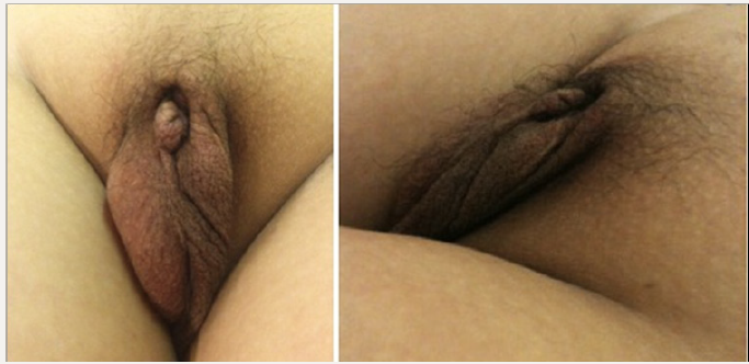
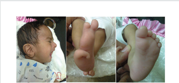
Figure 1: The girl had low set ears and deformity of the right foot with the presence of only three toes. There was no obvious abnormality of left foot, but the big toe was relatively large.

Lupinepublishers-openaccess-Surgery-Casestudies

R.J was first seen at about the age of four months during November 2018 because of poor feeding, failure to thrive, poor response to sounds, and poor head control. The girl also had low set ears, and deformity of the right foot with the presence of only three toes. There was no obvious abnormality of left foot, but the big toe was relatively large (Figure1). She was delivered at 38 weeks by cesarean section. She didn’t pass motion and was found to have imperforated anus. She had colostomy, and the surgeon reported that the sigmoid was not present. The five-centimeter colon ended at the pelvis, and cecum found on the left side. Cloaca treated with diversion colostomy. The parents were relatives and have three normal children. Echocardiography performed during the first month showed normal findings. Brain ultrasound performed on the fifth of August 2018 showed normal findings. Abdominal ultrasound was also performed on the fifth of August 2018 and showed small hypoplastic right kidney (18 x 12 mm) with normal shape. At the age of forty-six days (16, August 2018), a second abdominal ultrasound showed small hypoplastic right kidney. The left kidney had normal size.

Discussion

Authors from Germany, the Netherlands, the UK, the USA, Belgium, Italy , Switzerland and the Czech republic (Jürgen Kohlhase et al ,1998; Jürgen Kohlhase et al ,1999) defined Townes Brocks syndrome as a rare autosomal dominant malformation syndrome with a combination of anal, renal, limb and ear anomalies. Townes Brocks syndrome is a very rare genetic syndrome with 129 welldocumented patients reported in the medical literature [1,2]. In this paper the first case of this syndrome in Iraq is reported which is the case number 130.

Read More About Lupine Publishers Journal of Surgery Please Click on Below Link:
https://surgery-casestudies-lupine-publishers.blogspot.com/
Thursday, 4 May 2023

Lupine Publishers | Multidisciplinary Management of Elderly Cancer Patients: The Radiation Oncologist’s Point of View

 Lupine Publishers | Journal of Surgery & Case Studies


Abstract

Oncological treatments of elderly patients are extremely complex; so far there’s no agreement even on the definition of “geriatric patient”. From the point of view of global health, the problem is of the outmost importance as the number of older patients will increase dramatically in the next years, leading to a change in world epidemiology with a significant increase of chronic-degenerative diseases such as cancer. For this reason, it’s mandatory to provide clinical oncologists with multidisciplinary algorithms aiming at the best treatment of older cancer patients.

Materials and Methods

The complexity of oncological treatments for elderly patients starts from the very beginning, since the definition of “geriatric patient” is not univocal. From the point of view of public health, the problem is of the outmost importance. since nowadays there are 600 million people over 65, in 10 years their number will overcome 1 billion and in 2050 there will be nearly half billion people over 80 [1-3]. Such an increase of longevity will lead to a change in world epidemiology with a significant increase of non-communicable disease such as cancer. In 2030 the annual incidence of new cases of cancer in aged people will be 13,7 million and, which is even more important, nearly half of such cases will be in low-income countries [4]. Very few clinical trials (which are the cornerstone of Oncology) take older patient into account, and usually they are very selected cases [5-8], quite differently from every day’s practice, so there’s paucity of data about care of older patients, which makes clinical oncologist’s task even harder [5].Moreover, at least till few years ago, most guidelines and recommendations only consider chronological age in order to determinate the treatment’s choice, and this policy has led to over- or under-treatments [5]. With the purpose of optimizing the treatment’s choice it is imperative to focus on the concept of biological or functional age, in opposition to merely chronological age [6,7].

We should briefly review the physiological modifications caused by aging, which affects every organ and apparatus. Considering for instance Nervous System, there is a decrease of cortical volume and of synaptic density, which leads to a weaker memory and attention. Cardiovascular system is strongly affected, with a diminished cardiac output, increased arterial stiffness, slower modulation of cardiac frequency etc. [9]. Osteo-muscular apparatus is involved too, with a decrease of bone density leading to an increased risk of fracture and a sarchopenia which causes decreased physical activity with parallel increased fatigue and asthenia [10]. Additionally, for most of oncologic treatments, liver and kidney’s function is crucial; with their reduction, drug toxicity increases. In some aged patients an aforementioned change is plain; in other they can be silent in conditions of balance, becoming evident in stress situations such as a malignant disease and its treatments [1].

Biomolecular Markers of Aging

Aging is an extremely complex phenomenon, showing deep differences among individuals, consequently so far, it’s difficult to identify biological markers which enable us to divide subjects of the same chronological age into different functional ages. Several markers have been suggested, starting with markers of systemic inflammation such as CRP, D-dimer, IL 6 [8]. They are easily quantifiable, and they’ve been associated with functional decline in aged people, but their levels are influenced also by frankly pathological conditions like infections and cancer itself [11,12]. Markers of cellular aging have been considered too, such as telomeres or cell cycle components [8]. Dosing such markers is anyway extremely expensive, and moreover they have a significant interindividual variability. Another marker which could document a link between cancer and aging is P16 INK 4A, which has been showed to increase in aged breast cancer patients receiving chemotherapy [13]. Nevertheless, all of these markers are, so far, not completely validated and reliable.

Geriatric Assessment

As long as validated and reliable biomarkers are not established, the best way to assess a geriatric cancer patient is clinical evaluation [14-17]. Geriatric assessment is a multidisciplinary and multi parametric evaluation which takes into account physical aspect, nutritional status, neurological and cognitive status and even social support [18,19].

Comorbidities

When planning an oncological treatment at any age it’s mandatory to take into account comorbidities. This is mainly true in aged patient, beginning from the commonest pathologies such as cardiovascular diseases, diabetes, chronic renal failure, collagenopathies [20].

Polypharmacy

About 50% of aged patients are on 5 or more different medical therapies before undertaking an oncologic treatment, so it’s mandatory to evaluate all of these therapies and their potential interaction\interference with anticancer therapy [21].

Nutritional Status

Malnutrition and weight loss are deeply connected with cancer and its treatments and they have been shown to be linked to increased risk of toxicity and mortality [22].

Functional Status

All oncologists are familiar with ECOG and KPS scoring systems. In aged cancer patients it’s appropriate to integrate them with other evaluation systems [5,8] such as ADL (Activities of Daily Living) and IADL (instrumental activities of daily living). For instance, an extremely simple and reliable indicator of functional status is the number of falls. They are seldom taken into account, but they seem to be connected with oncological treatments’ toxicity.

Cognitive Status

The risk of cognitive decline increases with age. During anticancer treatment, it can cause for instance a diminished comprehension of its side effects which can be communicated with a delay, increasing the toxicity of the treatment itself, even in a serious way [1].

Psychological Status

Anxiety and depression worsen quality of life and precipitate functional decline, with a lower adherence to therapies [23].

Social Support

Many older people, so even aged cancer patients, live alone. It’s been documented that social isolation is linked to a significant higher mortality in cancer patients [24].

Screening Tools

Many screening tools have been validated with the purpose of identifying aged cancer patients who can take advantage of a multidimensional geriatric assessment [25,26]. Among the commonest ones we mention G8 and Vulnerable Elderly Survey 13 [18]. The final result of these screening and of the subsequent geriatric evaluation is the final decision to perform an oncological treatment (and its intensity) or not [7]. Sometimes it could be appropriate not to perform a treatment with curative purpose in an aged patient because of multiple comorbidities (which could lead to increased toxicities and a reduction in life expectancy). In other circumstances, on the opposite side, it could be an error not to undertake a treatment only because of chronological age. Patient’s preferences must be taken into account too; main international guidelines recommend to including patient in the therapeutic decision [27]. In this setting we must insert geriatric assessment; it’s been documented in literature that multidimensional geriatric evaluation has lead to significant changes in treatment planning, in most of cases with the aim of attenuate it [28]. At the present a multidimensional geriatric assessment is not often performed in the process of decision making regarding oncologic treatment of older patients. It’s been shown anyway that older cancer patients who have been evaluated in such a way have completed their treatment in a significant higher percentage, and with less modifications, compared to those who haven’t received it.

Radiotherapy

radiotherapy is the clinical discipline which aims at curing cancer by means of ionizing radiations; it could be employed as the sole therapeutic modality or in association with surgery and \or systemic therapies. [29,30]. A geriatric evaluation is strongly advisable for older patients who are candidates to radiotherapy, first of all for those treatments which consider its association with a systemic therapy, but also for the exclusive setting. Around 70% of cancer patients will require a radiation treatment, and this is especially true in older patients, as state of art radiotherapy techniques offer higher cure rates with less side effects. Moreover, treatment time can be reduced, and this can help patient with logistic difficulties (e.g. distance from radiotherapy facility) and their family\caregivers. This is true first of all in the palliative setting (e. g. Treatment of pain from bone metastases), but it could be accomplished even in the non-palliative setting, with the adoption of shortest scheduled.

Conclusion

The first dilemma of radiation and medical oncologists treating aged patients is how to decide if a patient is suitable for a given treatment and whether to treat patients with standard protocols or with adapted regimens. So, it’s advisable to include a geriatrician in multidisciplinary oncological teams (Tumour Board).

If it’s not possible, a good result can be achieved even with a conventional geriatric evaluation and a higher cooperation among specialists. Anyway, even after an effective evaluation has declared that an older patient is fit enough to undergo an oncological treatment, it’s mandatory to monitor such a patient in a closer and stricter way compared to a younger one [31].

Read More About Lupine Publishers Journal of Surgery Please Click on Below Link:
https://surgery-casestudies-lupine-publishers.blogspot.com/

Friday, 10 March 2023

Lupine Publishers | Inguinal Hernias Repair by Laparascopy. Repair of Inguinal Hernias by Laparascopy

 Lupine Publishers | Journal of Surgery & Case Studies


Abstract

Introduction: A series of 78 laparoscopic hernioplasties performed in the General Teaching Hospital “Enrique Cabrera”. Objective: To determine perioperative events, surgical complications and the evaluation of the pain referred by the operated patients.

Methods: Between January 2012 and December 2018, 78 hernioplasties were performed laparoscopic in 60 patients; 18 had bilateral inguinales hernias. He collected the variables: age, sex, type of hernia, perioperative events and complications, and a pain scale was applied. A database was filled and processed statistically.

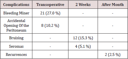
Results: The male sex predominated in a 5:1 ratio, the surgical time average was 53.5 minutes for unilateral hernias and 71.3 minutes for the bilateral ones. The most frequent complication in the transoperative period was bleeding lower in 27.0%, and in the postoperative period the hematoma was in 15.3%, it recurred two hernias (2.5%). At 15 days after surgery, 93.3% of theoperated did not complain of pain, but the social and labor reintegration was of only 34% of patients.

Conclusion: laparoscopic inguinal hernioplasty is a therapeutic option more, mainly in patients with bilateral and reproduced hernias.

Keywords:Laparoscopic Hernioplasty; Inguinal Hernia; Hernia Recurrence

Introduction

Since the concept of endoscopic inguinal hernia repair was first described by Ger R [1] in 1982, the endoscopic techniques are gone modifying, going through a time when failures and complications -united to high cost-exceeded initial enthusiasm [2]. Laparoscopic hernioplasty (HL) has been gaining popularity in the last decade, and numerous controlled studies appear in the literature comparing the laparoscopic techniques with conventional techniques [3-7]. In recent years, HL, despite consolidated as a therapeutic option to consider. The advantages of this have been demonstrated method in bilateral hernias, relapsed and in the active labor subject, that requires a precocious labor reintegration [5-7].

Methods

Between June 2012 and June 2018, a prospective descriptive study of Longitudinal section of 60 patients operated by hernia endoscopy of the region inguinal, in the Department of Surgery of the General Teaching Hospital “Enrique Cabrera”. The inclusion criteria were: - Patients who agreed with the type of surgical intervention and the study, and They gave their informed consent. Patients older than 30 years classified ASA I-III, without contraindications anesthetics for laparoscopic interventions.

a) Patients classified as Nyhus III and IV. Exclusion criteria.

b) Patients with previous surgical wounds in the inguinal region to operate, not dependent on inguinal hernias reproduced.

c) Inguinal hernias complicated, irreducible or slipped.

The surgical techniques were: laparoscopic inguinal hernioplasty completely extraperitoneal (TEP) of total extraperitoneal English and inguinal hernioplasty laparoscopic transabdominal preperitoneum (TAPP) preperitoneal), and one or the other was performed, at the discretion of the main surgeon. The TEP technique was executed with some variants such as: not using the trocar balloon, the preperitoneal space was decoloured by means of the 0º laparoscope, and the insufflation of CO2 at 13 mmHg. In patients with large herniated rings, placed a polypropylene cone in the hernia defect and then a 15 x 12 cm polypropylene prosthesis. There was no need to fix with clips the tights. The TAPP technique was performed on a regular basis [8]. In the immediate postoperative period, the scale of visual pain analog scale was applied (VAS), [9] and a value was assigned to pain through “caritas”, which starts very cheerful (value I) until very sad (value X). The quantification of pain was repeated in consultation at 7, 15 days and one month after surgery.

Results

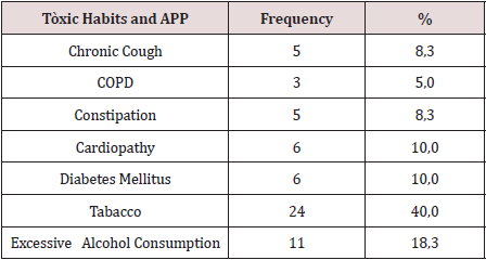
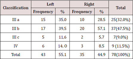
There were operated 78 hernias in 60 patients (18 patients [30.0%] suffered from bilateral hernias, 69 primary hernias and 9 reproduced hernias). The average of age was 55.6 years, the youngest patient was 30 years and the oldest was 77 years, but the majority (12 patients) were in the fifth decade of life. The male sex predominated in 82.9%, which represented a relationship man / woman of 5: 1. 42.9% of patients performed large efforts habitual physicists. The pathological history of the patients (Table 1). It is observed that 24 patients (40.0%) consumed tobacco, and in 11 an excessive consumption of alcohol was collected 18.3%. COPD: Chronic Obstructive Pulmonary Disease (Table 2). The distribution of the series according to the Nyhus classification. Right hernias predominated (55.1%), the indirect variety with large dilation of the ring and destruction of the posterior wall (IIIb) was the most frequent (37 hernias), and 7 femorales hernias and 9 recurrent hernias were operated 73 PET (93.6%) and 5 TAPP (6.4%) were performed. Two of the patients in whom a TEP technique was started were converted to a conventional prosthetic technique by accidental perforation of the peritoneum, passing the CO2 into the peritoneal cavity, and consequently, the loss of the preperitoneal surgical space. Of the 5 TAPP repairs, 3 of them were in the course of a laparoscopic cholecystectomy, and another was the conversion of a failed PET technique. The average surgical time of unilateral hernias was 53.5 min, with a minimum of 25 min and a maximum of 120 min. In bilateral repairs, the average surgical time was 71.3 min, and a minimum of 40 and a maximum was observed. of 110 minutes.

Table 1: Toxicos habits, and personal phalogical history (APP).

lupinepublishers-openaccess-surgery-case-studies-journal

Table 2: Distribution According to the Nyhus Classification.

lupinepublishers-openaccess-surgery-case-studies-journal

The hospital stay was less than 24 h in 50 patients (71.4%), in 5 it extended from 24 to 48 h, and in 5 to more than 48 hours. The most frequent complication in the transoperative period (Table 3) was minor bleeding in 21 repairs (27.0%) that gave rise to 12 hematomas (15.3%). No complications were observed after the second week, but two patients suffered recurrences (2.5%), more than two months after surgery. The application and evaluation of the VAS scale (Table 4). In the immediate postoperative period, after the patient recovered from anesthesia, 56 individuals (93.3%) were classified as VAS I, and 4 as VAS II. Twenty-four hours after surgery, 14 patients (23.3%) were classified as VAS I, 40 (66.6%) as VASII, 4 patients as VAS III and 2 VAS IV. In the consultation of the first week of postoperatively, 42 patients (70.0%) were classified as VAS I and [10] as VAS II, and two patients with moderate pain (VAS V) appeared in this period. Fifteen days after surgery, 56 individuals (93.3%) were VAS I and a month were 58 (96.6%). The incorporation to the usual activities, including work, was 3 patients a week after surgery, at 15 days they were 19 patients (31.6%) and 54 patients a month (90.0%).

Table 3: Complications.

lupinepublishers-openaccess-surgery-case-studies-journal

Table 4: Evaluation of the Visual Analogy Scale (VAS).

lupinepublishers-openaccess-surgery-case-studies-journal

Discussion

Currently, with the improvement of laparoscopic techniques, these are outlined as safe, reproducible and as a therapeutic option regardless of the age of the patient; nevertheless, the evaluation of the individual must be correct and meticulous in the preoperative period, specifically cardiorespiratory function, since with the TEP method a working space is created between the sheets of the crosssection sheet, richly vascularized, so that absorption and elimination of CO2 is greater than that produced in the peritoneal cavity during the performance of the pneumoperitoneum [10]. Although men predominated, there was a slight increase in women in the series with respect to other authors, [3-5] which could have been due to the inclusion in the study of 3 women who underwent the diagnosis of inguinal hernia, in the course of a laparoscopic cholecystectomy. In laparoscopic practice, it is not uncommon finding of hernia defects diagnosed in the transoperative, in men and women, the latter basically with a history of gynecological disorders. Although the usefulness of hernia repairs in asymptomatic patients is questioned in some articles, [11,12] the authors consider that it would be beneficial for the patient, if conditions permit, the repair of the hernia defect by the TAPP method. The relationship between the hernia disease and the physical efforts, is classic from the Cooper era [1]. In the series, 68% of the patients performed physical activities involving large and medium efforts, and also analyzing the multifactorial character in the pathogenesis of hernia disease, was striking, that approximately half of the patients operated on were smokers, a factor that influences the metabolism of collagen, significantly linked to hernia recurrences [13]. The majority of the repairs were by means of the PET technique, and we consider, as other authors [10,14 -19], that although the TAPP technique brings us closer to the area from a family perspective to the surgeon (peritoneal cavity) and facilitates the so-called “learning curve”, the hernial disease - considering it a parietal defect- should be given solution from this same plane to avoid the likelihood of serious complications of intrabdominal , and to leave the transperitoneal method as a tactical resource when the totally extraperitoneal method is unsuccessful.

The average surgical time was similar to other series [3-6]. It is known that this tends to decrease when the surgical team gains experience [16]. The largest surgical time recorded was in a patient, who was started with a PET technique, but Due to technical difficulties, it was converted to a conventional posterior repair. The fundamental complications were in relation to minor bleeding in the transoperative period and postoperative hematomas. In 3 patients it was necessary to drain the hematoma due to the discomfort caused, however, in the rest of the patients with hematomas and seromas they were treated with conservative measures. In two patients, the recurrence occurred 2 months after surgery, which was interpreted as a technical error. Our results coincide with numerous studies [3-7], that indicate the least postoperative pain of the minimum access techniques, as well as a prompt social and labor reincorporation of the patients. Despite the fact that 70% and 93.3% a week and 15 days postoperatively, respectively, had no pain or discomfort were minimal, only [18] individuals (30%) were incorporated into their usual activities before 15 days. These results contrast with other studies that report a return to work and social activities between 10-15 postoperative days, 4-17- although it is likely that some sociocultural factors are influencing these results. It can be concluded by noting that laparoscopic inguinal hernioplasty is another therapeutic option, mainly in patients with bilateral and reproduced hernias. In the series there were no major transoperative or postoperative complications, only minor bleeding and bruising were present. Most patients were not afflicted by pain 2 weeks postoperatively, however, return to social and labor activities after 15 days was low [18-20].

Read More About Lupine Publishers Journal of Surgery & Case Studies Please Click on Below Link:
https://surgery-casestudies-lupine-publishers.blogspot.com/

Saturday, 8 October 2022

Lupine Publishers | Aetiology and Prognosis of Pancreatic Pseudo Cysts Over A 10 Year Period

 Lupine Publishers | Journal of Surgery


Abstract

Introduction: Pancreatic pseudocysts often arise as a complication of acute or chronic pancreatitis with reported prevalence in chronic pancreatitis of 20-40%. Most common cause is alcoholic chronic pancreatitis (70-78%) then idiopathic chronic pancreatitis (6-16%), then biliary pancreatitis (6-8%). The aim of this study is to assess the demographic characteristic of patients diagnosed with pseudocysts, aetiology, characteristic features, and prognosis.

Methods: Prospective observational study to follow up patients diagnosed with pancreatic pseudocyst in 5 years (2006-2011). Data was collected in 2011 and patients followed up till June 2019. Radiology database searched for all cases that had a diagnosis confirmation by Computerised Tomography (CT). Total of 167 CT carried out for 119 patients. 35 patients were excluded due to absence of pancreatic pseudocyst after radiological re-evaluation.

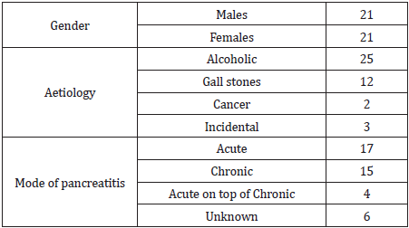
Results: In 5 years, 84 patients diagnosed with pancreatic pseudocysts on CT. 127 CT scans done mainly for follow up. 51(60.7%) males and 33 (39.3%) females; mean age= 57.8 years (20-93). 41 cases (48.8%) were associated with acute pancreatitis (8 on background of chronic pancreatitis). 21 cases (25%) were associated with chronic pancreatitis, 4 were associated with pancreatic malignancy while 18 (21.4%) were reported as incidental finding. The underlying factor was alcoholic pancreatitis in 37 patients (44%), gall stones in 19 patients (22.6%), pancreatic malignancy in 4 (4.8%) patients, biliary strictures in 2 patients and trauma in 2 patients. Idiopathic pancreatic pseudocysts were seen in 20 patients (23.8%), 18 of them were incidentally found on CT scan. 8 cases (40%) of idiopathic pseudocysts, were associated with non-pancreatic malignancies. 50% mortality (42 patients). 17 had acute pancreatitis, 15 had chronic pancreatitis, 6 were from the incidental finding and 4 from acute on top of chronic group. As for aetiology, 25 patients of the 42 had alcoholic pancreatitis, 12 had gall stones pancreatitis, 3 were unknown aetiology and 2 had pancreatic cancer.

Conclusion: Our study showed that alcoholic pancreatitis remains the most frequent underlying aetiology for pancreatic pseudocysts although it is not as common as previously reported. Idiopathic pseudocysts constituted a substantial number of this study with a higher than expected incidental pseudocysts. The association of pancreatic pseudocysts with malignancy needs to be further evaluated.

Introduction

Pancreatic pseudo-cysts often arise as a complication of acute or chronic pancreatitis. Previous reported prevalence of pancreatic pseudo-cysts in chronic pancreatitis ranges from 20% to 40%. Pancreatic pseudo-cysts most commonly arise in patients with alcoholic chronic pancreatitis (70% to 78%) (1, 2). The second most common cause is idiopathic chronic pancreatitis (6% to 16%), followed by biliary pancreatitis (6% to 8%) (3). Various imaging modalities are used in the diagnosis of pancreatic pseudo-cysts with Computed Tomography (CT) being the gold standard with 82% to 100% sensitivity and 98% specificity (4). There is wide variability in the range of spontaneous regression in the literature from 8 to 70%., with almost 40% of cysts that are less than 6 weeks old resolving without intervention compared to around 10% of the cysts older than 6 weeks (5). The aim of this study is to assess the demographic characteristic of diagnosed patients, aetiology of the pseudo-cysts, their characteristic features, prognosis and mortality rate.

Methods

This is a prospective observational study to follow up all consecutive patients diagnosed with pancreatic pseudo-cyst over a period of five years (2006 – 2011) in a district general hospital. Data collected retrospectively in 2011 to identify patients with pseudopancreatic cysts. The search was conducted using the radiology database of all cases had a diagnostic confirmation of pseudo pancreatic cyst by Computerized Tomography (CT). A total number of 167 CT scans were carried out for 119 patients. Radiological re-evaluation was conducted by a designated radiologist. 35 patients were excluded from the study due to absence of pancreatic pseudo-cyst after radiological re-evaluation. The remaining 84 patients were followed up till June 2019. The following demographic data were collected (age, gender, aetiology of pancreatitis, blood tests including amylase, liver function tests, white cell count, serum calcium and oxygen saturation). Mode of treatment and related complications. Radiological characteristics of the pancreatic pseudo cyst included: site, size, duration, calcification within the cyst. Other parameters observed prospectively were; mortality/ morbidity, further episodes of pancreatitis and duration between diagnosis and mortality/ morbidity. Subgroup analysis was conducted to look at prevalence of different aetiologies in both genders. Statistical analysis was conducted using Fisher Exact test, Mann-Whitney U test and the multivariate analysis was carried out using SPSS version 25 for Windows (SPSS Inc, Chicago, IL, USA). P value ≤ .05 was considered significant.

Results

In five years, period (2006 – 2011), 84 patients were diagnosed with pancreatic pseudo-cysts on CT scan. These patients had a total of 127 CT scans mainly for follow up. There were 51 male patients (60.7%) and 33 female patients (39.3%) with mean age of 57.8 years (20 – 93). In 41 cases (48.8%) the pseudo-cysts were associated with acute pancreatitis (8 on background of chronic pancreatitis). 21 cases (25%) were associated with chronic pancreatitis, 4 were associated with pancreatic malignancy while in 18 cases (21.4%) there was no obvious history of pancreatitis and the diagnosis was reported as incidental finding.

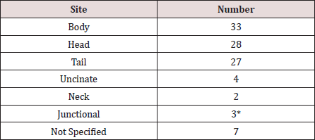
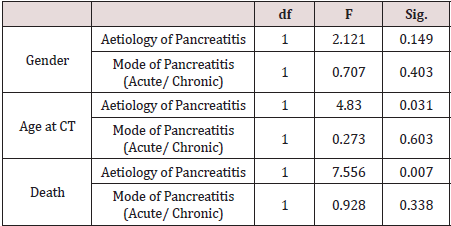
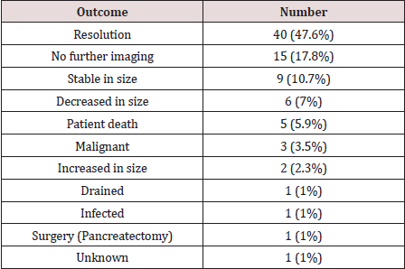
The underlying factor was alcoholic pancreatitis in 37 patients (44%), gall stones in 19 patients (22.6%), pancreatic malignancy in 4 (4.8%) patients, biliary strictures in 2 patients and trauma in 2 patients. Idiopathic pancreatic pseudo-cysts were seen in 20 patients (23.8%), 18 of them were incidentally found on the CT scan. Of note, in 8 cases (40%) of the idiopathic pseudo-cysts, were associated with non-pancreatic malignancies. In a subgroup analysis, alcohol was the commonest aetiology in male patients 29/51 (57%) while in female patients, incidental pseudo-cysts constituted 11/33 (33.3%), P= 0.23. Gall stones were the 2nd most common aetiology in females 10/33 (30.3%). Nine patients had more than one pseudo-cyst. The size of the pseudo-cysts varied significantly in reporting from small to huge pseudo-cysts with extension into the left thigh in one case. The body of the pancreas was the most common site (33 pseudo-cysts) followed by the head of the pancreas with 28, tail 27, uncinate 4, neck 2, not specified 7, 1 junction between body and tail, 2 junction between head and body (Table 1). The majority were managed conservatively with two drained percutaneously and two drained endoscopically. The 84 patients were followed up till June 2019 (mean follow up of 10 years). This showed that 42 patients (50%) died (male: female, 21:21), mean age of 61.8 (27- 93). 9 patients (21% of the mortality) died from complications related to the pancreatitis or due to complications from the pseudo-cyst like infection or bleeding into the cyst, with one patient dying from respiratory failure following laparoscopic cholecystectomy for gall stones pancreatitis (Table 2). The 9 patients represent 10.7% cause-related mortality. 17 of those 42 patients (40%) were from the acute pancreatitis group, 15 patients (36%) from the chronic pancreatitis group, 6 patients (14%) from the incidental finding group and 4 patients (10%) from the acute on chronic group. As regards the aetiology, 25 patients of the 42 (59.5%) were from the alcoholic pancreatitis group, 12 patients (28.5%) from the gall stones pancreatitis group, 3 patients (7%) from the unknown aetiology group and 2 patients (5%) from the pancreatic cancer group, Table 2. Multivariate analysis (MANOVA) yielded a highly significant association between the aetiology of pancreatitis and death, p = 0.007; however, there was no significant association between the mode of pancreatitis (acute or chronic) and death, p = 0.338, Table 3. Using death or alive dichotomy, chi square test confirmed the highly significant relationship between the aetiology of pancreatitis and death, p = .000. A two-way analysis of variance yielded a significant relationship between the aetiology of pancreatitis and its mode of onset (acute or chronic), p = 0.000 (Table 3). The duration of time form diagnosis of pancreatitis/ pancreatic pseudo-cyst to death was quite variable ranging from 8 to 3809 days (median 1018 days) for the whole cohort of mortality patients. The range for the cause specific cohort was 10 to 1424 days (median 112 days), p-value is .04006. 12 patients (14%) had further episodes of pancreatitis requiring admission to hospital. The outcome from the follow up of the pancreatic pseudo-cysts over the study period is shown in Table 4.

Table 1: Sites of the pseudo-cysts within the pancreas.

lupinepublishers-openaccess-surgery-case-studies-journal

*1 in junction between body and tail, 2 in junction between head and body.

Table 2: Demographics of mortality patients.

lupinepublishers-openaccess-surgery-case-studies-journal

Table 3: Multivariate analysis.

lupinepublishers-openaccess-surgery-case-studies-journal

Table 4: Pseudo-cyst follow-up outcome (June 2019).

lupinepublishers-openaccess-surgery-case-studies-journal

Discussion

Pancreatic pseudocysts are the commonest pancreatic cystic lesions and represent 75%-80% [1]. They are localized fluid collections rich in amylase and other pancreatic enzymes that gets surround by fibrous tissue wall not lined by epithelium [2]. Pseudocysts are a common clinical problem and arise as a complication of chronic pancreatitis in up to 40% of cases [3]. Alcoholic pancreatitis is the most common cause and account for over 75% of cases in some series [4]. The incidence of pancreatic pseudocysts is low and ranges between 1.6%-4.5% irrespective of the aetiology [5,6]. The pathogenesis of pancreatic pseudocysts is the disruption to the pancreatic duct as a result of pancreatitis or trauma which results in extravasation of pancreatic secretions. Two thirds of patients with pseudocysts have demonstrable connections between the cyst and the pancreatic duct. In the other third, an inflammatory reaction most likely sealed the connection so that it is not demonstrable [7]. In acute pancreatitis, fluid collections persisting for more than 4-6 weeks that are lined by a well-defined wall of fibrous or granulation tissue, would be regarded as acute pseudocysts [2,4]. In chronic pancreatitis the mechanism is less clear, but it could be as a consequence of an acute exacerbation of the underlying disease and/or blockage of a major branch of the pancreatic duct by a protein plug, calculus or localized fibrosis [8]. The clinical presentation is quite variable and while some patients could be asymptomatic, others might present with abdominal catastrophe as a result of; bleeding, infection or rupture of the cyst [9,10]. In our study, 2 patients had acute hemorrhage into the cyst from erosion into the splenic artery while 1 patient had infection of the cyst and all 3 patients died. Various imaging modalities are used in the diagnosis of pancreatic pseudo-cysts with Computed Tomography (CT) being the gold standard with 82% to 100% sensitivity and 98% specificity [11]. Endoscopic Ultrasound (EUS) has the highest sensitivity (93% to 100%) and specificity (92% to 98%) in differentiating acute fluid collections from pancreatic abscess and other pancreatic pseudocysts [12]. In our study 41 patients (48.8%) of the pancreatic pseudocysts were associated with acute pancreatitis and 21 cases (25%) were associated with chronic pancreatitis. However, it is worth mentioning that 8 cases of the 41 in the acute pancreatitis group had a background of chronic pancreatitis. 4 patients (19%) were associated with pancreatic malignancy while 18 patients (21.4%) were incidental finding on CT scans done for other reasons. These results are different from other studies showing the prevalence of pancreatic pseudocysts in acute pancreatitis to range from 6% to 18.5% [13,14]. The prevalence of pancreatic pseudocysts in chronic pancreatitis is 20%- 40% [15]. In our study, the most common underlying factor was alcoholic pancreatitis in 37 patients (44%), gall stones in 19 patients (22.6%), pancreatic malignancy in 4 patients (4.8%), biliary strictures in 2 patients and trauma in 2 patients. Idiopathic pancreatic pseudocysts were seen in 20 patients (23.8%), 18 of them were incidentally found on the CT scan. Of note, in 8 cases (40%) of the idiopathic pseudo-cysts, were associated with non-pancreatic malignancies. Results from other series showed that pancreatic pseudocysts are most common in patients with chronic alcoholic pancreatitis (up to 78%) [16], followed by idiopathic chronic pancreatitis (6% to 16%) and biliary pancreatitis (6% to 8%) [17]. The range of spontaneous regression of pancreatic pseudocysts ranges from 8% to 70% and the two major factors affecting this are; the size of the pseudocyst and the time since diagnosis [17]. In our study, 40 patients (47.6%) had spontaneous resolution of their pseudocysts and only 4 patients had drainage procedure for their cysts (2 percutaneously and 2 endoscopically) in the initial period of the study and 1 further patient having endoscopic drainage in the follow up period of the study. The body of the pancreas was the most common site with 33 pseudo-cysts, followed by the head of the pancreas with 28, tail 27, uncinate process 4, neck 2, not specified 7, 1 in the junction between body and tail, and 2 in the junction between head and body. 9 patients in our study had multiple pseudocysts. The results in the literature about the site of pseudocysts within the pancreas is variable, as some studies showed that most pseudocysts would be in or near the tail of the pancreas [18]. In another study, most extra pancreatic pseudocysts were located in the body and tail region, whereas most intrapancreatic pseudocysts were in the head of the pancreas [19]. The overall mortality in our study was 50% (42 patients), however the cause specific mortality was only 9 patients (10.7%). 59.5% of the whole mortality cohort had alcoholic pancreatitis while 28.5% had gall stones pancreatitis. The 9 cause specific mortality patients; 7 were alcoholic pancreatitis and 2 were gall stones pancreatitis. On multivariate analysis there was a statistically significant association between the aetiology of pancreatitis and death which was also confirmed on Chi-square testing. There was no statistically significant association between the mode of pancreatitis (acute or chronic) and death. The duration of time form diagnosis of pancreatitis/ pancreatic pseudo-cyst to death was quite variable ranging from 8 to 3809 days (median 1018 days) in the whole mortality cohort. In the cause specific mortality group, the range was 10 to 1424 days (median 112 days), this was statistically significant.

Conclusion

Our study showed that alcoholic pancreatitis remains the most frequent underlying aetiology for pancreatic pseudocysts although it is not as common as in other studies. The incidence of incidental pseudocysts with no history of pancreatitis is higher than that in the literature. Around 50% of pseudo pancreatic cysts in our study resolved spontaneously; therefore, conservative treatment has a big role in management of pancreatic pseudocysts. Bleeding or infection of a pseudo pancreatic cyst is an emergency associated with high mortality and should be managed promptly with laparoscopic and endoscopic approaches now gaining popularity over the surgical approach which is only used if the previously mentioned approaches fail.

Read More About Lupine Publishers Journal of Surgery Please Click on Below Link:
https://surgery-casestudies-lupine-publishers.blogspot.com/

Monday, 31 January 2022

Lupine Publishers | Use of Skin Grafts for the Correction of a Circumcision Complication

 Lupine Publishers | Journal of Surgery & Case Studies


Abstract

Background: Circumcision is largely performed and is considered to be technically simple and safe but it may occasionally lead to important complications.

Aim: To report the case of a patient who underwent a circumcision and presented a complication that obliged the use of skin grafts to recover the penis.

Materials and Methods: A 12-year old boy with neurogenic bladder underwent a circumcision using Plastibell device and presented a fasciitis of the lower abdomen and perineum with necrosis of the skin of the penis. He was submitted to a new surgical intervention where skin grafts from the abdomen were used to cover the body of the penis

Result: The cosmetic aspect of the penis was considered adequate at six months after the surgical procedure.

Conclusion: The use of skin graft could be a good and reliable alternative for different conditions resulting in buried penis.

Keywords: Circumcision, Male, Urinary bladder, Neurogenic, Urinary catheterization, Skin transplantation

Introduction

Circumcision is performed as a common ritual in Islamic, Jewish and other cultures and for phimosis correction. Patients with neurogenic bladder often have to undergo circumcision to facilitate intermittent catheterization. Although circumcision is considered to be a technical simple and safe procedure with no significant risk in neonates, it may occasionally lead to severe complications such necrotizing fasciitis or even penis amputation in older patients with commorbidities [1,2]. In this report, we present a case of a patient with neurogenic bladder who underwent a circumcision in order to facilitate the intermittent catheterization and presented a complication that obliged the reconstruction of the skin covering of the penis

Case Presentation

A 12-year old boy with neurogenic bladder has undergone a circumcision using Plastibell device in other service because the mother was having difficulty to perform intermittent catheterization. In the week following the surgery, as the child was obese, the penis sank into the pubic fat like a buried penis. A portion of the urine was eliminated by the “penis hole” but much of the urine leaked into the subcutaneous tissue without the mother noticing it because the child was incontinent. A week later the child was admitted to our Emergency Service presenting a fasciitis of the lower abdomen wall and perineum. He underwent drainage of the abdomen, removal of the Plastibell device, resection of all necrotic tissue and a cystostomy for urine drainage.

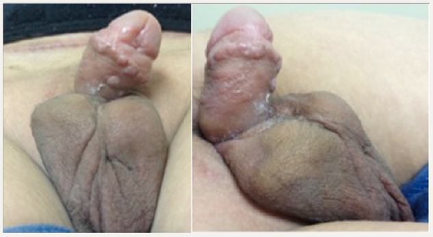
One year later he was submitted to a new surgical intervention. The penis shaft was completely exposed, and since there was not enough skin, the penis was covered with a skin graft from the abdomen (Figures 1 & 2). The cystostomy was closed and a laparotomy permitted the appendix to be mobilized to build a catheterization conduit using the Mitrofanoff technique for intermittent catheterization The graft had 6cm wide and and 10 centimeters long and was able to completely cover the body of the penis. A dressing with antibiotic ointment was applied to the penis and changed at the seventh day at the operating room. The cosmetic aspect of the penis was considered adequate at six months after the surgery (Figure 3).

Figure 1: Preoperative finding. The penis was sunk in the abdominal fat like a buried penis

Lupinepublishers-openaccess-Surgery-Casestudies

Figure 2: Immediate Postoperative Aspect - Note the conduct for urinary catheterization located in the right inguinal region. A skin graft from the abdomen was used to cover the penis.

Lupinepublishers-openaccess-Surgery-Casestudies

Figure 3: Postoperative Aspect after 6 Months.

Lupinepublishers-openaccess-Surgery-Casestudies

Discussion

Different techniques have been proposed for the treatment of buried penis and megaprepuce, which are conditions in which the body of the penis has a normal size but the skin coverage is insufficient [3]. These techniques, however, are only satisfactory if the skin of the penis or the scrotum allow the construction of flaps. In a previous publication we showed that the children with bladder exstrophy had a consistent improvement in the cosmetic appearance of the penis when it was covered with skin grafts [4]. The use of skin grafts allows a more appropriate coverage of the body of the penis when it is completely ungloved because they fit adequately between the glans and the base of the penis. Although the use of skin flaps increase the morbidity of surgery, require special care in handling it and increase the time of hospitalization, the aesthetic result can be very satisfactory (Figure 3).

Circumcision is widely used around the world, in neonatal period with the benefits outweighing the risks, often with preventive indications [5]. However the risks are increased in older children, especially in obese and with co morbidities like neurogenic bladder needing intermittent catheterization. In this case the fasciitis could be avoided if the patient was submitted to neonatal circumcision. Fortunately the infection affected only the skin and fascia, leaving untouched the cavernous tissue and glans, and could be adequately treated with a skin graft, being a good and reliable alternative. We think this case clearly shows that neonatal circumcision can avoid life threatening conditions due to delayed operations when performed by experienced surgeons.

Conclusion

The use of skin grafts could be a good and reliable alternative for different conditions resulting in buried penis.

Read More about Lupine Publishers Journal of Surgery & Case Studies Please Click on Below Link:
https://surgery-casestudies-lupine-publishers.blogspot.com/

Thursday, 2 December 2021

Lupine Publishers | Mini OPCAB Coronary Artery Bypass Surgery Plus Medical Treatment: An Option for High Risk Coronary Patients

 Lupine Publishers | Journal of Surgery


Abstract

MINI OPCAB surgery (Xiphoid Approach) is a surgical technique in which the left internal mammary artery is bypassed to the Anterior Descending Artery (ADA) by a medial inferior sternotomy Fourteen high risk patients with multiple coronary disease with a preoperative logistic Euroscore of 10.86 were operated and follow up with medical treatment and strictly control of risks factors MACE at 80 months was 0% and Survival at 7 years 82% (KM) Although is an alternative the combination of Mini OPCAB operation plus medical treatment in high risk. Patients with multiple vessels coronary disease, more experience is needed to confirm this initials results. Statistical analysis applied the student test (SPSS program), with p<0.05 were considered significant

Keywords: Mini Opcab; Minimally Invasive Coronary Surgery; Coronary High Risk Patients

Introduction

In high-risk patients with multiple vessel disease who are not candidates for conventional surgery with extracorporeal circulation or for percutaneous procedures as a single treatment, the alternative of left mammary artery to left internal descending artery bypass graft surgery without extracorporeal circulation offers advantages over medical treatment [1,2].

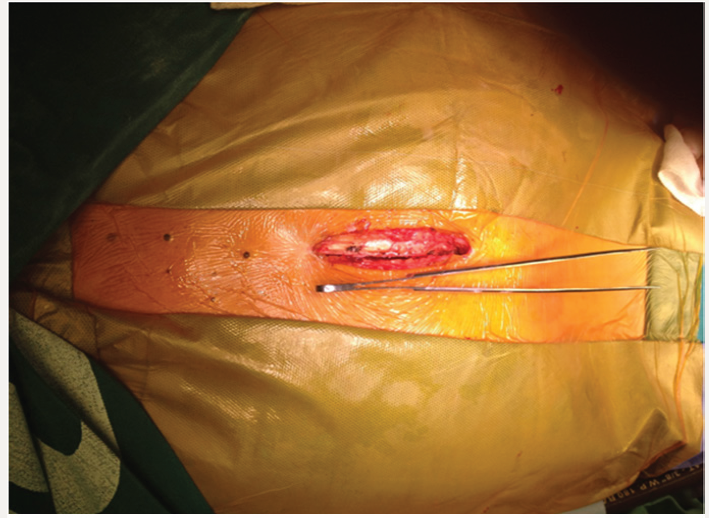
Figure 1:

Lupinepublishers-openaccess-Surgery-Casestudies

Figure 2:

Lupinepublishers-openaccess-Surgery-Casestudies

a) The MIDCAB operation

b) Is effective to treat high risk patients with multiple vessel disease. Greater long-term follow-up is necessary to clarify the indications and validate the procedure for this type of patients.

c) MINI OPCAB surgery (Xiphoid Approach) is a surgical technique in which the left internal mammary artery is bypassed to the Anterior Descending Artery (ADA) by a medial inferior sternotomy approach in the 3rd or 4th intercostal space, leaving intact the sternal manubrium (Figures 1 & 2). Long-term results have already been published, reaching 82% survival at 12 years (Kaplan-Meier).

d) This presentation describes the experience with this surgical technique in our institution [3,4].

Materials and Methods

Fourteen high risk patients with multiple vessel coronary artery disease with mean age of 71.07 years (±9.051, 95% CI), 21% women and mean preoperative Logistic EuroSCORE of 10.68 (±5.407, 95% CI), were operated-on in the last 7 years, followed up in our institution with strict medical treatment and control of risk factors.

Result

Operative mortality in this series was 0%, the incidence of perioperative infarction was 0%, the average duration of surgery was 2 hours and 20 minutes; 10 (71%) patients were extubated in the operating room and average hospital stay was 2 days and 11 hours. Following the intervention, one patient received a stent in the right coronary artery and another in the circumflex artery for presenting large arteries with severe lesions. In this group of patients, major adverse cardiovascular events were 0% at 80 months. Survival rate was 82% at 7 years (Kaplan-Meier); an 85-year-old woman died 5 years after surgery due to stroke.

Conclusion

The combination of a MINI-OPCAB surgery for bypass of the left internal mammary artery to the left anterior descending artery [5,6] together with an adequate medical treatment and a hybrid treatment when the right coronary artery or circumflex artery are of high caliber, is a viable option for elderly and high-risk patients. More experience is needed to confirm these initial results.

Read More About Lupine Publishers Journal of Surgery Please Click on Below Link:
https://surgery-casestudies-lupine-publishers.blogspot.com/
Thursday, 21 October 2021

Lupine Publishers | Surgical Management of Multiple Nevus Lipomatosis Superficialis: A Case Report

 Lupine Publishers | Journal of Surgery & Case Studies


Abstract

Nevus lipomatosis superficialis is a rare skin lesion characterized by the ectopic dermal deposition of adipose tissue. While benign, it typically presents in the second to third decade of life as regional discomfort. We present the case of multiple lesions requiring serial surgical management.

Presentation of Case: The patient was a 22-year-old male who presented with multiple, soft, warty lesions histologically consistent with nevus lipomatosis superficialis. He underwent excision of the largest mass and is undergoing multiple excisions for adjacent masses.

Discussion: Nevus lipomatosis superficialis is a rare connective tissue hamartoma of unclear etiology and multiple clinical subtypes. While management is typically surgical, efficacy of treatment options for multiple nearby lesions is not well characterized.

Conclusion: We review the literature for nevus lipomatosis superficialis and describe a case with multiple lesions of varying chronicity. Treatment must be chosen to optimize wound healing and cosmetic outcomes.

Keywords: Nevus lipomatosis superficialis; Multiple; Surgery; Skin; Excision

Introduction

Nevus lipomatosis superficialis (NLS) is a rare, benign hamartoma of the skin that was first described by Hoffmann and Zurhelle [1]. It is characterized by mature, ectopic adipose deposition in the dermis [2]. The classical form of NLS as described in 1921, is found on the buttocks, trunk, and thighs and described as multiple collections of soft, cerebriform, non-tender papules or nodules that are yellowish in color and may be pedunculated [3]. Solitary lesions have also been reported on the arms or scalp and are typically dome-shaped or sessile papules [4]. We report the surgical management in a young, otherwise healthy male with multiple pedunculated and sessile NLS lesions. Currently, there is no standardized surgical treatment for multiple nearby lesions.

Case Report

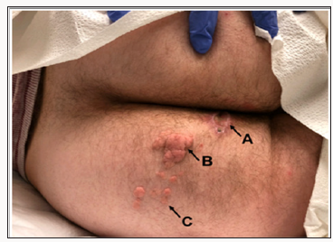
A 22-year-old white male presented to outpatient General Surgery with a 9-year history of a painless mass in the perianal region and left buttock. The mass gradually increased in size and was not associated with any skin changes over the surrounding areas. The mass had never been associated with bleeding, discharge, or itching. The patient reported a second mid-sized mass that had progressively increased in size over the past 4 years. Several smaller satellite masses had emerged in the past year. While the patient did have cosmetic concerns, the largest of the lesions was causing discomfort when sitting and requested the mass be surgically removed. Patients past medical history were unremarkable. Laboratory testing was wholly unremarkable and standard sexually transmitted infection screens were negative. He was not previously vaccinated against human papilloma virus. On physical examination, there was a 5.9 x 4 x 3.5 cm, fleshy, tanpink polypoid lesion arising at the 4 o’clock position approximately 2cm from the anal verge and extending 5cm along the radial axis of the left buttock. The largest lesion had a relatively narrow, pedunculated base and multiple papulo-nodules with a warty surface. Another, mid-sized sessile lesion similar in appearance was present 4cm lateral to the largest mass. Several smaller lesions were also present further lateral. All lesions were soft to palpation and nontender. There was no bleeding or discharge with manipulation. Differential diagnosis included anogenital warts and neurofibromatosis. The patient was offered surgical excision of the largest of the masses. Linear incision was made at the base of the mass, 2cm away from the anal margin radially out. Postoperative recovery was uncomplicated. Patient’s two-week post-operative check showed an appropriately healing wound (Figure 1). Pathology revealed nevus lipomatosis superficialis. There was no atypia or malignancy identified (Figure 2). Patient was subsequently offered serial-excisions for the remaining masses.

Figure 1: (A) Post-excision of largest (5.9 x 4 x 3.5cm) polypoid lesion (B) Sessile, cerebriform masses of varying chronicity including a 4-year-old medium-sized mass. And (C) Multiple 1-year-old smaller, satellite masses

Lupinepublishers-openaccess-Surgery-Casestudies

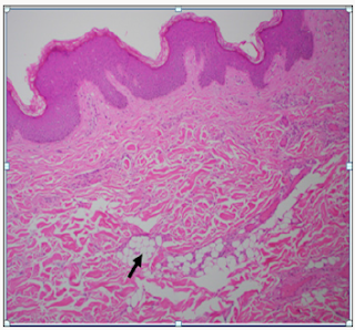
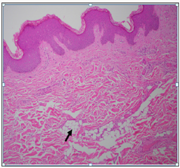
Figure 2: Spotty Involvement of Dermal Adipose Deposition (Arrow) Creating an Irregular Zone of Fat Splaying Apart Dermal Collagen.

Lupinepublishers-openaccess-Surgery-Casestudies

Discussion

NLS is a rare connective tissue hamartoma with altered epithelial elements with no sex predilection or familial trend [5]. While its incidence has not been reported, NLS descriptions are mostly limited to case reports. NLS may be congenitally present or develop later in life. There are two primary forms of NLS: the classical/multiple form and the solitary form. The lesions of the classical form are either congenital or fully form by the second or third decade of life [6], thought it has been reported to arise in the fifth decade as well [7]. Classical NLS is described as skin-colored papules that coalesce into plaques with cluster-like or linear distribution. Lesions are asymptomatic, unilateral, slow-growing over many years and may have smooth or cerebriform surface that is nontender [8]. Classical lesions most frequently distribute over the lower abdomen, the pelvic girdle, thigh, and gluteal surface. The second NLS subtype is the solitary form (pedunculated lipofibroma [9]) that may appear at any body surface and tends to affect older patients in the third to sixth decade of life [10,11]. The largest study of solitary form NLS is a retrospective case study of 13 cases in Tunisia [12]. A third, much less reported form of NLS is described as “Michelin tire baby syndrome,” an autosomal dominant deletion on chromosome 11 characterized by symmetric circumferential skin folds. These folds harbor underlying NLS and may affect the neck, legs, and arms; they are self-limited and resolve during childhood [13,14]. This is the first reported case of NLS with both sessile and pedunculated features with multiple lesions requiring serial surgery for optimal would healing and cosmetic outcome. Histologically, NLS lesions are classically characterized by ectopic mature adipocytes in the reticular dermis surrounded by dermal collagen fibers. Fat cells may extend to the papillary layer as larger fat lobules blur the boundary between dermis and hypodermis. Adipocytes may be either entirely mature or incomplete. The density of collagen, fibroblasts, and vasculature in the dermis is increased [15]. The epidermis can show acanthosis, elongation, rete ridge obliteration, hyperkeratosis, and hyper basilar pigmentation [16]. On electron microscopy, young adipocytes are of perivascular pericyte origin [17].

The etiology of NLS remains unclear. Originally, Hoffman and Zurhelle theorized that connective tissue degeneration incited dermal fat deposition [1]. In 1955, Holtz postulated that pericapillary lipoblast differentiated into mononuclear cells that grew into preadipose tissues. Others argue that focal heterotropic growths of aberrant adipose tissue occur during embryonic development [18]. Thus far, there have been no studies describing any cytogenic alterations in those with classical or solitary NLS, though Cardot-Leccia et al. [19] report a case of NLS with a 2p24 deletion. As in our case, NLS is typically asymptomatic. Ulceration has been reported secondary to external trauma or ischemia,11as well as café-au-lait macules, hypopigmented spots, leukodermic macules, and comedo-like changes may be present [15]. As such, the differential diagnosis for NLS includes condyloma acuminata, neurofibroma, lymphangioma, skin tag, Fordyce spots, granuloma annulare, and nevus sebaceous. Treatment for NLS is typically cosmetic, as malignant transformation is extremely rare [20]. Surgical excision, as in our patient, is the mainstay of treatment, as recurrence rate are low. Cryotherapy, ultrapulse CO2 laser ablation [21], topical fludroxycortide corticosteroids [7], and intralesional injections of phosphatidylcholine and sodium deoxycholate. Kim HS [22] have been reported as non-surgical options with positive clinical response. For optimal cosmetic results, patients with multiple, separated masses should be serially managed with adequate time for healing and scare formation after each excision.

Conclusion

NLS is a rare, benign skin tumor characterized by aberrant adipocyte deposition. While NLS is typically asymptomatic, lesions can cause discomfort and require surgical management. We report the case of a 22 year-old with multiple large lesions requiring serial surgery for optimal wound and cosmetic outcomes.

Read More About Lupine Publishers Journal of Surgery Please Click on Below Link:
https://surgery-casestudies-lupine-publishers.blogspot.com/
Thursday, 2 September 2021

Lupine Publishers | Double Malignancies: A Rare Entity

 Lupine Publishers | Journal of Surgery & Case Studies


Abbreviations: MPM: Multiple Primary Malignancies, SEER: Surveillance Epidemiology and End Results Programme, SPT: Second Primary Tumor, TT: Third Tumor, QT: Quadrant Tumor, SPM: Second Primary Malignancies, NAACCR: North American Association of Central Cancer Registries

Introduction

Patients which have diagnosed with a cancer, have a life time risk for developing another de novo malignancy depending on various inherited environmental and iatrogenic risk factors. Cancer victims could survive longer due to settling treatment modalities, and then would likely develop a new metachronous malignancy [1]. The incidence of multiple primary malignancies has not been rare at all. Screening procedures have especially been useful for the early detection of associated tumors, whereas careful monitoring of patients has treated for primary cancer, and then a good communication between patients and medical care team would certify not only an early detection for secondary tumors, but only finally & subsequently, an appropriate management [2]. Differentiation between multiple primary and multicentric cancers was addressed in the classification by Moertel CG [3]:

I. MPMNs of Multicentric Origin

a) The same tissue and organ.

b) A common, contiguous tissue shared by different organs.

c) The same tissue in bilaterally paired organs.

II. MPMNs of Different Tissues or Organs

III. MPMNs of Multicentric Origin Plus a Lesion (s) of a Different Tissue or Organ

In 2002, However in ‘A review of the definition for multiple primary cancers in the United States’ classified the association of different cancers in two categories depending on the timing of their discovery [4];

a) Synchronous in which the cancers occur at the same time or within two months and

b) Metachronous in which the cancers follow in sequence of more than two months apart.

Vaamonde [5]. reckoned the time factor as six months. In 2005, International Agency for Research on Cancer working Group has come out with International Rules for Multiple Primary Cancers [6].

Although the mechanism involved in the development of multiple primary cancers has not been clarified, some factors such as heredity, constitution, environmental and immunological factors, oncogenic viruses, radiological and chemical treatments have been implicated. Hereditary susceptibility explains only a small proportion of all second cancers though many hereditary cancer syndromes have been described. MPMNs can occur at any age. However, from the reviewed series, patients with MPMNs tend to be older than those with a single primary malignant neoplasm. In many autopsy series and clinical reports, the median age of 50- 94% of MPMN patients was over 50 years [7]. Multiple Primary Malignancies (MPM) were first described in 1879 by Billroth [8]. The neoplasms may be limited to a single organ or may involve multiple separate anatomical organs. The North American Association of Central Cancer Registries (NAACCR) classifies MPM into two categories:

a) Synchronous, in which the cancers occur at the same time (The Surveillance Epidemiology and End Results Programme (SEER) definition is within two months) and

b) Metachronous, in which the cancers follow in sequence, that is, more than six months apart [9].

According to Warren Gates criteria a diagnosis of MPM require the following criterias to be fulfilled

a) Each tumor should present a definite picture of malignancy

b) Each tumor should be histologically distinct

c) The possibility that one is metastasis of the other must be excluded [10].

Meta-analyses show the frequency of Second Primary Tumor (SPT) as 3-5%, Third Tumor (TT) as 0.5%, and fourth tumor, that is, Quadrant Tumor (QT), as 0.3%, in different organs and of different histogenesis. Metachronous primary malignancies are becoming increasingly common because of an increase in the number of elderly cancer survivors, greater awareness and improved diagnostic modalities [1]. The exact pathophysiology for MPM remains unknown. However certain factors have been postulated which includes the common carcinogen induced multiple cancers in a exposed epithelial surface, called as “Field-Cancerization” as seen in head-neck tumors. In addition other causative factors includes ionizing radiation, increased use of organ transplant, and the increasing use of newer treatment modalities like hormonal manipulation, target therapies, genetic manipulation, and immunomodulators [2]. In a study conducted by Chakrabarti [11] it has been reported that the over a period of 2 years, 12 cases of MPM were detected against a total of 1255 cases. Of these, five cases were synchronous malignancies and seven cases were metachronous. Head and neck was the commonest site of index malignancies with seven cases followed by the breast cancer with three cases and next gynaecological malignancies with two cases. Most common sites for Second Primary Malignancies (SPM) were head and neck with (four cases).

Male to Female ratio was 1:1.5 in the synchronous primary group and 1:1.3 in the metachronous group. Median age of presentation of the primary tumour was 52 years and 6 months. The age range for the SPM was 17-72 years with the highest incidence in the 6th decade of life. Studies have reported that that the relative risks of SPM ranges from 1.08 to 1.3. SPM are often missed during follow-up and are detected accidentally. According to various series, the onset of SPTs decreases the 5-year survival by 18-30% as compared to those with only a single tumor. The controversy between the lateral spread of clones vs multiple foci of independent alterations does not currently affect the surgical and medical management of theses premalignant and malignant lesions. In the future, the presence of altered clones at mucosal margins may be an indication for aggressive therapy, including chemoprevention or radiotherapy to treat altered clonal patches that are unable to be detected grossly and are beyond the initial scope of surgical excision [3]. The issue of whether those with an extensive visible mucosal field defect is more likely to benefit from chemotherapy, radiotherapy or chemoprevention is a complex one. Current management is often site specific: Recurrent oral premalignant disease is often treated by surgical excision, whereas diffuse high grade premalignant changes in the laryngeal mucosa are frequently treated with radiotherapy. Determination of the role for these and other treatment modalities for clinically occult, clonally altered patches of epithelium is likely to be a difficult issue, since treatment of mucosa with widespread visible alteration is already challenging [4]. The paucity of awareness about SPM has also prevented the formulation of population-based screening protocol. Multiple tumors that have been pathologically confirmed at the time of presentation should be evaluated and staged as independent tumors. The treatment plan should be decided after staging of both the primary and secondary tumors in view to attain maximum clinical response. Proper counselling and patient’s understanding of magnitude of the disease is paramount. Operable synchronous SPM can be operated in a single setting with minimal morbidity with better survival and is less taxing on the patient and his/her relative both psychologically and financially. A regular follow-up on the patient by the clinician increases the chances of early detection of metachronous SPM and the formulation of the treatment plan at the earliest with better overall survival

Conclusion

MPMNs are still elusive for want of proper guidelines regarding correct terminology and classification encompassing varying presentations of chronological, aetiological, clinical and histopathological combinations. Our case adds up to literature for further research. The possibility of occurrence of synchronous multiple primary malignancies should be considered during workup for any malignant condition to institute early intervention to achieve good outcome [12]. The possibility of multiple primary malignancies existence should always be considered during pretreatment evaluation. Screening procedures were especially useful for the early detection of associated tumors, preferably before clinical manifestations occurrence [12]. There were some evidences that screening would improve outcomes among patients who might develop second malignancies, although the data were limited. The optimal screening modalities and strategies for reducing mortality from second malignancies remained to be defined for most tumor sites [13]. The early diagnosis of secondary malignancies should not be neglected in patients treated for a primary malignancy, especially when the long clinical period before the diagnosis of subsequent tumors has been taken for management. With careful monitoring, secondary tumors could be detected earlier, and, with appropriate intervention, might be better managed, without compromising survival. Our data could guide oncologists towards a closer follow-up strategy in the management of patients treated for common tumors. Availability of data regarding incidence of MPM, particularly those from developing countries is very limited and hence further studies are needed. SEER is working in this direction with an aim to define and develop appropriate and reliable criteria’s for synchronous and metachronous cancers. It is imperative that patient with a primary malignant tumors should be thoroughly closely, and regularly followed. Genetic counselling, risk estimation, cancer screening and chemoprevention must be emphasized. Appropriate cancer prevention strategy in with proper emphasis on synchronous and metachronous cancer needs to be designed and incorporated in the National Control Programme as multiple primary cancers have unique, biological behaviour requiring specific diagnostic and therapeutic interpretation [14].

Read More About Lupine Publishers Journal of Surgery & Case Studies Please Click on Below Link:
https://surgery-casestudies-lupine-publishers.blogspot.com/