Showing posts with label OSMOAJ. Show all posts

Wednesday, 18 October 2023

Lupine Publishers | Surgical Repair of a Neglected Patellar Tendon Rupture

 Lupine Publishers | Journal of Orthopedics and Sports Medicine


Abstract

Neglected rupture of the patellar tendon is rare, as this type of injury is typically disabling in the acute setting. We present a 31-year-old male patient who sustained a left patellar tendon rupture while playing basketball. The diagnosis of patellar tendon rupture was neglected by the patient and care was delayed by 8 months. The proximally retracted patella and distally detached patellar tendon were brought back to their anatomic positions and repaired surgically while avoiding the use of autograft or allograft tissue due to fat interposition maintaining the patellar tendon length. This case report contributes to the scarce literature on surgical management of neglected patellar tendon rupture and presents a unique radiologic appearance of a chronic patellar tendon rupture.

Keywords: Patella; Athlete; Neglected; Delayed Repair

Introduction

Neglected ruptures of the patellar tendon are defined as ruptures presenting after at least six weeks and are often difficult to repair [1]. These injuries are rare, as they are often acutely disabling, and usually occur in patients under the age of 40 [2]. Rupture of the patellar tendon usually occurs near the inferior pole of patella, and most often occurs during sporting activities [3]. Surgical management of neglected patellar tendon rupture is more technically difficult to manage than acute ruptures, and the results are less favorable due to quadriceps muscle atrophy, adhesions, and proximal patellar migration [4]. Many different surgical techniques have been described for reconstruction of the disrupted extensor mechanism of the knee following neglected patellar tendon rupture. In this case we describe a simple technique of direct tendon reattachment with strong Ethibond suture reinforcement to allow immediate postoperative mobilization without the need for autograft or allograft tissue.

Case Report

A 31-year-old male presented to our orthopedic clinic for evaluation of his left knee after sustaining an injury 8 months earlier while playing basketball. He was seen in an urgent care clinic immediately following his injury, but was told his patella was displaced due to edema and should improve with time. He was unable to fully extend his knee (45 degree extension lag) following the injury and was using an over-the-counter knee brace to maintain knee extension in order to ambulate. Focused examination of the left knee revealed a superiorly displaced left patella and 45° to 120° passive range of motion with crepitus throughout. There was no pain to palpation of the knee, ankle, foot, or thigh, and the patient was neurovascularly intact. He had full strength in all muscle groups except the left quadriceps, which demonstrated grade 2 out of 5 strength. A chronic left patellar tendon rupture was suspected and confirmed by MRI (Figure 1). Operative management options were discussed, including primary vs. allograft repair of the patellar tendon.

All the risks and benefits of surgery were explained to the patient in detail, which the patient understood and consented to the procedure. A left femoral nerve block was performed for postoperative pain control and to prevent contraction of the quadriceps tendon stressing the repair. Patient was positioned supine on the operating room table and a bump was placed under the left hip. A midline incision was made ending medial to the tibial tubercle. Sharp dissection was performed down through the skin and subcutaneous tissues. There was noted to be a complete rupture of the patellar tendon just distal to the inferior pole of the patella with significant scar tissue surrounding the patellar tendon. There was rupture of both the medial and lateral retinacula. Adhesions were present laterally around the patellar tendon, additionally there were adhesions from the quadriceps to the femur proximally, these adhesions were carefully released in order to achieve necessary excursion and reapproximation. There was mild chondromalacia noted on the patella and trochlea of the femur. No fracture of the patella was appreciated. At that point, the tendon edges were gently debrided several millimeters down, back to healthy-appearing tendon. The inferior pole of the patella was then prepared using a #15 scalpel blade to resect up the periosteum from the anterior patella, then a rongeur and a curette were used to prepare down to the bleeding bony surface to improve tendon healing to bone. At that point, #2 Ethibond was used to run a total of 4 strands of suture coming out proximally on the patellar tendon in a running locking-type Krackow stitch. At that point, a drill was used for pilot holes in the inferior pole of the patella. Next, a tap was used in each of the pilot holes for the 3.5 mm suture Swivelock anchors (Stryker, Kalamazoo, MI). Then, the suture limbs were passed through the Swivelock suture anchor and tension was maintained by placing a polydioxanone (PDS) suture through the quadriceps tendon and manually pulling to approximate the inferior pole to the patellar tendon. There was excellent purchase of the anchors in the patella as well as approximation of the patellar tendon to the inferior pole of the patella. The repair was felt to take up excellent tension without undue stress to about 30 degrees of knee flexion and maintained full extension. At that point, the knee was then extended and the Ethibond sutures were passed proximally into the patellar periosteum and distally into the patellar tendon to reinforce our repair. There was excellent tracking of the patella within the trochlea without subluxation. The retinaculum was repaired. Subcutaneous tissues were closed with 2-0 Vicryl followed by running 3-0 Monocryl suture for the skin. Dermabond Prineo dressing was then applied. A sterile dressing followed by an Ace wrap was then applied, followed by a knee immobilizer locked in extension. The post-operative plan was to continue weight bearing as tolerated with early rehabilitation focusing on isometric strengthening of the quadriceps. The knee was to remain in full extension for six weeks, which was achieved using a hinged brace locked in full extension for ambulation, followed by a transition to graduated range of motion over the course of 12 weeks. At 4 months, the patient achieved 121 degrees of flexion with a 5 degree extension lag. In comparison, his nonoperative leg demonstrated 125 degrees of flexion with a 5 degree extension lag. No instability was observed in varus and valgus stress testing. The patient’s Knee Society Score (KSS) was 80/100.

Figure 1: Sagittal T2-weighted magnetic resonance imaging (MRI) of the left knee revealed a patellar tendon with avulsion of its origin from the distal pole of the patella (green arrow). The degree of patellar tendon retraction was decreased due to the presence of the large amount of infrapatellar fat (white arrow).

Lupinepublishers-openaccess-orthopedics-sports-medicine

Discussion

The major disability for our patient was an absent extensor mechanism of the left knee as a result of complete patellar tendon rupture. This was a unique presentation of a chronic patellar tendon rupture due to patient delayed presentation. Our primary goal was to restore this mechanism by repairing the patellar tendon. Patellar tendon rupture is usually unilateral and is commonly reported as a result of athletic injury [5], which were both seen in our patient. Patellar tendon rupture is acutely disabling and as such often treated primarily, however in rare cases chronic patellar tendon injuries are reported either as a result of missed injury or neglect. In our patient, the delay in treatment was due to a missed diagnosis immediately following injury by the urgent care communication. Early repair of patellar tendon ruptures is preferred and gives favorable results [3]. The results of reconstruction of neglected ruptures of the patellar tendon are less predictable because of quadriceps muscle atrophy and proximal retraction of the parapatellar soft tissues. Various reconstruction techniques, largely as case reports, have been reported by authors for neglected patellar tendon ruptures however there is no widely accepted method. Primary repair with autogenous graft augmentation using hamstring tendons or fascia lata has been most commonly seen [4]. External fixation using wires and pins has been reported as a solution for patients with an elevated patella and severe contracture of the quadriceps tendon [2]. Reconstruction with allografts consisting of an intact patellar tendon or Achilles tendon has also been used [6]. Mandelbaum et al. recommended Z lengthening for the quadriceps tendon and Z shortening for the patellar tendon with augmentation using the gracilis and semitendinosus tendons [7]. Our patient avoided the the use of allograft or autograft tissue, and a primary repair technique utilizing knotless anchors and suture tape [8]. However, given this patient’s anatomy the patellar tendon did not retract or enfold upon itself due to the presence of a large infrapatellar fat pad. Due to this presentation we were able to primarily repair his native patellar tendon. It is important to maintain the normal position of the patella intraoperatively as both patella baja and patella alta have been shown to negatively impact knee function [9]. In addition, maintaining patellar tendon length avoids any restriction of flexion or extension lag. It is also important to prevent excessive compression of the patella over the trochlea of the femur via retinacular release, which maintains smooth tracking and gliding of the patella and prevents anterior knee pain. In addition, the strength of the reconstruction can be confirmed intraoperatively with gentle passive knee movements up to 90°. This can also verify normal patella tracking over the femoral trochlea without undue pressure.

Conclusion

This case represents a unique presentation of a chronic patellar tendon rupture that presented 8 months following the injury but was still able to be repaired primarily. Additionally, we utilized suture anchors to strengthen our repair and tendon fixation into the patella.

Read More About Lupine Publishers Journal of Orthopedics and Sports Medicine Please Click on the Below Link:

https://lupinepublishers-orthopedics.blogspot.com/


Wednesday, 23 August 2023

Lupine Publishers | Effectiveness of Eccentric Exercise for the Management of Rotator Cuff Tendinopathy: A Critical Review

 Lupine Publishers | Journal of Orthopedics and Sports Medicine


Introduction

The rotator cuff (RC) mainly contains four muscles and their tendons: infraspinatus, supraspinatus, teres minor and the subscapularis [1]. These four muscles and their tendons surround the shoulder joint [2]. It is a group of muscles start from the scapular and attach around the humeral head, forming a cuff at the anatomical neck of the humeral head. The main function of the RC is to stabilize and activate the glenohumeral joint (ibid). The RC primarily performs three types of movement: abduction, external rotation and internal rotation. Previous studies report that there is no strong evidence that the RC contributes to glenohumeral movement (ibid). However, there are a large number of published studies that describe the link between RC and glenohumeral joint stability [3]. Hence, RC injury can result in loss of stabilization of the glenohumeral joint. RC tendinopathy (RCT) has been regarded as a kind of RC injury. RCT is a term used to describe a combination of pain and weakness in the RC tendon [4]. Also, it can be loosely described as excessive load resulting in impaired performance of the RC tendon when experienced with particular movements of the shoulder [5]. The prevalence of RCT is 14% in the general labor population [6]. In addition, a number of authors have considered the prevalence of RCT. It has been reported that prevalence is 19% among all ages (ibid). Therefore, the high prevalence of RCT cannot be underestimated. The diagnosis of RCT can be measured by ultrasound and magnetic resonance imaging (MRI). Its diagnosis also can be considered in association with pain symptoms [7-9].

Pathophysiology

There are many risk factors that can cause RC injuries. These include glenohumeral joint dynamic instability, joint abnormality and abnormal shoulder structure [10]. After a RC injury, the damaged tendon gradually develops microtears, calcification and fiber vascular proliferation [11]. The pathophysiology of RC injury can be attributed to the result of series of inflammatory reactions after soft tissue injury. However, RCT is characterized by different types of RC tendon injury. Tendinopathy is a common term used to describe pathological changes in tendons. Previous research has established that the pathology of RCT primarily contains intrinsic mechanisms, extrinsic mechanisms or combined mechanisms [2-7]. Intrinsic mechanisms can be defined as pathological changes directly affecting the tendon itself [12]. Excessive load is an important contributor to RC intrinsic tendinopathy. Healthy adult’s RC tendons consist of water, collagen, proteoglycan and glycosaminoglycan [13]. The largest proportion of RC tendon is type I collagen [14]. The collagen fibers of the tendons are confluent and form a cuff on the humeral head. It also forms a five-layer complex near the greater tuberosity. The first to fifth layers are superficial coracohumeral ligament, the main portion of tendon complex, dense fascicles, loose connective tissue and fibrocartilage, respectively. This composite structure seems to have excellent stability and elasticity. However, according to the intrinsic theory, the transformation of collagen bundles density and orientation could result in sheer forces between layers (ibid). Excessive load can cause changes and result in a strong force in the tendon. This increases with age and the impact of the disease can cause degenerative changes which can reduce adaptability in the tendon. There is an unambiguous relationship between degenerative changes and increasing age [15]. Excessive load plus degenerative changes lead to variation in biology and morphology of the RC tendon (ibid). There is a relatively small body of literature that is concerned with degenerative changes in RC tendons which contained calcification, fatty infiltration, tendon thinning and vascular proliferation [16]. Moreover, in an investigation into degenerative changes, [17] found that type III collagen has been increased that could decrease the elasticity of tendon during RC injury. Extrinsic mechanisms can broadly be defined as RC tendon injury that is potentially attributed to the friction on impact with external structures. These structures contain the humeral head and coracoacromial arch. Numerous studies have attempted to explain that 95% of RC injury was caused by impingement between the coracoacromial arch and humeral head [18]. Even in normal shoulder anatomy, a series of shoulder functions such as elevation and abduction can damage the RC tendon. There are a large number of published studies that describe the link between external impingement and RCT [19,20]. It has been thought that extrinsic impingement primarily results in pathological changes near the glenohumeral joint side of the RC tendon. The causes of intrinsic and extrinsic mechanisms are different, but both of them lead to an inflammatory response. After RCT, tendon healing is commonly divided into three parts [21, 22].

Firstly, the inflammatory phase lasts for one week. During this phase, vascular permeability increases, lymph and inflammatory cells flood into the lesion site. Secondly, in the proliferative phase, the influx of inflammatory cells produces some growth factors and cytokines which result in recruitment and proliferation of fibroblasts. Finally, in the remodeling phases, fibroblasts proliferate and begin to generate, orient, deposit and interconnect than become collagen [23] has shown that intrinsic factors or extrinsic factors alone do not increase the prevalence of RCT. In total 108 mature rats were equally divided into three groups: extrinsic compression group, overuse group and a combination of overuse and extrinsic compression group. Results from the studies indicated that no significant differences were found between separate overuse or extrinsic compression and RCT. Also, a positive correlation was found between a combination of overuse and extrinsic compression and RCT. This study suggested that a combination of intrinsic factors (overuse) and extrinsic factors could increase the possibility of RCT. However, these results came from study on animals and are difficult to directly utilize in clinical conditions. This is because human beings and animals have different anatomical and biological characteristics.

The Physiological Effects of Eccentric Exercise

Eccentric exercise is a type of muscle action characterized by contraction of a muscle during its lengthening due to an external load. To determine the effect of eccentric training, [24]. carried out an experiment. This study recruited 12 young male soccer players (mean=26 years) and implemented 3 sets of heels raising eccentric training twice a day for 12 weeks. Six participants have Achilles tendinosis and others were healthy one. To evaluate collagen synthesis and degradation, blood samples were taken 1 week before and after 12-week eccentric training programme through micro dialysis technique. Pain visual analogue scale (P-VAS) also was used to assess the level of pain before and after 12-week training. There are a large number of publish studies that describe the link between carboxyterminal propertied of type I collagen (PICP) increasing in blood and type I collagen synthesis. Also, data from several sources have identified the increasing carboxyterminal telopeptide region of type I collagen (ICTP) associated with the degeneration of type I collagen [25,26]. The findings of this study showed that PICP increased significantly in the blood after 12 weeks of training in injured tendon, but not changed in healthy tendon. ICTP have no significant difference before and after study in both health and injured. Many recent studies have shown the same findings as [23] experiment [27,28]. To eliminate bias, this study carried out the same starting time (9am) and controlled the room temperature (25︒C). In injured group, the level of pain also reduced after 12 weeks training. Combining the outcomes from blood samples and P-VAS, this study could provide strong evidence to support the effectiveness of eccentric exercise. However, although the study examined plasma concentrations of PICP and ICTP, the changes of collagen type I formation could not directly be observed. Previous studies suggested that MRI or ultrasound can observe the changes in collagen type I formation [29,30]. More medical examinations should be included in further studies. Also, the population of this study is professional athletes, instead of ordinary people. It may reduce the generalizability of this study. Further studies should focus on the ordinary population and carry out more clinical examinations.

One of the most influential accounts of eccentric training came from [31]. The findings of this study demonstrated significant neovascularization after eccentric training. 22 males and 8 females with Achilles tendinopathy participated in this investigation. Ultrasonography and colored Doppler were used to examine the neovascularization in the tendon before and after intervention. Participants were asked to complete 12 weeks of knee eccentric exercise twice per day, seven days per week. The result of this study indicated that eccentric training is likely to accelerate the neovascularization. Although this study focused on patients with Achilles tendinopathy, the benefit of eccentric exercise is likely to generalize to other tendinopathies, due to the similar anatomical, physiological and pathological feature of tendinopathies [32]. However, different gender ratios (22 males and 8 females) may result in bias of experimental outcomes. Compared to women, men are more likely to regenerate new blood vessels in the tendon area after eccentric training [33]. This is due to the difference in the physiological characteristics of men and women. In addition, there is no control group in this study. Although setting a control group is not ethically permissible, a control group could reduce cointervention bias and make the experimental results more reliable and valid. A systematic review that is often cited in research on the effect of eccentric exercise is that of [34] who found that eccentric training may promote increases in tendon stiffness. This study also reported that low intensity eccentric exercise is more effective than concentric exercise for increasing a tendon’s mechanical tension. The keywords “eccentric”, “tendon” and “performance” were used in its search strategy. 40 studies were included and total of 1150 participants (406 women and 744 men) were recruited in this study. Results of this study found that, compared to concentric exercise, muscle cross-sectional area (CSA) increases significant difference after eccentric exercise. Eccentric exercise can promote more CSA synthesis than concentric exercise [35]. also identified that increasing number of CSA were beneficial for the stiffness of tendons. In addition, this study highlighted low intensity eccentric exercise was beneficial for the maximal tendon force and stress, instead of high intensity eccentric exercise. The whole design of this study is rigorous and appropriate, given proper keywords and suitable search strategy. However, this study carried out narrative analysis which cannot provide strong evidence for the findings. In addition, the study recruits healthy participants which may not demonstrate the effect of eccentric training in clinical condition. Therefore, these findings should be translated with caution into clinical scenarios. Overall, these studies provided relevant evidence that eccentric exercise promote collagen synthesis, formation of new blood vessels and increases tendon stiffness. However, further study should focus on clinical condition and more rigorous experimental design.

The Effectiveness of Eccentric Exercise

Reported [36] positive correlation between tendon repairing and supraspinatus and infraspinatus eccentric exercise. Ten middleaged (mean age=54) patients with diagnosed RCT were recruited from two health care clinics. All the patients were provided with shoulder abduction and external rotation eccentric training which involved 90 repetitions per day. Shoulder pain was assessed by visual analogue scale (VAS) after the training programme, which was believed to be valid and reliable for evaluating level of pain. According to the result of this study, VAS significantly reduced from 57 to 29. Authors attributed this improvement to healing of tendon. This hypothesis seems to be consistent with the study of [23]. which found significantly improved collagen synthesis after eccentric training. In Susanne’ s study, ten middle-aged participants indicated small sample size and low generalizability. However, compared to middle aged people, the synthesis of young people’s collagen is significantly faster [37-39]. found that insulin-like growth factors that promote collagen synthesis increased significantly among young people (mean age =25) compared with elderly people (mean age 70 years). The design of this study is a single-subject research design which is appropriate for the study condition. Therefore, the outcomes produced by middle-aged people as research participants can be accepted. [40] investigated the effectiveness of eccentric training comparing with conventional exercise in clinical RCT. 36 patients diagnosed with RCT, were recruited and randomly divided into eccentric exercise group (20) and conventional exercise group (16).

The eccentric exercise group (mean age = 50.2) performed external rotator and abduction exercises for the shoulder. The conventional exercise group (mean age = 48.9) contained shoulder shrugs and stretching exercise for the pectoralis. Both groups completed 12 weeks of daily exercise at home. 14 weeks after the training programme (26th week), the Constant Murley Score (CMS) and Pain VAS were applied for measuring shoulder function and pain. Results of this study indicated that both eccentric and conventional exercise for the RC area improve significantly in shoulder function and pain after 26 weeks. The findings from this study also shown that there is no significant difference between eccentric training and conventional training for managing shoulder pain and function after 26 weeks. This study provided 12 weeks training and follow-up after 26 weeks, giving plenty time to examine the results. However, single-blinded instead of doubleblinded controlled trial measures were implemented in this study. This may lead to experimenter bias. In addition, although different language versions of the CMS showed good reliability and validity [41, 42], this instrument needs to have established standards [43]. Therefore, this study showed that in the long term, both eccentric and conventional exercise is beneficial for middle-aged people with RCT.

There is a published study describing the role of eccentric exercise for managing RCT [44]. A total of 11 patients with diagnosed RCT and preparing for arthroscopic subacromial decompression surgery were recruited in this study. All patients were randomly divided into 3 groups: control group, concentric RC exercise group and eccentric RC exercise group. The concentric group was asked to perform shoulder abduction exercises, 3 sets of 15 repetitions twice a day from 0° to 90°. The eccentric group was asked to perform shoulder adduction exercises, 3 sets of 15 repetitions twice a day from 90° to 0°. The control group were asked to carry out standard practice without normal treatment pathway. The Oxford shoulder Score and pain Visual Analogue Scale were used to measure shoulder function and pain at the beginning, after 4 weeks and after 8 weeks. Interestingly, results from this study did not indicate any statistically significant differences between all three groups. In addition, two individuals in the eccentric group determined to cancel the surgery and continue exercise at the end of study. Compared to [37]. study, a total of 8 weeks length of this study is relatively short. It may demonstrate that longer study could result in statistically significant differences [45-47] also found that a longer follow-up period of eccentric exercise can avoid shoulder surgery. To compare with eccentric exercise and concentric exercise, further study design should extend the length of the experiment or implement a follow-up period. However, 11 patients is a small sample size which cannot provide strong evidence to support the outcomes of the study. In addition, although individual cases show significant improvement and avoid surgery after this study, this result may not be generalizable. Overall, data from this study gave no significant differences between concentric exercise and eccentric exercise on managing RCT. Moreover, short-term eccentric exercise may not have obvious therapeutic effect. Further study should collect more samples and extend experimental time.

Conclusion

Overall, the present study was designed to determine the effect of eccentric exercise. The research findings clearly indicated that eccentric exercise can promote the synthesis of type I collagen, accelerate the neovascularization and increase the integrity of a tendons. The second aim of this study was to investigate the effectiveness of eccentric exercise on RCT. Generally, the investigations have shown that, in the long term, eccentric, concentric and conventional exercise are beneficial for patients with RCT. Compared to concentric and conventional exercise, although eccentric exercise may not have obvious therapeutic effect, evidence indicated that, in the long term, eccentric exercise should be more effective in managing RCT than other treatments. However, the findings in this assignment are subject to at least three limitations. First, different gender ratio and age stage could lead to bias. Secondly, small sample size could decrease the generalizability. Finally, single examinations method and lack of randomization show that results should be interpreted with caution. Nonetheless, therapists may still select eccentric exercise or combine them with other treatment to manage RCT in clinical condition. Further investigation should increase sample size, balance gender ratio and use diverse examination methods. Also, it is necessary to determine which level of intensity of eccentric exercise is more effective in treating RCT. In addition, accurate eccentric exercise should be investigated as the RC contains four muscles.

Read More About Lupine Publishers Journal of Orthopedics and Sports Medicine Please click on the Below Link: https://lupinepublishers-orthopedics.blogspot.com/


Monday, 3 July 2023

Lupine Publishers | Effect of Intensive Integrative Therapies on Pain, Disability and Quality of Life in Patients with Osteoarthritis Knees: A Comparative Observational Study

 Lupine Publishers | Orthopedics and Sports Medicine Open Access Journal


Abstract

Objective: To assess the differential effect of intensive yoga, yoga and naturopathy and yoga and Ayurveda management for Osteoarthritis of knees Design: Pre-post comparative study

Participants: Ninty-five individual prediagnosed with knee osteoarthritis according to ACR guidelines aged between 30-75 years were randomized into three groups, i.e., Yoga (n= 36), yoga and Naturopathy (n=30), Yoga and Ayurveda (n=29). All three group received their respective intervention for 1 week at arogyadhama, prashanti kutiram, S-VYASA.

Outcome measures: The primary outcome variables were visual analog scale (VAS) for pain on activity, Western Ontario and McMaster Universities Secondary Osteoarthritis index (WOMAC) and secondary outcome variables were anthropometric measurements and 6 min walk test were measured on day 1 and day 7.

Results: There was significant reduction in VAS scale on activity in yoga group (p < 0.001), whereas in Ayurveda group showed significant improvement in stiffness (p <0.001), and significant reduction in weight and pain on rest (p < 0.001) was in naturopathy group after 7 days of intervention.

Conclusion: IAYT practices combined with other therapies had better effect than alone IAYT. There were significant changes seen within groups.

Keywords: Knee Osteoarthritis; Integrative Approach of Yoga Therapy (IAYT); VAS; WOMAC; Ayurveda; Naturopathy

Introduction

Movement is a medicine for creating change in a person’s physical, emotional, and mental states. Disease is the destroyer of health wealth and mind. Lack of movement of knees leads to the later. The most common of lack of movement is Osteoarthritis (OA) OA knee mainly occurs in elders and middle age people and is more common among women and overweight subjects. The symptoms include pain, stiffness and decreased range of motion (ROM), which result in limited activity and reduced quality of life. The prevalence increases with age, and by the age of 65, approximately 80 percent of the US population is affected. It is the second most common rheumatological problem and most frequent joint disease with prevalence of 22% to 39% in India OA is a heterogenous disease, involving complex and interacting mechanical, biological, biochemical, molecular and enzymatic feedback loops with cartilage degeneration as the common, final event [1] despite this degeneration, OA is an active process and a network of mechanisms reacting to stress or injury on the joint. All joint features are affected in OA [2]. Structural changes include cartilage fibrillation, degeneration of articular cartilage, thickening of subchondral bone, osteophyte formation, synovial inflammation, degeneration of ligaments and meniscus, hypertrophy of joint capsule, cellular and molecular changes in nerves, as well as changes to periarticular muscle, bursa, fat pads [3]. The loss of cartilage and modifications to bone and synovial membrane contribute to an unfavorable biomechanical environment which increases stress on the joint and furthers the progression of cartilage degradation [4]. Non-steroidal anti-inflammatory drugs (NSAIDs), including cyclo-oxygenase II inhibitors and non-opioids analgesics such as acetaminophen have been the most popular method of managing pain in musculoskeletal tissues. Tradational System of medicine like Ayurveda, Naturopathy and Yoga also specify the symptoms and mode of treatment. So a observational study was planned to understand the effect of this traditional system of medicine.

Aim of the Study

a. To study the differential effect of three Indian systems of medicine on the outcome of OA knee management

Objective of the Study

a. To assess the differential effect of intensive yoga, yoga and naturopathy and yoga and Ayurveda management for Osteoarthritis of knees on pain and stiffness.
b. To assess the differential effect of intensive yoga, yoga and naturopathy and yoga and Ayurveda management for Osteoarthritis of knees on physical functions.
To assess the differential effect of intensive yoga, yoga and naturopathy and yoga and Ayurveda management for Osteoarthritis of knees on anthropometric measurements.

Material and Methods

Source of data

A total of 95 participants aged 30-75 years, were registered from Arogyadhama, a home-based health centre, S- VYASA, bangalore.

Sample size

The sample size was calculated with G-power software by fixing the alpha at 0.05 powered at 0.8 and an effect size of 0.71 based on the mean and SD of an earlier study.

Inclusion criteria

The inclusion criteria were patients clinically/radiologically diagnosed with mild to moderate OA knees according to American college of rheumatology (ACR) guidelines and with associated comorbidities were included in the study.

Exclusion criteria

Patients with severe OA knees, rheumatoid arthritis, autoimmune diseases, malignancies, knee surgery or kneearthroscopy and knee pain caused due to congenital dysplasia were excluded.

Ethical consideration

The study was approved by the Institutional Ethics Committee of S-VYASA University (approval letter no: RES/IECSVYASA/ 107/2017 dated 23rd October 2017). Signed informed consent was obtained from all the participants included in this study.

Design

Participants were randomly divided into three groups, i.e., Yoga alone, Yoga and Naturopathy, Yoga and Ayurveda group. All three groups underwent intervention of Integrative approach of yoga therapy (IAYT) and respective therapies for 6 days and assessments and treatment plans for participants were discussed with in-charge doctor and therapists. All groups continued their medication as per prescription.

Grouping and posology

Three groups pre-post comparative study.
Group A: Table 1 Standalone Yoga group

Table 1: Time table.

Group B: Table 2 Integrative Yoga and Ayurveda group

Table 1: Time table.

Group C: Table 3 Integrative Yoga and Naturopathy group

Table 3: Time table.

Group B: Table 2 Integrative Yoga and Ayurveda group

Assessment criteria

Primary outcome variables
a) Visual analog scale for pain on activity
b) WOMAC Indian version
c) 6 min walking distance test - The subjects were asked to walk on a flat surface for 6 minutes and the distance covered was expressed in meters.

Anthropometric measurements

a) Height: It was measured with stadiometer in centimeters (cm), and later converted into meters.
b) Weight: Weight was measured using research grade electronic weighing scale.
c) BMI: By using formula, weight in kg/ height in meter2, calculated the body mass
index of every individual.

Observations

Table 4

Table 4: Observations.


Results

Recapitulation

In the current study, variables were taken at baseline and following one week of intensive yoga, yoga and Ayurveda and yoga and naturopathy therapies. The primary outcome variable was pain pain assessed through Visual Analogue Scale (VAS) while resting, walking on plane surface, climbing stairs up and down as well as WOAMC scores. The secondary assessments included anthropometric measurements; and 6 min walking distance. We also recorded the vital parameters such as systolic and diastolic blood pressure, pulse rate and respiratory rates at baseline and at the end of one-week intervention for all three groups.

Yoga and ayurveda group

Within group comparison revealed that as compared to the baseline following variables showed significant improvement in weight, body mass index, 6-min walk test, WOMAC global score, WOMAC-pain, stiffness and physical functioning, visual analog score on rest, walking, climbing up and down. The values of within group comparison through paired sample t test are mentioned in Table 4.

Yoga and naturopathy group

Table 5: With-in group comparison changes in yoga and ayurveda group.

Within group comparison revealed that as compared to the baseline following variables showed significant improvement in weight, body mass index, systolic blood pressure, respiratory rate 6-min walk test, WOMAC global score, WOMAC- pain, stiffness and physical functioning, visual analog scale on rest, walking and climbing up and down. The values of within group comparison through paired sample t test are given in Table 5.

Yoga group

Within group comparison revealed that as compared to the baseline following variables showed significant changes in weight, Diastolic blood pressure, Pulse rate, 6 MIN walking distance, WOMAC global scores, WOMAC-pain, stiffness and physical functioning, visual analog score on rest, walking and climbing up and down. The values of within-group comparisons through the paired samples t-test are mentioned in Table 6.

Table 6: Within group changes in yoga and naturopathy group.

Table 7: Within group changes in yoga group.

Abbreviations

WT: Weight, DBP: Diastolic Blood Pressure, PR: Pulse Rate, WGS: WOMAC Global Score, STF: Stiffness, PF: Physical Functioning, VAS: Visual Analog Scale, WK: Walking, DW: Down, BMI: Body Mass Index, SBP: Systolic Blood Pressure, RR: Respiratory Rate, WOMAC
Table 8 presents the changes in all three groups at baseline and at the end of one week. None of the variables between groups were found significant both at baseline and following the one-week intervention duration.

Table 7: Results of the study.

Outcome Measures

All participants were assessed for primary and secondary outcomes twice, at baseline (day 1) and end of study period, day 7.

Primary outcomes

i. Visual Analog Scale (VAS): The visual analogue scale is a psychometric response scale which was used to assess pain on climbing stairs (20 staircases), climbing down from stairs (20 staircases) as well as following 6 min walking on flat surface. It was be used as an instrument to measure subjective quantification of pain. When responding to a VAS item, respondents was be asked to specify their level of pain by indicating a position along a continuous line between two endpoints.
ii. WOMAC: The WOMAC has been extensively evaluated in populations suffering from osteoarthritis (N Bellamy, Buchanan, Goldsmith, Campbell, & Stitt, 1988). The Indian version of WOMAC was used to assess the disability and quality of life [5]. Western Ontario and McMaster Universities Secondary Osteoarthritis index (WOMAC) is widely used in the evaluation of Hip and Knee Osteoarthritis. It is a self-administered questionnaire consisting of 24 items divided into 3 subscales assess the three dimensions of pain, disability and joint stiffness in knee and hip osteoarthritis [6].

Secondary outcome measures: Anthropometric Measurements

i. Height: Was measured in Centimeters (cm)
ii. Weight
iii. BMI: 6- min walk test
iii. BMI
iv. 6-min walk test

Discussion

Modus operandi of yoga and naturopathy therapy

Possible mechanism of improvement in yoga and naturopathy group is detoxification treatment, diet restriction and physical activity which help them more in reducing pain and improve the physical functioning. Yoga and naturopathy integrated therapies are found to reduce inflammatory markers in patients with chronic inflammatory disorders [7]. This could be the mechanism of action for improvement in the outcomes of OA knees through Yoga and Naturopathy.

Modus operandi of ayurveda therapy

The possible mechanism of action behind reduction of all the symptoms is due to medicated oil application frequently but the long-term effects of such applications need to be studied Ayurveda and Naturopathy therapies was planned by respective doctors.

Conclusion

Although statistically insignificant, the current study revealed that integration of naturopathy to yoga therapy was effective in reducing weight, and pain, whereas integrating Ayurveda to yoga therapy could lead to better reduction in stiffness in patients with OA knees. Such non-significant trends could be tested using larger sample size with prolonged intervention period in future studies.

Strength of The Study

The strengths of the study are:
a) This multidisciplinary study encompasses the fields of yogic science, Ayurveda and Naturopathy.
b) No earlier study has reported integration of yoga with Ayurveda and naturopathy for OA knees.
c) Because the duration of intervention was short, acceptability and adherence to therapy was good
d) As integration were delivered through a standard protocol, it could be reproduced in the exact way for all cases.


Read more about Lupine Publishers Journal of Orthopedics and Sports Medicine Click on the below link: https://lupinepublishers-orthopedics.blogspot.com/


Tuesday, 21 March 2023

Lupine Publishers| Painfull Posterior Cruciate Ligament Ganglion Cyst. A Case Report

Lupine Publishers| Journal of Orthopedics and Sports Medicine


Introduction

Ganglion cysts (GC) are benign tumor-like lesions usually going out from mucinous degeneration of collagenous structures [1,2]. They could occur in several anatomic areas but GC arising from cruciate ligaments are rare [3,4] with a prevalence of 0.36% or 0.8% respectively when diagnosed by magnetic resonance (MR) or by arthroscopy [3,5-6] However other studies of GC prevalence refer ranges from 0.2% to 1.9% [7-9], with posterior cruciate ligament ganglion cysts (PCLGC) being five times less frequent than those identify in anterior cruciate ligament [5,10]. This lesion is mainly diagnosed in people aged 20-40 years-old and a male predominance has been reported [5,11-13]. The etiology of PCLGC is not clear. They could appear from synovial herniation or congenital translocation of synovial cells. Mesenchymal stem cells proliferation with cysts formation or mucoid degeneration occurring in areas suffering chronic injuries are also reported [7,14-16]. Many of PCLGC are asymptomatic. When symptomatic the main clinical symptoms and signals includes knee pain and / or movement restrictions [17].

The knee joint could present a slight effusion, restriction to extension and particularly in extreme flexion [1,4]. The common classification of cruciate ligament cysts is supported on the position of the cyst, anterior, posterior or between cruciate ligaments [7]. MR is the gold standard for detecting GC1. Recently observation by ultrasonography is considered useful for identifying and locating the lesion, as well as being a conservative approach to treat cystic lesions [1].

Case Report

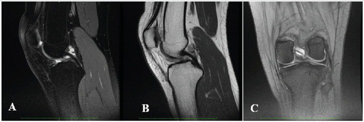
A 17-year-old Caucasian female, a soccer player, presented with a 9 month history of left knee pain, mainly in the posterior and medial aspects of the knee, combined with slightly back swelling. Pain was exacerbated with exercise, especially squatting, and partially alleviated with rest. She had no history of a knee major traumatic event (Figure 1). She complained of knee pain on soccer playing, one of the main reasons to suspend this practice. She alsorefers no confidence on demanding tasks.

Figure 1: Knee MRI of PCL GC; A: T2 sagittal view FS; B: T2 sagittal view FSE; C: T2* coronal view.

Lupinepublishers-openaccess-orthopedics-sports-medicine

In clinical examination we identified a light quadriceps atrophy, compared to the opposite side, and symmetry in active range of motion (ROM). Passive ROM was limited (10 degrees) and painful in extreme flexion and symmetric in hyperextension. Meniscal tests and varus-valgus stress tests were negative. Some tenderness on palpation of the popliteal aspect but no pain or even tenderness was verified in medial or lateral joint line. Plain radiographs were normal. A knee magnetic resonance image (MRI) was then performed regarding the potential diagnosis of a joint cyst so it was acquired weighted T2 sagittal sequences with and without fat suppression and T2* coronal. The MRI revealed a high-signal well-defined, ovoid shaped formation extending along PCL on both T2-weighted images and fat-suppression sequences, measuring 18mmm length compatible with a PCL cyst. Considering that in the last consultation there is no pain or appreciable impairment of mobility, and also the athlete does not intend to continue the practice of soccer, the choice was made for conservative treatment. Proprioceptive closed-chain training and quadriceps and hamstrings muscle strengthening were prescribed for 3 months, 3 x week. Currently she runs 5 x week (40min per day) without pain and functional disability. A new appointment will be made in six months.

Discussion

Etiology and pathogenesis of PCLGC are unclear, however it is proposed that repetitive microtrauma of joint and soft tissue can promote expansion of mucin from ligament fibers and acting as a potential trigger [20]. Recognition of PCLGC as a clinical entity leading knee pain and impairment is increasing due to the sensitivity of MRI to identify intra-articular abnormalities. The typical finding is an ovoid fluid filled cystic lesion which can frequently be multilocular in the intercondylar notch of the knee [22,25]. In our case report MRI shows a cystic multilocular mass with fluid signal intensity within the synovial layer of the PCL. Although most knee cysts are asymptomatic, in some case they could be a relevant source of pain [20,21]. Clinical manifestations of a knee cyst are mostly dependent on the pathologic process involved, along with its location, size, mass effect, and relationship to surrounding structures [26]. The typical presentation of symptomatic PCLGC include posterior knee pain, restriction of ROM, stiffness and mild swelling [20,21].

Limited ROM is a typical finding with an intra-articular ganglion arising from the PCL, mainly with inability and pain to extreme flexion due to the compression of the cyst mass between the PCL and the posterior joint capsule. With this clinical picture in mind, athletes between 20 and 40 years old who present knee pain with restriction on hyperextension or full flexion, with no previous macrotraumatic report or knee instability, should raise a high level of suspicion for intra-articular ganglion cysts. Only symptomatic PCLGC need to undergo treatment. There a broad spectrum of treatments described for these lesions, from a rehabilitation program focused on ROM, strengthening and proprioception to avoid kinetic impairment, to ultrasound or CT-guided aspiration or infiltration, or even arthroscopic excision. Treatment choice must take into account several criteria such as level of activity, time for recovery, risk of joint damage and recurrence of the cyst. Arthroscopic treatment has demonstrated good outcomes with up to 95% of patients reporting good results and associated with the lowest recurrence rate, but it needs an hospitalization, anesthesia and a longer recovery period, which can become a major problem when we are dealing with competitive sports [23,24].

Athletes require quick return to play with minimal side effects, so we need to take into account less invasive treatments like US or CT-guided procedures, or even load management in addition to a rehabilitation program.

Conclusion

CCP is a rare and often asymptomatic condition. Its pathogenesis and prognosis are still unclear. In a young adult with posterior knee pain (popliteal aspect), no history of major event, limited ROM (hyperextension and extreme flexion), meniscus and ligament test negative and no confidence in demanding tasks it is important to think about this condition. The therapeutic option stems from the patient’s characteristics, but US or CT- guided puncture should be considered.

Read More about Lupine Publishers Journal of Orthopedics and Sports Medicine Please Click on Below Link: https://lupinepublishers-orthopedics.blogspot.com/


Monday, 20 March 2023

Lupine Publishers| Outpatient Latarjet Procedure: Early Complications and Feasibility Validation

  Lupine Publishers| Journal of Orthopedics and Sports Medicine


Abstract

Background: The Latarjet procedure may be amenable to outpatient elective surgery as it is often performed on young and healthy patients. Thus, ambulatory surgery for Latarjet procedure is recently rapidly increasing in France with very few validation studies. This feasibility study presents early adverse events following outpatient Latarjet surgery compared to Latarjet surgery performed as an inpatient procedure.

Hypothesis: There is no difference between outpatient or inpatient Latarjet procedure.

Patients and Methods: Thirty patients operated on an outpatient basis and prospectively followed were compared to 30 patients operated on an inpatient basis. All procedures were performed by the same surgeon. Complication rates as well as clinical outcomes at one year were compared between groups.

Results: Post-operative hematomas which did not require surgery occurred more frequently in outpatient group in which no drain was used. No other differences occurred between groups. All outpatients but one was satisfied with the procedure.

Discusion: The latarjet procedure was found to be safe when performed on an outpatient basis. The addition of wax to the base of the coracoid seemed to diminish hematoma formation.

Level of Evidence: level III

Keywords: Shoulder; Latarjet; Instability; Complications; Ambulatory surgery

Introduction

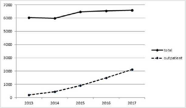
Outpatient surgery provides benefits to patients including a decreased exposure to nosocomial infections, a higher rate of satisfaction [1], and a up to a 68% decrease in direct costs [2]. The Latarjet procedure is now frequently performed on an outpatient basis in up to one third of cases in France in 2017 (Figure 1).

However, the literature regarding the feasibility of outpatient Latarjet surgery is very poor and there is still a need of validation studies [3]. Our aim was to confirm the feasibility of the outpatient Latarjet procedure by comparing the incidence of adverse events and clinical outcomes between patients who underwent inpatient or outpatient Latarjet surgery. Our hypothesis was that the outpatient procedure is both feasible and safe.

Patients and methods

Patients

Study inclusion criteria were as follows: patients were considered if they had a diagnosis of recurrent anterior shoulder instability; were deemed candidates for surgical stabilization; had not undergone prior shoulder surgery, and did not have any significant shoulder co-morbidities. All patients underwent primary surgery for anterior shoulder instability using transfer of the coracoid process (Latarjet procedure). All procedures were performed by a Single Surgeon (SZ). Thirty consecutive patients underwent surgery on an outpatient basis between 2013 and 2017 and were prospectively followed. This group was compared to 30 patients who underwent the Latarjet procedure on an inpatient basis between 2007 and 2012 by the same surgeon.

Surgical protocolA standardized general anaesthesia protocol was followed. An additional interscalene block was administered under ultrasound guidance (single bolus of 20ml of 0.375% ropivacaine) associated with 8mg of a dexamethasone intravenous injection. An open minimally invasive technique was used. The osteotomy of the coracoid process was performed through a deltopectoral approach after the coraco-acromial ligament and pectoralis minor tendon were released and following conjoint tendon exposure and dissection. All harvested coracoid grafts were a minimum of 20mm in length. The subscapularis tendon and muscle was split horizontally. Following glenohumeral capsulotomy, bone on the ventral aspect of the coracoid process and on the anteroinferior aspect of the glenoid rim was decorticated. Any remaining anteroinferior bone bankart fragments were resected. The ventral aspect of the coracoid graft was fixed to the inferior portion of the anterior scapular neck such that the transplant was level with the anterior glenoid rim. The coracoid process was drilled with two 3.5mm holes and fixed with two 3.5 diameter cortical screws; whilst the glenoid neck was drilled with 2.5mm holes to enable compression.

Care was taken to avoid lateral overhang of the graft across the joint line as described by Alain et al. [4]. No additional capsular suture was used. The subscapularis tendon was closed lateral to the graft. Traction on the coracoid graft was avoided all along the procedure to decrease the risk of musculocutaneous nerve injury. The wound was closed in layers with continuous absorbable skin suture. All inpatients had a suction drain inserted. No drain was used in the outpatient group. Sling immobilization was used for one week following surgery. Simple activites of daily living (shower, eating, writing) were immediately permitted. Following one week, self-assisted stretching in all planes was permitted. Running and swimming were allowed after two months, and high-risk sports (rugby, judo…) were allowed after 4 months. All patients were assessed on post-operative day one (by telephone for outpatients); further assessments took place at 1 week, 1 month, 4 months and 12 months post-operative.

Evaluation criteria and statistical analyses

Readmission rates and early complications were recorded. Shoulder range of motion, recurrent instability, persistent subjective apprehension and shoulder pain were compared between groups at one year. Satisfaction rate with the outpatient protocol was assessed. Continuous variables were compared with the independent t-test and categorical variables with the Fisher exact test; statistical significance was set at 0.05.

Results

The 2 groups were comparable at baseline (Table 1). Mean hospital stay in the inpatient group was 2.2±0.4 days. One admission for one night occurred in the outpatient group due to dizziness which resolved without further treatment. No complications occurred related to the interscalene block. There were no reoperations, no nerve injuries and no infections in the series.

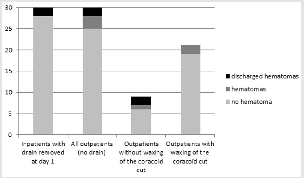
In the inpatient group, drained blood volume prior to drain removal was negligible in 18 patients, less than 100cc in 11 patients and > 100 cc in one patient. All drains were removed on post-operative day one. Seven hematomas occurred within the first 3 weeks following surgery: two in the inpatient group and five in the outpatient group (Figure 2). Four of these hematomas, two in each group, discharged and healed spontaneously. All others healed spontaneously without fistulization. One of these hematomas occurred in a patient with the Factor V Leiden defect (outpatient group). Three of these hematomas, with fistulization twice, occurred in the first 9 patients in the outpatient group and induced a change in the surgical technique. In the following 21 patients, prior to closure, the osteotomy of the coracoid feet was explored, washed and waxed prior to closure during which time a blood clot was typically found. Wax was never used for patients in the inpatient group.

Figure 1: Latarjet procedures statistics in France between 2013 and 2017 (ATIH, technical agency of information on hospitalization, www.atih.sante.fr).

Lupinepublishers-openaccess-orthopedics-sports-medicine

Figure 2: Hematomas formation by group. There was more hematomas in the outpatient group (p<0.01), all hematomas healed spontaneously.

Lupinepublishers-openaccess-orthopedics-sports-medicine

All outpatients but one were satisfied with the procedure. One patient indicated that he would have preferred a one night hospital stay due to postoperative discomfort. At final 12 month followup, no patient had experienced further instability. Six patients answered positively for subjective persistent apprehension in both group (20%). One third of patients of each group have reported occasional shoulder pain. Loss of external rotation was found in half of patients of each group (Table 1).

Read More about Lupine Publishers Journal of Orthopedics and Sports Medicine Please Click on Below Link: https://lupinepublishers-orthopedics.blogspot.com/


Monday, 6 February 2023

Lupine Publishers| Haglund Syndrome – A Case of Bilateral Involvement

 Lupine Publishers| Journal of Orthopedics and Sports Medicine


Abstract

Haglund syndrome is a cause of posterior heel pain and occurs commonly in adolescent girls who wear high heels with restrictive heel counters and may occur in people with rheumatoid arthritis. Is characterized clinically by thickening of the soft tissues at the Achilles tendon insertion or a painful “pump bump” and retrocalcaneal bursitis, Achilles tendinitis, and a prominent posterosuperior calcaneal border or bursal projection. We report a case of a 59 years-old woman with bilateral Haglund syndrome which did not needed surgical treatment, being the conservative treatment enough to improve the patient’s quality of life.

Keywords: Calcaneus; Magnetic resonance imaging; Foot diseases

Introduction

Haglund syndrome is a cause of posterior heel pain and was first described by Patrick Haglund, in 1928, to occur in patients who had a prominence of the posterosuperior surface of the calcaneus [1]. It occurs commonly in adolescent girls who wear high heels with restrictive heel counters and may occur in people with rheumatoid arthritis [1]. Haglund syndrome has also been termed “pump bump,” “winter heel,” “knobby heels,” “calcaneal altus,” “highbrow heels,” and “cucumber heels” [1]. Haglund syndrome is characterized clinically by thickening of the soft tissues at the Achilles tendon insertion or a painful “pump bump” and retrocalcaneal bursitis, Achilles tendinitis, and a prominent posterosuperior calcaneal border or bursal projection [1-4].

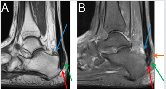
Figure 1: Sagittal section MRI in T1 sequence in A and T2 STIR sequence in B of the right ankle demonstrating posterior calcaneal spur (red arrow), liquid in retrocalcaneal bursa (blue arrow), liquid in subcutaneous calcaneous bursa (orange arrow), and calcaneal tendinopathy (green arrow).

Lupinepublishers-openaccess-orthopedics-sports-medicine

Figure 2: Sagittal section MRI in T1 sequence in A and T2 STIR sequence in B of the left ankle demonstrating posterior calcaneal spur (red arrow), liquid in retrocalcaneal bursa (blue arrow), liquid in subcutaneous calcaneous bursa (orange arrow), and calcaneal tendinopathy (green arrow).

Lupinepublishers-openaccess-orthopedics-sports-medicine

Case Presentation

59 years-old woman with pain and swelling in both feet for four months that get worse when use slippers and when she wake up and put her feet in the floor for the first time in the day. The patient denies other complaints and surgeries. At physical examination refers pain at the palpation of in the posterior and inferior regions of the ankle. The Magnetic Resonance Imaging (MRI) demonstrates posterior and plantar calcaneal spurs, liquid in retrocalcaneal and subcutaneous calcaneous bursa, and calcaneal tendon with heterogeneous signal and thickened (1.4cm at left and 1.2cm at right), markedly in their insertion with adjacent bone edema (Figures 1 & 2). The set of findings are compatible with Haglund syndrome. The patient started conservative treatment with non-steroidal anti-inflammatories, change of footwear and physiotherapy, presenting a good response, not requiring surgery.

Discussion

Haglund syndrome is caused by mechanically induced inflammation of the Achilles tendon and its bursa, from abnormally high pressure between the bursal projection of the calcaneus, the Achilles tendon, and the bursa [1,5]. Patients with bursitis have erythema and swelling over the bursa and tenderness to direct palpation [2] besides pain with dorsiflexion of the foot [6]. Clinically, by physical examination alone, it may be difficult to distinguish Haglund’s syndrome from other causes of hindfoot pain such as Reiter’s disease, rheumatoid arthritis, or isolated local conditions such as superficial tendon Achilles bursitis secondary to poor shoe fit [7]. Plain radiograph in a lateral standing position is useful to assess the presence of a prominent bursal projection of the calcaneum, the Haglund deformity [3]. Loss of a lucent retrocalcaneal recess is an important indication of underlying retrocalcaneal bursitis [3]. The Achilles tendon is swollen, and dystrophic calcifications may also be seen [3].

MRI may be required for ambiguous or clinically equivocal cases [3]. The cardinal soft tissue abnormalities, namely Achilles tendinopathy, and retrocalcaneal and retroachilles bursitis are more easily and directly depicted by MRI [3]. The detection of marrow oedema within the prominent bursal projection is likely to support the repetitive mechanical compression and inflammation as the pathological mechanism in this condition [3]. Treatment of Haglund deformity, with or without bursitis, targets decreasing the pressure and inflammation with openheeled shoes, anti-inflammatory or analgesic medications, and corticosteroid injections [2]. Physical therapy may also help reduce pain. In recalcitrant cases, surgery to remove the Haglund deformity may be necessary [2].

The nonsurgical treatment, combined with modifications in daily shoe wear, is an appropriate initial treatment for pain relief of Haglund’s syndrome [7]. Using ultrasound guidance to inject the retrocalcaneal bursa is a simple, reliable method of ensuring accurate delivery of medication into the bursa and avoiding intratendinous injection [7].

Read More about Lupine Publishers Journal of Orthopedics and Sports Medicine Please Click on Below Link: https://lupinepublishers-orthopedics.blogspot.com/

Wednesday, 21 December 2022

Lupine Publishers| Digit Ratio and Soccer

 Lupine Publishers| Journal of Orthopedics and Sports Medicine


Abstract

The ratio between the lengths of index finger in relation to the length of ring finger of a palm is noticed to as second to fourth digit ratio. Digit ratio tented to shows the quantity of male hormone, to which an individual is exposed in the womb of the mother. Several investigations establish the negative relationship between lower digit ratio and various sports performance as the lower digit ratio settle the high prenatal testosterone hormone. The exposure of prenatal androgenisation masculinizes the human body that impacts the efficiency of sports. Soccer player perceives numerous situations and makes instantaneous decisions. Experienced players are reading the game and anticipating the next move of the opponent. Previous studies have demonstrated the link between cognitive ability and the digit ratio. Similarly, players have numerous cognitive skills to dictate soccer game such as spatial intelligence, awareness, and visual spatial ability. Here, we explore the possible causes of negative associations between lower second to fourth digit ratio and soccer performance. We also think soccer-specific skill performance is likely to be associated with lower digit ratio.

Keywords: Digit ratio; Testosterone; Estrogen; Soccer

Introduction

The peripheral blood cannot be extracted in utero from fetuses [1]. The second to fourth digit ratio (2D:4D) was therefore suggested as a prenatal testosterone marker [2]. Manual second to fourth digit ratio (2D:4D) at the end of the first trimester of pregnancy is believed to be a biomarker of the balance between prenatal testosterone and prenatal estrogen hormone [2-4]. Thereafter, the digit ratio (2D:4D) probably remain unchanged throughout the life [2]. However, Manning (2002) indicated that the digit ratio (2D:4D) was set particularly between week 8 and 12 at the end of the first trimester. The male fetuses mainly produce large quantities of testosterone hormone, primarily from their testis and adrenal glands [5]. This influences brain and other organ systems development [2-6]. In general, the length of index (2D) and ring fingers (4D) in women is about the same (digit ratio=1.00), whereas in men the ring finger is generally slightly longer (digit ratio=0.98) [7]. The length difference between the two digits is higher for men than for women [8-9]. Testosterone influences the growth of the ring finger (4D), whereas estrogen exposure stimulates the growth of the index finger (2D) [7]. The ratio of the index finger to the ring finger (2D:4D) has been shown to be a sexually dimorphic trait [9-10]. Additionally, the ratio of digits (2D:4D) measured by the length of index finger divided by the length of ring finger [7-11]. Therefore, researchers found an index of prenatal testosterone exposure relative to prenatal estrogen exposure [7-12]. The prenatal androgen is likely to be increase, if the digit ratio goes down [7]. Several studies portray the second to fourth digit ratio in which prenatal testosterone hormone was associated. The researches recommend that the lower ratio of digit is a noninvasive feasible indicator for sport success rate Manning and Taylor [13] ; Manning and Hill [14] ;Manning et al. [8]; Hone and McCullough [15] ; Longman et al. [16]; Sudhakar et al. [17]; Sudhakar et al. [18]; Bennett et al. [19]; Kim [20]. As because, adult lower digit ratio (2D:4D) promote the masculine feature [2]. The testosterone (T) is a steroid hormone that develops and maintain masculine feature of human body [21]. The specific aim of this review study was to explore the relationship between digit ratio (2D:4D) and soccer performance.

How does the digit ratio (2d:4d) fixed?

The adult finger length ratio is becoming a widely used research tools to know the tentative trait of prenatal androgens, a diversity of physiological and psychological conditions, athletic ability and sexual orientation [10,22,23]. The differentiation of gonads, fingers, and toes is influenced by HOXA and HOXD genes. HOXA and HOXD genes are also necessary for finger length development and differentiation [24]. Congenital Adrenal Hyperplasia (CAH) is an anomalous hormonal environment that does not function correctly with the adrenal glands [25]. The 21-hydroxylase deficiency, results in the production of surplus quantities of masculine hormones by the adrenal glands [26]. However, researchers Okten and his colleagues studied digit ratio (2D:4D) and 21-hydroxylase deficiency in male (right palm) patients and reported lower digit ratio confirm the 21-hydroxylase deficiency than female and male controls. Women with CAH had a much lower second to fourth digit ratio than women without CAH on the right hand and on the left hand, men with CAH had a much lower digit ratio (2D:4D) than men without CAH [27]. Similarly, researchers [28] reveal the relationship between low digit ratio and CAH. This characteristic also supports a combination of low digit ratio and elevated Fetal Testosterone concentrations [29].

Relationship Between Digit Ratio (2d:4d) With Sports Performance

Researchers [20] widely reviewed the most correlational studies and postulated that low second to fourth digit ratios (high prenatal testosterone and low estrogen hormone) could be a determinant of high sport performance. However, the high performance of rugby depends on low digit ratios [19]. The researchers also discovered differences in the low right-left digit ratios to be a determining factor in elite rugby performance. Keshavarz and his team (2017) studied on three male groups of Wrestlers; they are:

a) World class elite Greco-Roman wrestlers.
b) Collegiate non-elite wrestlers.
c) Sedentary age matched control.

The lower right- and left-hand digit ratios of world class wrestlers were predictors of high wrestling performance compared to other groups [30]. The achievement of the competition phase in team sports was also associated with the ratio of digits (2D:4D). The second to fourth digit ratio was therefore likely to have an impact on the possible athleticism [31]. Similarly, lower digit ratio (high prenatal androgens) has been shown to indicate the sport performance of soccer, surfing, sprinting, endurance, hand grip strength, rowing, kabaddi, swimming, Tennis [8,13-18,32].

Digit Ratio as Soccer Performance Determinant

High prenatal testosterone and low prenatal estrogen hormones are likely to be a strong predictor of soccer performance [7]. Competitive achievement is a major objective of soccer in connection with prenatal androgenization [31]. This prenatal situation influences the judgment of the visual perception [13]. Therefore, according to [7], “Striking a moving opponent or ball requires fine judgment of distance. Determining the exact point of impact demands an accurate perception of the surface of the target as it moves through space” (p.128). However, researchers studied on different types of soccer players and noticed ‘professional’, ‘International’ and ‘1st team players’ had lower digit ratio (2D:4D) than the ‘control group’, ‘youth team’ and the ‘players who had not represent their country’ respectively [13]. Similarly, the International presence of the player in a match is greater for the lower digit ratio individuals [7]. The lower digit ratio could therefore provide an additional discriminator to help estimate soccer capability. Prenatal testosterone exposure also influences professional soccer players’ aggressive behavior. Researchers indicated that exposure to adult and prenatal testosterone detects the number of fouls per match that confirm the aggressiveness of players [33]testified by a low second-to-fourth digit ratio (2D:4D . Furthermore, aggression guarantees the dominant behavior that is essential in competitive sport.

Association among Digit Ratio, Visual-Spatial Ability and Left Handedness

Digit ratio is a putative indicator of sport performance differences [34]. A study concerning several sports related psychological variables (mental toughness, aggression, optimism scale, coping strategies, and goal orientations) with masculine digit ratio reported high scores of optimistic dispositions than those with feminine digit ratio. The study also claims that mental toughness partly determined on gestation period [34] that benefited for gender, age and sporting experience [35]. Mental rotation score test [36] can measure the visual-spatial intelligence [36-38]. Manning and Taylor found negative association between lower digit ratio and high mental rotation scores in males. So, the visual spatial intelligence may partially develop on intrauterine life [13]. High prenatal testosterone exposure is likely to associated with handedness [39,40]. Left-handed people dominated by the right hemisphere and assists visual spatial ability [41]. Interestingly, androgenisation exposure influence the right palm more than the left palm [27,42,43]. Right palm digit ratio is also significantly connected with several psychological and behavioral traits compared to the left palm digit ratio [43].

Cognitive Abilities Influence on Soccer Performance

Most team sports, particularly in soccer players, need to pursue numerous situations that are changing quickly [44]. Elite players perceive the situations and make the appropriate choice at the right moment [45,46]. Therefore, technical and tactical ability influence the outcome of the match [47,48]. Elite players perform the technical and tactical skills better in compared non-elite counterparts [49]. However, researchers postulated that distinguished correlations in male between more masculine digit ratio (lower digit ratio) and in visual-spatial ability [13,22]. On the other side, females have prone to more feminine digit ratios (higher second to fourth digit ratio) should relate to higher scores for depression (Repeat). Therefore, high prenatal testosterone exposure is likely to be predictor of soccer performance as well as cognitive ability [13]. Research also shows that human behavior and the status of cognition can result from prenatal androgenization [50].

Conclusion

Most correlational study reveals the negative relationships between lower digit ratio and sports performance. Low second to fourth digit ratio (2D:4D) can be an indicator in scouting potential athletes especially soccer players. In multifaceted aspects, lower digit ratio is likely to be a potential indicator of soccer specific skill performance. Further studies are required to clarify whether lower second to fourth digit ratio could predict the soccer skill performance in multifaceted aspects including passing, dribbling, control, shooting and decision making within a dynamic situation. In addition, we realized that sporting success might be depended on our hands’ fingers length ratio a long with other variables.

Read More about Lupine Publishers Journal of Orthopedics and Sports Medicine Please Click on Below Link: https://lupinepublishers-orthopedics.blogspot.com/

Wednesday, 9 November 2022

Lupine Publishers| How to Pick a Good Surgeon

Lupine Publishers| Journal of Orthopedics and Sports Medicine


Abstract

A shoulder surgeon has a shoulder injury and requests a specific shoulder surgeon colleague to operate. Instead, the patient is sent to an expert shoulder surgeon with an international reputation who delivers many lectures and writes numerous papers and book chapters. Surgery is performed but the shoulder gets infected and the overall result is unsatisfactory. After a legal challenge, the patient is allowed to go to the surgeon originally requested based on the patient’s own insights into the profession. Revision surgery is performed and iatrogenic damage is discovered from the first surgery. However, the result of this revision surgery is good. This anecdotal case contains many aspects of healthcare decision making, control, and culpability which will be further elucidated here.

Keywords: Picking a Surgeon; Good Surgeon

Introduction

I am sure many of us have either heard or been asked the question, “How do you pick a good surgeon”. Of all the academic questions we deal with on a daily basis as surgeons/physicians, this question perplexes us beyond our educational background due to the multitude of types of information needed to truthfully answer. Now, consider how hard this decision is to make for the lay person/patient who does not have the same insights into the inner workings of the profession. Before being able to pick a good surgeon, we need to define what makes a good surgeon. A surgeon is a medically trained doctor who has chosen to specialize in pathologies that can be treated with a surgical option but also has the knowledge and ethics to know when to treat non-surgically. However, most pathologies have a spectrum ranging from near normal to end-stage and management options that often depend on several factors: stage of the pathology, chronological versus physiological age, activity level, co-morbidities, availability of resources, etc. Therefore, anyone dealing with a specific pathology (e.g. osteoarthritis or shoulder instability) should not only understand the whole spectrum of the pathological process and its variants, but also be adept at managing these different stages with “stage specific solutions” instead of employing a “one operation fits all” philosophy. Picking a surgeon for oneself or one’s family has several pathways, some controlled by the patient, and some by the agency responsible for the financial settlement.

There are no standard pathways for this choice, but when trying to understand what factors are of influence, we are able to identify many.

Commonly Used Factors When A Patient Seeks A Surgeon

a) A primary caretaker (family member/family doctor/ friend) recommends a surgeon due to personal experience or word of mouth recommendation [1,2]. While this sort of recommendation can be based on limited interactions with the surgeon, and his/ her outcomes, someone who has had a prolonged interaction would be able to gain a better insight into the outcomes from that surgeon. However, when limited interactions are the basis of a recommendation, this may prove to be detrimental to the patient since, for example, a surgeon who once performed a successful carpal tunnel surgery may not be the best option for performing a shoulder replacement.

b) An insurance company/workers compensation board will limit choice from surgeons within their approved network based upon whom they have an agreed terms and conditions contract [3]. This network is not based purely on quality of surgical skill, but is more based on a financial agreement with the insurer [3]. The financial entity does not have the responsibility of auditing the surgeon’s outcomes, and hence can rarely make the choice easier for the patient except to limit the choice to a specific group of surgeons.

c) A surgeon with a higher rating on social media platforms is viewed as a better option than one with a lower rating [4]. Very few people understand how social media ratings come about. Individual ratings tend to be either strongly positive or strongly negative. Unhappy patients are more likely to complain, and thus there is a bias toward negative spontaneous social media ratings. Social media provides a platform from which the angry patient can voice their discontent. Happy patients are generally more interested in getting on with their lives following a successful surgery than devoting a lot of time to grandstanding on social media. Conversely, many well-run private practices are very efficient at collecting ratings from happy patients while avoiding collecting ratings from unhappy patients in their clinics. The latter practice may balance the equation by counteracting the negative bias or perhaps tilt responses to excessively positive. Social media ratings by patients are also more determined by their overall customer experience than their surgical outcome. Factors such as wait time, parking, office atmosphere, and interpersonal interactions with the surgeon and staff have been shown to have a stronger impact on social media ratings than quality of the outcome. This is logical since humans have daily interpersonal interactions which provide a standard for comparison, unlike a surgical procedure which is generally limited to a small number of times in an individual’s life. Hence, social media may focus on extraneous factors and can be manipulated. Thus, online patient ratings are not the best indicator of a good surgeon. If every patient had to input a rating, usefulness might be improved and this may represent a direction for the future.

d) Speed of Surgeon [5]. Is a slow surgeon better than fast surgeon, or vice versa? Some surgeons have a reputation for being very quick while others have a reputation for being very slow, or sometimes described as meticulous. Inorder to fully appreciate this issue, one needs to understand what a particular pathology requires in order to resolve the symptoms. Taking a very simple example, carpal tunnel release, some surgeons take 4 minutes to complete this through a 1 cm incision, versus others take 90 minutes or more with a 5 cm incision or an endoscopic technique. While a fast surgery may miss some steps, although all being achievable for a carpal tunnel surgery within the 4 minutes, the same surgery with a larger incision and a long operative time may have a greater risk of infection and scarring, due to many more surgically performed strokes of the knife. The latter refers to the surgeon performing more surgery than is necessary to achieve the result, under the label of being “meticulous” and “precise”. Hence, performing more surgery than is necessary to treat the pathology is detrimental to the outcome. Therefore the speed of the surgeon is not always easy to understand without the context of the surgical results achieved by that surgeon.

e) Communication skills [6]. A surgeon who can communicate well is better than a surgeon who is not a good communicator [6]. Communication is a central and under-valued aspect of any surgeon’s skill set. The most important factor in healthcare is the needs of the patient. Hence, it is the patient who needs to understand the problem, and how to fix it, to a level compatible with his/her educational background. Communication between the surgeon and the patient allows the necessary information to be patient-centric, in order that confidence is built within that relationship [6]. A surgeon who is able to communicate well with all the patient demographics and educational levels in his/her practice should be viewed in a positive light, while the abrupt, minimal communicator cannot justifiably impart the necessary information. If a patient is not made to understand the whole process, he/she cannot be an effective part of the treating team, and patients should be considered as part of the team.

f) Academic impact [7]. “A surgeon who writes a lot of papers is better than one who doesn’t write papers”. Surgery, like any athletic pursuit or manual trade (carpenter, motor mechanic, etc.) is a dexterous skill, combined with knowledge and the translation of that knowledge to the physical activity. As any athlete knows, it takes a very long time to train one’s body to perform a physical task at a certain level, more than 90% of their time. So if an athlete spends a significant amount of time reading about a skill, and less time translating the skill into his/her own physical performance, the results are failure in competition. Surgeons are no different. Those who spend significant amounts of their time in research, giving talks, writing papers, advocacy and committee work, etc. have less time to see and diagnose patients, operate and treat patients, follow up and assess patients’ outcomes etc. Although there is not a welldefined parameter to judge the optimal times for cerebral versus dexterous activities, the principle of the athlete is relevant to the surgeon.

g) Education at high profile institutions [7]. Those with educational backgrounds in high profile institutions are considered with greater regard than those with lesser academic profile institutions [7]. It should be constantly remembered that surgery is an intricately intermingled cerebral and dexterous skill. Being overweight in either may not compensate for achieving the best results. High profile institutions achieve their profile by many different means, but none achieve it by the quality of the training they provide to the next generation of surgeons. The institutions select their surgeons based on qualities important to the mission of the institution, and it can be assumed the trainees who are trained there will both actively and passively achieve some of those qualities. However, great institutions can produce poor quality surgeons and lesser institutions can produce outstanding surgeons so it is not a guaranteed assurance to rely on the surgeon’s educational institution’s reputation.

h) Specialty Board Certification [8]. The American Board of Medical Specialties (ABMS) and its 24 member boards are organizations established to ensure consistency of training and practice within established guidelines for surgical specialists. These organizations seek to ensure that surgeons have a specific knowledge base and have completed requisite numbers of surgical procedures during their training. In certain competitive areas such as cosmetic surgery, physicians lacking ABMS certification seek to deceive prospective patients by creation of non-ABMS boards which do not require any specific training and allow surgeons to join simply by paying a fee. In orthopaedic surgery the ABOS (American Board of Orthopaedic Surgery) seeks to ensure this quality with didactic exams and a review of cases performed by the surgeon. While these boards try to ensure quality, they do not have the capacity to be stringent enough and would require considerable investment to improve quality of candidates. So a “Board Certified” surgeon may not always be a better choice than a non-board certified surgeon, but the certification ensures a minimum quality.

i) Availability of the surgeon [9]. Some consider a surgeon who is easily available for new appointments to be better than those who are really difficult, or have a very long waiting list. Similar to a restaurant, would you prefer to eat at an empty restaurant or one where it is always busy? The three ‘A’s of medical practice priority used to be (and should be) “Ability, Affability, Availability”. Unfortunately, with the societal shift in attitudes the order has reprioritized to “Availability, Affability, Ability” [9].

j) Intellectual honesty. One of the most important qualities for a medical professional is the ability to honestly assess one’s own level of knowledge and skill. Not all surgeons are equal, just like not all tennis players or restaurants are equal. Hence, when a surgeon is unable to perform a certain operation that may be the optimal treatment for a particular patient, it takes intellectual honesty for that surgeon to communicate this discrepancy and refer the patient to another surgeon who is able to perform that optimal procedure. It is dishonest to perform a procedure that is not in the best interest of the patient just because it is a procedure that the surgeon is able to perform, regardless of the suitability of the performed procedure to the presenting pathology. An example of this is when surgeons perform reverse shoulder replacements instead of anatomical total shoulder replacements despite the presence of all the factors that can make an anatomical replacement successful. Although a reverse replacement is an easier procedure to perform, it sacrifices an intact rotator cuff. The price of an inappropriate operation is paid at a later date by the patient, with very fewer good options if the reverse shoulder replacement fails.

k) Volume and institutional protocols [10]. Frequent performance of a procedure by a surgeon naturally leads to technique refinements if the surgeon is thoughtful about the process. Likewise, the operating room staff and other associated individuals such as anesthesiologists become familiar with the surgeon’s routine and optimize their functions accordingly. Further, some surgical procedures benefit from ancillary services such as specialized nursing care, physical therapy or occupational therapy working in a coordinated manner immediately following the procedure. In these instances, the good surgeon’s skills are only one component of a successful surgical outcome, albeit an important one. Surgeons who perform low volumes of surgery are likely to be less dexterous than those who perform high volumes of surgery, with a required debate regarding the definition of high, low, and adequate volumes to maintain dexterous skills.

l) Word of mouth reputation [1,2]. The natural course of surgical practice is that the surgeon reaps what he sows. That is, if a surgeon performs an operation successfully on one patient, that patient is more likely to tell their friends and their referring physician about their experience [1,2]. Those patients and providers are more likely to send other patients, and thus forms the basis of “word of mouth” referenced in the first paragraph. This leads to a snowball effect on the surgeon’s practice which grows over the course of a career. Name recognition for surgeon grows over time and hence the effect of word of mouth becomes more powerful for them individually. Name recognition would seem to imply a larger pool of satisfied patients, but this is not always true as other factors can lead to name recognition such as advertising. Further, it is likely this effect is more powerful in smaller communities with fewer providers compared to larger communities.

m) Access to specialized equipment [10]. Specialized equipment may allow an appropriately-skilled surgeon the ability to perform a procedure in a less invasive, safer, or more efficient manner. The recent innovations in robotic surgery provide an example for certain procedures. However, it is important to note that just because a technology is new or “hi-tech”, it is not necessarily better or safer than established methods. For instance, endoscopic carpal tunnel release has no superior data compared to minimal incision release. Furthermore, technology presents a constantly changing environment and yet it can take years for a particular innovation to mature to its fullest potential or fade out.

Read More About our Lupine Publishers Journal of Orthopedics and Sports Medicine Please Click on Below Link: https://lupinepublishers-orthopedics.blogspot.com/