Showing posts with label journal of orthopaedics. Show all posts

Monday, 3 July 2023

Lupine Publishers | Effect of Intensive Integrative Therapies on Pain, Disability and Quality of Life in Patients with Osteoarthritis Knees: A Comparative Observational Study

 Lupine Publishers | Orthopedics and Sports Medicine Open Access Journal

Abstract

Objective: To assess the differential effect of intensive yoga, yoga and naturopathy and yoga and Ayurveda management for Osteoarthritis of knees Design: Pre-post comparative study

Participants: Ninty-five individual prediagnosed with knee osteoarthritis according to ACR guidelines aged between 30-75 years were randomized into three groups, i.e., Yoga (n= 36), yoga and Naturopathy (n=30), Yoga and Ayurveda (n=29). All three group received their respective intervention for 1 week at arogyadhama, prashanti kutiram, S-VYASA.

Outcome measures: The primary outcome variables were visual analog scale (VAS) for pain on activity, Western Ontario and McMaster Universities Secondary Osteoarthritis index (WOMAC) and secondary outcome variables were anthropometric measurements and 6 min walk test were measured on day 1 and day 7.

Results: There was significant reduction in VAS scale on activity in yoga group (p < 0.001), whereas in Ayurveda group showed significant improvement in stiffness (p <0.001), and significant reduction in weight and pain on rest (p < 0.001) was in naturopathy group after 7 days of intervention.

Conclusion: IAYT practices combined with other therapies had better effect than alone IAYT. There were significant changes seen within groups.

Keywords: Knee Osteoarthritis; Integrative Approach of Yoga Therapy (IAYT); VAS; WOMAC; Ayurveda; Naturopathy

Introduction

Movement is a medicine for creating change in a person’s physical, emotional, and mental states. Disease is the destroyer of health wealth and mind. Lack of movement of knees leads to the later. The most common of lack of movement is Osteoarthritis (OA) OA knee mainly occurs in elders and middle age people and is more common among women and overweight subjects. The symptoms include pain, stiffness and decreased range of motion (ROM), which result in limited activity and reduced quality of life. The prevalence increases with age, and by the age of 65, approximately 80 percent of the US population is affected. It is the second most common rheumatological problem and most frequent joint disease with prevalence of 22% to 39% in India OA is a heterogenous disease, involving complex and interacting mechanical, biological, biochemical, molecular and enzymatic feedback loops with cartilage degeneration as the common, final event [1] despite this degeneration, OA is an active process and a network of mechanisms reacting to stress or injury on the joint. All joint features are affected in OA [2]. Structural changes include cartilage fibrillation, degeneration of articular cartilage, thickening of subchondral bone, osteophyte formation, synovial inflammation, degeneration of ligaments and meniscus, hypertrophy of joint capsule, cellular and molecular changes in nerves, as well as changes to periarticular muscle, bursa, fat pads [3]. The loss of cartilage and modifications to bone and synovial membrane contribute to an unfavorable biomechanical environment which increases stress on the joint and furthers the progression of cartilage degradation [4]. Non-steroidal anti-inflammatory drugs (NSAIDs), including cyclo-oxygenase II inhibitors and non-opioids analgesics such as acetaminophen have been the most popular method of managing pain in musculoskeletal tissues. Tradational System of medicine like Ayurveda, Naturopathy and Yoga also specify the symptoms and mode of treatment. So a observational study was planned to understand the effect of this traditional system of medicine.

Aim of the Study

a. To study the differential effect of three Indian systems of medicine on the outcome of OA knee management

Objective of the Study

a. To assess the differential effect of intensive yoga, yoga and naturopathy and yoga and Ayurveda management for Osteoarthritis of knees on pain and stiffness.
b. To assess the differential effect of intensive yoga, yoga and naturopathy and yoga and Ayurveda management for Osteoarthritis of knees on physical functions.
To assess the differential effect of intensive yoga, yoga and naturopathy and yoga and Ayurveda management for Osteoarthritis of knees on anthropometric measurements.

Material and Methods

Source of data

A total of 95 participants aged 30-75 years, were registered from Arogyadhama, a home-based health centre, S- VYASA, bangalore.

Sample size

The sample size was calculated with G-power software by fixing the alpha at 0.05 powered at 0.8 and an effect size of 0.71 based on the mean and SD of an earlier study.

Inclusion criteria

The inclusion criteria were patients clinically/radiologically diagnosed with mild to moderate OA knees according to American college of rheumatology (ACR) guidelines and with associated comorbidities were included in the study.

Exclusion criteria

Patients with severe OA knees, rheumatoid arthritis, autoimmune diseases, malignancies, knee surgery or kneearthroscopy and knee pain caused due to congenital dysplasia were excluded.

Ethical consideration

The study was approved by the Institutional Ethics Committee of S-VYASA University (approval letter no: RES/IECSVYASA/ 107/2017 dated 23rd October 2017). Signed informed consent was obtained from all the participants included in this study.

Design

Participants were randomly divided into three groups, i.e., Yoga alone, Yoga and Naturopathy, Yoga and Ayurveda group. All three groups underwent intervention of Integrative approach of yoga therapy (IAYT) and respective therapies for 6 days and assessments and treatment plans for participants were discussed with in-charge doctor and therapists. All groups continued their medication as per prescription.

Grouping and posology

Three groups pre-post comparative study.
Group A: Table 1 Standalone Yoga group

Table 1: Time table.

Group B: Table 2 Integrative Yoga and Ayurveda group

Table 1: Time table.

Group C: Table 3 Integrative Yoga and Naturopathy group

Table 3: Time table.

Group B: Table 2 Integrative Yoga and Ayurveda group

Assessment criteria

Primary outcome variables
a) Visual analog scale for pain on activity
b) WOMAC Indian version
c) 6 min walking distance test - The subjects were asked to walk on a flat surface for 6 minutes and the distance covered was expressed in meters.

Anthropometric measurements

a) Height: It was measured with stadiometer in centimeters (cm), and later converted into meters.
b) Weight: Weight was measured using research grade electronic weighing scale.
c) BMI: By using formula, weight in kg/ height in meter2, calculated the body mass
index of every individual.

Observations

Table 4

Table 4: Observations.


Results

Recapitulation

In the current study, variables were taken at baseline and following one week of intensive yoga, yoga and Ayurveda and yoga and naturopathy therapies. The primary outcome variable was pain pain assessed through Visual Analogue Scale (VAS) while resting, walking on plane surface, climbing stairs up and down as well as WOAMC scores. The secondary assessments included anthropometric measurements; and 6 min walking distance. We also recorded the vital parameters such as systolic and diastolic blood pressure, pulse rate and respiratory rates at baseline and at the end of one-week intervention for all three groups.

Yoga and ayurveda group

Within group comparison revealed that as compared to the baseline following variables showed significant improvement in weight, body mass index, 6-min walk test, WOMAC global score, WOMAC-pain, stiffness and physical functioning, visual analog score on rest, walking, climbing up and down. The values of within group comparison through paired sample t test are mentioned in Table 4.

Yoga and naturopathy group

Table 5: With-in group comparison changes in yoga and ayurveda group.

Within group comparison revealed that as compared to the baseline following variables showed significant improvement in weight, body mass index, systolic blood pressure, respiratory rate 6-min walk test, WOMAC global score, WOMAC- pain, stiffness and physical functioning, visual analog scale on rest, walking and climbing up and down. The values of within group comparison through paired sample t test are given in Table 5.

Yoga group

Within group comparison revealed that as compared to the baseline following variables showed significant changes in weight, Diastolic blood pressure, Pulse rate, 6 MIN walking distance, WOMAC global scores, WOMAC-pain, stiffness and physical functioning, visual analog score on rest, walking and climbing up and down. The values of within-group comparisons through the paired samples t-test are mentioned in Table 6.

Table 6: Within group changes in yoga and naturopathy group.

Table 7: Within group changes in yoga group.

Abbreviations

WT: Weight, DBP: Diastolic Blood Pressure, PR: Pulse Rate, WGS: WOMAC Global Score, STF: Stiffness, PF: Physical Functioning, VAS: Visual Analog Scale, WK: Walking, DW: Down, BMI: Body Mass Index, SBP: Systolic Blood Pressure, RR: Respiratory Rate, WOMAC
Table 8 presents the changes in all three groups at baseline and at the end of one week. None of the variables between groups were found significant both at baseline and following the one-week intervention duration.

Table 7: Results of the study.

Outcome Measures

All participants were assessed for primary and secondary outcomes twice, at baseline (day 1) and end of study period, day 7.

Primary outcomes

i. Visual Analog Scale (VAS): The visual analogue scale is a psychometric response scale which was used to assess pain on climbing stairs (20 staircases), climbing down from stairs (20 staircases) as well as following 6 min walking on flat surface. It was be used as an instrument to measure subjective quantification of pain. When responding to a VAS item, respondents was be asked to specify their level of pain by indicating a position along a continuous line between two endpoints.
ii. WOMAC: The WOMAC has been extensively evaluated in populations suffering from osteoarthritis (N Bellamy, Buchanan, Goldsmith, Campbell, & Stitt, 1988). The Indian version of WOMAC was used to assess the disability and quality of life [5]. Western Ontario and McMaster Universities Secondary Osteoarthritis index (WOMAC) is widely used in the evaluation of Hip and Knee Osteoarthritis. It is a self-administered questionnaire consisting of 24 items divided into 3 subscales assess the three dimensions of pain, disability and joint stiffness in knee and hip osteoarthritis [6].

Secondary outcome measures: Anthropometric Measurements

i. Height: Was measured in Centimeters (cm)
ii. Weight
iii. BMI: 6- min walk test
iii. BMI
iv. 6-min walk test

Discussion

Modus operandi of yoga and naturopathy therapy

Possible mechanism of improvement in yoga and naturopathy group is detoxification treatment, diet restriction and physical activity which help them more in reducing pain and improve the physical functioning. Yoga and naturopathy integrated therapies are found to reduce inflammatory markers in patients with chronic inflammatory disorders [7]. This could be the mechanism of action for improvement in the outcomes of OA knees through Yoga and Naturopathy.

Modus operandi of ayurveda therapy

The possible mechanism of action behind reduction of all the symptoms is due to medicated oil application frequently but the long-term effects of such applications need to be studied Ayurveda and Naturopathy therapies was planned by respective doctors.

Conclusion

Although statistically insignificant, the current study revealed that integration of naturopathy to yoga therapy was effective in reducing weight, and pain, whereas integrating Ayurveda to yoga therapy could lead to better reduction in stiffness in patients with OA knees. Such non-significant trends could be tested using larger sample size with prolonged intervention period in future studies.

Strength of The Study

The strengths of the study are:
a) This multidisciplinary study encompasses the fields of yogic science, Ayurveda and Naturopathy.
b) No earlier study has reported integration of yoga with Ayurveda and naturopathy for OA knees.
c) Because the duration of intervention was short, acceptability and adherence to therapy was good
d) As integration were delivered through a standard protocol, it could be reproduced in the exact way for all cases.


Read more about Lupine Publishers Journal of Orthopedics and Sports Medicine Click on the below link: https://lupinepublishers-orthopedics.blogspot.com/


Monday, 11 October 2021

Lupine Publishers| A Rare Case of Fracture Dislocation of Hip: Is There a Pip kin's Type 5?

 Lupine Publishers| Orthopedics and Sports Medicine


Abstract

Fractures of the femoral head associated with neck fracture and posterior dislocation of the hip are uncommon. Pip kin and later Brumback classified these complex fracture dislocations. However in today's era patient have more atypical injury pattern. We encountered a case which has never been described in literature and cannot be fit into any previously described classification system. We present the radiological details and management.

Keywords: Femoral Head Fracture; Trochanter Fracture; Posterior Dislocation; Acetabular Fracture; Sciatic Injury; Pipkin's Types; Total Hip Replacement

Introduction

Modern day accidents not only produce unpredictable but at times unclassifiable injuries. Fractures of the femoral head associated with posterior dislocation of the hip are uncommon as such [1]. Pip kin was the one who sub classified Epstein-Thomas type V fracture-dislocations into four additional subtypes which is still most widely followed [2]. Later Brumback et al. further classified femoral head fractures emphasizing hip stability, with type "B" injuries being unstable [3]. The paucity of these cases is the main constraint to make an algorithm of management. Nevertheless the results depend on early anatomical reduction with the potential threat of osteonecrosis irrespective of approach [4,5]. We hereby describe a case of posterior dislocation of hip which is novel and never been described before. The purpose is to make the fraternity aware of such a case which is unclassifiable even though the management protocol may not be grossly different.

Case

A 52 year old migratory laborer had an accident at civil construction site where he fell down from a height of 20 feet landing first on knees (in kneeling position). He presented to emergency department with severe pain in right groin, and inability to move the left lower limb. On examination the limb was shortened, externally rotated and a bony mass felt in the loin which did not move with the movement of distal thigh. There was severe tingling and decreased sensation in front and lateral aspect of leg and patient was unable to dorsiflex his ankle and toes. His pulsations were normal but he had had transient hypotension with tachycardia which corrected with initial resuscitation. First X ray showed fracture about right hip (Figure 1) mostly a trochanteric fracture but a careful evaluation showed incongruity of head and a "vacant" on super lateral acetabulum. A CT scan was followed wherein the fracture was better delineated. A posterior dislocation of hip with fracture about the trochanter was seen. The head was also fractured and the infra foveal part could be seen lying the acetabulum. A fracture line also ran through the posterior wall of acetabulum which was not more than 25% of the wall and also was undisplaced (Figure 2a- 2c). The patient was admitted and limb placed in Thomas splint and prepared for next morning. No attempts to reduce the dislocation were made.

Under general anesthesia, the patient was positioned laterally and a Moore's posterior approach was taken guided by dislocation and wall fracture. The gluteus maximus had a huge rent and external rotators were torn, the head was seen indenting the sciatic nerve (bowstring effect) which was contused but intact (Figure 3a). The neck was ostetomised with help of saw at the appropriate level and the calcar was reconstructed using encirclage wiring (Figure 3b). Ethibond was used to suture the greater trochanter. Next 2 temporary K wires were used in the ace tabular walls and reaming done and cement less cup size 52 was fixed augmented with 2 screws (Figure 3c). The K wires were removed after the cup was found stable. The femur was prepared and uncemeneted corail stem size 11 was inserted and size 36x0 femoral head was found stable. C arm was used to confirm the placement of implants (Figure 3d). All soft tissues were closed in best possible way, the hip was stable (Figure 3e). An abductor brace was applied post operatively. Postoperatively day one faradic stimulation was started and patient was allowed side tuning. Check X ray was done which showed a reasonable reconstruction (Figure 4). The patient was allowed sitting with non weight bearing mobilization from day 3 after the pain had subsided. 3 weeks after surgery the patient was discharged when he went back to his home state and never came back for follow up.

Discussion

Femoral head fractures in combination with posterior dislocation of the hip are rare presentation. Since first reported by Birkett in 1869 only a small number of cases have been reported world widen [6]. Epstein et al.'s found about 10% in their series. It was Pip kin who subs classified these fractures and various other classifications have been proposed by several authors who claim to be improved version [5, 7]. Pipkin's classification remains most popular and widely accepted. They divide them into 4 types with type 3 having associated ace tabular fractures and type 4 having associated neck fractures. Our case is unique with fracture in the trochanteric area. The trochanteric area has never been described. The ace tabular fracture is also an addition actually a combination pattern of Pipkin's 3 and 4. We believe that there should be a type 5 including a both neck and ace tabular fracture in association of head fracture and a subtype T wherein fracture configuration is in trochanteric region instead of classical neck.

The cases of type 3 or 4 are very sporadic for any protocol to be made. In younger individual attempts to fix the neck fragment, often augmented with vascularised fibular graft has been made in past but long term follow up is lacking. Osteonecrosis is a complication and surgeons have tried different approaches with varying results [8,9]. In our case closed reduction was not possible and moreover the impeding ischemia to sciatic pushed.com for an early operative intervention. Taking into consideration- a difficult fracture pattern, age, urgency of surgery, peripheral set up hospital and an invincible avascular necrosis; we thought a Total hip arthroplasty (THA) was an appropriate solution than open reduction. Yet the challenges of choosing the right implant from the whole Pandora box of prosthesis implants remained. In surgery, the aim was to stick to principles of bone preservation like calcar reconstruction and thus avoiding the distal fixation stems which were reserved for future revision if a situation arose. A constrained hip would have more advantages in this circumstance of soft tissue rent, but the large head option of 36 was stable enough in our case. We admit management to be debatable with expertise hands. Final outcome does depend on return of sciatic function and proper occupational therapy since he was a laborer. A draw back in our report remains that despite best attempts to persuade the patient to visit nearest ortho clinic; he never turned up but telephonically did tell to having a over the counter sitting job in the village and satisfied with his hip.

Conclusion

The case highlights a rare injury and attempts to give an expansion to the present Pipkin’s classification. Even though the demerit of this report is a lacking follow up X ray and clinical picture, the initial presentation is worth to be noted among the practioners and scholars.

Read More About Lupine Publishers Journal of Orthopedics and Sports Medicine  Please Click on Below Link: https://lupinepublishers-orthopedics.blogspot.com/
Wednesday, 2 June 2021

 Lupine Publishers| Orthopedics and Sports Medicine Open Access Journal (OSMOAJ)


Mini Review

Osteogenesis Imperfecta (OI) is an inherited disorder of bone characterized by bone fragility and increased fracture risk affecting approximately 25,000 children and adults in the.com [1]. OI is caused by different gene mutations involving the synthesis of type I collagen alpha chains. Recently, mutations affecting post translational processing of type I collagen as well as several non-collagenous proteins (SP7. Osterix) involved in the Wnt signalling pathway have been recognized. To date, 19 genes are implicated in osteogenesis imperfecta phenotypes [2]. In individuals with OI fractures occur throughout the lifetime, more frequently at the extremes of age. Fractures in OI may first be recognized in ultrasound studies during pregnancy but fracture risk while increased in childhood, tends to decrease following puberty only to increase around age 50 in both women and men. Compared to adult osteoporosis, there have been no systematic studies evaluating optimal daily intake of calcium or vitamin D on BMD or fracture incidence in adults. Current use is highly variable in adults who: a) either do not regularly take supplements or, b) take calcium and vitamin D without, measurement of serum 25(OH)D levels or urinary calcium excretion. Both hypercalcuria and renal stones occur in adults with OI although the incidence is not reported. Furthermore there is little documentation of vitamin D levels in adults with OI.

An extensive literature describes effective pharmacologic treatment of OI in children. Initially based on treatment with a second generation bisphosphonate pamidronate, treatment options have expanded to include third generation bisphosphonate zoledronic acid as well as denusomab, a RANK ligand inhibitor [3]. Teriparatide (hrPTH) use is restricted in children.

However, the indications for pharmacological treatment in adults with OI is less well defined than in children. Symptoms directing the physician towards initiating treatment include ongoing fracture rate, generalized bone pain and limitation in mobility affecting activities of daily living.

For that reason, physicians tend to rely on current adult osteoporosis treatment guidelines [4]. Treatment regimens include bisphosphonates used orally (alendronate, residronate) or by parenteral administration (pamidronate, denusomab and teriparatide). As an example, a recent study comparing teriparatide vs.intravenious neridronate, a third generation bisphosphonate, reported that while BMD changes at the lumbar spine at 24 months were 5.1% with teriparatide vs -1.6% with neridronate (P<0.001) there was no significant difference in new fractures during the 24-month treatment period [4]. Note; There was no control group in this study [5]. Treatment with anti-sclerostin antibody, recently shown effective in improving bone density and decreasing fracture rate in adult osteoporosis, is the subject of a current phase 3 treatment protocol (Mereo Pharmaceutical).

Fractures are the hallmark of OI and thus, orthopaedic care is the mainstay of treatment: Coordination of orthopaedic and medical care is important in order to limit fractures and the time constrained by disability. It is useful for the orthopaedic surgeon to know about prior treatment with drugs such as bisphosphonates or teriparatide when surgical repair of a fracture is required as prolonged treatment with these drugs will alter bone quality and may affect repair. In OI the incidence of lifetime surgical intervention in adults approximates an estimated 60-70%. Surgical repair of fractures or long bone deformities in adults with OI may present various difficulties. Low bone mineral density, skeletal or joint deformities, or instrumentation from previous surgeries complicates surgical stabilization of fractures. Furthermore, the adult with OI may present with post-traumatic or accelerated degenerative joint disease, kyphoscoliosis, and spondylolisthesis. Pre and post operative anaesthesia care, including the process of intubation, is a major problem particularly in patients with severe, restrictive pulmonary disease or tracheal deformity

Orthopedic surgery in OI may have 4 major roles: to treat fracture; to prevent recurrent fractures by adding long stabilizing implants; to correct deformity of long bones or spine; and to treat degenerative joint disease [4]. In all these cases, internal fixation by long intramedullary implants is preferred since it minimizes stress changes in the bone. Continued improvements in implants have decreased the risk of re-operation. Spine surgery is indicated for severe scoliosis or kyphosis to prevent pulmonary restriction or slippage of the lumbosacral joint. Fixation with multiple pedicle screws is an advance over the past decade which provides better correction and stability than wires or hooks [6]. Basilar invagination at the craniocervical junction is a silent but progressive threat in a minority or patients which may cause spasticity and weakness. In severe cases surgical decompression and stabilization is indicated.

Arthritis of large joints is more common in OI than in the general population, and occurs at an earlier age. It most commonly affects the hips and the knees. Protrusion of the femoral head in the acetabulum may accompany hip disorders. Although conservative care is the mainstay of treatment, total joint arthroplasty has been successful in many patients with type 1 OI. Over time, orthopedic/ medical issues tend to pose an increasing problem for the adult with OI. Skeletal deformities impair daily activities pulmonary function tends to decline with age and the risk of fracture increases with age. It important that the patient can rely on an accessible, knowledgeable, interactive and affordable program of health care.

Read More About  Orthopedics and Sports Medicine Open Access Journal (OSMOAJ) Please Click on Below Link: https://lupinepublishers-orthopedics.blogspot.com/


Thursday, 29 April 2021

Lupine Publishers| Role of Visco-Supplementation Following Micro- Fracture in Focal Chondral Lesions

 Lupine Publishers| Orthopedics and Sports Medicine Open Access Journal (OSMOAJ)


Abstract

Introduction: Cartilage lesions pose a significant problem to surgeons, at best being replaced by poorer quality fibrocartilage post micro-fracture. Hyaluronic acid has shown to inhibit joint degeneration and reduce joint inflammation. This study wishes to evaluate the outcome of visco-supplementation following micro-fracture.

Material & Method: Twenty one consecutive patients with chondral lesion less than 2 sq cm were treated with micro-fracture and post operatively randomly divide into two groups. One group was supplemented with intra-articular hyaluronic acid at 3 weeks post surgery. The patients were followed up at 3, 6 & 12 months for WOMAC score and IKDC score.

Results: The WOMAC score and IKDC score in the visco-supplementation group were significantly better than those of the non visco-supplementation group at 6 months (p=0.0001). At 12 months although the scores were significantly better than at 6 months, there was no significant difference between the two groups (WOMAC p=0.86, IKDC p=0.347).

Conclusion: Hyaluronic supplementations following micro-fracture ensure an early improvement in functional scores following micro-fracture. A longer follow up is necessary to evaluate its role in preventing further joint degeneration.

Keywords: Micro-fracture; Visco-supplementation; Hyaluronic acid

Introduction

Athletic injuries often lead to full-thickness chondral defects, which have limited intrinsic healing potential [1,2]. These lesions if not managed adequately predisposes to joint degeneration [2]. Symptomatic lesions are often managed with micro-fracture to attempt cartilage repair [3]. Bleeding from the sub-chondral bone within the defect and subsequent clot formation covers the exposed bone. Also, mesenchymal stem cells that migrate into the clot promote formation of fibrocartilagenous repair tissue [4]. This filling of the defect restores the congruity of the joint leading to symptomatic relief [5]. The viscoelastic property of the synovial fluid is credited to Hyaluronic acid, which is often used in the non operative treatment of early osteo-arthritis [6]. Hyaluronic acid has been shown to inhibit degenerative changes within chondrocytes, reduce synovial inflammation and enhance cartilage proteoglycan content along with inducing chondrogenic differentiation from mesenchymal cells [7,8]. This study aims to compare the functional outcome of micro-fracture with and without visco-supplementation.

Materials and Method

In a prospective level II study, patients with chondral lesions of ICRS grade III-IV measuring less than 2sq cm were managed with micro-fracture and randomly divided into two groups. One group was supplemented with a single shot intra-articular Hyaluronic acid at 3 weeks post surgery in the form of 6ml hylan G-F 20, [9] the other group did not receive any visco-supplementation. Patients more than 45 years of age and having generalised chondral changes were excluded from the study. Multi-ligament injured patients and those with unmanaged single ligament injury or unmanaged meniscus injuries were also excluded from the study. There was no sex restriction. All the patients were put on continuous passive motion from the first post operative day unless they had under gone an associated PCL reconstruction or meniscus repair surgery, in which case PCL and meniscus rehabilitation protocols were followed. The patients were kept non weight bearing for 6 weeks followed by full weight bearing. WOMAC score and IKDC score were evaluated at 3, 6 and 12 months post surgery.

Results

The Visco-supplementation group (Group A) had 9 patients and the group with no supplementation (Group B) had 12 patients. All our patients were between 21-40 years of age (Mean: 29.3 years) with 14 males. Two had associated PCL reconstruction surgery, 17 underwent ACL reconstruction of which five underwent partial medial menisectomy and two partial lateral menisectomy. Five patients underwent a meniscus repair procedure. Eight of group A had grade 3-4 ICRS lesion on the weight bearing area of medial femoral condyle while one had involvement of the lateral femoral condyle. In group B 8 had involvement of the medial femoral condyle. Eight had injured their knee in a game of football, five while dancing, six while playing cricket and another two due to fall on stairs. None were lost to follow up. None of the patients reported any re-injury between the surgery and latest follow up. There were no complication in any of the patients and no one required any additional surgical intervention.

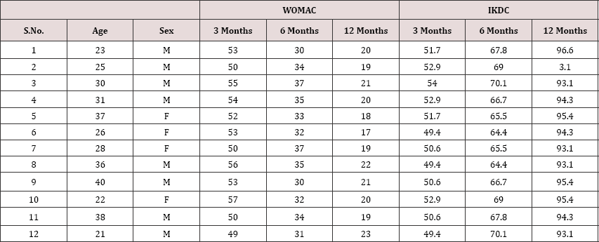
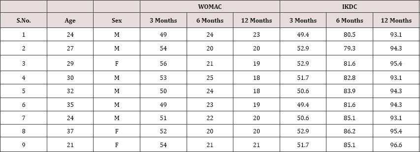
The WOMAC score in group A ranged from 54-49 (mean- 52) at 3 months, which improved to (mean-22.2) at 6 months. The WOMAC score at 12 months showed significant improvement from the 6 month follow up score (p=0.024), however there was no significant difference between group A and B at 12 months (p= 0.86). In group B the WOMAC score at 6 months was better than that at 3 months (P=0.0001) (Table 1). But comparing with group A, group B showed significantly poorer results at 6 months (p=0.0001).

Table 1: WOMAC and IKDC scores of individual patients of group B at 3 months, 6 months and 12 months.

Comparing the IKDC scores, the group A patients showed supplementation however seemed to wane away by 12 months significantly better results compared to group B at 6 months with no significant difference in IKDC scores between the two follow up (p=0.0001) (Table 2). The beneficial effect of visco- groups (p=0.347).

Table 2: WOMAC and IKDC scores of individual patients of group A at 3 months, 6 months and 12 months.

Discussion

Joint degeneration and poor healing potential of chondral lesions pose a challenge to surgeons managing these lesions. Various techniques have been described aiming to fill the defect and obtain a congruent joint. Micro-fracture is one of such procedures in which the sub-chondral bone plate is penetrated to cause fibrin clot formation and later conversion to fibrocartilage [10]. Variable outcomes of micro-fracture have been described in literature with almost 70%-90% of patients showing improvement in function [11]. The durability of the fibrocartilage has however been put to question with mid-term results showing a decline in the functional scores [12,13]. Augmentation of micro-fracture to improve the quality and durability of the repair tissue and ensuring a more hyaline like tissue is the current interest topic of various researchers. Strauss E et al. [10] showed a positive effect of hyaluronic acid supplementation on repair tissue both histologically as well as grossly.

Intra-articular basic fibroblast growth factor when used without hyaluronic acid induces poor repair tissue besides undesirable side effects. hyaluronic acid injection along with basic fibroblast growth factor results in significantly better tissue quality [7]. Hyaluronic acid has been increasingly used in the non-operative management of osteoarthritis [14,15]. Rabbit model studies have demonstrated hyaluronic acid to reduce arthritic changes by inhibiting degenerative changes in chondrocytes and cartilage matrix. It also decreases synovial inflammation and increases proteoglycan contect of cartilage [7]. Hyaluronic acid has also been shown to induce chondrogenic differentiation in mesenchymal stem cells [8].

Tytherleigh-Strong et al. [6]. Demostrated significantly higher aggregate moduli in the visco-supplementation group in an ovine model. Jansen et al showed chondroprotective effect of hyaluronic acid, preventing apoptosis of chondrocytes just adjacent to the lesion [16]. Although there are several animal studies demonstrating beneficial effect of hyaluronic acid in cartilage healing, not much is available on their role in human patients [10].

This study aimed to evaluate the role of visco-supplementation in functional outcome following micro-fracture in focal chondral lesions. The visco-supplemented group fared significantly better than the non-supplemented group at 6 months. The beneficial effect however seemed to wane away by one year with both groups having comparable functional scores and subjective outcomes at 12 months follow up.

Limitations

Our sample size is small as we purposefully selected only lesions which were less than 2sq cm in size. Credibility is added to our study by comparing the outcomes of micro-fracture supplemented with hyaluronic acid with those without. No patients were lost to follow up in this series. We however, do not have any follow up MRI or repeat arthroscopy to ascertain the healing status at the repair site. The follow up is also a short term follow up 1 year. A longer term follow up is needed to assess the long term outcomes of viscosupplementation in preventing joint degeneration.

Conclusion

Supplementing micro-fracture with hyaluronic acid injection ensures quicker return of better functional state. In the longer term however this beneficial effect seems to wane off.


Thursday, 24 October 2019

Lupine Publishers | On A Criminal Trace of Crippling and Often Even Lethal False Diagnosis: Lege Artis Cz

Lupine Publishers | Journal of Orthopaedics  

Case Report

In 2010, the Czech Republic participated in the World Health Organization WHO Research to determine the quality of medical decision making in determining diagnosis and to determine an adequate individual choice of patient treatment. The conclusions were really frightening: the relative frequency of fatal medical decision-making errors is 10%, with a statistical accuracy of the tolerance estimate of +/- 2%, with a statistical reliability of conclusions of about 95%. Approximately rewarding results are found in all OECD countries - in all types of outpatient or inpatient health services. In real terms, this means that approximately every 10th accidental visit of the patient to the doctor will end up with an additional health problem. For example, a late-diagnosis of B-type jaundice, a medical prescription of an inappropriate medication that contravenes the health of a particular patient, or contraindicates the contraindication effects of other concurrent medications, or a radiologist misinterprets an X-ray image-with overlooked by emerging stomach cancer, overlooked severely fatal osteoporosis-disturbed bone, a preventive standard examination of the colonoscopy will occur with careless medical manipulation of the colon perforation probe in a hitherto healthy patient. In general, they are seriously threatened patients, sometimes even seriously endangered patient lives.
If the above mentioned documented WHO research results refer to the whole Czech pupil - that is, for the total population of the Czech Republic, 10.6 million living people and an average of at least one annual patient visit to any doctor in the Czech Republic. approximately 1 million citizens are probable expected to be expected to receive approximately one million people instead of the actual assistance needed in the Diagnosis and appropriate individual treatment, likely to have an unexpected, addictive - crippling health risky next complication - and some patients even due to a deteriorated health condition, probably some of them die sooner - many years before the bio statistic mean survival estimate for the remaining years would be the same-if the medical error did not become the correct diagnosis or treatment. The Providers Health care and medical staff said: It is fatal legal yearly irrevocable Facts - but I have said NO!

On a Criminal Trace of Repeated Mass Medical Mistakes With False Coded Working with Regards Lege Artis CZ

The First Criminal suspicious for Mass Illegal Medical Workflow CZ

If approximately one million Patients CZ are seriously threatened annually with incorrect medical decision-making contrary medical Knowledge’s WHO, logically at least Sum 50,000 Patients CZ are unnecessarily mutilated yearly. They are mostly casual preliminary dying - a long time ago from the adequate Cluster with similar Patients CZ with the same Diagnoses, Age, similar variant of Treatments they are growing differenced Health consequences and ever-increasing causal next health risks to die after medical mistakes - All these cases should end annually with ignoring of the Czech Justice Courts, supervising Criminal Police CZ, supervising from State Penalty Offices CZ - and survivors or heavily crippled patients of the Czech Republic should receive from the responsibly Health instance CZ of the Providers nets of Health Services of the Czech Republic - approximately there are ignored 3 million compensation for financial satisfaction per 1 case per annum.
The total should therefore be a probable compensation in the astronomical annual amount yearly - 50 thousand persons CZ x 3 million crowns - the result is 15 x 10x10x10x10x10x10x10x10x10x10 = 150 milliard CZK / year!!! Thus, in the Czech Republic’s national income about Sum CZK 330 Milliard, almou nearly half of the annual income is a waste of medical work. This is for the Healthcare of the Czech Republic, for the Provider of Health Services and their Employees, for the Judiciary CZ, for the State Budget of the Czech Republic, for the Criminal Police of the Czech Republic, for the Public Prosecutor’s Office of the Czech Republic unprivileged by national, political.
However, we have a very novel solution in the Czech Republic - whether you are an individual patient crippled and dying for legal or illegal medical mistakes very Cheap: All fatal cases will be placed in the category of “Unwanted inevitable medical and legal results of medical processes” with the unique cover false slogan LEGE ARTIS CZ. It is similar as a Italy Mafia with more strong laws, more servos as a OMERTA, who has taken a keen interest in all the injured patients in the medical, security, criminal and custody organizations: You are either a healthy, unnecessarily frozen or unnecessarily dying patient in the Czech Republic - but absolutely under the monotonic motto of observing Medical cases with false wisdom declination the principles of LEGE ARTIS CZ. So that any private truth criminalist evidence of Patients CZ, when that you are the Victim of a criminal way of providing medical health care will come out monotonously: You will almost never receive adequate financial justice satisfaction, because it is only derived of your preliminary “informed Patient consent” to planned risky and probably often including very technically illegal medical practice with Medical Devices, often with explicitly wrongly misused The Medical Devices- you have only one direction right Cementary route: sooner or later, without any patient comments to Court dealing CZ: to die without delay as a false result of the absolutely indifference responsible medical work “LEGE ARTIS CZ” with content many fatal explicit illegal technical partial medical mistakes [1-3].
Annual unnecessary severe mutilation of at least about 40 to maybe perhaps 50,000 patients in the Czech Republic with a much earlier causal death - this is a pre-agreed probable loss of life and health of the Czech Patients, which is the number of deaths and astronomically large national economic losses similar to the lives of Victims of the secret national continual war Physicians CZ - against their own patients in the Czech Republic, often absolute out of legal technical usage Medical Devices in hospital net CZ in regards to Technician Requirements of Laws EU/CZ.

The Perspective for Solutions to Limit and to Prevency the Mass Repeated Medical Mistakes in OECD

I know namely reliable coherent scientific processing to limit the Mass repeated similar medical mistakes with more efficiency sharing the best medical experiences only as perfect redefined scientific principles “LEGE ARTIS CZ“ - with more effective managing medical workflow with more effective usage samplings, clustering, validated medical processing, continual testing elementary partial medical activities of partial medical processing step by steps“ with more efficiency implanting Artificial intelligence, with more regards to Technician requirements of Laws EU/CZ for usage Medical Devices - with acquired my decision making method S_T_A_R_S in daily medical workflow - see the Literature Antonín Cuc: The Utility model 21532 CZ 2010, Czech Office for Industrial Laws. The Equipment for Retrieval and Search of sufficient statistic information to repeated similar strategic decision making with risk and computer support “the opened Sources to usage for Medicine US since April 17, 2017!
As a scientist, I am well educated in the field of Cybernetics and Statistics and in the field of H+S for Medical Devices, Technician requirements of Laws EU/CZ for legal processing usage Medical Devices in Providers Health care nets!

The précised and Redefined Logical Criminalist Definition Lege Artis Medical Processing in Health Care OECD

For example, violation of the Technical Requirements of the Laws on the Legal Safe Use of Medical Devices, for example in contravention of the Technical Requirements of EU / the use of Medical Devices, contrary to the requirements of related harmonized European standards, contrary to the instructions of the manufacturers of medical devices and contrary to the certified use of their installation and measuring instruments, contrary to the obligation to prevent known hazards declared in the Protocols on Medical Devices as products with use management conformity assessment rules “CE”.

The Logical Substantiation in Medical Processing OECD

In the Binary Logics so as in the Criminalist Logics for the conjunctions partial mixed procedural phenomenon’s with evaluating True, False - there are sum resulting evaluation the coherent sequential processing always resulting in evaluation F A L S E, when there are occurrence at least the One partial unit with evaluation with partial well criminal evidenced FALSE, definitely!
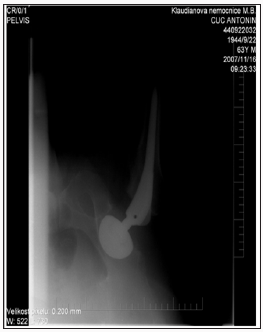
I am just needless dying Patient CZ because the implanting processing set Total Hip Arthroplasty since November 13, 2007 contained many illegal processing medical mistakes contrary functional assembling product processing for set THA Bicontact S, no cemented, nominal dimension 13 mm N, B.Braun Germany
- with false coaxial ties between the metallic Stem and my right femoral bone in the Surgery hall without preliminary Clinic Plan, with illegal declination coaxial ties 13,68 grads in sagittal Direction, out of acceptable assembling tolerance +/- 1 grad - The post operational first RTG images from date November 16, it was the sufficient criminal evidences about false illegal placement the spice the Stem in coordinate Xi, Yi, Zi - out of the firm installing radiologic Masks - but it should be controlled by the Laws and by the product ovoid on the Orthopaedic screen with the same scaling.
The wisdom false Court Medical Message from the Knowing Medical Institute - The Central Army Hospital Prague, CZ No. 36 C 181/2009-123 since the March 27, 2012 defined all Medical Orthopaedic Processing in Limits Lege Artis CZ, The fatal unhappy was defined not as Diagnose Fausse route stem on the Orthopaedic Surgery Hall - but as the results of the false medical Hypotheses in Complot of the Radiologic, Orthopads Doctors from the Orthopady Clinic and from the Knowing Medical Institute as Post operational tragic Event by Patient fallen“, despite such substitutions in truth Criminal traceing, Radiology, Geometry 3 D, Health Patient Documentation EHRs.
I laid down my life so that you and your physicians and technicians can work together in a responsible manner and in the real truth of God and in the same truth of the criminalist evidences.
I became an unnecessary Merthyr of human Stupidity and irresponsibility that threatens humanity and the patients of the Czech Republic who are denied the Constitutional Human Rights of the Czech Republic: for life, for a fair Court trials, for legal health care CZ. I believe that Medicine.com will never accept the falsity of the current approaches of „LEGE ARTIS CZ“, as now submitted to the world public by the Czech courts, the Criminal Police of the Czech Republic, the network of the State Prosecutor’s Offices of the Czech Republic.
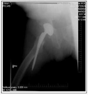
I am just placing the principal private RTG images for Forensic reconstruction Crash of set THA Bicontact S, no cemented in Geometry 3 D since date November 13, 2007 on the Orthopaedic Surgery Hall of the Regional Hospital Mladá Boleslav. Most of Patients with similar Crash are dying in duration since 3 till 7 days after overlooked FAUSSE ROUTE STEM, I was reoperation till 17 days after Poly trauma on the Surgery Hall, when the preoperational THA processing was realised in the same profile of Surgeon catting as in first THA, but the Spice of the Stem was created met he blooding and full plegic dysfunction Nervus is achidici with destructions my structure muscles in right calf, I am just frequential happened fallen with risk the repeated breaking the proximal femoral bone with just 8x worse probabilities and I am dying earlier about many years as other Patients CZ in Cluster with right legal first THA surgery.
Figure 1:
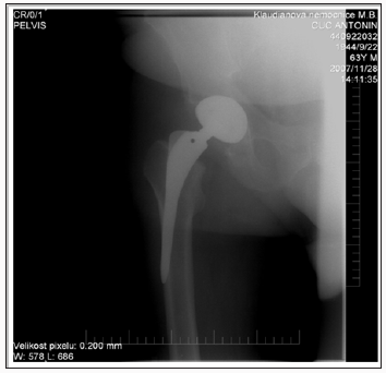
My Orthopads, my Judge, the forensic doctors from the Knowing Medical Institute CZ, Departments of Criminal Police CZ, State Penalty Offices CZ are smiling to my nearly Death within forced me many growing casual orthopaedic and leukemic heavy injuring LEGE ARTIS CZ (Figure 1). The virtual „Shorten of the Length the Axes of metallic Stem “It is caused the false placement with fatal declination in sagittal Direction in angle 13, 68 grads, there are false installing position of the spice Stem on coordinates Xo, Yo, Zi - it is illegal „to observing the RTG image in hands and by intuitive views“, when the Orthopads should taken the Orthopaedic screen and comparison the placement the Spice contrary firm radiologic Mask for the stem!
The Doctors said by illegal daily habits: We are working Lege artis, only a few orthopaedic patient are sometimes legal dying! The Criminal police CZ gave legal agreements so as the Knowing Medical institute CZ too - The mortal injured Patient CZ on the Prosector Hall haven´t commentaries! We are dying needless and very Cheap and frequently in CZ, always Lege Artis CZ (Figure 2). This is the Image from date November 28, 2007 too, this is forensic nonsense to prented this Criminal traceing could be realised as Patient fatal post operational happened! The last moving of the Stem was realised in Surgery hall November 13, 2007 in Regional Hospital Mladá Boleslav CZ.
Figure 2:
In Court trials CZ the Judge could believe for medical illegal Stupidities each of workaday. No Protests from Patients CZ are accepted in legal care for Constitutional Human Rights CZ by Ombudsman CZ- despite me is the Sate investigator for occupational mortalities in Branche Medical Devices. My Death is very awaiting CZ Events, like a public Execution of a disagreeable world Scientist in 21st Century (Figure 3).
Figure 3:


Saturday, 14 September 2019

Lupine Publishers | Role of Visco-Supplementation Following Micro- Fracture in Focal Chondral Lesions

Lupine Publishers | Journal of Orthopaedics

Abstract

Introduction: Cartilage lesions pose a significant problem to surgeons, at best being replaced by poorer quality fibrocartilage post micro-fracture. Hyaluronic acid has shown to inhibit joint degeneration and reduce joint inflammation. This study wishes to evaluate the outcome of visco-supplementation following micro-fracture.
Material & Method: Twenty one consecutive patients with chondral lesion less than 2 sq cm were treated with micro-fracture and post operatively randomly divide into two groups. One group was supplemented with intra-articular hyaluronic acid at 3 weeks post surgery. The patients were followed up at 3, 6 & 12 months for WOMAC score and IKDC score.
Results: The WOMAC score and IKDC score in the visco-supplementation group were significantly better than those of the non visco-supplementation group at 6 months (p=0.0001). At 12 months although the scores were significantly better than at 6 months, there was no significant difference between the two groups (WOMAC p=0.86, IKDC p=0.347).
Conclusion: Hyaluronic supplementations following micro-fracture ensure an early improvement in functional scores following micro-fracture. A longer follow up is necessary to evaluate its role in preventing further joint degeneration.
Keywords: Micro-fracture; Visco-supplementation; Hyaluronic acid

Introduction

Athletic injuries often lead to full-thickness chondral defects, which have limited intrinsic healing potential [1,2]. These lesions if not managed adequately predisposes to joint degeneration [2]. Symptomatic lesions are often managed with micro-fracture to attempt cartilage repair [3]. Bleeding from the sub-chondral bone within the defect and subsequent clot formation covers the exposed bone. Also, mesenchymal stem cells that migrate into the clot promote formation of fibrocartilagenous repair tissue [4]. This filling of the defect restores the congruity of the joint leading to symptomatic relief [5]. The viscoelastic property of the synovial fluid is credited to Hyaluronic acid, which is often used in the non operative treatment of early osteo-arthritis [6]. Hyaluronic acid has been shown to inhibit degenerative changes within chondrocytes, reduce synovial inflammation and enhance cartilage proteoglycan content along with inducing chondrogenic differentiation from mesenchymal cells [7,8]. This study aims to compare the functional outcome of micro-fracture with and without visco-supplementation.

Materials and Method

In a prospective level II study, patients with chondral lesions of ICRS grade III-IV measuring less than 2sq cm were managed with micro-fracture and randomly divided into two groups. One group was supplemented with a single shot intra-articular Hyaluronic acid at 3 weeks post surgery in the form of 6ml hylan G-F 20, [9] the other group did not receive any visco-supplementation. Patients more than 45 years of age and having generalised chondral changes were excluded from the study. Multi-ligament injured patients and those with unmanaged single ligament injury or unmanaged meniscus injuries were also excluded from the study. There was no sex restriction. All the patients were put on continuous passive motion from the first post operative day unless they had under gone an associated PCL reconstruction or meniscus repair surgery, in which case PCL and meniscus rehabilitation protocols were followed. The patients were kept non weight bearing for 6 weeks followed by full weight bearing. WOMAC score and IKDC score were evaluated at 3, 6 and 12 months post surgery.

Results

The Visco-supplementation group (Group A) had 9 patients and the group with no supplementation (Group B) had 12 patients. All our patients were between 21-40 years of age (Mean: 29.3 years) with 14 males. Two had associated PCL reconstruction surgery, 17 underwent ACL reconstruction of which five underwent partial medial menisectomy and two partial lateral menisectomy. Five patients underwent a meniscus repair procedure. Eight of group A had grade 3-4 ICRS lesion on the weight bearing area of medial femoral condyle while one had involvement of the lateral femoral condyle. In group B 8 had involvement of the medial femoral condyle. Eight had injured their knee in a game of football, five while dancing, six while playing cricket and another two due to fall on stairs. None were lost to follow up. None of the patients reported any re-injury between the surgery and latest follow up. There were no complication in any of the patients and no one required any additional surgical intervention.
The WOMAC score in group A ranged from 54-49 (mean- 52) at 3 months, which improved to (mean-22.2) at 6 months. The WOMAC score at 12 months showed significant improvement from the 6 month follow up score (p=0.024), however there was no significant difference between group A and B at 12 months (p= 0.86). In group B the WOMAC score at 6 months was better than that at 3 months (P=0.0001) (Table 1). But comparing with group A, group B showed significantly poorer results at 6 months (p=0.0001).
Table 1: WOMAC and IKDC scores of individual patients of group B at 3 months, 6 months and 12 months.
Comparing the IKDC scores, the group A patients showed supplementation however seemed to wane away by 12 months significantly better results compared to group B at 6 months with no significant difference in IKDC scores between the two follow up (p=0.0001) (Table 2). The beneficial effect of visco- groups (p=0.347).
Table 2: WOMAC and IKDC scores of individual patients of group A at 3 months, 6 months and 12 months.

Discussion

Joint degeneration and poor healing potential of chondral lesions pose a challenge to surgeons managing these lesions. Various techniques have been described aiming to fill the defect and obtain a congruent joint. Micro-fracture is one of such procedures in which the sub-chondral bone plate is penetrated to cause fibrin clot formation and later conversion to fibrocartilage [10]. Variable outcomes of micro-fracture have been described in literature with almost 70%-90% of patients showing improvement in function [11]. The durability of the fibrocartilage has however been put to question with mid-term results showing a decline in the functional scores [12,13]. Augmentation of micro-fracture to improve the quality and durability of the repair tissue and ensuring a more hyaline like tissue is the current interest topic of various researchers. Strauss E et al. [10] showed a positive effect of hyaluronic acid supplementation on repair tissue both histologically as well as grossly.
Intra-articular basic fibroblast growth factor when used without hyaluronic acid induces poor repair tissue besides undesirable side effects. hyaluronic acid injection along with basic fibroblast growth factor results in significantly better tissue quality [7]. Hyaluronic acid has been increasingly used in the non-operative management of osteoarthritis [14,15]. Rabbit model studies have demonstrated hyaluronic acid to reduce arthritic changes by inhibiting degenerative changes in chondrocytes and cartilage matrix. It also decreases synovial inflammation and increases proteoglycan contect of cartilage [7]. Hyaluronic acid has also been shown to induce chondrogenic differentiation in mesenchymal stem cells [8].
Tytherleigh-Strong et al. [6]. Demostrated significantly higher aggregate moduli in the visco-supplementation group in an ovine model. Jansen et al showed chondroprotective effect of hyaluronic acid, preventing apoptosis of chondrocytes just adjacent to the lesion [16]. Although there are several animal studies demonstrating beneficial effect of hyaluronic acid in cartilage healing, not much is available on their role in human patients [10].
This study aimed to evaluate the role of visco-supplementation in functional outcome following micro-fracture in focal chondral lesions. The visco-supplemented group fared significantly better than the non-supplemented group at 6 months. The beneficial effect however seemed to wane away by one year with both groups having comparable functional scores and subjective outcomes at 12 months follow up.

Limitations

Our sample size is small as we purposefully selected only lesions which were less than 2sq cm in size. Credibility is added to our study by comparing the outcomes of micro-fracture supplemented with hyaluronic acid with those without. No patients were lost to follow up in this series. We however, do not have any follow up MRI or repeat arthroscopy to ascertain the healing status at the repair site. The follow up is also a short term follow up 1 year. A longer term follow up is needed to assess the long term outcomes of viscosupplementation in preventing joint degeneration.

Conclusion

Supplementing micro-fracture with hyaluronic acid injection ensures quicker return of better functional state. In the longer term however this beneficial effect seems to wane off.

For more Lupine Publishers Open Access Journals Please visit our website:
http://lupinepublishers.us/
For more open access orthopedics journal articles Please Click Here:
https://lupinepublishers.com/orthopedics-sportsmedicine-journal/

To Know More About Open Access Publishers Please Click on Lupine Publishers

Monday, 22 April 2019

Journal of orthopaedics-Lupine Publishers


Judo is a combat sport often related to injuries since it is associated with a significant direct contact [1,2]. Although injuries occur, judo is considered one of the most safest sports activity due to low injury rates [3], being in fact considered by UNESCO as the best sport for children and young people between 4-21 years old. Nowadays, because of the early competitive practice, specialization and intensive training load among young athletes, we are seeing different injury patterns and severity in these ages [2,4,5]. The potential vulnerability of these athletes due to the physical and physiological processes of growth exposed to high levels of activity, is a reason of concern [4,6] which must stimulate the development of this specific knowledge, in particular the analysis of risk factors in order to provide preventive injury programs and educative measures. With this study, we intend to reinforce the epidemiologic knowledge specific to judo in these particular ages, and, by knowing the risk factors and more common injury features and patterns, use them to help.com developing the strategies to control and prevent injuries.


For more Lupine Publishers Open Access Journals Please visit our website: http://www.lupinepublishers.com/

For more journal of orthopaedics articles Please Click Here: https://lupinepublishers.com/orthopedics-sportsmedicine-journal/