Showing posts with label Open access journal of Neurology. Show all posts

Monday, 28 November 2022

Lupine Publishers| Paraparesis caused by Aneurysmal Bone cyst (ABC)

 Lupine Publishers| Journal of Neurology and Brain Disorders


Abstract

Through the study of a case of spinal aneurysmal bone cyst managed in the Department of Neurosurgery at Hassan II University Hospital of Fez we focus our discussion of various aspects of this rare disease.

Our study concerns a male patient admitted to the emergency department for spinal cord compression that necessitated a spinal MRI. It showed T4 vertebral compression, hypointense T1, hyperintense T2 with hematic fluid levels causing spinal cord compression. The patient underwent surgical treatment with good clinical outcome. Histological study revealed a spinal aneurysmal bone cyst.

Keywords: Aneurysmal cyst; MRI-surgery; Embolization

Introduction

The aneurysmal bone cyst is a benign lesion with an aggressive character. It represents 1.4% of primary bone tumors, its spinal localization remains uncommon (3-30%) and spinal syndrome is the most frequent reason for consultation [1]. Plain radiography and CT have a significant role in its diagnosis, but MRI remains the investigation of choice. The management of spinal aneurysmal bone cyst remains controversial.

Case

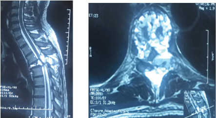
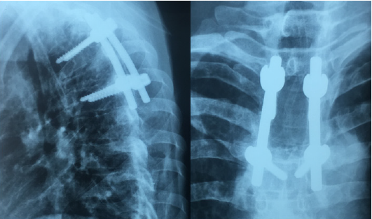
A 40 years old man, chronic smoker (a packet/day) was admitted to the emergency for a spinal syndrome for 2 years that was complicated 2 months before admission by the appearance of a grade D paraparesis, hypoesthesia with a sensory level at xiphoid process and urinary incontinence. The spinal MRI showed compaction of T4 vertebra with a heterogenous hyperintensity on T2 and STIR, a hyperintensity on T1 with heterogeneous enhancement of hematic fluid levels causing spinal cord compression at same level (Figure 1). The management consisted of performing a laminectomy of T3-T4 with pedicle screw and rod fixation of T3 and T5 vertebra (Figure 2). The postoperative course was marked by improved clinical signs. The Histopathology report came back in favor of aneurysmal bone cyst.

Figure 1: Spinal T2 weighted MRI showing (a) sagittal view with T4 compression and (b) Axial view with fluid level spaces.
Lupinepublishers-online-journal-of-neurology-and-brain-disorders

Figure 2: Spinal X-ray Lateral and anteroposterior showing post-operative pedicle screw and rod system.
Lupinepublishers-online-journal-of-neurology-and-brain-disorders

Discussion

Aneurysmal Bone Cyst (ABC) is a benign cystic lesion composed of blood-filled spaces, devoid of coating endothelial, muscle or elastic, surrounded by fibroblasts, described for the first time by Jaffe and Lichtenstein in 1942 [2]. Its incidence is 0.14 per 100,000 individuals with a female predominance (ratio 1.16) [3]. For Lichtenstein [4], the ABC is a secondary bone lesion due to hemodynamic imbalance resulting in a significant capillary dilatation, resulting from an increase in venous pressure. This increase in venous pressure might be due to venous thrombosis or abnormal arteriovenous communication. Biesecker et al [5] measured the intracystic pressure which was identical to that of arterioles and concluded that this hypertension is due to an arteriovenous fistula following a secondary lesion. Some authors [6-8] think that the appearance of an aneurysmal bone cyst would be secondary to impaired intraosseous vascular flow in the phase of consolidation following trauma. However, several cases of familial aneurysmal bone cysts [9,10] have been reported, hence orienting towards a genetic origin and indeed the genetic and immunohistochemical studies [11] have demonstrated a translocation on chromosome [7].

Aneurysmal bone cyst is most frequently found in the long bones especially the metaphyseal region [12,13], while the spinal location remains rare and affects the cervical, thoracic, lumbar and sacrum, with no case of coccygeal bone ABC reported [14]. Clinically, spinal ABC may be asymptomatic [1], but usually it manifests itself as a spinal disorder, with or without spinal cord compression signs. MRI remains the examination of choice which shows a welldemarcated bone lesion, hypointense on T1, hyperintense on T2 with a peripheral border taking contrast, defining spaces with fluid level [4]. Surgery that involves wide excision represents the preferred treatment of choice for ABC with a recurrence rate of 25% and multiple complications such as deformation and growth disorders [15]; For these reasons, selective arterial embolization was introduced into the management of this entity that proves similarly effective and with fewer complications, representing the treatment of choice in cases where the patient shows no deficit or unstable associated fractures [16]. Radiation therapy, which was an important pillar in the therapeutic arsenal, was gradually abandoned because of the risk of exposure and development of radiation-induced sarcomas [17]. Other therapeutic protocols have been proposed: the intralesional injection of calcitonin, and doxycycline Ethibloc [17-20], however they have not proven effective as with other therapeutic methods. Currently, research [21-23] has focused on the interest of Denosumab, a monoclonal antibody used in the treatment of giant cell tumors, and the intrinsic stimulation by the mesenchymal stem cells and have both shown promising results [24,25].

Conclusion

Aneurysmal bone cyst is a benign disease with unpredictable course, and thus what makes its treatment a challenge especially in locations where access is difficult. Surgery and embolization currently remain the treatment protocol of choice. Research on Mesenchymal stem cells and Denosumab have demonstrated promising results in the therapeutic management of ABC.

Read More About Lupine Publishers Journal of Neurology and Brain Disorders Please Click on Below Link: https://brain-disorders-lupine-publishers.blogspot.com/

Tuesday, 13 August 2019

Eating Disorders in Developing Countries | Lupine Publishers

Open Access Journal of Neurology | Lupine Publishers


Editorial

Eating disorders, once thought to be a set of rare diseases, found predominantly in females, are being increasingly seen in both males and females. They include binge eating disorder (BED), anorexia nervosa (AN), bulimia nervosa (BN), pica, rumination disorder, avoidant/restrictive food intake disorder, and a group of other specified feeding or eating disorders [1]. Eating disorders occur predominantly in females and the onset often follows puberty [2].
The number of people coming to psychiatrists with problems of anorexia and bulimia has shown a steady rise. It has been estimated that the lifetime prevalence of DSM-IV anorexia nervosa, bulimia nervosa, and binge eating disorder are 0.9%, 1.5%, and 3.5% among women, and 0.3% 0.5%, and 2.0% among men in USA [3]. In Ontario prevalence rates of of BN was 1.1% in women and 0.1% in men [4]. The prevalence rates for AN and BN in Zurich was 0.7% and 0.5% respectively [5]. In South Australia the prevalence of BN and BED was 0.3% and 1% respectively [6]. Across six European countries the prevalence rates for AN, BN, and BED were 0.48%, 0.51%, and 1.12% respectively [7]. Eating disorders as a group are psychiatric disorders with the highest mortality rate, resulting in about 7,000 deaths a year in 2010 [8]. What was earlier thought as a disease pertaining to a few developed countries is now being increasingly seen in developing countries. Moreover, patients of all ages and sexes are coming with these problems.
This has been attributed to acculturation and exposure through the media to Western standards of attractiveness and body size [2,9]. Increasingly, the population of developing countries is adopting Western views on thinness and the dietary habits associated with that state [9]. The point prevalence of AN, BN and BED in China and Japan is 1.05 and 0.43; 2.98 and 2.32; and 3.58 and 3.32 respectively [10]. The prevalence of eating disorder is believed to have increased in India over the past three decades as indicated by sporadic case reports. [11-12]. Though no epidemiological studies have been carried out using the Global Burden of Disease Study 2016 approach it was estimated that the prevalence rate of AN for males was 10/100,000, whereas for females, it was 37.2 and combined prevalence was 22.3/100,000[13].
The cause of eating disorders is not very clear as it is seen that biological and environmental factors appear to play a role. Many studies have shown a possible genetic predisposition causing eating disorders due to Mendelian inheritance [14]. A role for serotonin 4 receptors has been proposed [15]. Some studies suggest that both early pubertal timing and advanced pubertal development in girls are associated with increased rates of eating disorders. On the other hand, findings in boys suggest a smaller role for puberty in the development of eating disorders. Results from twin and animal studies suggest that the female-specific risk is due, at least partly, to genetic factors associated with puberty related estrogen activation [16]. People having previous disorders like anxiety, obsessive compulsive disorder etc have increased vulnerability to developing an eating disorder [17-18]. Cultural idealization that beauty is the same as thinness is also believed to contribute to the etiology [19]. Self-respect is being linked to the body shape and size. Social change and globalization are causing the shift in the thinking of the people in urban cities.
The children are being exposed to social media these days. A significant link has been shown between self-objectification, body dissatisfaction, and disordered eating, as the beauty ideal is altered through social media [20]. Organic cause and other psychiatric disorders must be ruled out before diagnosing eating disorder. The test to be done should include neuroimaging [21]. Beck Depression Inventory [22] just to name a few. Treatment differs according to type and severity of eating disorder, and more than one treatment option is used [23]. Treatment includes pharmacotherapy and psychotherapy. Medicines which can be given are cyproheptidine, [24] pimozide [25] and zinc supplementation [26] for anorexia nervosa, naltrexone for binge eating [27]. Cognitive behavioral therapy, [28] Dialectical behavior therapy [29] family therapy [30] are some of the therapies which show promising results. Most of the patients and their families do not know that they are suffering from a psychiatric disorder and may go to physicians for physical complaints. They are very reluctant and rarely come to psychiatrists. Patients should be encouraged to seek psychiatric treatment through media and other social awareness programmes.

For more Lupine Publishers Open Access Journals Please visit our website:
https://lupinepublishersgroup.com/


For more Open Access Journal of Neurology Please Click Here:
https://lupinepublishers.com/neurology-brain-disorders-journal/


To Know More About Open Access Publishers Please Click on Lupine publishers

Wednesday, 12 June 2019

Lupine Publishers-Journal of Neurology & Neurosurgery


Immunoglobulin G4-related disease (IgG4-RD) is an increasingly recognized immune-mediated condition comprised of a collection of disorders that share particular pathologic, serologic, and clinical features [1,2]. These disorders were previously thought to be unrelated [3-5]. The commonly shared features include tumor-like swelling of involved organs, a lymphoplasmacytic infiltrate enriched in IgG4-positive plasma cells, and a variable degree of fibrosis that has a characteristic “storiform” pattern. In addition, elevated serum concentrations of IgG4 are found in 60 to 70 percent of patients with IgG4-RD. IgG4-related sclerosing cholangitis (IgG4-SC) is a characteristic type of sclerosing cholangitis, with an unknown pathogenic mechanism. Patients with IgG4-SC display increased serum IgG4 levels [6] and dense infiltration of IgG4-positive plasma cells with extensive fibrosis in the bile duct wall [7]. Circular and symmetrical thickening of the bile duct wall is observed in the areas without stenosis that appear normal on cholangiography, as well as in the stenotic areas [8]. IgG4-SC has been recently recognized as an IgG4- related disease. IgG4-SC is frequently associated with autoimmune pancreatitis (AIP). IgG4-related dacryoadenitis/sialadenitis and IgG4-related retroperitoneal fibrosis are also occasionally observed in IgG4-SC [9-12]. However, some IgG4-SC cases do not involve other organs. IgG4-SC is most common in elderly men. Obstructive jaundice is frequently observed in IgG4-SC.A number of diseases, such as, Cystic fibrosis, Chronic obstructive Choledocholithiasis, Biliary strictures (secondary to surgical trauma, chronic pancreatitis), Anastomotic strictures in liver graft, Neoplasms (benign, malignant, metastatic), Infections, hypertonic saline instillation in the bile ducts, Post-traumatic sclerosing cholangitis, Systemic vasculitis, Amyloidosis, Radiation injury, Sarcoidosis, Systemic mastocytosis, Hypereosinophilic syndrome, Hodgkin’s disease, may easily be confused with IgG4- related sclerosing cholangitis, or coexist in a patient [13]. In this case, report 57 years male patient presented with jaundice, fatigue, weight loss, oral moniliasis and right sided neck swelling. He was misdiagnosed as Cholangiocarcinoma. To know more go through the below link.


For more Lupine Publishers Open Access Journals Please visit our website: https://lupinepublishers.us/


Wednesday, 31 October 2018

Physiology of Sleep and Clinical Characteristics: (OJNBD) - Lupine Publishers


Physiology of Sleep and Clinical Characteristics by Miranda Nava Gabriel in Online Journal of Neurology and Brain Disorders in Lupine Publishers

Sleep is defined as a natural decrease in the perception [1] of the external environment that occurs periodically and reversibly, but retaining a certain degree of reactivity towards the environment and autonomous functions [2]. Sleep is considered an active process of biological, cyclical and, it is essential for survival. Most adults require an amount of sleep between 7-8 hours per day; however, there are individual variations regarding the schedule, duration, and internal structure of the dream [3].

http://lupinepublishers.com/neurology-brain-disorders-journal/abstracts/physiology-of-sleep-and-clinical-characteristics.ID.000114.php
http://lupinepublishers.com/neurology-brain-disorders-journal/fulltext/physiology-of-sleep-and-clinical-characteristics.ID.000114.php
http://lupinepublishers.com/neurology-brain-disorders-journal/pdf/OJNBD.MS.ID.000114.pdf