Showing posts with label Journal of Neurology. Show all posts

Tuesday, 26 September 2023

Lupine publishers | Continuous or Intermittent? Which Regimen of Enteral Nutrition is Better for Acute Stroke Patients? a Systematic Review and Meta-Analysis

 Lupine Publishers | Journal of Neurology and Brain Disorders

Abstract

Background and purpose: Enteral nutrition via nasogastric tube in acute stroke patients with dysphagia is an important determinant of patient outcomes. It is unclear whether intermittent or continuous feeding is more efficacious. The aim of this review is to examine the current evidence comparing the effectiveness of intermittent versus continuous feeding in stroke patients in terms of nutritional status, gastrointestinal intolerance and other complications.

Methods: A systematic review of randomized controlled studies comparing intermittent with continuous nasogastric feeding in acute stroke patients was conducted in accordance with PRISMA (Preferred Reporting Items for Systematic Reviews and Metaanalyses) guidance using predefined search terms. The search was conducted in MEDLINE and EMBASE up to 1st March 2019. Two independent reviewers assessed study quality using the Joanna Briggs Institute Critical Appraisal Tool. Meta-analyses were conducted, where appropriate, using a random-effects model to pool risk ratio with corresponding 95% CI.

Results: Three studies including a total of 184 patients were identified. All three were medium to low quality. The definition of intermittent enteral nutrition within each study varied considerably in terms of volume, rate and mode of delivery. Achievement of nutritional targets was the same for both feeding patterns in the one study it was reported. Only aspiration pneumonia and diarrhea were measured by all three studies. There was no significant difference in the incidence of aspiration pneumonia (RR 0.91, 95% CI 0.53-1.57, p=0.74, I2=50%) and diarrhea (RR 1.74, 95% CI 0.70-4.30, p=0.23, I2=42%) between the two patterns of feeding. Other outcomes including, vomiting, gastric retention, mortality, pre-albumin and nasogastric tube complications showed no significant differences.

Conclusion: There is very little and low-quality evidence to inform patterns of enteral feeding after stroke. The available evidence shows no significant difference in nutritional achievement and complications between intermittent and continuous nasogastric tube feeding in acute stroke patients.

Keywords: Stroke; Enteral; Nutrition; Nasogastric; Dysphagia

Background

Dysphagia occurs in up to 50% of patients following a stroke [1- 4] and increases the risk of pneumonia almost ten-fold [5]. Strokerelated pneumonia is associated with longer length of hospital stay, worse levels of disability and increased mortality [6-9]. In most dysphagic patients, adaptation of the consistency of diet and fluids is sufficient to ensure that the swallow is safe. However, in a small proportion insertion of a Nasogastric Tube (NGT) is required to ensure safe and adequate nutrition. Despite this, more than twothirds of NGT-fed stroke patients still develop pneumonia [10] Gastric dysmotility is a well-documented phenomenon that occurs in critically ill patients, including acute stroke patients, whereby incomplete gastric emptying results in stasis, heightening the risk of reflux and aspiration of gastric contents [10-13]. NGT bolus feeding was first described by Morrison et al. [14] in 1895 for children with Diphtheria, who received 6-ounce bolus feeds 3 times a day via NGT. However, it wasn’t until 1910s when Morgan et al. [15] and Jones et al. [16] began administering their enteral feeds “drop by drop” rather than as a bolus. Contemporaneously, the regimen most frequently used in most patients requiring enteral feeding is continuous (i.e. low volume pumped feed lasting 16-24 hours without interruption). However, recent attention has been afforded to examining whether a discontinuous feeding strategy - often described as either intermittent or bolus (i.e. high volume of feed administered over a short period multiple times a day) - could reduce patients’ risk of pneumonia and achieve better nutrition and digestive tolerance.

Intermittent feeding reflects normal human feeding patterns more closely than continuous feeding. A period of fasting interrupted by the ingestion of a discrete meal causes gastric distension and subsequent stimulation of gut motility, secretion of digestive enzymes and metabolic responses to nutrient loading [17- 18]. This physiological gastrointestinal response to intermittent feeding has been demonstrated in healthy adults, neonates and intensive care populations [17-20]. While there are good theoretical reasons to assume that intermittent feeding is more physiological, most stroke patients in the UK receive nasogastric feeding continuously, as there are concerns that intermittent feeding may be less well tolerated. Guidance and practice relating to enteral feeding after stroke differs between countries; with the American Heart Association [21] and the Royal College of Physicians [22] not addressing the issue, Australian Guidelines allowing for both options [23] and intermittent feeding described as “traditional” in China [24]. The aim of this systematic review is to determine whether there are differences in the achievement of adequate nutrition, gastrointestinal tolerance, and metabolic stability between intermittent and continuous nasogastric feeding.

Methods

This systematic review and meta-analysis were prepared according to PRISMA (Preferred Reporting Items for Systematic Reviews and Meta-Analyses) guidelines [25].

Criteria for Considering Studies for this Review

The inclusion criteria for this review were:
a. Population: Acute stroke patients aged 18 or more with a nasogastric tube receiving enteral nutrition
b. Intervention: Intermittent enteral nutrition: by bolus, gravity systems or infusion pump several times a day with a rest between feeds
c. Control: Continuous enteral nutrition: with gravity systems or infusion pumps, without interruption for a minimum period of 12 hours/day
d. Outcomes: Nutritional status, aspiration pneumonia, diarrhea, vomiting, gastric distension, gastric retention, hyperglycemia, pre-albumin, mortality, length of stay, and NGT complications
e. Study Design: Randomized controlled trials or pseudo-randomised controlled trials (a study without true randomisation) that compared continuous and intermittent enteral feeding methods.

Search Strategy

A literature search was performed using MEDLINE (1966 – 1st March 2019) and EMBASE (1974– 1st March 2019). Studies were searched for using the terms enteral, nutrition, nasogastric, gastrointestinal, feeding as Medical Subject Heading (MeSH) and free text terms. These were combined with the set operator “AND” with following terms: intermittent, continuous as both MeSH and free text terms. Publications were restricted to those studying adult populations, defined as greater than 18 years old, with a documented diagnosis of stroke according to accepted international criteria [26]. This search strategy is described in Appendix 1. The reference lists of all eligible studies that were identified were also comprehensively searched for studies not identified using the initial search strategy. This search was performed independently by two reviewers.

Selection of studies

Two reviewers (GDP and ET) assessed the studies independently for inclusion using the title and abstract. In cases where relevance could not be determined solely from the abstract, the full text was consulted. Any disagreements were resolved by consensus with a third reviewer (CR).

Data extraction and management

Data extraction was done manually by two reviewers (GDP and ET). Differences were discussed and adjudicated in faceto- face meetings. Foreign language papers were translated, and descriptions of each study were derived. This included authors, year of publication, type of participant, location, study design, sample size, age and gender of participants, exclusion criteria, when feeding was started, monitoring period, nasogastric tube size, type of feed and definitions of each intervention. In addition, data was extracted for definition and results of each outcome from all studies.

Assessment of risk of bias in included studies

Methodological quality of the studies was assessed using the Joanna Briggs Institute Meta-Analysis of Statistics Assessment and Review Instrument (MAStARI) Critical Appraisal tool for experimental studies [27].

Data synthesis

The studies presented in this review all fitted the conceptual definitions of intermittent and continuous enteral nutrition, as outlined in the inclusion criteria. However, there were differences in the volume, rate and temperature of nutrient delivered. In addition, two of the studies did not use true randomisation. Taking into consideration these limitations, a meta-analysis has been carried out with the outcome’s diarrhea and aspiration pneumonia, as these were the outcomes assessed by all studies. Narrative synthesis was used where outcomes did not allow meta-analysis. The meta-analysis was performed using Review Manager (RevMan) Version 5.3. Copenhagen: The Nordic Cochrane Centre, The Cochrane Collaboration, 2014. Data was extracted from all three studies for the outcome’s diarrhea and aspiration pneumonia. We calculated risk ratios (RR) and 95% CIs using the Mantel–Haenszel model. Statistical heterogeneity among trials was assessed by the I2 test, with I2 >50 representing possible substantial heterogeneity. The meta-analysis was performed with a random‐effects model irrespective of the level of heterogeneity as the included trials varied considerably in a number of methodological features.

Results

Study selection

Figure 1: PRISMA flowchart for study selection process.


The PRISMA flow-chart for study selection is shown in Figure 1. Following the removal of duplicates the number of potentially relevant studies identified from this search was 1,377. Four studies met the criteria of relevance and no studies were added following a secondary manual search. On review of the full-texts, one study [28] was excluded due to a cross-over study design with no washout period and the outcomes reported were not clinically relevant to this review. Three studies [25] [29,30] remained including a total of 184 patients.

Study characteristics

Table 1 shows the characteristics of the included studies and patients. Two studies were conducted in China [24,25,29] and one in Turkey [30]. Population sizes (52-69) and age (mean 61-69 years) were similar in all three. A summary of the studies is given in Appendix 2.

Table 1: Characteristics of included studies. SD: Standard Deviation; M: Male; F: Female; NG: Nasogastric; BMI: Body Mass Index.


Risk of bias and quality of the evidence

Appendix 3 shows details of the quality assessment with moderate risk of bias (9/13 quality criteria fulfilled by Wang, and 6/13 by Chen and Gungor respectively). Only one study (Wang) was truly randomized (random numbers table), while Chen used alternate assignment for allocation of treatment groups, and Gungor randomized patients into two groups taking into account the age and gender, with no more detail has been given regarding how they randomised. Wang randomised patients using a random number table. Blinding of participants and assessors was not feasible due to the nature of the intervention and the outcomes measured. Only Chen commented on removal of patients from the study for clinical reasons. Four patients were excluded within three days of enrolment because of left ventricular failure, cerebral herniation, gastrointestinal haemorrhage, and respiratory failure respectively. It was not reported whether these patients were included in an intention to treat analysis. The other two studies did not refer to removal of patients after allocation to treatment groups. Chen was the most comprehensive in demonstrating similar baseline characteristics using age, gender, Glasgow Coma Scale, [31] the Acute Physiology, Age, Chronic Health Evaluation- II scale, [32] the National Institutes for Health Stroke Scale, [22] and the Barthel index [33]. Gungor used age, gender and a stroke subscale, and Wang only used age, gender and the Glasgow Coma Scale. The only outcome measure that is likely to be unreliable is the assessment of gastric distension in Wang’s study. This was ascertained by palpation combined with measurement of abdominal circumference, a method which is not validated and has no defined criteria.

Delivery of the feeds

Feed was given via wide bore NGTs (16 and 14 F for Wang and Gungor respectively). Details of administration given in Appendix 4. Continuous enteral nutrition was delivered via an infusion pump in all three studies, initially at less than 50 ml/h increasing to 75-100 ml/h as tolerated. Gungor started at a slower rate (10 ml/h) than the other two studies and increased feeding rates more gradually. Two studies (Wang and Gungor) continued feeding overnight without a period of rest, while Chen discontinued the feed for a period of 7 hours overnight. Intermittent regimens were considerably different between the studies. Wang delivered each feed (200-300 ml) over 10-15 minutes at a rate of 800-1800 ml/h, while Gungor infused a smaller volume of feed (120 ml) over a longer period of time (30-60 min) at a much slower rate of 300-600 ml/h. Wang administered the feed manually with a 50 ml syringe, which may have resulted in an even quicker administration time by the nurses than appreciated by the assessors of the study.

Outcomes

The effects of intermittent and continuous feeding on clinically relevant outcomes are given in Table 2. Definitions for key outcomes are detailed in Appendix 5.

Table 1: Comparative effects of intermittent and continuous enteral nutrition on clinically relevant outcomes. g/L: grams per litre.


Achievement of nutritional targets

This was only reported in one study [29]. There was no significant difference in achievement of the nutritional target and in levels of pre-albumin between intermittent and continuous feeding.

Complications of nasogastric feeding

One of the three studies (Chen) showed a significantly higher incidence (58.3%) of pneumonia with continuous feeding than with intermittent feeding (33.3%), with no difference in the other two studies. Diarrhea was significantly more frequent with intermittent feeding (64.0% vs. 14.3%) in Wang, but not in the other two studies. Wang also reported significantly more hyperglycaemia with intermittent feeding. No significant differences were found for vomiting (Gungor), gastric retention (Gungor, Wang), and NGT complications (Gungor).

Other outcomes

One study (Gungor) reported mortality and length of stay. No significant differences were identified between feeding patterns.

Meta-analysis

Figure 1: Meta-analysis of intermittent compared with continuous enteral nutrition on the incidence of aspiration pneumonia and diarrhea in acute stroke patients.


CI: Confidence interval; Chi2: Chi-squared test; Tau: Tau test

Only aspiration pneumonia and diarrhea were assessed by all three studies and could be included in the meta-analysis. There was no significant difference between intermittent and continuous feeding in either incidence of aspiration pneumonia (RR 0.91, 95% CI 0.53-1.57, p=0.74, I2=50%) or diarrhea (RR 1.74, 95% CI 0.70- 4.30, p=0.23, I2=42%). A funnel plot is not presented here as there were only 3 trials. This is analysis is displayed in Figure 2.

Discussion

The systematic review identified three studies comparing intermittent and continuous nasogastric feeding including 184 acute stroke patients. There was no significant difference between feeding regimes for most outcomes in individual studies with the exception of pneumonia, which was higher with continuous feeding in one study [29] and diarrhea, gastric distension and hyperglycemia, which were seen more frequently in another study [24]. The only outcomes which were assessed by all three studies and could be included in the meta-analysis were aspiration pneumonia and diarrhea, neither of which were significantly different in the two feeding regimens. Intermittent feeding would be expected to improve achievement of nutritional goals, as it is closer to normal feeding patterns allowing for more physiological gastrointestinal and metabolic responses. There is insufficient evidence to determine the effect of feeding pattern on the achievement of nutritional goals in this patient group. In the one study [29], where nutritional goals were addressed, no significant difference was found. Studies in intensive care patients found that calorific objectives were more likely to be achieved with intermittent than with continuous enteral nutrition [34,35] and this was confirmed through systematic review [21]. Furthermore, studies examining these two methods of administering enteral nutrition in older adults on general wards also found no discernible difference in the calories achieved [36,37] This was in keeping with the results observed from this review.

Aspiration pneumonia is a major complication of dysphagic stroke and may be affected by the pattern of feeding. Our metaanalysis did not find a significant difference in pneumonia between intermittent and continuous feeding. In all three studies the minimum incidence of aspiration pneumonia in acute stroke patients fed by NGT was regardless of intervention. Chen was an outlier with almost twice the incidence of aspiration pneumonia in the continuous group, and this difference might have been due to chance. However, this was the only study to specify that they recruited patients within 7 days of admission, and this could have ensured that patients hadn’t had a significantly long starvation period in which gastric dysmotility would have developed. Interestingly, it was the only study which discontinued feeding during the night, a practice usually considered to reduce the risk of pneumonia. Studies of intermittent versus continuous feeding in other settings give mixed results with a reduction of pneumonia with intermittent feeding in intensive care,38 but no difference in older people nursed on general wards [37]. Gastrointestinal tolerance is a major determinant of choice of feeding pattern. There was no significant difference in the incidence of diarrhea in our meta-analysis. Looking at individual studies, Wang consistently reported more gastrointestinal and metabolic adverse effects in the intermittent feeding group than with continuous feeding with a significantly higher incidence of diarrhoea, gastric distension, and hyperglycaemia. While this might have been a chance effect, it could have been due to differences in the delivery of the feeds. They gave intermittent feeding manually via a 50 ml syringe rather than by pump and at a much higher rate (200-300 ml over 10-15 minutes). Wang was the only study to warm their feed to body temperature (37 degrees in the intermittent group and 40 degrees in the continuous group to allow for slower infusion rates).

This would be expected to improve tolerance [39-41] especially with the larger volumes in the intermittent feeding group [42]. Both gastric distention and retention are known to be affected by gastric motility/emptying, which has been shown to be improved by the use of intermittent enteral nutrition in healthy adults[17,18]. However, in intensive care patients, where gastric dysmotility is common, studies have consistently demonstrated no difference between intermittent and continuous nutrition [43-48]. Several previous studies, largely conducted in intensive care, demonstrated that gastrointestinal tolerance was similar with intermittent and continuous enteral nutrition. However, [36] 1992 found a very high frequency of diarrhea in older adults on intermittent compared with continuous feeding (96% v 66%, p <0.008).36 Not to the same extent, this finding was also reported by Hiebert et al 1981 in adult patients with burns.44 However, in a systematic review in intensive care patients by Martinez 2014 [20] there was no significant difference between intermittent and continuous enteral nutrition with regards to gastrointestinal tolerance. This is corroborated by our results, which has also shown no significant difference in incidence of diarrhoea when comparing intermittent with continuous enteral nutrition.

The measurement of gastric residual volume (GRV) is not standard practice for acute stroke patients admitted in the UK, although it is carried out in patients on intensive care units. Two of the studies used GRV to assess gastric retention as an outcome measure; this will have required large bore NGT (French 14- 16 as described in Wang and Gungor). These size NGT are not normally required for standard feeding regimens and would have facilitated the faster rates of feeding seen in these studies. Glycaemic responses to feeding were only assessed in one study (Wang), where hyperglycaemia was found to significantly more common with intermittent feeding. In this study, blood glucose was measured every 4 hours and a blood glucose of more than 8.0 mmol/L was documented as an episode of hyperglycaemia. It has previously been shown that increasing gastric emptying heightens postprandial glycaemic excursions, [49] which is likely to be the case in intermittent feeding. In an of itself, hyperglycaemia potentiates the slowing of gastric emptying [50,51] which is an important factor considering its sequential impact on gastric retention. However, this may not mean that the overall glycaemic control is worse than with continuous feeding, which would be better assessed through 24-hour blood glucose monitoring. The limitations of the review are the small number of studies, the limited number of participants, and the moderate quality of the evidence. There is a risk of bias which was evident when significant findings in individual studies were no longer evident in systematic review. While the interventions and populations where comparable, there were variations in the definition of intermittent and continuous feeding and delivery of feeds which may have accounted for some of the differences observed between individual studies.

Conclusion

In conclusion, there are only few studies comparing intermittent with continuous feeding in stroke patients, and these are of low quality with small sample sizes. The definitions of intermittent enteral nutrition varied, and the findings were inconsistent. Based on this review, no definitive conclusion can be made as to which method of delivery of nutrition by nasogastric tube is safer and more effective in acute stroke patients. Further research is warranted to address this.


Tuesday, 1 August 2023

Lupine Publishers | Concomitant Cervical Spine Infection with Mycobacterium Tuberculosis and Pyogenic Bacteria Causing Spinal Cord Compression

 Lupine Publishers | Journal of Neurology and Brain Disorders


Case Report

A 57-year-old man presented to the emergency room with neck back pain for about 2 months, unresponsive to nonsteroidal antiinflammatory drugs and progressive course of upper and lower extremity weakness with no sphincter dysfunction. The patient had no predisposing risk factors such as recent spinal surgery, trauma, instrumentation, distal site of infection, immunosuppression, diabetes. He was apyrexial. Physical examination showed marked mid neck tenderness, no palpable masses were felt, no lymph nodes were felt. Neurological examination of his extremities, spasticity was positive, and power was decreased 3/5 in both lower extremities, 2/5 in both upper extremities. Bilateral Babinski signs were present and deep tendon reflexes were increased.

Full blood count and biochemistry showed white blood cell count (WBC) 10,269/L (neutrophils 71.3%; lymphocytes 21.8%; monocytes 2.2%; WBC 4.4 to 11.3/L); C-reactive protein 13.86 mg/dL (0.1 to 6 mg/dL). Magnetic Resonance imaging of the cervical spine showed the collapsed body of C4 with epidural abscess formation, complicating with spinal cord compression. He underwent urgent anterior cervical decompression and evacuation of anterior epidural abscess with fusion. The material underwent histologic examination and aerobic, anaerobic, fungal, mycobacterial cultures. A tuberculous granuloma was detected on histology. Ziehl-Neelsen stain confirmed the diagnosis. Cultures also detected Staphylococcus aureus. Treatment was started with rifampin (600 mg), Isoniazid (300 mg), ethambutol (25 mg/kg), pyrazinamide (25 mg/kg), and levofloxacin 750 mg for two months. This was followed by seven months of isoniazid and rifampin. The patient was referred to rehabilitation. One year later, the patient is able to walk independently, and the back pain is gone.

Figure 1: T2-weighted axial MRI showing an intraductal cystic lesion lateralized to the left and protruding in the adjacent neuro foramina. Squeezing the cervical spinal cord

Spondylodiscitis can be etiologically classified as pyogenic, granulomatous (tuberculosis, brucellosis, or fungal infection), or parasitic. Pyogenic spondylodiscitis commonly affects the lumbar column and more rarely affects the thoracic and the cervical column [1,2] (Figure 1). S. aureus is the predominant pathogen in pyogenic spondylodiscitis, followed in older people by enterobacteria, mainly Escherichia coli, Proteus, Klebsiella, and Enterobacter [2-4]. Mycobacterium tuberculosis is the most common cause of spondylodiscitis worldwide. Tuberculosis affects mostly the thoracic spine and involves two or more vertebral segments. The main contamination routes are hematogenous spread, external inoculation, or involvement from adjacent tissue [5]. Isolation of pyogenic bacteria from an abscess may guide the clinician to disregard the possibility of spine tuberculosis. It is recommended, therefore, to made mycobacterial culture and histopathological examination for all suspicious cases even when there is positive culture of pyogenic bacteria.

Read More About Lupine Publishers Journal of Neurology and Brain Disorders Click on the Below Link: https://brain-disorders-lupine-publishers.blogspot.com/


Monday, 17 April 2023

Lupine Publishers| Hyponatremia in Psychiatric Inpatients: A Native Pilot Study

 Lupine Publishers| Journal of Neurology and Brain Disorders


Abstract

Introduction: Hyponatremia is one of the most frequent ion and water disorders and severe hyponatremia is associated with well-known clinical symptoms and manifestations. In the present assessment the incidence and clinical profile of hyponatremia have been probed among a great sample of non-western psychiatric inpatients and compared with the available data in literature regarding prevalence and other associated clinical characteristics.

Methods: All inpatients with idiopathic hyponatremia during the last sixty-four months had been included in the present study. Clinical diagnosis, as well, was in essence based on ‘Diagnostic and Statistical Manual of Mental Disorders, 5th edition (DSM-5)’. Statistical significance had been defined as a p value ≤0.05.

Results: While the annual incidence of hyponatremia in current evaluation was around 0.01%, the annual incidence of mortality due to hyponatremia was around 0.001%. It was significantly more prevalent among male psychiatric inpatients (p<0.04) and patients with duration of illness in excess of one year (p<0.04). Furthermore, it was meaningfully more evident among schizophrenic patients (p<0.007), in comparison with remaining primary psychiatric disorders. There was no significant relationship between hyponatremia and symptomatic profile, or serum level of sodium and occurrence of seizure.

Conclusion: Hyponatremia was significantly more prevalent among male patients and cases with duration of illness in excess of one year. Furthermore, it was meaningfully more evident among schizophrenic patients.

Keywords: Hyponatremia; Psychiatric disorders; Psychotropic drugs; Schizophrenia

Introduction

Hyponatremia (serum sodium concentration < 136mEq/L) is one of the most frequent ion and water disorders. It is generally due to disproportionate renal water retention. Severe hyponatremia (<125mEq/L) is associated with well-known clinical symptoms and manifestations. However, even mild reductions in sodium blood levels have been shown to be associated with increased mortality and with the risk of falls and fractures. The diagnosis of hyponatremia, although requiring simple clinical and laboratory tests, may be complex and difficult [1]. Hyponatremia is a prevalent and potentially dangerous medical comorbidity in psychiatric patients, too [2]. Hyponatremia can occur in the context of water intoxication, where water consumption exceeds the maximal renal clearance capacity along with a low serum and urine osmolality. Cross‐sectional studies of chronically ill, hospitalized psychiatric patients have found the prevalence of water intoxication to be approximately 5% [3]. It may occur, as well, due to drug‐induced Syndrome of Inappropriate Antidiuretic Hormone (SIADH), where the kidney retains an excessive quantity of solute‐free water. In this situation, serum osmolality is low and urine osmolality is relatively high. The prevalence of SIADH has been estimated to be as high as 11% in acutely ill psychiatric patients [4]. Risk factors for antidepressant induced SIADH (increasing age, female gender, medical co‐morbidity and polypharmacy) seem to be less relevant in the population of patients treated with antipsychotic drugs [5]. Overall prevalence of antipsychotic‐induced hyponatremia has been estimated at 0.004% [6] and 26.1% [5] of patients. Mild to moderate hyponatremia presents as confusion, nausea, headache and lethargy.

As the plasma sodium falls, these symptoms become increasingly severe and seizures and coma can develop. So, while monitoring of plasma sodium is desirable for all those receiving antipsychotics, signs of confusion or lethargy should provoke thorough diagnostic analysis, including plasma sodium determination and urine osmolality [3]. Prevalence of polydipsia in a population of chronic psychiatric patients can be as high as 6 to 17% [7]. Schizophrenia represents 80% of cases reported [8]. Early onsets of psychiatric disorder and long duration of that or poor response to psychopharmacotherapy have been identified as significant risk factors for appearance of hyponatremia [9]. Patients with hyponatremia may be asymptomatic or present with nausea, anorexia, muscle cramps, weakness, fatigue, confusion and disorientation. Severe hyponatremia may result in serious neurologic sequelae such as coma and seizures, and death. Advanced age, too, appears to be a risk factor for this adverse effect, as does the concomitant use of diuretics [10]. Like other psychotropic medications, it is suspected that atypical antipsychotics can induce hyponatremia by either stimulating antidiuretic hormone release from the brain or enhancing antidiuretic hormone activity in the kidneys [10]. Currently, there are no reliable estimates of incidence or risk of hyponatremia from atypical antipsychotic drugs in older adults [11]. On the other hand, while acute hyponatremia is characterized by onset of symptoms <48h, chronic hyponatremia develops over >48h and most patients have chronic hyponatremia. The serum sodium concentration is usually above 120meq/L. Brain adapts itself to hyponatremia by generation of idiogenic osmoles. This is a protective mechanism that reduces the degree of cerebral edema; it begins on the first day and is complete within several days. Hence in chronic hyponatremia patients may appear asymptomatic [12]. Mild chronic hyponatremia is not benign as previously thought and can directly contribute to increased morbidity and possibly, mortality [13,14]. In the present assessment the incidence and clinical profile of hyponatremia have been probed among a great sample of non-western psychiatric inpatients and compared with the available data in literature regarding prevalence and other associated clinical characteristics.

Methods

Razi psychiatric hospital in south of capital city of Tehran, as one of the largest and oldest public psychiatric hospitals in the Middle East, which has been established formally in 1917 and with a capacity around 1375 active beds, had been selected as the field of study in the present retrospective assessment. For evaluation, all inpatients with idiopathic hyponatremia during the last sixty-four months had been included in the present study. Clinical diagnosis, as well, was in essence based on ‘Diagnostic and Statistical Manual of Mental Disorders, 5th edition (DSM-5)’ [15].

Statistical Analysis

Analysis of dependent variables had been accomplished by ‘t-test’ and appraisal of independent variables had been explored by means of ‘comparison of proportions’. Statistical significance, as well, had been defined as a p value ≤0.05. MedCalc Statistical Software version 15.2 was used as statistical software tool for analysis.

Results

As said by results, among 20118 psychiatric patients hospitalized in razi psychiatric hospital, during the last sixty-four months (April of 2014-August 2019), eighteen patients had been diagnosed as hyponatremic, whether symptomatic or asymptomatic, during their inpatient treatment, based on laboratory checkups. So, the annual incidence of hyponatremia in the current inpatients’ evaluation was around 0.01%. In spite of referral to intensive care unit and given medical treatment, one elderly chronic female patient died by reason of hyponatremia. Accordingly, in the present survey the annual incidence of mortality because of hyponatremia was around 0.001%. Also, with regard to gender difference and in keeping with results, while there was no significant difference regarding age between male (mean: 43.92±9.51y/o) and female (mean: 52.50±8.34y/o) patients (t = 1.962, p< 0.08, CI 95%: -18.29, 1.13), hyponatremia was significantly more prevalent among male psychiatric inpatients (z= 2.00, p<0.04, CI 95%:0.006, 0.660). besides, concerning chronicity of psychiatric disorders, comparison of proportions showed that hyponatremia was significantly more prevalent among psychiatric patients with duration of illness in excess of one year (p<0.04) (twelve patients in the chronic wards versus six patients in the acute districts of the hospital). Moreover, with respect to prevalence of primary psychiatric disorders among patients, who have experienced hyponatremia during the last 64 months, schizophrenia (n=13) was significantly more prevalent in comparison with remaining disorders [mental retardation (n=1), schizoaffevtive (n=1), bipolar disorder (n=2) and major depressive disorder (n=1)] (z=2.66, p<0.007, CI 95%: -0.11, 0.77). While, eleven patients had different clinical symptoms due to hyponatremia (Table 1), seven cases were asymptomatic, and diagnosis had been confirmed based on merely coincidental checkup. Quantitatively, analysis revealed no significant difference between those groups (z=1.33, p<0.18, CI 95%: -0.54, 0.10). Also, though mean total plasma level of sodium was around 121.72±4.97, there was no significant difference between serum level of sodium in seizure cases (mean total= 119.75±1.29) and non-seizure patients (mean total=122.64±5.06) (t = 1.929, p< 0.28, CI 95%: -6.07, 0.29).

Read More About Lupine Publishers Journal of Neurology Please Click on Below Link:
https://brain-disorders-lupine-publishers.blogspot.com/

Tuesday, 14 February 2023

Lupine Publishers| CeA & BNST: D2R &Impulsivity

 Lupine Publishers| Journal of Neurology and Brain Disorders


Introduction

Impulsivity

Impulsivity is defined as a predisposition to performa with slight or no forethought or deliberation of the consequences and is a most important element of several psychiatric ailments [1]. It transpires in a various form, which can be branded as:
a) Choice Impulsivity: Acting to attain a less important instant reward against a superior late reward.
b) Impulsive Action: The incapability to prevent an initiated action.
c) Reflection Impulsivity: A deed lacking sufficient evaluation of information.
d) Attention Impulsivity: Diminished capacity to persevere a germane behaviour avoiding other distractions [2].

Impulsivity and addiction

The pathological trademark of addiction disorder is Impulsivity. It is principally thought that Addiction can be caused due to diminished inhibitory control [3]. Impulsivity is closely associated with drug dependence and abuse. The tendency to pick a subordinate, but instantaneous, reward over a significant outcome which occurs late. Addiction is nothing but the discrepancies in regulating the inhibitions due to disability to properly constrain actions or thoughts that lead to impetuous actions. There is a clear overlap amongst addiction and impulsivity, which leads to the conclusion that there is an overlapping neurobiological mechanism on which these processes depend on. Undeniably, it is suggested that the dopaminergic transmission and corticostriatal system is the communal neurobiological substrate for these behaviours. Addiction is demonstrated by various studies that display a noteworthy reduction in striatal dopamine transmission that is determined as pre-synaptic dopamine release and dopamine D2 receptor binding [2].

Amygdala

Amygdala is modest in dimensions, as it is includes many interrelated nuclei burrowed in the deeper parts of temporal lobe. BLA is the basolateral complex of the amygdala that is constitutes BM-basomedial, LA- lateral, BA- basal cell groups. principal neurons of BLA are glutamatergic and inhibitory interneuron. CeA is the central nucleus of amygdala which consists of CeL the lateral subdivision and CeM the medial subdivision. CeA neurons are principally GABAergic. the CeL projects to CeM. Intercalated cells are the primary source of inhibition is a group of interconnected GABAergic neurons, connecting the BLA and CeA [4].

CeA: Central Amygdala

Extended amygdala: The neurotransmission inside the intangible macrostructure in the basal forebrain is accredited to several long-standing emotional conflicts connected to alcohol abuse and addiction. The major constituents of the extended amygdala are the CeA, BNST (bed nucleus of stria terminalis, and NAc (nucleus accumbens) [5]. The inputs of extended amygdala include afferents from hippocampus, limbic cortices Basolateral Amygdala (BLA). They also demonstrate similarity in morphology and has overlapping and interconnected neural circuits. Whereas the primary outputs to various areas that produce behaviours associated with anxiety and fear such as lateral brain stem regions and hypothalamus. The extended amygdala constitutes the neurotransmitters connected with positive reinforcing effects of abusal of drugs and the key apparatuses of the brain stress systems connected to negative reinforcement of dependence [6]. The CeA causes incorporation of anxiety and fear related information. it is also responsible for reward-related actions. These behaviours are modulated by Dopamine 2 Receptors (D2Rs) located in the CeA [1].

BNST: A constituent of extended amygdala the BNST controls various physiological functions such as anxiety, goal-directed behaviours, feeding behaviour, fear formation. The interaction between the stress, anxiety, and reward systems are mediated by the Central Amygdala projections to the BNST [1].

D2R (Dopamine 2 Receptor): CeL the lateral nucleus of the CeA and capsular nucleus of the CeA has the D2R-expressing neurons. D2R mediates the signalling in CeA which influences the VTA and BNST. The Impulsive behaviour is controlled by D2R-expressing neurons of the CeA → BNST pathway. The mechanism underlying impulsivity is the dysfunctional dopaminergic neurotransmission, in D2R. Impulsive behaviors are controlled by the dopaminergic signalling in the CeA which is the central neural locus that acts via D2R-positive neurons that projects from the CeA to the BNST [1].

Conclusion

The important point of convergence for the neuroadaptation behaviours is the synaptic transmission and the special neuronal circuitry in the CeA-BNST. Research in this fundamental system (CeA) is the goal for therapeutic testing for impulsivity and addiction can be projected by effects of drugs on synaptic transmission. Comprehensive clinical trials are mandatory to explicate the mechanism of CeA-BNST circuitry regulating the impulsivity. This will form the foundation for therapeutic interventions targeted over addiction and impulsivity allied neuropsychiatric disorders.

Read More About Lupine Publishers Journal of Neurology Please Click on Below Link:
https://brain-disorders-lupine-publishers.blogspot.com/


Friday, 3 February 2023

Lupine Publishers | Paraparesis caused by Aneurysmal Bone cyst (ABC)

 Lupine Publishers | Journal of Neurology


Abstract

Through the study of a case of spinal aneurysmal bone cyst managed in the Department of Neurosurgery at Hassan II University Hospital of Fez we focus our discussion of various aspects of this rare disease.

Our study concerns a male patient admitted to the emergency department for spinal cord compression that necessitated a spinal MRI. It showed T4 vertebral compression, hypointense T1, hyperintense T2 with hematic fluid levels causing spinal cord compression. The patient underwent surgical treatment with good clinical outcome. Histological study revealed a spinal aneurysmal bone cyst.

Keywords: Aneurysmal cyst; MRI-surgery; Embolization

Introduction

The aneurysmal bone cyst is a benign lesion with an aggressive character. It represents 1.4% of primary bone tumors, its spinal localization remains uncommon (3-30%) and spinal syndrome is the most frequent reason for consultation [1]. Plain radiography and CT have a significant role in its diagnosis, but MRI remains the investigation of choice. The management of spinal aneurysmal bone cyst remains controversial.

Case

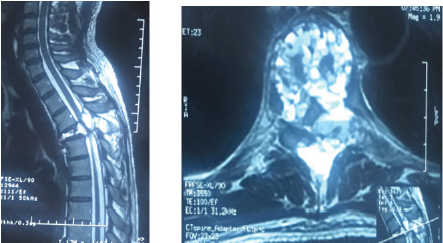
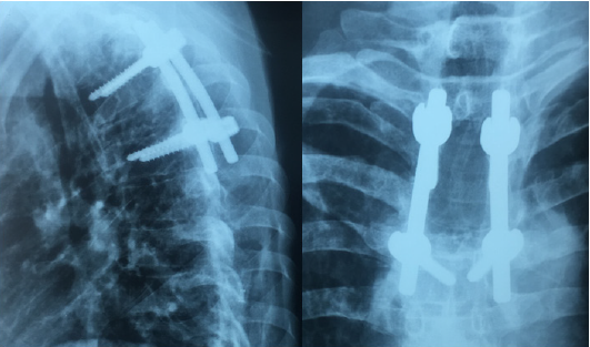
A 40 years old man, chronic smoker (a packet/day) was admitted to the emergency for a spinal syndrome for 2 years that was complicated 2 months before admission by the appearance of a grade D paraparesis, hypoesthesia with a sensory level at xiphoid process and urinary incontinence. The spinal MRI showed compaction of T4 vertebra with a heterogenous hyperintensity on T2 and STIR, a hyperintensity on T1 with heterogeneous enhancement of hematic fluid levels causing spinal cord compression at same level (Figure 1). The management consisted of performing a laminectomy of T3-T4 with pedicle screw and rod fixation of T3 and T5 vertebra (Figure 2). The postoperative course was marked by improved clinical signs. The Histopathology report came back in favor of aneurysmal bone cyst.

Figure 1: Spinal T2 weighted MRI showing (a) sagittal view with T4 compression and (b) Axial view with fluid level spaces.

Lupinepublishers-online-journal-of-neurology-and-brain-disorders

Figure 2: Spinal X-ray Lateral and anteroposterior showing post-operative pedicle screw and rod system.

Lupinepublishers-online-journal-of-neurology-and-brain-disorders

Discussion

Aneurysmal Bone Cyst (ABC) is a benign cystic lesion composed of blood-filled spaces, devoid of coating endothelial, muscle or elastic, surrounded by fibroblasts, described for the first time by Jaffe and Lichtenstein in 1942 [2]. Its incidence is 0.14 per 100,000 individuals with a female predominance (ratio 1.16) [3]. For Lichtenstein [4], the ABC is a secondary bone lesion due to hemodynamic imbalance resulting in a significant capillary dilatation, resulting from an increase in venous pressure. This increase in venous pressure might be due to venous thrombosis or abnormal arteriovenous communication. Biesecker et al [5] measured the intracystic pressure which was identical to that of arterioles and concluded that this hypertension is due to an arteriovenous fistula following a secondary lesion. Some authors [6-8] think that the appearance of an aneurysmal bone cyst would be secondary to impaired intraosseous vascular flow in the phase of consolidation following trauma. However, several cases of familial aneurysmal bone cysts [9,10] have been reported, hence orienting towards a genetic origin and indeed the genetic and immunohistochemical studies [11] have demonstrated a translocation on chromosome [7].

Aneurysmal bone cyst is most frequently found in the long bones especially the metaphyseal region [12,13], while the spinal location remains rare and affects the cervical, thoracic, lumbar and sacrum, with no case of coccygeal bone ABC reported [14]. Clinically, spinal ABC may be asymptomatic [1], but usually it manifests itself as a spinal disorder, with or without spinal cord compression signs. MRI remains the examination of choice which shows a welldemarcated bone lesion, hypointense on T1, hyperintense on T2 with a peripheral border taking contrast, defining spaces with fluid level [4]. Surgery that involves wide excision represents the preferred treatment of choice for ABC with a recurrence rate of 25% and multiple complications such as deformation and growth disorders [15]; For these reasons, selective arterial embolization was introduced into the management of this entity that proves similarly effective and with fewer complications, representing the treatment of choice in cases where the patient shows no deficit or unstable associated fractures [16]. Radiation therapy, which was an important pillar in the therapeutic arsenal, was gradually abandoned because of the risk of exposure and development of radiation-induced sarcomas [17]. Other therapeutic protocols have been proposed: the intralesional injection of calcitonin, and doxycycline Ethibloc [17-20], however they have not proven effective as with other therapeutic methods. Currently, research [21-23] has focused on the interest of Denosumab, a monoclonal antibody used in the treatment of giant cell tumors, and the intrinsic stimulation by the mesenchymal stem cells and have both shown promising results [24,25].

Conclusion

Aneurysmal bone cyst is a benign disease with unpredictable course, and thus what makes its treatment a challenge especially in locations where access is difficult. Surgery and embolization currently remain the treatment protocol of choice. Research on Mesenchymal stem cells and Denosumab have demonstrated promising results in the therapeutic management of ABC.

Read More About Lupine Publishers Journal of Neurology Please Click on Below Link:
https://brain-disorders-lupine-publishers.blogspot.com/

Tuesday, 23 August 2022

Lupine Publishers| 3rd Nerve Palsy After Microsurgical Clipping of Basilar Top Aneurysm

 Lupine Publishers| Journal of Neurology and Brain Disorders


Abstract

Postoperative oculomotor nerve palsy is a rare complication that occurs in the setting of micro¬surgical aneurysm clipping. While a number of theories have been postulated to explain the development of postoperative oculomotor nerve palsies, the underlying pathophysiology of such complications still remain to be elucidated. In this report, we present a case of postoperative isolated ipsilateral oculomotor nerve palsy after clipping of basilar tip aneurysm which we believe may be attributed to periperative oculomotor nerve manipulation related neuropraxia.

Keywords:Oculomotor Nerve Palsy; Basilar Apex Aneurysm; Postoperative

Introduction

Oculomotor nerve is the third cranial nerve that enters the orbit through the superior orbital fissure and controls muscles that drive most movements of the eye and raise the eyelid. Oculomotor nerve is derived from the basal plate of the embryonic midbrain. Cranial nerves IV and VI also participate in the control of eye movement. Oculomotor nerve palsy is an eye condition resulting from damage to the third cranial nerve or a branch there of. As the name suggests, oculomotor nerve supplies the major¬ity of the muscles that control eye movements. Therefore, damages to oculomotor nerve will render affected individuals unable to move his or her eye normally. Unilateral oculomotor nerve palsy is often encountered in the setting of ipsilateral aneurysms located at the Posteri¬or Communicating Artery (PcomA), Internal Carotid Artery (ICA) or their junction (PcomA/ICA). There have been some reports on ocu¬lomotor nerve palsy as a result of aneurysms in basilar tip, anterior artery or anterior communi¬cating artery. In the present report, we describe a case of basilar apex aneurysm presenting with ipsilateral postoperative oculo¬motor nerve palsy that is thought to be attrib¬uted to peroperative oculomotor nerve handling followed by neuropraxia.

Case Report

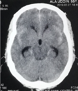
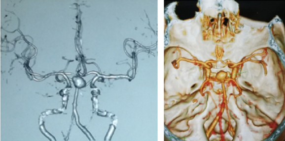
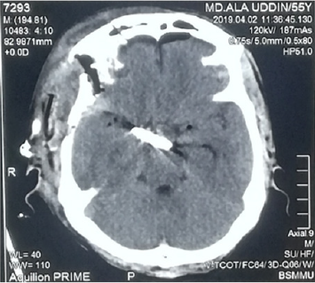
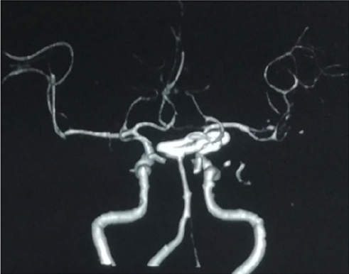
A 55years old hypertension male with no other significant past medical history or contributing family history complained about sudden severe headache for one hour followed by several episodes of vomiting and unconsciousness for 05 hours on admission at our hospital. Physical examination results were within nor¬mal limits except for considerable neck stiffness, positive Kernig sign and Brudzinski’s sign. Computed Tomography (CT) scan demonstrated diffuse subarachnoid hemorrhage, which was especially concentrated in ambient, sylvian fissure, interpeduncular and suprasellar cisterns with intraventricular extensions and mild triventricular hydrocephalus (Figure1). Computed tomography of the head show¬ing diffuse subarachnoid hemorrhage especially concentrated in ambient, sylvian fissure, interpeduncular and suprasellar cisterns with intraventricular extensions and mild triventricular hydrocephalus. CT Angiography (CTA) revealed a large saccular basilar tip aneurysm (approximately 10.7×9.5×10.5mm) (Figures 2A & 2B). The aneurysm was micro surgically clipped (Figures 4) via the right Orbitozygomatic approach (Figures 3A & 3B). Postoperatively the patient developed complete right third nerve palsy characterized by the presence of dilated pupil, ptosis and downward deviation and abduction of the eyeball. CT scan revealed no postoperative intracranial hematoma and the surgical clip was in the proper location. However, compression of the basal cisterns and assessment of the ventricular system were noted (Figure 5). Postoperative CTA showed no existence of another aneurysm or vasospasm (Figure 6). The patient was discharged 02 weeks after the surgery. At this point, his mydriasis and eye lid drooping were still present to a lesser extent. At one-month follow-up, the patient’s pupil was slightly contracted and reflexed reluctantly to direct and indirect light stimulation.

Figure 1: Computed tomography of the head show¬ing diffuse subarachnoid hemorrhage especially concentrated in ambient, sylvian fissure, interpeduncular and suprasellar cisterns with intraventricular extensions and mild triventricular hydrocephalus.

Lupinepublishers-online-journal-of-neurology-and-brain-disorders

Figure 2A & 2B: Computed tomography angiography show¬ing a large saccular basilar tip aneurysm (approximately 10.7×9.5×10.5mm). The arrow indicates the aneurysm.

Lupinepublishers-online-journal-of-neurology-and-brain-disorders

Figure 3A & 3B: Right sided Orbitozygomatic craniotomy (one piece), extradural anterior clinoidectomy & clipping of basilar apex aneurysm through trans-sylvian approach.

.
Lupinepublishers-online-journal-of-neurology-and-brain-disorders

Figure 6: Postoperative CTA showed no existence of another aneurysm or vasospasm and the surgical clip was in the proper location.

Lupinepublishers-online-journal-of-neurology-and-brain-disorders

Figure 7: On postoperative day two, the patient developed a complete right third nerve palsy.


Discussion

There are many proposed hypotheses pertain¬ing to the etiology of oculomotor nerve palsy before and after aneurysm surgery. Direct com¬pression by the aneurysm is the most classic and common cause. Other causes include direct injury to the third nerve intraoperatively [1-6], microvascular ischemia [7-9], focal hemato¬ma formation [3], vasospasm [8-12], anomaly of the vessels along the oculomotor nerve [10], elevated intracranial pressure and herniation, compression by intracranial structures other than aneurysms [7], and undetermined ori-gins [11]. In the present case, compression by aneurysm may not be possible because of the anatomi¬cal distance between the two entities. Postoperative CT, CTA decrease the chances of hematoma formation, vasospasm and elevated intracranial pressure. Although we cannot rule out the possibility of ischemic injury to the oculomotor nerve. Small vessel ischemic injury to oculomotor nerve usu¬ally exhibits pupilsparing [9] whereas our patient demonstrated a blown pupil. So, we believe that, probably from coarse dissection of the cavernous sinus dura and as well as direct anterior clinoidectomy or from heat of the low power drill may be responsible for the 3rd nerve palsy of our patient.

Conclusion

The novel use of intramuscular cerebrolysin and citicoline in a patient with kernicterus was safe and effective.

The Neuroreparative Effects of Cerebrolysin have Been Attributed to [3-5]:

a) Inhibition of apoptosis.

b) Improving synaptic plasticity and induction of neurogenesis.

c) Augmenting the proliferation, differentiation, and migration of adult.

d) subventricular zone neural progenitor stem cells, contributing to neurogenesis.

e) Induction of stem-cell proliferation in the brain.

Citicoline (cytidine diphosphate choline) is a mononucleotide made of ribose, pyrophosphate, cytosine and choline is a watersoluble naturally occurring substance that is generally grouped with the B vitamins. It is also considered a form of the essential nutrient choline. It is a safe substance with generally minor side effects which may include digestive intolerance after oral administration [6]. An accumulating research evidence suggests that citicoline is endowed with interesting pharmacological properties that can make it useful in the treatment of various disorders that has no universally accepted effective treatment including neurological conditions such as Parkinson’s disease, brain ischemia, hemorrhagic stroke, Alzheimer’s disease; and ocular condition such as glaucoma, nonarteritic ischemic neuropathy and amblyopia [6].

The Neuro-Protective Effects of Citicoline Were Attributed to the Followings [6]:

a) Preservation of cardiolipin and sphingomyelin

b) Preservation of arachidonic acid content of phosphatidylcholine and

c) phosphatidylethanolamine.

d) Partial restoration of phosphatidylcholine levels.

e) Stimulation of glutathione synthesis and glutathione reductase activity.

f) Reduction of phospholipase A2 activity.

g) Increasing glucose metabolism in the brain.

h) Increasing cerebral blood flow.

Reducing oxidative stress and preventing excessive inflammatory response in the brain by inhibiting the release of free fatty acids and reducing blood brain barrier breakdown. Enhances cellular communication by increasing the availability of neurotransmitters, including acetylcholine, norepinephrine, and dopamine. Lowering increased glutamate concentrations and increasing the decreased ATP concentrations induced by ischemia. Citicoline increases dopamine receptor densities, and therefore could improve memory impairment resulting from poor environmental conditions. Citicoline could also improve focus and mental energy and could be useful in the treatment of attention deficit disorder. Citicoline has also been shown to improve visual function in patients with glaucoma [7].

Conclusion

The novel use of intramuscular cerebrolysin and citicoline in a patient with kernicterus was safe and effective.

Read More About Lupine Publishers Journal of Neurology and Brain Disorders Please Click on below Link: https://brain-disorders-lupine-publishers.blogspot.com/

Tuesday, 3 May 2022

Lupine Publishers| Understand the Insomnium

 Lupine Publishers| Journal of Neurology and Brain Disorders


Editorial

In the international classification of sleep disorders we place insomnia as the first point, which we define as the inability to initiate, maintain or even consolidate sleep; this is best explained in the following paragraphs. When we arrived at the bed we put on our sleepwear, some pyjamas, other rompers, and the least with underwear to feel comfortable; the TV goes off, we kiss good night and turn off the light, and that’s when it starts, if man, that difficulty to fall asleep immediately, to see the roof, hear the horn of the passing truck and not be able to finish that moment of every day; and go that one despairs, we get bad and the time passes until eleven or even twelve, even more, of the night and that is when we can finally close our eyes, which I do not even tell dear reader the obviousness of the next day he is sleepy during working hours; it is what is called insomnia of onset.

Well, well, but he could sleep and start enjoying his dream, but suddenly, without just cause, so out of nowhere he wakes up one, two or three times in the night, just by opening his eyes, and above all accompanied by Think, and think, and think, what do you think about? In whatever: that the payment of those horrible things called tuition, other less beautiful calls alimony, or the classic car letters, undelivered work, together with the boss the next day, exams, legal circumstances in general; you just met the intermediate or maintenance insomnia. It’s okay, he could sleep and more or less reached a good quality of sleep, but always in the early morning, at a time that is not of God, like 3 or 4 in the morning, he wakes up and cannot for any reason to go back to sleep, and feel worse than bad, anxious, badly rested and knowing from within that he will not go back to sleep in what remains of the time in bed; We now know about insomnia of consolidation, late or terminal.

These in turn can be classified by their duration in transient, when it does not exceed two weeks; acute when it is greater than two weeks but less than six months and chronic when it exceeds six months.

In the world there are about 10% of the adult population who suffer from insomnia, of them more than half use drugs that help in their treatment as hypnotics, antidepressants or sedatives; The consequences of insomnia in any of its varieties translates into fatigue, concentration difficulties, increased incidence in work accidents, irritability, gastrointestinal disorders and increased behaviour problems. The treatment is based on treating the cause that triggers said insomnia, including removing the factor that causes stress, exercise, eating habits with weight control and measures of sleep hygiene; The drug must be at the discretion of the attending physician.

Read More About Lupine Publishers Journal of Neurology and Brain Disorders Please Click on Below Link: https://brain-disorders-lupine-publishers.blogspot.com/


Friday, 19 February 2021

Lupine Publishers | Cerebral Autosomal Dominant Arteriopathy with Subcortical Infarcts and Leukoencephalopathy (CADASIL) Syndrome: A Case Report

 Lupine Publishers | Journal of Neurology and Brain Disorders


Abstract

Cerebral autosomal dominant arteriopathy with sub cortical infarcts and leukoencephalopathy (CADASIL) is a rare autosomal dominant genetic disease. A35 year old women presented with progressive loss of memory left sided weakness and hemiplegic and non insulin dependent diabetes mellitus with characteristic MRI findings is reported due to its rarity.

Keywords: CADASIL Small vessel disease; Cognitive impairment

Introduction

Cerebral small vessel disease (SVD) is increasingly being recognized as the cause of stroke, impaired cognitive function and mood disorders in geriatric age group. Commonly SVD is sporadic due to old age and high blood pressure, but occasionally SVD has a monogenic cause. The best known among these is cerebral autosomal dominant arteriopathy with sub cortical infarcts and leuko encephalopathy(CADASIL), a rare type of autosomal dominant cerebral angiopathy. It causes recurrent sub cortical ischemic events and vascular dementia which appear as diffuse white matter abnormality on neuro imaging. It involves mainly the small cerebral vessels; histopathology reveals non-atherosclerotic and non-amyloidal angiopathy. [1] The 1st case of aforementioned symptoms was reported 30 years ago in a Swedish family [2] and the term CADASIL was coined in early 1990's [3] Since then CADASIL has been diagnosed in number of families and ethnic groups all over the world.

The prevalence of the condition varies between 2 and 5 in 100,000. Clinically the condition manifests in early or middle adulthood with variety of complaints ranging from migraine, with or without aura, which occur in 30-40% individual, mood disorders, usually depression seen in 30 % people, along with recurrent ischemic events, transient or permanent, accompanied by progressive cognitive impairment leading to dementia and premature death [4-5].

Case Report

This 35 years old married female patient was brought to the psychiatry outpatient department by her family members with the complaint of gradual decline of memory, difficulty in walking due to residual left sided motor weakness and co morbid Non Insulin dependent Diabetes Mellitus. There was no history of hypertension, depression or psychosis or impaired pain sensation. No history of seizures. EEG done earlier did not reveal any abnormality. There was past history of two episodes of CVA in last 5 years. Family history was not significant for headache but there was history of premature death of uncles. Physical examination showed thin built poorly nourished lady. There was no pallor, cyanosis, icterus, edema or generalized lymphadenopathy. Systemic examination of central nervous system revealed grade 3 motor weakness in left upper and lower limbs. Cranial nerves and fundi were normal. There were no sensory deficits or cerebellar signs. Examination of other systems was normal. Score on Mini Mental Status Examination was 12 out of 20. Relevant investigations including hemoglobin, blood sugar, HbA1c, liver function tests, SGOT, SGPT, alkaline phosphatase, blood urea, serum creatinine, were within normal limits. CT Scan brain showed a lacunar infarct in left external capsular region and age inappropriate diffuse cerebral atrophy. MRI of brain revealed diffuse cerebral atrophy with prominent sulcal spaces with periventricular and subcortical arteriosclerotic white matter changes. Small areas of acute to sub acute infarct in rest of the bilateral parietal and frontal lobes support diagnosis of CADASIL Syndrome.

Discussion

CADASIL gene was first identified on the chromosome 19 in the year 1996 [6] Subsequent studies revealed the mutation of NOTCH3 gene on chromosome 19q12. This gene codes for transmembrane receptor protein which is located on surface of smooth muscle cell which surrounds arteries. When pathological NOTCH3 receptor protein got accumulated in cerebral arteries it resulted in varied symptoms of CADAIL [7].

Patient with CADASIL can present with a variety of psychiatric manifestation in 20-41% of cases. The psychiatric manifestations range from agoraphobia, psychoses, personality disorders, alcohol and substance use disorders, adjustment disorder to episodic mood disturbances and bipolar affective disorder [4,8].

CADASIL should be suspected in individual with history of transient ischemic attacks, migraine, severe mood disturbances or late onset affective disorder with headache and neurological symptoms [8-9]. MRI studies shows hyper-intense lesion and ischemic lesion in Basal Ganglia and frontal location of white matter is associated with mood disturbances in patient of CADASIL [10-11].

To conclude it is advisable to do MRI brain in patients presenting with late onset Psychiatric Disturbances and a diagnosis of CADASIL should be considered as a possible differential diagnosis whenever a marked leukoencephalopathy is reported [12].

Read More Lupine Publishers Neurology and Brain Disorders Journal Articles:
https://brain-disorders-lupine-publishers.blogspot.com/

Wednesday, 12 June 2019

Lupine Publishers-Journal of Neurology & Neurosurgery


Immunoglobulin G4-related disease (IgG4-RD) is an increasingly recognized immune-mediated condition comprised of a collection of disorders that share particular pathologic, serologic, and clinical features [1,2]. These disorders were previously thought to be unrelated [3-5]. The commonly shared features include tumor-like swelling of involved organs, a lymphoplasmacytic infiltrate enriched in IgG4-positive plasma cells, and a variable degree of fibrosis that has a characteristic “storiform” pattern. In addition, elevated serum concentrations of IgG4 are found in 60 to 70 percent of patients with IgG4-RD. IgG4-related sclerosing cholangitis (IgG4-SC) is a characteristic type of sclerosing cholangitis, with an unknown pathogenic mechanism. Patients with IgG4-SC display increased serum IgG4 levels [6] and dense infiltration of IgG4-positive plasma cells with extensive fibrosis in the bile duct wall [7]. Circular and symmetrical thickening of the bile duct wall is observed in the areas without stenosis that appear normal on cholangiography, as well as in the stenotic areas [8]. IgG4-SC has been recently recognized as an IgG4- related disease. IgG4-SC is frequently associated with autoimmune pancreatitis (AIP). IgG4-related dacryoadenitis/sialadenitis and IgG4-related retroperitoneal fibrosis are also occasionally observed in IgG4-SC [9-12]. However, some IgG4-SC cases do not involve other organs. IgG4-SC is most common in elderly men. Obstructive jaundice is frequently observed in IgG4-SC.A number of diseases, such as, Cystic fibrosis, Chronic obstructive Choledocholithiasis, Biliary strictures (secondary to surgical trauma, chronic pancreatitis), Anastomotic strictures in liver graft, Neoplasms (benign, malignant, metastatic), Infections, hypertonic saline instillation in the bile ducts, Post-traumatic sclerosing cholangitis, Systemic vasculitis, Amyloidosis, Radiation injury, Sarcoidosis, Systemic mastocytosis, Hypereosinophilic syndrome, Hodgkin’s disease, may easily be confused with IgG4- related sclerosing cholangitis, or coexist in a patient [13]. In this case, report 57 years male patient presented with jaundice, fatigue, weight loss, oral moniliasis and right sided neck swelling. He was misdiagnosed as Cholangiocarcinoma. To know more go through the below link.


For more Lupine Publishers Open Access Journals Please visit our website: https://lupinepublishers.us/


Wednesday, 6 March 2019

Sleep is for Life: an Essential Part of Everyday Life: (OJNBD)- Lupine Publishers


Sleep is essential for basic survival as well as for optimal physical and cognitive performance in both human beings and animals. Sleep is a normal human function that is detrimental to sustaining life yet; individuals are affected differently by their sleep schedule. However, the community at large often underestimates sleep and its importance, therefore leading people to not be as concerned with a proper night’s sleep, thereby preventing them from performing at peak efficiency. Sleep plays a vital role in learning and when a person fails to obtain enough sleep the night prior, neurons in the brain might not fire properly, the body becomes out of synch, and it can even lead to accidental physical injuries. As many studies have been conducted, the majority have seemed to come to similar conclusions: a lack of sleep can have detrimental side effects on the human mind and body and by regularly obtaining enough sleep each night; a person can function more efficiently.


For more Lupine Publishers Open access journals please visit our website
For more journal of neurology open access journals articles please click here

Wednesday, 31 October 2018

About the Trigemino-Cardiac Reflex, their Association with Sleep Bruxism and the Reason Why Clinicians Treating Sb Should Consider a Careful Examination in Order to Decide Properly How to Adequately Follow these Patients: (OJNBD) - Lupine Publishers


Although well established as a neurogenic reflex pursuing a cardiovascular protective control in physiological conditions, trigemino-cardiac reflex (TCR) mechanisms and its clinical significance remains unclear This powerful brainstem oxygen-conserving autonomic reflex usually manifests as sudden onset of hemodynamic changes on heart rate (HR) and blood pressure (BP) and has been associated to arrhytmias, asystole, apnea and disturbed gastric mobility [1].