Lupine Publishers| Journal of Ophthalmology

Abstract
Visual impairment (VI) is a condition of reduced visual performance that cannot be remedied by refractive correction (spectacles or contact lenses), surgery or medical methods. Visual Impairment is a broader term that encompasses both Low vision and Blindness. The prevalence of visual impairment is estimated to be around 2.2 billion in the world as per World Health Organization (WHO). Causes of Visual Impairment differs significantly in different regions. Saying that, still cataract and refractive errors are main causes of visual impairment globally. Owing to its negative impact on the individual’s dailies, professional life, productive time, confidence, and self-respect, VI needs to be addressed timely and appropriately. Visual impairments do have profound consequences for the individual, however effective rehabilitation services can restore independence and therefore ameliorate these consequences and restore the individual’s quality of life.
Keywords: Visual Impairment; Blindness; low Vision; Rehabilitation; Optical Devices; Non-Optical Devices
Introduction
Impairment of any kind (sensorial or motor) is a bane for any human being. With visual impairment being one of the leading causes of sensorial impairment, it is necessary to acknowledge its prevalence, causes and methods to avoid them, if possible. Also, at the same time, it is important to understand that the term “visual impairment”, “blindness” and “Low vision” are not synonymous; rather visual impairment includes low vision as well as blindness. Simply, Visual impairment (VI) is a condition of reduced visual performance that cannot be remedied by refractive correction (spectacles or contact lenses), surgery or medical methods [1]. Various definitions of visual impairment have been given by various concerned authorities. International Statistical Classification of Diseases and Related Health Problems (ICD) defines visual impairment categories primarily on the basis of recommendations made by a World Health Organization (WHO) Study Group in 1972 [2] and defines as:
• low vision is defined as visual acuity of less than 6/18,
but equal to or better than 3/60, or a corresponding visual field
loss to less than 20 degrees in the better eye with best possible
correction
• blindness is defined as visual acuity of less than 3/60, or
a corresponding visual field loss to less than 10 degrees in the
better eye with best possible correction.
Since, the ICD definition of visual impairment is based on best corrected visual acuity (BCVA), which exclude uncorrected refractive error as a cause of visual impairment, leading to substantial underestimation of the total visual impairment burden by about 38%, Lalit Dandona [3] and colleagues debated a revision in these definitions and defined:
a person with low vision as one who has impairment
of visual functioning even after treatment and/or refractive
correction, and has a visual acuity of less than 6/18 to light
perception, or a visual field of less than 10° from the point of
fixation, but who uses, or is potentially able to use, vision for the
planning and/or execution of a task [4].
• blindness is defined as visual acuity of less than 6/60,
equivalent to central visual field of 20° [5,6].
• A guideline has been instated to define “Legal Blindness”
by Social Security Act to help people receive government assistance, such as Social Security disability benefits. Legal
Blindness is defined as:
Remaining vision in the better eye after best correction is
20/200 (6/60) or less OR contraction of the peripheral visual fields
in the better eye (A) to 10 degrees or less from the point of fixation;
or (B) so the widest diameter subtends an angle no greater than 20
degrees [7].
Some other definitions and types of blindness adopted by
different countries and institutions are:
Avoidable Blindness
It is the blindness that could be either treated or prevented by known, cost effective means. It can either be preventable or curable. It includes:
• Cataract
• Glaucoma
• Diabetic retinopathy
• Corneal infections
• Ocular trauma
• Refractive error
Curable Blindness
It is the stage of blindness where the damage is reversible by prompt management. E.g.: Cataract
Preventable Blindness
It is the type of blindness in which the loss of vision that
could have been completely prevented by effective preventive and
prophylactic measures
Example: Xerophthalmia
Manifest Blindness
It restricts the accomplishment of tasks for daily living leading
to impairment in mobility.
Vision less than 1/60 to just perception of light in the better eye
with best correction is considered as manifest blindness
Absolute Blindness
It restricts the ability of even perceiving the light. E.g.: Optic atrophy.
Economic Blindness
It is the level of blindness that prevents an individual from earning his wages. Vision less than 6/60 to 3/60 in the better eye with best correction is considered as economic blindness
Social Blindness
It hampers an individual from socially interacting with the family and peer groups in a satisfactory manner. Vision less than 3/60 to 1/60 in the better eye with best correction is considered as social blindness.
Classification:
The classification of visual impairment varies worldwide [8]. Since ICD is considered the standard worldwide classification therefore, the ICD definitions of visual impairment are used most often for worldwide estimates of visual impairment. The ICD-11 classification of visual impairment (which specifies all levels of vision loss, for each eye, and is used for coding a visual impairment as the diagnosis) is listed in the table below [2] (Table 1):
Table 1:
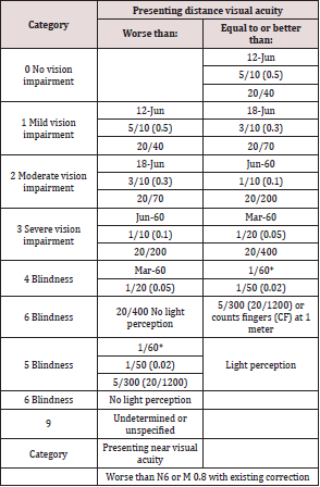
In the above table, it should be noted that in the revision of the ICD-10 categories of visual impairment proposed in 2003 by a WHO consultation on the development of standards for characterization of vision loss, low vision is replaced by two categories: moderate visual impairment (presenting visual acuity less than 6/18 but equal to or better than 6/60) and severe visual impairment (presenting visual acuity less than 6/60 but equal to or better than 3/60) [9]. Also, the previous classifications of visual impairment considered visual acuity as the only determining factor for categorizing visual impairment; loss of function due to hemianopia, loss of contrast sensitivity, photophobia, visual distortion, diplopia, or visual perceptual difficulties were not considered. These factors have been addressed on ICD-10 and ICD-11. Vision impairment results as an involvement of more than one factor which may include visual acuity, visual field, and contrast sensitivity. Therefore, these are the most clinically useful quantifiers of visual impairment [8].
Prevalence and Incidence:
According to World Health Organization, globally, at least 2.2
billion people have a vision impairment or blindness, of whom
at least 1 billion have a vision impairment that could have been
prevented or has yet to be addressed [10], among whom 1 billion
people includes those with moderate or severe distance vision
impairment or blindness [10] However, with the introduction of the
global initiative to eliminate avoidable blindness (vision 2020: the
right to sight), many agree that the initiative is in the right direction
to reduce the prevalence of avoidable blindness [11]. Estimates
of the number of visually impaired persons vary, depending upon
the criteria used [12]. In 2002, it was estimated that with bestcorrected
vision there were 161 million individuals with VI globally
[13]. However, when the prevalence of uncorrected refractive error
was included, this value increased substantially to 314 million
individuals with VI [14]. This implies that an additional 153 million
individuals were visually impaired from uncorrected refractive
error alone [14]. This statistics stress upon the underestimation of
total visually impaired population if uncorrected refractive error is
to be looked. Thus, the importance of correction and management
of refractive error is significantly high for the management of visual
impairment [15].
An interesting fact is that, the majority of the visually
impaired population are among the elderly and shows female
preponderance. The incidence of visual impairment increases with
age; more than two-thirds of persons with low vision are over
the age of 65 [16] The global prevalence of blindness is greater in
women than in men [17], especially among high-income regions
which may be attributed to longer life expectancies and lack of
access to health care services especially in rural areas [18]. Visual
impairment secondary to uncorrected refractive error starts at a
younger age than cataract [19], therefore childhood blindness
must not be undermined. Vision impairment, particularly among
children interferes with most life experiences [20]. World Health
Organization (WHO) estimates the number of blind children in the
world to be approximately 1.4 million, out of which three quarter
resides within developing and underdeveloped countries like
Nepal.20 According to mid-term review of Nepal Blindness Survey
2010, an estimated 1,013,041 children less than 16 years of age
(prevalence among under 16 age group assumed to be 10% based
on different studies varying from 3 to 20%); 1,164,053 persons
between 16-35 years of age have uncorrected refractive error for
distance (estimated prevalence 15%) [21].
Causes and Risk Factors:
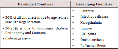
Not only the ocular morbidities, but also the systemic health conditions including trauma can pre-dispose a person to ocular abnormalities, rendering them visually impaired. The causes of visual impairment are numerous, including not only congenital and acquired ocular conditions, but systemic diseases with ocular complications and neurological insult and trauma. The major causes of visual impairment in vary widely from region to region, being largely determined by socioeconomic development, and the availability of primary health care and eye care services20 The Vision 2020- Right to Sight has identified five priorities for elimination of preventable blindness and they are: Cataract, Trachoma, Onchocerciasis, Childhood blindness and Refractive errors and low vision services. However, the causes of visual impairment are largely determined by the economy of the country. Refractive error is one of the most common causes of visual impairment around the world and the second leading cause of treatable blindness19 which remain as a common cause in both developed and developing countries. Globally, the leading causes of blindness and moderate and severe VI include uncorrected refractive error, cataract, and macular degeneration [22]. The major causes of blindness in developed and developing countries are shown in the table below (Table 2):
Table 2:
The incidence of these conditions increases with age. So, increasing age is the single- most risk factor for visual impairment. However, the causes of visual impairment among children are different from those of adults. Among children, the causes of vision impairment vary considerably across countries; for example, in developing countries congenital cataract is a leading cause, whereas in developed it is more likely to be retinopathy of prematurity, uncorrected refractive errors remain a common cause [20]. Though the global burden of childhood blindness is insignificant in comparison to that of adult, children have life time of blindness ahead. So, the proper cause and management should be instituted more readily among pediatric age groups. According to Nepal Blindness Survey (NBS), 1981 and Mid-term Review of NBS, 2010, the major causes of childhood blindness and the main avoidable causes in Nepal are:
• Cataract
• Glaucoma
• Retinopathy of prematurity is emerging in major cities
• Refractive errors
• Low vision, which encompasses visual impairment and
blindness from untreatable causes
Impact:
Visual impairments are pervasive in their effect on the individual’s entire life: social, occupational, recreational, and familial life. Blindness, of all chronic illnesses and disabilities, is the second leading cause of inability to work, with visual impairments being historically correlated with underemployment, unemployment, and poverty [23]. Recent studies have also suggested that sensory impairments, including visual impairment increase the risk for mortality, especially among the elderly. Vision loss can affect one’s quality of life (QOL), independence, and mobility and has been linked to falls, injury, and worsening of mental health, cognition, social function, employment, and educational attainment. The economic impact of vision loss is also substantial. Individuals with VI experience more symptoms of depression than those without. The implications of childhood blindness and VI may be more significant because of greater life expectancies [10].
Rehabilitation:
It has been reported that proper management of individuals with VI can provide the same quality of life as that of normally sighted individuals [24]. Therefore, rehabilitation should be made accessible to every visually impaired person. A visually impaired person needs following types of rehabilitation:
Medical Rehabilitation:
This includes early identification of the visually impaired
people and timely management which can be either medical or
surgical management. Medical rehabilitation not only includes
correction of refractive error, but prevention of any predisposing
factors like early cataract surgery, timely management of retinal and
optic nerve disease to stop progression and preserve the residual
useful vision, vitamin A supplements among children. Low Vision
Aids form an important part of medical rehabilitation of visually
impaired people so that they can benefit from their residual useful
vision. They are of two types, optical & non-optical.
1. Optical type: Spectacle magnifiers, Handheld and stand
magnifiers, Microscopic lenses, Telescopic lenses, Projection
devices etc.
2. Non-optical type: Large print books, Type writers, Typo
scopes, Special illumination devices, Talking books etc.
Educational Rehabilitation:
Educational rehabilitation includes education avenues provided to the visually impaired in the residential blind schools with the facility of Braille system of education. Louis Braille’s invention of Braille alphabet system in 1832 provided a tremendous impetus to the education of visually impaired children throughout the world. After gaining primary education from these schools, these children are placed in regular schools in the general education system. This system of education that is, first Braille from special blind school and then learning from general schools in the presence of regular teachers as well as a resource teacher is known as integrated system of education. It aims at normalizing the life and education of visually impaired children in the least restrictive environment along with sighted children in general schools.
Vocational Rehabilitation:
Vocational rehabilitation aims at helping the visually impaired earn their livelihood and live as useful citizens. They are taught simple occupations like candle making, chalk making, book binding, chair canning etc. Rehabilitation intervention can affect coping patterns over time and that direction and magnitude of such an effect may depend on the type of rehabilitation received.
Psychosocial Rehabilitation:
It includes mobility training, training for basic life skills and social support. Mobility training can be provided with cane or guide person or devices. Visually impaired needs to have a good and efficient training in the use of his remaining senses so that he acquires some amount of independence in his mobility. Basic survival skills should be taught. The social support extended by family and community is very important.
Conclusion
The burden of visual impairment is prevalent in every country, be it developing or developed. Visual impairments do have profound consequences for the individual, however effective rehabilitation services can restore independence and therefore ameliorate these consequences and restore the individual’s quality of life. Therefore, proper counseling, timely referrals and appropriate low vision aids, trainings and rehabilitations are very important aspects of eye care and patient management.
Read More About Lupine Publishers Journal of Ophthalmology Please Click on Below Link:
https://ophthalmology-lupine-publishers.blogspot.com/


