Lupine Publishers is a multidisciplinary, scholarly Open Access publisher focused on Genetic, Biomedical and Remedial missions in relation with Technical Knowledge as well. Our journals maintains a scrupulous, methodical, fair peer review System. Besides, quality control is riveted in each step of the publication process. https://in.linkedin.com/company/lupinepublishers
Showing posts with label Peer reviewed Journal. Show all posts
Showing posts with label Peer reviewed Journal. Show all posts
Fractures of the femoral head associated with neck fracture and
posterior dislocation of the hip are uncommon. Pip kin and later
Brumback classified these complex fracture dislocations. However in
today's era patient have more atypical injury pattern. We encountered a
case which has never been described in literature and cannot be fit into
any previously described classification system. We present the
radiological details and management.
Modern day accidents not only produce unpredictable but at times
unclassifiable injuries. Fractures of the femoral head associated with
posterior dislocation of the hip are uncommon as such [1]. Pip kin was
the one who sub classified Epstein-Thomas type V fracture-dislocations
into four additional subtypes which is still most widely followed [2].
Later Brumback et al. further classified femoral head fractures
emphasizing hip stability, with type "B" injuries being unstable [3].
The paucity of these cases is the main constraint to make an algorithm
of management. Nevertheless the results depend on early anatomical
reduction with the potential threat of osteonecrosis irrespective of
approach [4,5]. We hereby describe a case of posterior dislocation of
hip which is novel and never been described before. The purpose is to
make the fraternity aware of such a case which is unclassifiable even
though the management protocol may not be grossly different.
Case
A 52 year old migratory laborer had an accident at civil construction
site where he fell down from a height of 20 feet landing first on knees
(in kneeling position). He presented to emergency department with
severe pain in right groin, and inability to move the left lower limb.
On examination the limb was shortened, externally rotated and a bony
mass felt in the loin which did not move with the movement of distal
thigh. There was severe tingling and decreased sensation in front and
lateral aspect of leg and patient was unable to dorsiflex his ankle and
toes. His pulsations were normal but he had had transient hypotension
with tachycardia which corrected with initial resuscitation. First X ray
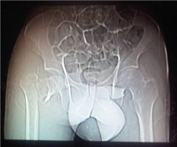
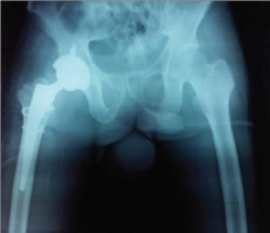
showed fracture about right hip (Figure 1) mostly a trochanteric
fracture but a careful evaluation showed incongruity of head and a
"vacant" on super lateral acetabulum. A CT scan was followed wherein the
fracture was better delineated. A posterior dislocation of hip with
fracture about the trochanter was seen. The head was also fractured and
the infra foveal part could be seen lying the acetabulum. A fracture
line also ran through the posterior wall of acetabulum which was not
more than 25% of the wall and also was undisplaced (Figure 2a- 2c). The
patient was admitted and limb placed in Thomas splint and prepared for
next morning. No attempts to reduce the dislocation were made. Figure 1.
Figure 2(a).
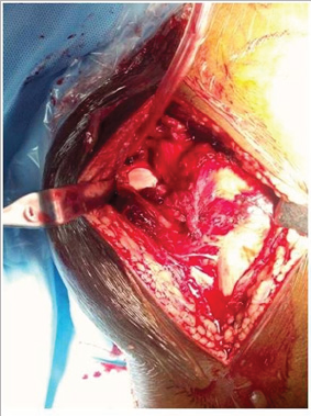
Figure 3(a).
Under general anesthesia, the patient was positioned laterally and
a Moore's posterior approach was taken guided by dislocation and wall
fracture. The gluteus maximus had a huge rent and external rotators were
torn, the head was seen indenting the sciatic nerve (bowstring effect)
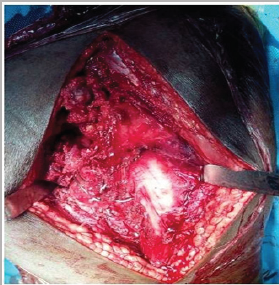
which was contused but intact (Figure 3a). The neck was ostetomised with
help of saw at the appropriate level and the calcar was reconstructed
using encirclage wiring (Figure 3b). Ethibond was used to suture the
greater trochanter. Next 2 temporary K wires were used in the ace
tabular walls and reaming done and cement less cup size 52 was fixed
augmented with 2 screws (Figure 3c). The K wires were removed after the
cup was found stable. The femur was prepared and uncemeneted corail stem
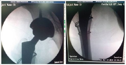
size 11 was inserted and size 36x0 femoral head was found stable. C arm
was used to confirm the placement of implants (Figure 3d). All soft
tissues were closed in best possible way, the hip was stable (Figure
3e). An abductor brace was applied post operatively. Postoperatively day
one faradic stimulation was started and patient was allowed side
tuning. Check X ray was done which showed a reasonable reconstruction
(Figure 4). The patient was allowed sitting with non weight bearing
mobilization from day 3 after the pain had subsided. 3 weeks after
surgery the patient was discharged when he went back to his home state
and never came back for follow up. Figure 3(b).
Femoral head fractures in combination with posterior dislocation of
the hip are rare presentation. Since first reported by Birkett in 1869
only a small number of cases have been reported world widen [6]. Epstein
et al.'s found about 10% in their series. It was Pip kin who subs
classified these fractures and various other classifications have been
proposed by several authors who claim to be improved version [5, 7].
Pipkin's classification remains most popular and widely accepted. They
divide them into 4 types with type 3 having associated ace tabular
fractures and type 4 having associated neck fractures. Our case is
unique with fracture in the trochanteric area. The trochanteric area has
never been described. The ace tabular fracture is also an addition
actually a combination pattern of Pipkin's 3 and 4. We believe that
there should be a type 5 including a both neck and ace tabular fracture
in association of head fracture and a subtype T wherein fracture
configuration is in trochanteric region instead of classical neck.
The cases of type 3 or 4 are very sporadic for any protocol to be
made. In younger individual attempts to fix the neck fragment, often
augmented with vascularised fibular graft has been made in past but long
term follow up is lacking. Osteonecrosis is a complication and surgeons
have tried different approaches with varying results [8,9]. In our case
closed reduction was not possible and moreover the impeding ischemia to
sciatic pushed.com for an early operative intervention. Taking into
consideration- a difficult fracture pattern, age, urgency of surgery,
peripheral set up hospital and an invincible avascular necrosis; we
thought a Total hip arthroplasty (THA) was an appropriate solution than
open reduction. Yet the challenges of choosing the right implant from
the whole Pandora box of prosthesis implants remained. In surgery, the
aim was to stick to principles of bone preservation like calcar
reconstruction and thus avoiding the distal fixation stems which were
reserved for future revision if a situation arose. A constrained hip
would have more advantages in this circumstance of soft tissue rent, but
the large head option of 36 was stable enough in our case. We admit
management to be debatable with expertise hands. Final outcome does
depend on return of sciatic function and proper occupational therapy
since he was a laborer. A draw back in our report remains that despite
best attempts to persuade the patient to visit nearest ortho clinic; he
never turned up but telephonically did tell to having a over the counter
sitting job in the village and satisfied with his hip.
The case highlights a rare injury and attempts to give an expansion
to the present Pipkin’s classification. Even though the demerit of this
report is a lacking follow up X ray and clinical picture, the initial
presentation is worth to be noted among the practioners and scholars.
For more Lupine
Publishers Open Access
Journals Please visit our website:
Background: Altered patellar alignment is associated with
anterior knee pain and multitude of conditions that affect the
patellofemoral joint. The study aim was to report the incidence of
patella altaand patella baja in indian population with anterior knee
pain and to investigate whether the normal limits of the patellar height
ratios are applicable in indian population. Methods: Lateral radiographs of 528 knees with anterior knee
pain were performed. Patellar heights were measured to calculate the
Insall-Salvati (IS), Insall-Salvati Modified (MIS), Blackburne-Peel
(BP), Caton Deschamps (CD) ratios. Results: The overall mean LT/LP ratio was 1.03 (SD 0.37) with
incidence of 16.2% (86/528) for patella alta with Insall-Salvati. Mean
MIS was found to be 1.68 (SD 0.018) and incidence of patella alta as per
MIS ratio was 14.3% (76/528). Blackburne-Peel and Caton-Deschamps
ratios revealed mean ratios of 0.88 (SD 0.15) and .99 (SD 0.16) and the
incidence of patella alta 17.8% with BP ratio and 10.2% with CD ratio
respectively. Conclusion: Incidence of patella alta was found to be 17.8%
with BP ratio, 16.2% with IS ratio, 14.3% with MIS ratio and 10.2% with
CD ratio. The BP and IS ratio were the most sensitive with MIS ratio
being the more specific one and CD ratio the least sensitive.
Anterior knee pain remains one of the commonest musculoskeletal
complaint with patients, and evaluation of patellar height is done
commonly as many conditions are associated with abnormal patellofemoral
relationship. The lever arm function of the patella plays an important
role in knee extensor mechanism and improves the quadriceps strength by
30-50% [1]. The joint reaction force of the patellofemoral joint varies
with the patellar height. A high riding patella or patella alta is
associated with patellofemoral malalignment and a reduced patellofemoral
contact often leading to patellofemoral pain or instability [2-3]. A
low riding patella or patella baja is often associated with limited knee
range of motion, patellofemoral arthritis and Osgood Schlatter disease
[4]. Numerous methods of patellar height have been described in the
literature: Blumensaat [5] Insal-Salvati [6], modified Insal-Salvati
[7], Blackburne-peel [8], Caton-Deschamps [9], DeCarvalho [7] and
Koshino [10]. These ratios are based on the ratio of the patellar length
to the reference point from the tibia. A very few studies have compared
the different indices for the patellar height analysis regarding their
reproducibility and reliability.
The study objective was to analyse the commonest methods for
measuring the patellar height in patients with anterior knee pain and
the aim of this study was to report the incidence of patella alta and
patella baja and investigate whether the patellar height ratios have
significant variations in adult indian population in which sitting on
the ground, kneeling, and squatting is common.
528 lateral x-rays of the knee (212 male and178 females) were
collected between the time period August 2017 to February 2018 from
patients with anterior knee pain, with difficulty or pain on squatting
or sitting. Patients with underlying knee pathologies, knee deformities
and knee surgery were excluded. Institutional review board approval was
obtained prior to conducting the study. Lateral radiographs of the knee
set in 30° of flexion were taken with assistance of a goniometer. 30° of
flexion of knee results in better visualization of the tibial
insertion. Radiographs were perpendicular to the film and centered on
the joint at a distance of 100cm.
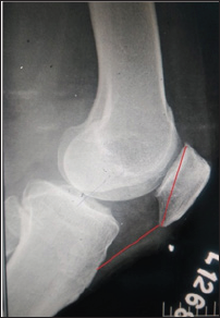
Figure 1: IS.
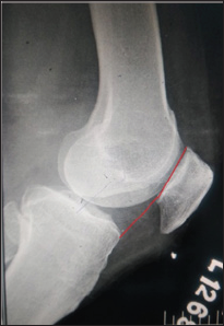
Figure 2: CD.
The measurements were made with the digital imaging software (Radiant
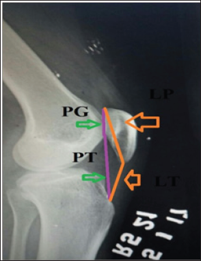
diacom viewer) by a single experienced physician. The patella length
was measured from the superior articular pole to the inferior non
articular pole (Figure 1). The distance from the origin of the patellar
tendon at the inferior pole of patella to its insertion on the tibial
tubercle was taken as the patellar tendon length. The Insall-Salvati
index is calculated as ratio of LT/LP, where LT is the length of the
patellar tendon and LP is the patella length (Figure 1). The
Blackburne-Peel index (Figure 2) is calculated as PP/PG where PP is the
perpendicular height of the distal part of the joint surface of the
patella to a line projected anteriorly to the surface of the tibial
plateau and PG is the length of the articular surface of patella. The
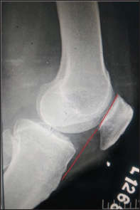
ratio PTG/PG, in which PTG is the distance from the lower edge of the
articular surface of the patella to the anterosuperior angle of the
tibia, and PG is the length of the articular surface of the patella is
calculated as Caton-Deschamps index (Figure 3). The Modified
Insall-Salvati index consists of the ratio PT/PG, wherein PT is the
distance from the lower edge of the joint surface of the patella to the
insertion of the posterior or deep surface of the patellar tendon in the
tibial tubercle, and PG is the length of the joint surface of the
patella (Figure 4).
The overall mean LT/LP ratio was 1.02 (SD 0.37). Comparison between
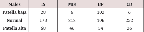
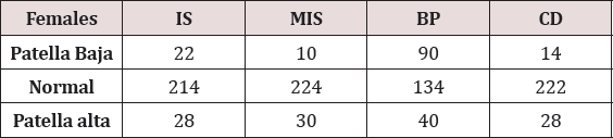
genders revealed that the mean LT/LP ratio was higher in males than
females with a mean of 1.04 (SD 0.29) and 1.01 (SD 0.46), respectively
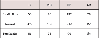
(Table 1 & 2). Using criteria of defining abnormal patellar position
(1.00±20%) based on Insall's study, the overall incidence was 16.2%
(86/528) for patella alta (Tables 3 & 4). The mean PT/PG ratio was
1.68 (SD 0.018). The MIS ratio was higher in males with mean of 1.70(SD
0.29) than females with a mean of 1.66 (SD 0.29). The incidences of
patella alta as per MIS ratio were 14.3% (76/528). Blackburne -Peel and
Caton- Deschamps ratios revealed mean ratios of 0.88 (SD 0.15) and .99
(SD 0.16). Males were found to have a higher mean in both the ratios
compared to the females (BP ratio: 0.89, SD 0.12 and CD ratio: 1.0, SD
0.18 in males and BP ratio: 0.88, SD 0.05 and CD ratio: 0.98, SD 0.06 in
females). The incidence of patella alta were 17.8% and 36% with BP
ratio and CD ratio respectively.
Vast majority of the studies in literature are often quoted with the
data obtained from the Caucasian population. There are none, if at all
few studies of patellar height being done in Asian population. There are
morphological changes in patella of the Caucasian and the Asian
population which make the patellar height ratios even more significant.
The present study is an observational study done with aim of assessing
the patellar alignment in anterior knee pain patients.
The position of the patella has an important role on patellofemoral
function. Abnormalities in patella position have thus been associated
with anterior knee pain and many extensor mechanism disorders. While
many techniques have been developed to measure patellar position such as
the Blacburne's ratio, the Insall Salvati ratio still remains one of
the most popular, largely because it is easy to use and remember [7,8]
.Despite its popularity, recent studies have suggested that the current
normal ranges should be extended, as the ratios may not be applied to
ethnicities outside western regions [11,12].
The Insall-Salvati (IS) method uses the length of the patellar
ligament in relation to the length of the patella6. The patellar
morphology and morphological differences in the anterior tuberosity of
the tibia (ATT) directly affect the measurements made using this method.
Grelsamer and Meadows [7] developed the modified Insall-Salvati (ISM)
method based on the length of the joint surface. Difficulty in
identifying this parameter is considered to be the main measurement
bias. The Modified Insall -Salvati ratio has its advantages over the IS
ratio that it can efficiently find out the patella alta in patients with
small patellar articular surface which is not taken into account in IS
ratio (Figure 5). Digital radiography seems not to present greater
details for this anatomical reference. The Blackburne-Peel (BP) method
exchanges the reference point of the ATT for the joint surface of the
tibial plateau, while keeping the joint surface of the patella. Although
Berg et al. [13] found that this was the most accurate and reproducible
method in conjunction with the IS index, and Seil et al. [13] ranked it
as the second most reproducible method in conjunction with the IS
index, we did not obtain similar results in our analysis, such that it
was only better than the ISM index. Lack of definition of the reference
line of the tibial plateau, such as which condyle to use as the
reference, or whether this line runs parallel to the joint surface or
perpendicular to the long axis of the tibia, contributes towards lower
concordance with this method. The method of Caton et al. [2], which uses
the joint surface of the patella and the angle of the tibial plateau as
references, also presents difficulty regarding identification of the
joint surface, as well as presenting a certain amount of variability in
defining the angle of the tibial plateau. Despite these factors, this
method was the one that showed greatest concordance in the study by Seil
et al. [13].
Figure 5.
The mean IS ratio was 1.02(SD 0.37) with the incidence of patella
alta being 16.2%. The incidence of patella alta calculated by height
ratios was found to be lesser than that of the published literature. Out
of the ratios analysed, it ws found out that the IS ratio and BP ratio
had the highest sensitivity for patella alta wherein the CD ratio had
the lowest sensitivity. The difficulty in analysing the anatomy of the
proximal tibia may be attributed to the decreased sensitivity of CD
ratio. MIS ratio is helpful in situations where in the patellar
articular length is decreased compared to that of the length of patella
(Figure 5). The study had the most sensitive ratio as BP ratio followed
with IS ratio, MIS ratio and lastly CD ratio which is comparable to the
study by Seil et al. [13]. It was interesting to note that, patella baja
were present in a proportion of patients with knee pain signifying the
requirement of understanding the knee biomechanics and further
evaluation of the patellofemoral joint for the articular cartilage.
Patella alta was found to have a incidence of 16.2% as per Insall-
Salvati ratio and needs to be kept in mind in paients with anterior knee
pain.
For more Lupine
Publishers Open Access
Journals Please visit our website:
The volar shearing distal radius fractures associated with subluxation or dislocation of the carpus were first described by Barton in 1838. To our knowledge there is no literature regarding spontaneous radiocarpal dislocation during an osteosynthesis recovery of a Barton's fracture. This article describes the treatment and outcome of a case of a patient submitted to radius osteosynthesis with an anatomic volar distal radius plate. After the cast removal he started to feel pain, with no history of associated trauma. Imaging showed a Dumontier type II dorsal radiocarpal dislocation. It was reduced through a double approach of the radius, and a radiocarpal fixation with 3 Kirschner wires was performed. There were no perioperative complications. The imobilization and the kirschner wires were maintained for 7 weeks. After removing the cast and the kirschner wires, the patient presented flexion of 20°and extension of 15°, which improved up to flexion 30° and extension 60°, supination 60°, with preserved pronation, 24 months after the trauma. At this time, the patient reports no pain, feeling of instability or signs of nerve compression. The DASH disability score result was 3, 1 and the Mayo Wrist Score was 93. The radiograph control did not show any relevant alteration. In the presence of a comminuted distal radius fracture two-dimensional and even three-dimensional computed tomographic scans may well provide important additional information preoperatively and in selected cases a temporary external fixation may prevent displacement of volar fragment.
The volar shearing distal radius fractures associated with subluxation or dislocation of the carpus were first described by Barton in 1838. They represent complex injuries, challenging to treat due to, in part, the anatomy of the volar surface of the distal part of the radius [1-4]. To our knowledge there is no literature regarding spontaneous radiocarpal dislocation during an osteosynthesis recovery of a Barton's fracture. The purpose of this report is to expose a case with the aforementioned characteristics.
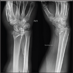
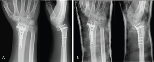
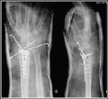
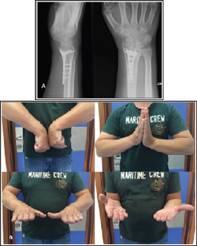
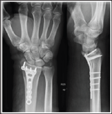
A 48 year-old male, caucasian, policeman, right handed, attended the emergency service due to right wrist pain and functional impotence. Five weeks earlier he had suffered a road accident resulting in a fracture of the distal right radius (AO subtype 23-B3) (Figure 1) and a tibial plafond fracture. At a foreign hospital that received him, he was submitted to radius osteosynthesis with an anatomic volar distal radius plate and in the lower right limb a temporary joint-bridging external fixation was placed during 1 week, and was posteriorly substituted by screws and plate fixation in the tibia and fibula. The wrist cast was removed 4 weeks after surgery in a private hospital in our country. After this episode he began to feel pain, without any history of associated trauma. He had no other previous history of interest. The physical examination revealed a "dinner fork” deformity, with a volar translation of the carpus. The skin was intact. There was no neurovascular deficit. Imaging showed a Dumontier type II volar radiocarpal dislocation (Figure 2A). At the emergency department we conducted closed reduction through traction after infiltration of a local intra- articular anesthetic, without success nevertheless (Figure 2B), so we decided to proceed to surgical treatment. Intraoperatively, through a volar approach of the radius, a bone fragment distal to the plate was visible. A dorsal approach was needed to reduce the dislocation. A radiocarpal fixation with three Kirschner wires was performed, two of them fixing the volar fragment of the distal radius, and one stabilizing the radiocarpal joint. One screw of the plate was removed due to its' intraarticular location. The distal radioulnar joint was evaluated under dynamic fluoroscopy and was apparently stable. Immediate postoperative radiographs confirmed a concentric reduction and stable fixation of the radiocarpal joint (Figure 3). Postoperatively, the patient was placed in an arm cast with free elbow and metacarpophalangeal (MP) joints. There were no perioperative complications. The immobilization and the kirschner wires were maintained for 7 weeks. After removing the cast and the kirschner wires, the patient presented flexion of 20°and extension of 15°, which improved up to flexion 30° and extension 60°, supination 60°, with preserved pronation, 24 months after the trauma. At this time, the patient reports no pain, feeling of instability or signs of nerve compression. The DASH disability score result was 3,1 and the Mayo Wrist Score was 93. The radiograph control did not show any relevant alteration (Figure 4).
Figure 1: Simple AP and lateral radiograph obtained upon arrival at the emergency service at the foreign hospital.
Figure 2: (A) Posteroanterior (PA) and lateral radiographs of the right wrist show volar dislocation of the carpus at our emergency department (B) Postreduction attempted failed PA and lateral radiographs of the right wrist.
Figure 3: Postoperative PA and lateral radiographs of the right wrist show maintained reduction of the radiocarpal joint after radiocarpal fixation with 3 Kirschner wires.
Figure 4: Radiographic and clinical results after 24 months. AP view and lateral view (A); Comparative photographs of both hand in flexion, extension, pronation, supination (B).
Dislocations of the radiocarpal joint are high-energy injuries, extremely infrequent, with an incidence of 0.2% of all dislocations [5-6]. There are two classifications that are important in therapeutic decision: Moneim et al. [7] and Dumontier et al. [5]. The management of radiocarpal dislocations varies, since there are different therapeutic options and no long-term follow-up data. Most authors propose a surgical treatment, but nonoperative management has been successful and also reported positive functional results in several series [5].
Figure 5: Postoperative PA and lateral radiographs of the right wrist after surgery at the foreign hospital.
On the other hand, articular fractures can be associated with a spectrum of radiocarpal dislocation. Muller et al. classified articular fractures resulting from a shearing force to the wrist as type B. Jupiter subdivided these fractures in 3 subgroups, in which B3 is volar marginal articular fracture (Barton's fracture)[8]. Open reduction with internal fixation is a commonly used method for the treatment of these intra-articular injuries. Our patient had a 23-B3 distal radius fracture (Figure 1). At the foreign hospital that received him, he was submitted to radius osteosynthesis with an anatomic volar distal radius plate. Postoperative lateral radiographs of the right wrist, provided to.com, showed non- anatomical reduction of fragments after the fixation (Figure 5). Due to surgery to the right lower limb the patient was immobilized with an arm cast during four weeks. After it's removal he began to feel pain, with no history of associated trauma. Imaging showed a Dumontier type II volar radiocarpal dislocation (Figure 2A). There are few data reporting loss of fixation of volar shearing fractures of the distal part of the radius [2]. To our knowledge, no reports have documented spontaneous radiocarpal dislocation after distal radius osteosynthesis.
Some authors reported favorable results after distraction plating of comminuted distal radius fractures. In the published reports, the main indication for distraction plating over traditional internal fixation is the presence of articular or metaphyseal comminution, which makes anatomic fixation difficult, and for polytraumatized patients, where the injured upper limb is required to aid lower-extremity weight bearing [9-10]. We hypothesized that the non-anatomical reduction of fracture and the need for aid lower-extremity weight bearing resulted in loss of fixation and consequently caused radiocarpal dislocation. Our case had a volar dislocation of the radiocarpal joint. At first, we performed a closed reduction under the fluoroscopy, but we were not successful. We needed a double approach to reduce the dislocation, and the volar fragment. After that we perform the radiocarpal and the volar fragment fixation with kirschner wires. The distal radioulnar joint was evaluated under dynamic fluoroscopy and was felt to be stable. Using a simple surgical technique we obtained a good functional result after 24 months of follow-up.
This report brings attention when evaluating volar shearing fractures. In the presence of a comminuted fracture two-dimensional and even three-dimensional computed tomographic scans may well provide important additional information preoperatively and in selected cases a temporary external fixation may prevent displacement of volar fragment.
For more Lupine
Publishers Open Access
Journals Please visit our website: