Ocular manifestations of rosacea are commonly nonspecific and
variable. The etiology of the inflammation is not fully known
and there is no definite diagnostic test for the disease. We often under
diagnose the disease, despite the potential for serious
sightthreatening
sequelae. When evaluating patients with rosacea, dermatologists and
family physicians should obtain a careful history
of eye complaints and examine the eyelid margins thoroughly. Patients
with moderate-to-severe ocular findings will benefit from
evaluation by an ophthalmologist. Controlling symptoms and preventing
potential risk of blindness should be the aim of early
diagnosis and prompt intervention. The mainstay of treatment is good lid
hygiene, topical metronidazole, topical azithromycin eye
drops, topical cyclosporine, and oral tetracycline.
The aim of this review is to shed light on the signs of ocular
rosacea, dermatologists and family physicians must be familiar with,
the understanding of the pathogenesis of this old disease of skin and
the eye since the original observation by Von Arlt in 1864 and
its treatment.
Quality of Evidence
A Pubmed search of English-language literature from August 1931 to
April 2018 revealed a combination of prospective cohort
studies and descriptive studies, with a predominance of descriptive
studies based on clinical experience. The MeSH terms; eye, skin,
rosacea was used, as well as the non-MeSH terms ocular and
oculocutaneous. The articles selected for assessment were the ones that
provided complete clinical reviews of relevance, i.e., they provided a
thorough review of the clini¬cal presentation of ocular rosacea,
its pathophysiology, diagnosis and treatment options.
Introduction
In dermatology, oculocutaneous diseases such as seen in bullous
diseases, atopic dermatitis, and rosacea are often encountered.
Many eye structures and the epidermis share the same ectodermal
origin that can partially explain the association. Since the original
observations of Von Arlt in 1864, we know that the eyes are
likely to be involved in rosacea. Von Arlt reported cases of this
disease associated with conjunctivitis and shallow, round ulcers
of the cornea. He recognized that treatment of the skin condition
improved the state of the eye [1].
Ocular involvement with rosacea is an under-appreciated and
under diagnosed [2-5]. In patients with ocular and cutaneous
rosacea, approximately 53% have the cutane¬ous eruption first,
20% with eye findings first and 27% both simultaneously at onset.
Peak incidence for ocu¬lar rosacea is later than for cutaneous
rosacea, commonly occurring in sixth and seventh decades. While
cutaneous rosacea affects women twice as often as men, ocular
rosacea manifests no sex predilection [6,7]. A strong correlation
exists between the degree of ocular involvement and the tendency
to flush [8].
Ocular findings can be considered as either minor or major;
however, minor findings are more common [4]. Ophthalmic findings
are nonspecific on their own and are inde¬pendent of the severity
of the skin disease [6,9,10]. Initial ocular complaints consist of
sensation of a foreign body, tearing, pain, photophobia, pruritus,
stinging, burning, and blurred vision [9,10-13].
Meibomian gland dysfunction occurs in about 50% to 93% of
rosacea patients, which is the hallmark of the disorder [13,14]. Other
ocular manifestations include chronic staphylococcal blepharitis, telangiectasia of the lid margin, recurrent chalazae, chronic
conjunctivitis, dry eye, peripheral corneal neovascularization,
marginal corneal infiltrates with or without ulceration and
episcleritis [15,16]. The most common ocular findings in adults
are hyperemia of the conjunctiva and corneal neovascularization
[15]. However, in children the most common ocular findings are
meibomian gland disease, recurrent chalazae and conjunctivitis
[16]. Dermatologists and primary care physicians must be aware
of the signs and symptoms of the major complications of rosacea
and know when to refer patients for ophthalmological evaluation
and care. Corneal neovascularization, erosion, ulceration and
perforation are major complications of rosacea and are better dealt
by ophthalmologists. This review covers the pathophysiology, the
major and minor clinical manifestations, the diagnosis and therapy
of ocular rosacea.
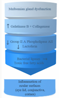
Pathophysiology: Figure 1
Meibomian Gland Disease
Meibomian glands (MGs) are rows of enlarged sebaceous
glands running along the lid margin posterior to the eyelashes.
They produce lipid secretions that retard tear evaporation and
are essential for the maintenance of the structural and refractive
integrity of the ocular surface. MGD is common in fair-skinned
individuals in association with rosacea and old age (Figure 2).
Figure 1: Pathophysiology of ocular rosacea.
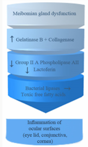
Figure 2: MGD with obliteration of some meibomian
glands orifices.
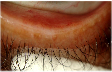
Figure 3: Showing stagnant sebum at meibomian orifices
of lower lid.
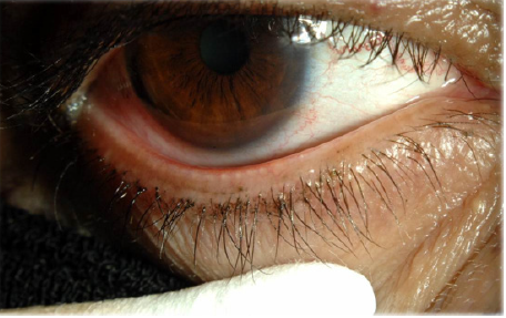
Figure 4: Blepharitis, erythema, edema, discharge and
crusting.
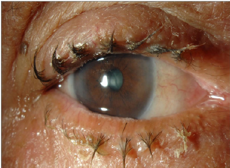
MGD (Figures 3 & 4) is a common cause of blepharitis, meibomian
keratoconjunctivitis, keratoconjunctivitis sicca and chalazae that
occurs in (100%, 60%, 37% and 57% respectively) in patients with
rosacea [17-22]. In addition to MGD, 5-10% of patients with ocular
rosacea develop vascular telangiectasia [19,23] and irregularity of
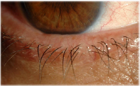
their lid margins and conjunctiva hyperemia [13,23-25] (Figures
5 & 6). There is a generalized sebaceous gland dysfunction that
involves the MGs in rosacea patients [18]. Alterations in lipid
secretions (meibum) and abnormal keratinization of the MGs duct
orifices have a profound effect on the quality of tear film and on the
ocular surface [21]. Sebum excretion rate is not elevated in ocular
rosacea [22].
Figure 5: Showing telengiectasia of lid margin.
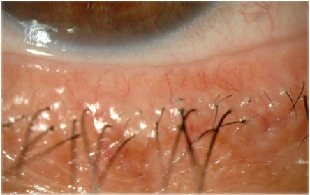
Figure 6: Showing irregularity of internal lid margin.
Gelatinase B
Gelatinase B, which is a member of the family of matrix
metalloprotienase (MMP), is secreted in high levels by the epithelium
and stromal keratinocytes of the cornea in response to elevated
levels of interleukin-1 alpha (IL-1). Clearance of interleukin-1
alpha (IL-1) in the tear fluid of patients with ocular rosacea is
disrupted, that leads to higher than normal levels of the cytokine
[26]. MMP and collagenase degrade extracellular matrix and lead
to recurrent epithelial erosions, vascularization and epithelial
basement membrane dystrophy of the cornea that eventually leads
to blindness [19,23,24,27,28]. These corneal changes respond to 4
weeks therapy with tetracyclines through reduction of the activity
of MMP [29]. Gelatinase B has been implicated as causative factor
in sterile corneal ulceration, which makes it a possible therapeutic
target to prevent sight threatening complication associated with
ocular rosacea [28].
Aqueous Tear Secretion
The amount of aqueous tear secretion by Schirmer’s test for dry
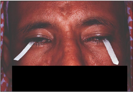
eyes is markedly reduced in patients with rosacea [30] (Figure 7).
Less than 5-10mm of wetting of the inserted Whatman filter paper
is suggestive of dry eyes. The paper is inserted in the middle and
outer third of the lower eyelid with or without topical anesthesia,
after 5 minutes of eye opening and blinking as necessary, the
wetting is measured [16].
Figure 7: Schermir test for dry eye.
The tear breaks up time TBT, that measures the stability of the
tear film, is abnormal in patients of rosacea; dry spots appear in
less than 10 seconds on the cornea. The test is done by installing
fluorescein dye into the lower eye fornix, patient is asked to blink and
stop. By the use of colored light, the time interval between the last
blink and dry spot appearance on the cornea is noted by stopwatch.
Dry spots appear as black lines or spots [30]. Schirmer’s test and
TBT, are two useful tests, used to diagnose dry eye syndrome. Intact
tear film quantity and quality as proven by normal Schirmer’s and
TBT tests, are required to maintain healthy epithelial cells of the
ocular surface. Due to the impaired secretion of the meibomian
glands in patients suffering from rosacea, instability of the tear film
and collateral damage of the cornea and conjunctiva may occur.
Group IIA Phospholipase A2 (GIIAPLA2)
Human tears contain several natural antimicrobial proteins
including group IIA phospholipase A2 (GIIAPLA2), lysozyme,
lactoferrins, defensins and gamma globulins [30-33]. Altered
secretions of these natural antimicrobial peptides may be important
in the pathogenesis of rosacea. GIIAPLA2 is a lipolytic enzyme
capable of killing a broad spectrum of gram-positive bacteria
[34,35]. The level of GIIAPLA2 is markedly reduced in patients of
rosacea with chronic conjunctivitis predisposing them to bacterial
infections [36]. The low values could be explained by increased
consumption due to long standing continuous staphylococcal
stimulation [37].
Lactoferrin
Secondary to chronic meibomitis and blepharitis Lactoferrin
level is also low in patients with ocular rosacea. The low
concentration of lactoferrin may play a role in the ocular surface
inflammatory components of rosacea [14].
Bacterial Lipases
Elevated levels of Staphylococcal lipases act upon wax and
sterol esters from MG producing toxic free fatty acids, which are
another source of inflammation in ocular rosacea [38,39].
Others
Infestation of eye lashes by Demodex mite and the symbiotic B
Oleronius bacteria living inside the mite are strongly correlated with
blepharitis and facial rosacea secondary to strong host immune
response to proteins produced by B. oleronius [40,41].
Culture independent analysis based on 16s rRNA gene sequences
of tear samples from patients with blepharitis showed increase in
Staphylococcus organisms and decrease in Propionibacterium.
Other ocular pathogens that are considered normal microbial
communities such as Streptophyta, Corynbacterium and
Enhydrobacter are detected in abundance. It seems that Blepharitis
may be induced by mixed microbial flora, pollens, dust and soil
particles that are the main source of Streptophyta, Corynbacterium
and Enhydrobacter.
Goblet cells are mucous producing, which is central to
stabilizing tear film layer. There is a significant decrease in the
number of goblet cells in patients presenting with dry eyes [35,42].
Abnormalities of goblet cell number or function could cause eye
symptoms in rosacea sufferers.
Clinical Presentation
The most common symptoms of ocular rosacea are nonspecific
and include foreign body, gritty or dry sensation, burning,
tearing, or the redness. Frequently the symptoms are out of
proportion to the minimal eye findings. Telangiectasia of the lid
margins, meibomian gland dysfunction, and blepharitis are the
common forms of lid involvement [11]. Lempert reported that
57 % of all patients over age 19 scheduled for chalazion excision
had rosacea [12]. Conjunctival involvement in ocular rosacea
is usually in the form of a mild bulbar conjunctival hyperemia.
Cicatrizing conjunctivitis, conjunctival granuloma, and phlyctenular
conjunctivitis are less common conjunctival manifestations [13].
The most common form of corneal involvement is in the form of
a superficial punctate keratopathy which usually occurs in the
lower 1/3 of the cornea. Peripheral epithelial nodular elevations,
recurrent epithelial erosion syndrome, corneal neovascularization
with peripheral thinning, and corneal ulcer are the other forms of
corneal involvement [13]. Episcleritis, scleritis and vitritis can also
be associated with rosacea.
Ocular rosacea commonly affects bilaterally the eyelids,
conjunctiva, and the cornea. A dermatologist should inquire about
the following symptoms such as dry eyes, burning, itching, ocular
irritation and grittiness, redness, photophobia, blurry vision,
stabbing pain foreign body sensation occurring during sleep or
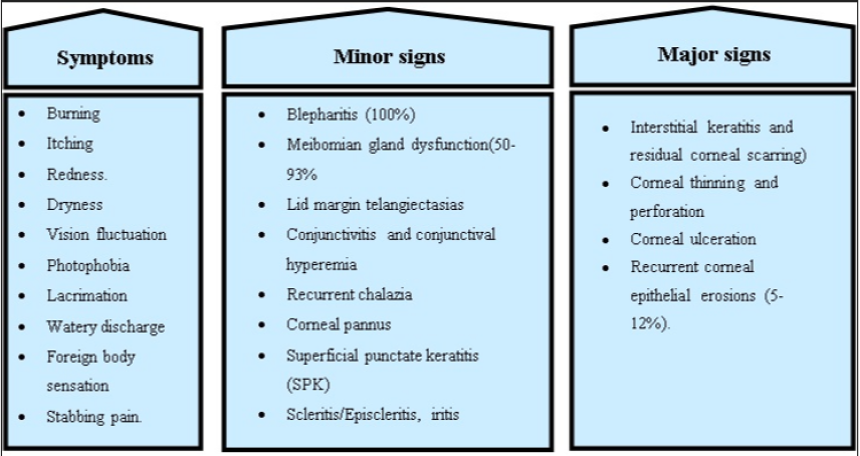
upon awakening or both (Figure 8). Patients with ocular symptoms
are better referred for ophthalmological evaluation. It is of utmost
importance to diagnose dry eye syndrome and manage it to avoid
corneal vascularization and ulceration that are sight threatening
complications.
Figure 8: Clinical symptoms and signs of ocular rosacea.
Eyelids and Meibomian Gland
Examination of the eyelid is very important looking for crusts,
thickening, rounding, erythema, telangiectasia and irregularity of
the lid margin (Figures 4-6), and dilatation of conjunctival vessels.
Blepharitis presents with erythema and scales or crusts around
eyelashes without scarring (Figures 4,9-12) and predisposes
to chalaziae (Figures 13 & 14) that usually present with a short
history of recent lid discomfort, followed by acute inflammation
(redness, tenderness, and swelling). Rosacea should be considered
in patients with recurrent chalaziae since it was almost twice as
common as those with only single occurrence of chalazia [43].
Figure 9: Rose Bengal staining, dry eyes.
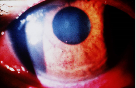
Figure 10: Showing anterior Blepharitis, telengiectasia of
lid margin, MGD.
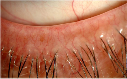
Figure 11: Mixed anterior and posterior blepharatitis. Note
the inspisated meibomian gland secretion.
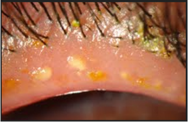
Figure 12: Hordeolum internum of lower eyelid (acute
chalazion), externum of upper eyelid (Stye).
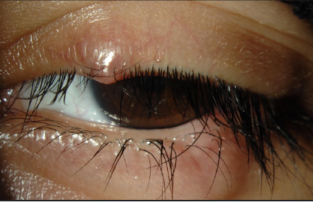
Figure 13: Multiple chalazia.
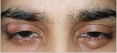
Figure 14: Inspisated meibomian gland secretion.
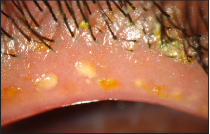
The symptoms of MGD are non-specific and include burning,
red eye, irritation, itching, dryness, and fluctuation of vision. Upon
examination, the lid margin is frequently erythematous, irregular,
thickened, or rounded with dilatation of palpebral conjunctival
vessels and telangictasia around the glandular orifices (Figure
14). The orifices are often plugged with yellowish solid secretions
(Figure 6). Since telangiectasia of the lid margin frequently occurs
in rosacea [4-49], therefore lid margin examination is very useful in
diagnosis of ocular rosacea.
Conjunctiva
Conjunctivitis presenting with photophobia, profuse
lacrimation and together with involvement of interpalpebral conjunctival vessels may occur [23]. The discharge of rosacea is
watery in nature, but if becomes mucopurulent, infection should
be excluded [23]. Chronic diffuse conjunctival hyperemia and
granuloma and rarely cicatrization that resemble trachoma can
occur [48,50-56]. It is important to remember that the severity of
ocular disease is not correlated with cutaneous severity [57].
Cornea
Recurrent corneal epithelial erosions have been reported to
occur in 5% to 12% [13, 23].
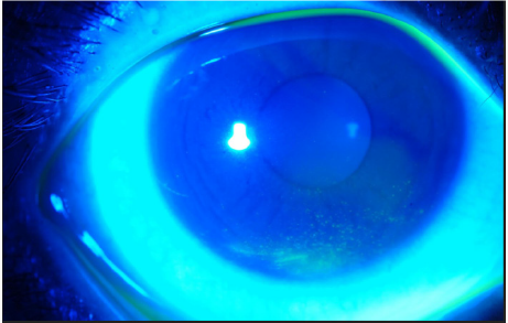
Figure 15: Severe punctate epitheliopathy stained with
Fluorescein. Note anterior migration of muco-cutaneous
junction on upper eyelid margin.
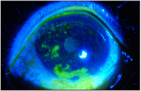
Figure 16: Peripheral corneal infiltrate. Note conjunctival
hyperemia and blepharitis.
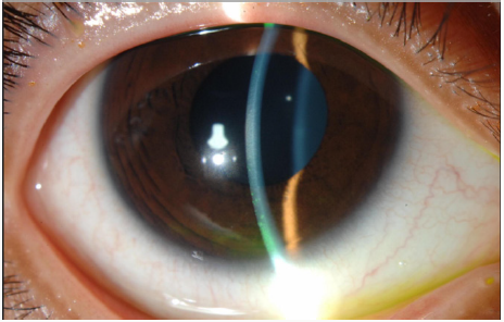
Symptoms of corneal erosions include stabbing pain and
foreign body sensation occurring during sleep or upon awakening
or both. It includes the presence of superficial punctate keratitis
(Figures 15-18) typically on the lower third of the cornea and
always accompanied by neovascularization [28]. Microbiological
evaluation should be considered even in cases of sterile keratitis
prior to the treatment with topical steroid, so to prevent possible
associated fungal keratitis [53]. When vascular invasion of the
peripheral cornea occurs, along with subepithelial infiltrates, this
type of inflammatory keratitis allows neovascularization to spread
circumferentially if left untreated [44-47,54] and blindness may
unfortunately occur. Corneal infiltrate (Figure 9), phlyctenules
(episcleral nodules) appearing like a small pump or vesicle, edema,
peripheral panus, corneal thinning and ulceration or perforation
may also occur [49,50,52-54]. Uncommon ocular findings include
kerato-conus like features such as high asymmetric astigmatism
and inferior corneal thinning, as well as herpes like dendritic
corneal lesions [44,54].
Figure 17: Mild punctate epitheliopathy inferiorly located.
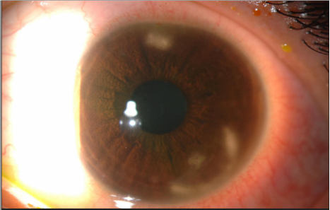
Figure 18: Mild punctate epitheliopathy stained with
Fluorescein.
Ocular Rosacea in Children
Overall, rosacea is rare in children and may be misdiagnosed
as viral or bacterial infections [55]. There is no estimate in the
literature of ocular rosacea in children, but under diagnosis occurs
frequently in this age group [55].
Children usually present with recurrent chalaziae,
blepharoconjunctivitis and generalized ocular surface irritation [43]
that antedates any cutaneous findings. The case series of pediatric
ocular rosacea by Nazir included six patients with age range from
3 to 12 years. In all cases, the patients had chronic irritation of
at least 6 months duration with no response to various topical
antibiotics and anti-allergy agents [55]. It appears to be commoner
in girls than boys, and findings are usually bilateral, although they
may be asymmetric. Cutaneous changes are uncommon but when found they are predominantly papulopustular with underlying
telangiectasia [56].
There are no criteria set for diagnosing ocular rosacea; however,
it may be diagnosed after excluding other conditions like herpes
simplex keratitis, bacterial infection, or allergic and atopic disease
[55]. The presence of lid margin telangiectasia, along with MGD and
long history of ocular irritation should point toward a diagnosis
of ocular rosacea [55] (Figure 5). Fortunately, treatment of ocular
rosacea by oral doxycycline or erythromycin tailored to the age of
the patient is helpful but relapses could occur [55].
Diagnosis
The diagnosis of ocular rosacea should be considered when
a patient has one or more of the following symptoms and signs,
foreign body sensation, burning or stinging, dry eye, itching, light
sensitivity, blurred vision, bloodshot appearance (interpalpebral
conjunctival hyperemia) telangiectases of the conjunctiva and lid
margin, blepharitis, conjunctivitis, and irregularity of the eyelid
margins. MGD presenting as chalaziae or chronic staphylococcal
infection are common signs of rosacea-related ocular disease
(Figures 10 & 11). Some patients may have decreased visual acuity
caused by corneal complications such as punctate keratitis (Figures
12 & 13), corneal infiltrates/ulcers, or marginal keratitis. There are
no specific test or procedures for diagnosing ocular rosacea. When
skin findings are present, routine eye examination is sufficient for
establishing the diagnosis.
Slit Lamp Examination
Slit lamp biomicroscopic examination is performed by
ophthalmologists. It is a useful method to inspect the anterior
segment of the eye (i.e., lids, lashes, meibomian gland orifices,
conjunctiva, cornea, anterior chamber, iris, and lens) both with and
without vital dyes (Figure 12).
It is well suited for diagnosing conditions such as eye lid
diseases, keratoconjunctivitis and corneal epithelial defect.
By the use of flourescein dye, which does not stain the
epithelium, any uptake of the dye means corneal stromal exposure.
Flourescein is readily available as a strip, which requires wetting
with normal saline or a 2% sterile single unit dose solution. It
absorbs light in blue wavelength and emit green light. The wetted
strip or the solution is applied to lower conjunctival surface. It stains
broken epithelium and the edges of necrotic tissues of the cornea
and conjunctiva. Other dyes such as Rose Bengal or Lissamine green
can be used to stain corneal erosions and ulcers (Figure 9).
Supplemental Testing
No single tear quantity or tear quality test alone is capable of
assessing tear film or ocular surface integrity. Diagnosis is more
likely to be accurate when it is based on two or more abnormal test
results. Tear quantity test include Schirmer tear test. Tear quality
and stability tests include Tear film breakup time (BUT), Lactoferrin
concentration tests and Lysozyme radial diffusion assay.
Meibography
The meibomian glands can be examined using in vivo
transillumination biomicroscopy with infrared photography
“meibography.” This is a technique which involves the
transillumination of lower lid by everting it over a transilluminator
probe. Slit lamp infrared photography shows light transmission
by the ducts and hypotransilluminescent spots representing
acini. Narrowing or occlusion of the gland orifices, dilatation and
distortion of the glands, has been observed with this technique.
Chalaziae or cysts appear as enlarged dark spots [57].
Impression Cytology
Impression cytology (IC) refers to application of a cellulose
acetate filter to the ocular surface to remove the superficial
and deep layers of the epithelium. These cells are subjected
to histological, immunohistochemical or molecular analysis.
Histological examination of IC in rosacea showed decreased
goblet cells, which results in unstable tears and over expression of
inflammatory markers like HLA-DR and ICAM-1 expression by flow
cytometry [58].
Histology
The histology and immunopatholgy of epibulbar conjunctival
biopsy showed infiltration mainly with T-helper /inducer (CD4)
cells, phagocytic cells and antigen presenting cells. The T-helper
cells in rosacea outnumbered the T-suppressor (CD8) in a ratio
of (CD4/CD8=1.6) while the ratio in normal individuals is 0.85.
The substance propria contained large numbers of chronic
inflammatory cells and occasional granuloma formation. The
conjunctival inflammation resembles a type IV hypersensitivity
reaction [59,60]. Similarly, in the skin, T Helper cells outnumbered
T suppressor cells around extrafolliclar Demodex parts, which
support the hypothesis that a cell-mediated immune response
plays an important role in the pathogenesis of rosacea [60].
Glycomics Analysis
Ocular rosacea patients without skin lesions presents
a diagnostic challenge. Analysis of tear glycomics (O-linked
oligodisaccharides) after tear stimulation by Schirmer paper
is considered a diagnostic marker of the disease. Mass spectra
analysis of glycomics yields a sensitivity of 100% and specificity of
95.2%, which makes the test an objective diagnostic marker [61].
Therapy
Dermatologists and health providers should always inquire
about ocular symptoms, if the patients fail to report them, due
to the potential risk of sight threatening ocular complications if
not detected and treated early. When patients report any ocular
symptoms, referral to ophthalmology is mandatory for baseline
evaluation. Situations like dry eyes, if left unmanaged can result
in severe corneal assault and resultant vision impairment. The
quality of studies evaluating ocular rosacea treatment according
to Cochrane database Review has been insufficient. Treatment of ocular
rosacea can be divided into topical or systemic. The level
of evidence will be mentioned with every treatment based on the
criteria adapted from the American Academy of Ophthalmology
Practice Guidelines (Table1).
Table 1: Grading of clinical evidence.

Topical Therapy (Table 2)
Therapy of ocular rosacea is aimed at control of symptoms
and prevention of complications. Most physicians use morning
lid hygiene with baby shampoo or using commercially available
lid scrubs that are less irritant than baby shampoo to remove
meibomian gland clogging, bacterial flora and debris that
accumulates overnight [62-64]. Lid scrubs is done by using diluted
baby shampoo or the commercially available lid scrubs on a cotton
tipped applicator and running it along the lid margin. Romero et
al reported in non-randomized uncontrolled prospective study
(clinical study level II) that 6 weeks of lid hygiene with hot normal
saline compresses and artificial tears (preservative free) improved
symptoms in MGD and TBT [64]. Lid scrubs with tea tree oil 50%
every other day have been found to be helpful in the eradication
of Demodex mite infestation of the eye lashes that contribute to
the occurrence of anterior blepharitis [37,65]. Gao and Kheirrkhah
reported that daily tea tree oil shampoo with weekly 50% tea tree
oil massage are useful in eradicating Demodex in 4 weeks [65,66].
Table 2: Topical ocular rosacea treatments [36,47,52,58,61-78].
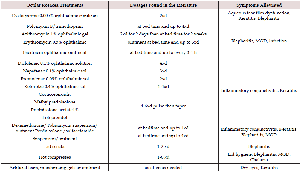
MGD= Meibomialn gland dysfunction.
Warm compresses with a minimum temperature increase of
5 degrees above body temperature for 5-10 minutes and digital
massaging of the lid margin. The patient should apply traction
on the lateral canthus to immobilize the upper and lower eyelids
followed by gentle pressure upward or downward with the fingers
of the opposite hand to helps facilitate the excretion of MG [63,64].
Olson et al reported that 5 minutes eyelids hot compresses (40º)
improved MGD in 80% (clinical studies level II) [67,68].
For the treatment of chalaziae, a self-administered technique
called the “4 fingers times 10 massage” can be beneficial. This
technique is performed as follows: At the conclusion of a bath or
shower, the patient warms his or her hands under hot water. Using
1 drop of baby shampoo (which does not sting eyes), the patient
works up lather, places the index finger over the closed lids at the
lid margin, and vigorously massages the lid back and forth 10 times.
The patient then repeats the procedure with the middle, ring, and
little fingers. Acute chalazia are also managed by hot compresses
for 5-10 minutes, 4 to 6 times daily. Surgical intervention may be
required for persistent chalaziae. There is evidence to support the
use of Intralesional corticosteroids for chalazia (clinical studies
level II) [69,70].
Punctate keratitis is managed by a variety of topical treatments
including preservative free artificial tears, anti-bacterial or antifungal
agents for secondary infections after obtaining cultures
should be used. Short term use of topical steroids helps decrease
inflammatory keratitis, conjunctivitis and lid disease. Cyclosporine
0.05% ophthalmic emulsion, an inhibitor of the T-cell function,
should be considered when treating ocular rosacea induced
keratitis [71]. In a study conducted by Schechter (clinical study level
I) topical cyclosporine 0.05% emulsion twice daily for 3 months
was found to be more effective than preserved and preservative
free artificial tears for the treatment of, rosacea-associated dry
eyes, lid and corneal changes [72]. In cases of chronic dry eyes,
ophthalmic cyclosporine twice daily for 3 months has been proven
to improve tear quantity and quality, MGD, lid margin telangiectasia
and corneal staining (clinical studies level I) [72-74].
Topical ophthalmic steroids when used in conjunction with
oral doxycycline have been reported to be effective in prevention of
recurrent corneal erosions associated with ocular rosacea (clinical
study level I) [72]. The potential for increased intraocular pressure,
cataract and thinning of the eyelid skin, limit its use for short
duration [75]. If opportunistic bacterial or fungal infection occurs,
topical steroid should be avoided [74,75].
For significant blepharitis with erythema, adding an antibiotic
ointment or combination steroid/antibiotic ointment is common
[53]. Topical azithromycin 1% eye drops for 4 weeks proved to be
effective in controlling adult blepharitis with mild burning sensation
after instillation and controlling childhood ocular rosacea at 1.5%
concentration 3 days twice daily every 10 days (clinical study level
III) [76-78]. Topical cyclosporine 0.05% ophthalmic emulsion
used twice daily has been shown to reduce posterior blepharitis
[78]. A single RCT that compared the efficacy of cyclosporine
emulsion 0.05% with artificial tears for conjunctival and corneal
ocular rosacea related pathology. The study showed significant
improvement of the ocular changes, Schirmer and Tear break up
time mean values after 3 months use of cyclosporine emulsion
compared with artificial tears.
Topical metronidazole gel 0.75% to 1% applied to the lid margin
twice daily for 12 weeks combined with lid hygiene may be a safe
and effective means of treating rosacea blepharitis (clinical studies
level II) [79]. Metronidazole is a broad-spectrum antibiotic with
anti-parasitic and anti-inflammatory effects. For sterile corneal
ulcers, anti-inflammatory drugs play a role in decreasing ocular
inflammation and preventing tissue damage. Diclofenac 0.1%
ophthalmic solution 4 times daily can be used with caution [73].
Local irritant effects of topical ophthalmic NSAIDs include
conjunctival hyperaemia, burning, stinging and corneal anaesthesia.
A more serious complication includes corneal ulceration and
full-thickness corneal melts due to usage of Diclofenac sodium
ophthalmic solution. NSAID-induced corneal injury is possibly
caused by epithelial hypoxia or the effect on corneal matrix
metalloproteinase and direct toxicity of the excipients such as
surfactants, solubilizers and preservatives found in topical NSAID
ophthalmic preparations. Ophthalmic NSAIDs should not be used
with gentamicin which might increase NSAID absorption and
toxicity. The concurrent use of NSAIDs with topical corticosteorids
in situations such as corneal inflammation has increased the risk
for precipitating corneal erosions and melts and such concurrent
use should be employed in an appropriate and judicious manner
[80].
Systemic Therapy (Table 3)
The choice of topical, systemic or combination therapy usage
is guided by the ocular manifestation of the disease. Corneal
neovascularization and tear film insufficiency are more responsive
to systemic therapy whereas, meibomian gland dysfunction
requires combination therapy [53]. Long term intake of 3000-
4000 mg of omega 3 fatty acids have been reported to be safe and
improve meibomian gland secretion, once symptoms are controlled
maintenance dose of 2000 mg/day can be initiated. Beneficial
effects of omega 3 require six weeks of consistent intake to be
recognized [80]. A recently published RCT (clinical studies level I)
of the use of omega 3 at a dose of 2000 mg 3 times daily for one
year in patients with MGD, showed improvement of symptoms and
objective clinical measures of the treatment group and to lesser
extent on the placebo group [81-84].
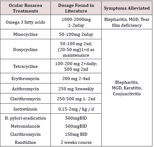
Table 3: Oral ocular rosacea treatments [5,9,49,52,60,73-77,80-98].
Significant ocular disease requires oral antibiotics such as
the tetracyclines family, that have been found effective because
of their anti-inflammatory properties [85]. It is not clear if the
anti-inflammatory properties of tetracyclines are primarily due
to inhibition of bacterial lipases, the reduction of the number
of bacteria present, inhibition of Phospholipase A2 and matrix
metalloproteinase or a combination of these and other unknown
factors [85-88]. Studies reported significant improvement in ocular
rosacea signs and symptoms including dryness, itching, blurred
vision, photosensitivity, erythema, and lid margin telangiectasia
with the use of doxycyline (clinical studies level II) [9,52,89]. The
optimal dose, duration of therapy for ocular rosacea by tetracycline
and doxycyline has not been established [88-89]. Dosages and
treatment duration is determined by the treating physician and
tailored according to patient response. Systemic therapy can be
tapered after symptomatic relief. If therapy is tapered too quickly;
recurrence is high especially within the first 3 months [9,50,85-88].
Reported doses by some authors for tetracycline 500 mg twice
daily for 4-6 weeks, the dose may be subsequently tapered to one
tablet every other day according to response [55]. Doxycycline
at a dose of 50-100 mg can be given twice daily for one month
then 50-100 once daily for 1-2 months before tapering [61]. In a
randomized controlled study reported by Yoo (clinical studies level
I), low dose doxycycline 20 mg twice daily was found to be effective
in controlling eye symptoms and improvement of Schirmer and
TBT [90].
Alternative systemic therapy includes erythromycin,
azithromycin, metronidazole, and low dose isotretinoin
Azithromycin a macrolide antibiotic with high antimicrobial
and anti-inflammatory properties has shown promising results
in reducing eyelid disease and conjunctival hyperemia, without
side effects [81,82]. Due to its high conjunctiva and tear fluid
concentration that persist for 2 weeks after 1 g oral dose, pulse
dose of 1 g. weekly for 3 weeks was found effective in treating
meibomitis (clinical studies level II) [91-93] Clarithromycin, was
reported to be as effective as doxycycline, with fewer side effects
[83]. Metronidazole has immunosuprssive, anti-inflammatory and
antimicrobial effects. Metronidazole can cause stomach upset and
alcohol cannot be consumed while taking it [81,83].
Low dose isotretinoin can be initiated in cases resistant to
antibiotics and should be used with caution since isotretinoin
could cause keratitis and keratoconjunctivitis [5]. Tetracycline
is contraindicated in children until 8 years of age as it may cause
permanent discoloration of teeth and interfere with tooth enamel
development in patients younger than 7 years [94,95]. Erythromycin
has anti-inflammatory effect through inhibition of synthesis of the
bacterial enzymes [94,95], improves the meibomian gland function
and TBT and decrease punctate keratopathy [96].
In patients younger than 8 years, oral erythromycin showed
success in treating ocular rosacea [55,61,97,98]. Nazir et al
reported excellent response of ocular rosacea in children with oral
erythromycin (clinical studies level III) [55]. For older children,
tetracycline and doxycycline may be used in dosages similar to
adults [55]. There is a 25% relapse rate in 1 month and a 66% rate
in 6 months after cessation of tetracycline, therefore prolonged
treatment may be required [55]. Patients with H. pylori infection,
the use of 2 weeks course of metronidazole, clarithromycin, and
ranitidine improved the ocular manifestations of rosacea more
than cutaneous (clinical studies level III) [99]. A variety of surgical
techniques have been used for the repair of corneal defects due
to ocular rosacea such as keratoplasty or the use of amniotic
membrane transplantation [100-103].
Conclusion
Unfortunately, ocular rosacea is frequently under diagnosed
by dermatologists and undiagnosed by ophthalmologists. Patient
fail to complain to dermatologists of minor eye symptom and
ophthalmologists do not examine the facial skin to establish
the diagnosis. Dermatologists and family physicians should
meticulously inquire about ocular symptoms and refer patients
to ophthalmology for further management. Symptoms of ocular
rosacea include dry eyes, burning, itching, ocular irritation and
grittiness, redness, foreign body sensation, photophobia, blurry
vision, stabbing pain, foreign body sensation occurring during
sleep or upon awakening or both. Telangictasia of the lid margin,
MGD, chalazia and blepharitis are the most common forms of
lid involvement. Conjunctival involvement in ocular rosacea is
usually in the form of a mild bulbar conjunctival hyperemia.
Cicatrizing conjunctivitis, conjunctival granuloma and phylctenular
conjunctivitis are less common conjunctival manifesttations.
The most common form of corneal involvement is in the form of
a superficial punctate keratopathy which usually occurs in the
lower 1/3 of the cornea. Recurrent epithelial erosion, corneal
neovascularization with peripheral thinning, and corneal ulcer are
the other forms of corneal involvement. Examination of the eyelid
is very important looking for erythema, telangiectasia, crusts,
irregularity of the lid margin, thickening rounding, and dilatation of
the conjunctival vessels.
The need for properly conducted, well randomized controlled
trials is urgent, since there exists no definite conclusion about any of
the therapies available nowadays. Strong clinical evidence suggests
that topical cyclosporine 0.05% emulsion is more effective than
preserved and preservative free artificial tears for the treatment
of, rosacea-associated dry eyes, lid and corneal changes upon any
slight suspicion for ocular involvement, patients should be referred
for ophthalmological evaluation. Therefore, a clinician’s increased
awareness of ocular finding in patients with rosacea may aid in the earlier diagnosis and treatment, preventing permanent eye
impairment.
Acknowledgment
Many thanks to the colleagues from the department of
Ophthalmology, King Abdulaziz University Hospital, King Saud
university for providing the clinical pictures. Without their
assistance the paper would have been deficient. We express our
gratitude namely to: Dr. Yasser AL Faky, MD, FRCS and Dr. Sameer
Mansouri MD, FRCS.
Read More About Lupine Publishers Journal of Respiratory & Skin Diseases Please Click on Below Link:
https://lupine-respiratory-skin-and-diseases.blogspot.com/