Showing posts with label DDIPIJ. Show all posts

Monday, 9 October 2023

Lupine Publishers | Plant Extracts as An Alternative to Synthetic Medicines

 Lupine Publishers | Journal of Drug Designing & Intellectual Properties


Abstract

Mankind first medicines were extracts from plants. However, over the years, mankind has resorted to the use of synthetic medicines. Whilst, synthetic medicines have fulfilled their roles and save millions of people around the globe, there is still a need to promote the use of herbal medicines, considering that synthetic medicines have severe side effects when administered and the cost of synthesising drugs, is an expensive endeavors for developing countries like Guyana. Guyana has an adundant of diverse flora, whose extracts have been shown by us and others to have potent and selective antimicrobial activities and antidiabetic activities. However, there is a need to promote the isolation of antimicrobial, antidiabetic and anti-cancer drugs from plants, an elucidation of their structure and modulation of their medicinal activities. This entails significant finance to aid in the purchase of requisite equipment etc. in Novel Drug Discovery. The Guyanese Diaspora and the Entrepreneurship Sector can promote such research in Guyana via the provision of finance to purchase “State of the Art” equipment to conduct such research or the establishment of a National Herbal Research Institute. Such an institute will promote further research on our current herbal medicines and also on the discovery of new Herbal medications.

Keywords: Medicines antimicrobial activities Antidiabetic activities Anti-cancer activities Diaspora Entrepreneurship

Introduction

Mankind first medicines were extracts from plants. Plants have had a long history in modern medicine [1-18]. For example, long before the advent of modern medicine, the Peruvians were fending off malaria with the Cinchona bark from the “quinaquinas” tree. It was given as a cure to a Spanish countess who fell ill with the disease in Lima in around 1630. Then Bernabe Cobo, a Spanish missionary and naturalist was the first to bring the bark to Europe. Quinine was purified from the bark by a team of French pharmacists19. Over the years, mankind has resorted to the use of synthetic medicines. Whilst, synthetic medicines have fulfilled their roles and save millions of people around the globe, there is still a need to promote the use of herbal medicines, considering that synthetic medicines have severe side effects when administered and the cost of synthesising drugs, is an expensive endeavours for developing countries like Guyana.

Antimicrobial endeavours

Guyana, has an adundance of diverse flora, whose extracts have been shown by us and others to have potent and selective antimicrobial activities against human pathogens such as E.coli, S.aureus, K. pneumoniae, B. subtilis and Candida albicans. We have also shown that selected plant extracts exhibited antidiabetic activities. Our initial efforts to evaluate the antimicrobial properties of plant extracts focuses on the leaves and stem extracts of plants. These were inclusive of those of Samanea Saman, Smilax schomburgkiana, Mormordica charantia, Phyllanthus Acidus, Sphagneticola Trilobata and Doliocarpus Dentatus [10-15]. Later on, we focused our antimicrobial studies of fruits. The idea is that a fruit is edible and so an evaluation of its antimicrobial properties, singly and in combination, would eventually lead us to find some good natural antimicrobials, without the need for clinical trials. In all our studies on antimicrobial activity of leaves, stems and fruit extracts, the diameter of Zone of Inhibition, DZOI and the Area of Zone of Inhibition, AZOI were used as guage of antimicrobial studies. Amongst the fruits investigated were passion fruit (passiflora edulis), Vicia faba L [16-20]. In search for natural antimicrobials, our aim is to find leaves, stems and fruit extracts that would be selective and potent as synthetic antimicrobials such as penicillin, aminoglycosides, and tetracyclines etc.

Antidiabetic endeavour

Diabetes mellitus is a chronic health problem around the world and in Guyana with long term consequences that are potentially preventable. It is a heterogeneous group of disease, characterized by high blood glucose levels resulting from impaired insulin secretion, impaired insulin action, or both [20-27]. In a hyperglycemic state, the body tries to remove excess glucose by excreting in the urine. This increases urine output, causing glycosuria and result in frequent thirst. In addition, the body is deprived of glucose energy and seeks alternative energy sources such as fats and muscle tissues, leading to weight loss [23]. A diminishing growth effect and increased predisposition to certain infections, may also be present with chronic hyperglycemia 4. These combinations along with polyuria, polydipsia, polyphagia, and blurry vision produces the common symptoms of diabetes [24]. As this disease progresses, vascular damage ensues leading to severe diabetic microvascular and macrovascular complications [25]. Therefore, diabetes covers a wide range of diseases which are the major causes of chronic morbidity and death in diabetic subjects [26,27].

Selected plants are a potential complementary source of hypoglycemic drugs to combat diabetes, in addition to the use of synthetic drugs: insulin, metformin etc. We have shown that the aqueous extract of the fruits of Psidium guajava, Averrhoa bilimbi and the peel of Tamarindus indica exhibited hypoglycemic effect on normoglycemic guinea pigs. Administration of extracts of Psidium guajava, Tamarindus indica and Averrhoa bilimbi resulted in a marked hypoglycemic (reduction) activity in blood glucose levels when compared to the control and Glibenclamide treated group on the 12th day: Psidium guajava (90 ± 3.0 to 75.7 ± 3.5mg/dL), Tamarindus indica (89.0 ± 5.6 to 70.7 ± 2.1mg/dL) and Averrhoa bilimbi (110.0 ± 9.2 to 86.7 ± 10.0mg/dL). Glibenclamide also resulted in a reduction (88.0 ± 2.0 to 67.3 ± 3.5mg/dL) as compared to the control [27].

Antidiabetic effect of the ethanolic extract of Phyllanthus emblica fruits in evan rats has been reported and is dose dependent [28]. The anti-hyperglycemic effect of Quercetin, a major constituent of the methanolic extracts of Phyllanthus emblica fruit in Streptozotocin (STZ) induced diabetic rats were determined [29]. There are several herbs that are used to treat diabetes in Guyana and internationally [30-34]. Locally Momordica charantia [30], an herbaceous, tendril-bearing vine contains a compound called charantin, which have hypoglycaemic effect. It has been found to increase insulin sensitivity. A daily dose of 100mg per kilogram of body weight is comparable to 2.5mg/kg of the anti-diabetes drug glibenclamide taken twice per day. Other compounds in M. charantia have been found to activate the AMPK, the protein that regulates glucose uptake. The whole plant is used as a decoction for diabetes. The dry leaf and stem are boiled, and the water drunk as an anti-diabetic. The fruit is cooked and eaten as an anti-diabetic. Use with almond oil for a vulnerary. Tablets of M. charantia extract are sold in the United Kingdom.

Phyllanthus niruri [30] contains alkaloids, flavonoids and triterpenes. The whole plant is used as a blood purifier (bitter tonic) to reduce blood sugar level. It’s available as capsules. Cajanus cajan [30]. Pigeon peas contain high levels of protein and the important amino acids methionine, lysine, and tryptophan. Leaves and flowers are boiled for a diuretic and diabetes remedy. The whole plant of Desmodium barbatum [30] is use to reduced blood sugar level. The wood of the plant, Telitoxium [27]. is used for diabetes. The active adaptogenic constituents of Tinospora cordifolia [30] are diterpenoid compounds: polyphenols, and polysaccharides, including arabinogalactan polysaccharide. T. crispa and T. rumphii are used in Thailand and Philippines for treatment of diabetes. A decoction of the leaves of Azadirachta indica [30], Neem is used as a bitter tonic for treating diabetes and jaundice. Internationally, the juiceof the leaves of Abrus precatorius [31-34] are given to diabetic patients. Achyranthes aspera [32-34] is used as a decoction in the treatment of diabetes mellitus. Likewise, the roots of Catharanthus roseus is used as decoction in the treatment of diabetes mellitus. The entire plant ofCentella asiatica [32-34] is also used as a decoction in the treatment of diabetes mellitus. [32-34] Curcuma longa is a rhizomatous herbaceous perennial plant which is used in the treatment of diabetes mellitus. Phyllanthus emblica [32-34]. Edible fruits are antidiabetic in nature. Piper bettle [32-34] lea extract is used in the treatment for diabetes mellitus. Sphaeranthus indicus [29-31].

Anticancer endeavours

Table 1: Some selected anticancer plants and their active constituents.

lupinepublishers-openaccess-drug-designing-journal

While plants have been tested over the years for their anticancer activities abroad and proven positive, such research is lacking in Guyana. There is a need to screen plants in Guyana for their anticancer activities. This stem from the fact that cancer is on the rise in Guyana. Cancer has been a constant battle globally with a lot of development in cures and preventative therapies. Cancer is the uncontrolled growth in human cells and will result in the formation of tumours of malignant cells with the potential to be metastatic35. Treatments include chemotherapy, radiotherapy and the use of synthetic drugs such as cis-platin. Using chemotherapy can put patients under a lot of strain and further damage their health. Thus, there is a need for an alternative complimentary medicine in herbal treatments36.However, this research is hampered by the lack of cancer research equipment. There needs to be the establishment of a cancer research unit in Guyana, to assess the anticancer proficiency of plants. (Table1). shows a list of plants tested abroad for their anticancer proficiency [37-45].

Diaspora role in promoting herbal medicines in Guyana

However, there is a need to promote the isolation of antimicrobial, antidiabetic and anti-cancer drugs from plants, an elucidation of their structure and modulation of their medicinal activities. Also, an investigation of their activities. This entails significant finance to aid in the purchase of requisite equipment etc in Novel Drug Discovery. A separate research laboratory, with modern equipment and instrumentation is necessary at the University of Guyana to realize the above. Also, lacking in Guyana is the equipment to conduct anticancer and antiviral studies A step can be taken further by the Government in the establishment of a National Herbal Research Institute to further explore our herbal folklore medicines and undiscovered herbal medicines. Such an institute will promote further research on our current folklore or traditional herbal medicines and also on the discovery of new Herbal medications. The Guyanese Diaspora, Entrepreneurship Sector and Foreign investors can promote such research in Guyana via the provision of finance to purchase “State of the Art” equipment to conduct such research. Our whole objectives is that we can be a leading exporter of herbal medicines in the Caribbean and one of the best in the world. Guyana has a richbiodiversified flora, whose herbal proficiency needs to be further investigated.

Read More About Lupine Publishers Journal of Drug Designing & Intellectual Properties Please Click on the Below Link:

https://lupine-publishers-drug-designing.blogspot.com/

Monday, 14 August 2023

Lupine Publishers | Danger of Buying Medicines in Any Place Other Than the Pharmacy

 Lupine Publishers | Journal of Drug Designing & Intellectual Properties


Introduction

There are medicines registered as such-and subject to the legislation of pharmaceutical specialties-and products that, without being registered as medicines, are sold and actively promoted, giving them medicinal virtues.
Among the medications it is necessary to distinguish those of prescription or prescription sale, of those without prescription or over the counter (over the counter or OTC counter medications in Anglo-Saxon terminology). On these there is the wrong an installed assumption among the general public, which considers “over-the-counter” medicines safe.
All medications without exception, used in excessive doses or for too long periods, in situations where they would not be indicated, can cause side effects, collateral, undesirable or adverse effects, generate interactions with other drugs or substances, induce behaviors of abuse or dependence and even delay the diagnosis of a condition that requires medical care.
Thus, for example, according to a study by Morlans, M., Laporte, J. et al., About 13% of all the etiological burden of terminal nephropathy that leads to dialysis or transplantation in our environment is due to the chronic use of painkillers, almost always in the framework of self- medication.
The sale of any medicine outside the pharmaceutical establishments is highly risky for the health of population. As the Argentine Pharmaceutical Confederation (COFA) warns, 20 percent of the drugs consumed in the country are purchased outside pharmacies, such stores include kiosks, supermarkets, service stations, gyms and even hotels accommodation. Sometimes this also applies to prescription drugs.
The traceability of a drug is the monitoring of the route that the product takes at all stages of its marketing, it is essential to achieve a correct use of the medications (pharmacovigilance, detection of chronic patients, patient monitoring) as well as to establish an action Therapeutics free of complications. It is also a tool that uses the health system to detect counterfeits, adulterations on medications. When separating from the original sales channel, traceability becomes difficult, if not impossible [1].
There is also a legal vacuum over their control by the national authority, since ANMAT has no jurisdiction over establishments that are not authorized by the Ministry of Health. In other words, the referred “sales channels” (kiosks, warehouses, service stations) are outside their jurisdiction and competence.

Advertising

The impact of the advertising of medicines on the behavior of consumers is undeniable, and hence the potential risks it represents for the health of the population. The National Academy of Medicine warned on repeated occasions about “advertisements that encourage the use of treatments and medications without medical supervision, and the proliferation of advertising messages advising medications and therapeutic procedures of benefit sometimes not established and sometimes exaggerated, with the purpose to encourage their consumption”.
The World Health Organization (WHO) created the “Ethical criteria for the promotion of medicines”, a theoretical framework where recommendations are made on advertisements aimed at the public “should help the population to make rational decisions about the use of medicines that are legally available without a prescription.
According to studies carried out by the Maimonides University and the Argentine Institute of Pharmaceutical Care (IADAF), the advertising of medicines encourages self-medication or drug misuse, a problem that takes 700 lives per year in the country.
In a note from health issues journalist Pedro Lipcovich, ( “Today we are addictive”) analyzes a television message of aspirin (acetylsalicylic acid) mentioning the striking omission of any of its therapeutic properties as anti-inflammatory, anti-fever, anticoagulant, associating its consumption “with ageneric welfare state and high social and labor performance”.
We passively assist in the promotion of bad eating habits, widely publicized, to increase the sales of a medicine that supposedly counteracts the effects. The induction to the conduct of immediacy in the installation of a treatment according to the symptoms, gives results: everything can be solved with a tablet and even some dietary supplements are preferred to be marketed as medicines because they “sell more”. But this attitude has its When a teenager is offered a “tablet” of ecstasy, with the promise of well-being and pleasure, it is utopian to think he could say “no.”
To all this must be added the lightness with which we assist in the prescription of medications. The mother, the coworker, the hairdresser, the kiosk and even the doctor’s office, among patients, recommend medications.
Advertising and active promotion by mass media, increases irresponsible self-medication and irrational use of medications. Medicinal drugs or medicines thus leave their place of social good, essential for public health, and become a consumer good.

Drug dependence

Numerous medications are used to produce changes or changes in mood, alone or in association with drugs or substances of abuse. According to the 2008 National Survey on Prevalence of Psychoactive Substance Consumption, conducted by INDEC, almost 4% of the population between 16 and 65 years of age consumes tranquilizers.
It cannot be taken into account that almost all drugs of abuse today were initially medications, which due to their pharmacological and toxicological characteristics (tolerance, abuse, dependence) began to be used for their psychoactive effects, being then restricted or withdrawn in your pharmacological employment. Such is the case of cocaine, some opiates (morphine, codeine), amphetamines, anesthetics (ketamine), ephedrine, popper, etc.
The Abuse Medicines Observatory (WCO) in Spain conducted a study that verified the non-medical consumption (substance abuse) of a total of 27 substances. 22% consumed analgesics, some 12% anxiolytics or tranquilizers (benzodiazepines); 10% stimulants and the rest a wide range of substances.
Majority use patterns included mixtures with drugs and alcohol and abuse of tranquilizers. Medications have always been part of the “drug culture” in non-therapeutic uses, in order to experience new sensations or obtain better yields. The modalities of drug abuse change and it could be said that some of them “become fashionable”. Currently, the “crazy jar” is the most common form of alcohol and medication use-mostly benzodiazepines- among teenagers and young adults.
According to data from the Toxicology Unit of the “Juan A. Fernández” Hospital of the City of Buenos Aires, during 2008, 2182 consultations related to acute poisoning by substances of abuse were attended; 292 of them were due to abuse of some type of medication and another 205 were due to the association of some medication with alcoholic beverages [2].
The equation between inappropriate promotion of drug use, forms of facilitated or uncontrolled accessibility and patterns of inappropriate use and substance abuse constitutes an increased risk trilogy, which exposes the potentially most vulnerable population.

Read More about Lupine Publishers Journal of Drug Designing & Intellectual Properties Please Click on the Below Link:

https://lupine-publishers-drug-designing.blogspot.com/


Monday, 19 June 2023

Lupine Publishers | Insecticidal Activity of Plant Extracts

 Lupine Publishers | Drug Designing & Intellectual Properties International Journal


Abstract

Insecticides are a group of compounds with heterogenous toxicity, whose intended purpose is to kills insects. Synthetic insecticides are known for their very harmful environmental and health impacts. This therefore generates a need for a safer solution. Botanicals are a special group of insecticides with a natural origin, obtained primarily from plant parts. There is a continued interest to screen plants for their insecticidal activities in light of the great destruction done to crops that affect the livelihood of the populace worldwide, especially in countries, whose GDP depends primarily on agriculture. Guyana, a country on the mainland of South America is no exception. Agricultural produce is now threaten by emerging new species of insect pests, such as in the rice sector. Thus, there is a need to screen Guyana’s richly biodiversified flora for its insecticidal activities and to promote research in biological control of crop pests and boost the agro industry.

Keywords: Insectides; environmental; Guyana; biodiversified flora

Introduction

Background

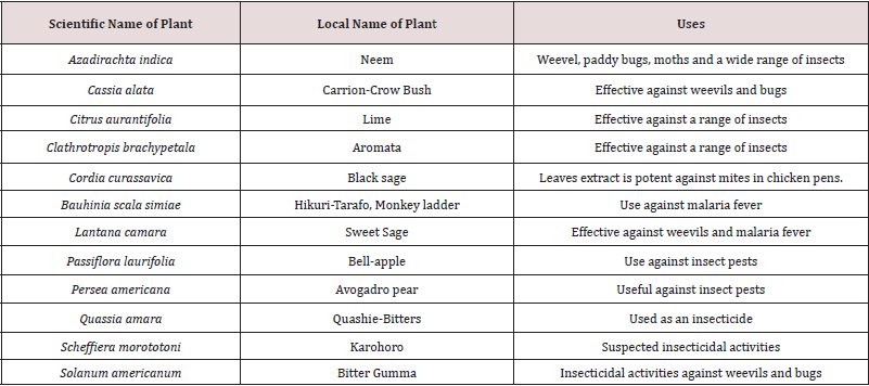
Insects, though useful in many ways to mankind, have had negative effects in the field of agriculture. They have attacked plant parts such as stored grains, seeds, flowers, leaves, stems, fruits etc and have had a significant effect on the agroeconomy of many countries [1-3] . One way to control insects deleterious effects on crops is to utilize chemical pest control, which employs potent chemical pesticides to curb, reduce or eliminate pests and thus sustain crop production throughout the world. However, most synthetic insecticides used to date are deleterious to human health and the environment [4]. Thus, an alternative strategy is necessary. That alternative strategy is the use of plant extracts and phytochemicals as natural antifeedant, insect deterrents and repellent [4]. Plant extracts are safe alternatives that are of low cost, convenient to use and environmentally friendly. Plant products have been successfully exploited as insecticides, insect repellents and insect antifeedants [1-3]. In addition, natural products insectides have been isolated from plants and serve the basis for structure mimicry synthesis. Figure 1 show a list of structure of some isolated natural products from crude plant extracts. Figure 2 shows a notorious insecticidal natural product, Azadirachtin Many countries whose GDP (Gross Domestic Product) depends on agriculture, have in recent times being affected by insects. Guyana, a country on the mainland of South America is of no exception. Agricultural produce is currently threaten by emerging new species of insect pests. One notable example is in the field of rice cultivation. Recent research in Guyana has shown that rice production is threatened by paddy bugs, leaf miners, water weevils and caterpillars [6]. Natural insecticides from Guyana flora, should be a good choice, apart from the synthetic analog imported. Thus, there is a need to screen Guyana’s richly biodiversified flora for its insecticidal activities and to promote research in natural pesticides control. There is not much report of plant being screened for their insectidal activities here, even though research is currently being done. So, this area of research needs intensification in Guyana. In addition, the crude plant extracts can be subjected to chromatographic separation, which will lead to the isolation of known and unknown natural products which will continue to provide the platform for novel insectidal discoveries and structure activity relationship.

Figure 1: The structure of some selected isolated insecticides.

Figure 2: Azadirachtin, a notorious insecticidal isolate from Azadirachta indica.

Brief survey of some selected natural insectides

Internationally, there are many reports of plant extracts being used for their insecticidal properties. Several will be briefly reviewed here. For example, Morinda citrifolia leaf extract was tested for larvicidal activity against three medically important mosquito vectors such as malarial vector Anopheles stephensi, dengue vector Aedes aegypti, and filarial vector Culex quinquefasciatus Insecticidal activity of essential oil [7], extracted from Morinda lucida was tested on pulse beetle Callosobruchus maculatus, which is a pest that causes serious damage to several pulses. The insecticidal activity was compared with two pesticides, Phostoxin and Primo-ban-20. Results clearly indicate that M. lucida essential oil can be used as an effective alternative for pulse beetle C. maculatus control, and it could be tested against other pulse beetles affecting Asia and Africa and throughout the world, thereby reducing use of synthetic pesticides [8].

The insecticidal and antifeedant activity of the ethanolic extracts from Allium rotundum L has been noted against the larvae of the L.decemlineata [9]. It was found that the ethanolic extracts from the aerial part possessed moderate level (40.0%) of insecticidal properties against the larvae of the L. decemlineata and low against the imago (6.7-13.3%0) [9]. The petroleum ether, ethanol and aqueous extracts from the leaves of Artemisia herbalba, Asso, Euclayptus camaldulensis Dehnh and Rosmarinus officinalis L were investigated for their insectidal activity against 3 to 4 days old Myzus persicae individuals at 1,2.5, 5 and 10% and observations were made after 24 hours. Etheric extracts of all plants were effective and induced mortalities (100%, 53% and 60% respectively) at the highest concentration. However, the ethanolic and aqueous extracts did not show any significant insecticidal effect [10] Sixteen aromatic plant extracts from three species belonging of the Asteraceae family, were tested for their insectidal activity against adults and larvae of confused flour beetle Tribolium confusum du Val (Coleoptera Tenebrionidae).

The methanolic and ethyl acetate extracts of Mantisalca duriaei Briq. Cavill. and petroleum ether, chloroformic and methanolic extracts of Raconteur acute DC, significantly induced larval growth. Antifeedant properties were detected in methanolic extracts of M. duriaei and R. acaule, petroleum ether and chloroformic extract of R. acaule and ethyl acetate extract of M. duriaei. For all extracts, mortality was higher for larvae than adults. Values of 83% and 77% were obtained using petroleum ether and methanolic extracts of R. acaule. These results suggest that M. duriaei and R. acaule may be used in grain storage against insect pests [11]. Leaf oil of Psidium guajava L obtained from Soxhlet extraction was tested for insecticidal effect and phytochemical screening against khapra beetle, Trogoderma granarium Everts (Coleoptera: Dermestidae. The application of the oil at the rate of 0.5ml gave significantly (p < 0.05) higher percentage mortality than the control [12].

A study was conducted to determine the insecticidal activity of essential oils from oregano, Origanum onites L. (Lamiales: Lamiaceae), savory, Satureja thymbra L. (Lamiales: Lamiaceae), and myrtle, Myrtus communis L. (Rosales: Myrtaceae) against three stored-product insects such as the flour moth Ephestia kuehniella Zeller (Lepidoptera: Pyralidae), the Indian meal moth Plodia interpunctella Hübner (Lepidoptera: Pyralidae) and the bean weevil Acanthoscelides obtectus Say (Coleoptera: Bruchidae). A. obtectus was the most tolerant species against the essential oils. However, the insecticidal activity of the myrtle oil was more pronounced than other oils tested against A. obtectus adults. The essential oils of oregano and savory were highly effective against P. interpunctella and E. kuehniella, with 100% mortality obtained after 24hrs [13].

Table 1: Some selected plants from the Guyanese flora with folklore insecticidal activities.

lupinepublishers-openaccess-drug-designing-journal

Six different indigenous plants were screened for antifeedant and insecticidal activities against fourth instar larvae of Epilachna beetle, Henosepilachna vigintioctopunctata, a severe pest on brinjal. Amongst the plants screened, Achyranthes aspera showed higher activity against the selected pest. Ethyl acetate extracts of A. aspera showed higher antifeedant index and insecticidal activity against fourth instar larvae of H. vigintioctopunctata. Preliminary phytochemical analysis revealed that the presence of alkaloid and quinines in the ethyl acetate extracts indicating higher percentage of activities [14]. The insecticidal activities of four local plants extracts Rhazya stricta Decne, Lantana camara L., Ruta chalepensis L. and Heliotropium bacciferum Forssk against subterranean termites Psammotermes hybostoma (Desneux) were reported. Of the four extracts, the hexane extract of R. stricta was more pronounced, having an acute (24hr) and chronic (48hr) LC50s of 194.8 and 147.4ppm, compared to 221.7 and 149.9; 288.9, 185.6 and 391.3 and 244.5ppm for L. camara, R. chalepenesis and H. bacciferum respectively [15]. There are other reports of plant extracts used for their insecticidal activities [16-22]. There are few reports of plants from the Guyana flora with insecticidal activities, even though research has been pursued in that direction. Table 1 shows a list of ten plants with selected insecticidal activity from the richly biodiversified Guyana flora.

Conclusion

Insects are indeed a threat to mankind, especially in the field of agriculture. Many countries, whose economy depends on the agro-industry are threaten with new emerging destructive insects. Guyana is of no exception. Whist synthetic insecticides can do the job in many cases, there use is deleterious to the environment etc. Insecticides derived from plant extracts is far more environmentally friendly and far less expensive to make compare to synthetic ones. From an economical point of view, it would be best to use the aqueous extract of selected plants with insecticidal activities. There are many plants with folklore insecticidal activities in Guyana and their extracts: organic and aqueous need insectidal investigation. Also, the isolation of natural products from the crude plant extracts can lead to the discovery of new isolates and it would form the basis for continued novel drug discovery.

Read more about Lupine Publishers Journal of Drug Designing and Intellectual Properties Click on the below link: https://lupine-publishers-drug-designing.blogspot.com/


Tuesday, 28 February 2023

Lupine Publishers| Affordability Issues of Biotech Drugs in Low- and Middle-Income Countries (LMICs)

  Lupine Publishers| Journal of Drug Designing & Intellectual Properties


Abstract

According to World Bank publication Disease Control Priorities: Improving Health and Reducing Poverty (3rd edition, 2017), nearly 20% total health expenditure globally came from out-of-pocket payments in 2014. The same was nearly 40% total health expenditure for low-income countries, 56% for lower-middle-income countries, and 30% for upper-middle-income countries (WHO, 2016). One third of the world’s population lacks timely access to quality-assured medicines while estimates indicate that at least 10% of medicine in low- and middle-income countries (LMICs) are substandard or falsified, costing approximately US$ 31 billion annually (Global Health, 2018). Surprisingly, 80% of global cardiovascular deaths occur in LMICs which is (partly) due to the lack of access to healthcare including skilled human resources, equipped facilities and medicines (Global status report on noncommunicable diseases, WHO, 2010). Price of drugs, vaccines, and diagnostics is a major burden in LMICs round the globe. Cost of biotech drugs are even higher due to high cost incurred by the pharmaceutical companies for clinical trial. Biotech drugs have completely changed the management of several diseases, including cancer and autoimmune diseases. Although essential but their affordability is still a burning issue, especially in LMICs.

Keywords: LMICs; Out-of-pocket expenditure; Cancer treatments; Biotech drugs; Pharmaceutical patents; Biosimilars

Abbreviations: PIVI: Partnership for Influenza Vaccine Introduction; NITAGs: National Immunization Technical Advisory Groups; OOP: Out-of-pocket; LMICs: Low-and Middle-Income Countries; RCT: Randomized Control Trial; HPV: Human Papillomavirus; GDP: Gross Domestic Product; EML: Essential Medicine List; R&D: Research and Development; BRICS countries: Brazil, Russia, India, China and South Africa; NCDs: Non-communicable diseases; CVDs: Cardiovascular Diseases

Mini Review

(Figure 1) Pharmaceutical companies invest in the development and testing of their drugs including by funding clinical trials. Furthermore, pharmaceutical companies also spend a large amount of money on advertising. For instance, in 2016 US$6.7 billion was spent on direct-to-consumer pharmaceutical advertising alone in the USA [1]. Worldwide spending on medicines reached $1.2 trillion in 2018 and will exceed $1.5 trillion by 2023, according to “The Global Use of Medicine in 2019 and Outlook to 2023” [2,3]. Although, access to essential medicines is problematic for one third of all persons worldwide [4]. Limited access to essential medicines (EMs) for treating chronic diseases is a major challenge in low- and middle-income countries (LMICs) [4,5]. Average public sector availability of even low-cost generic medicines ranges from 30% to 55% across 36 LMICs [6]. Price of drugs, vaccines, and diagnostics is a major burden in 105 middle income countries round the globe, comprising of 70% of the world population, 75% of the poor [7]. While public hospitals offer free or subsidized treatment including essential medicines, the high patient caseloads, underfunding and inefficient medicine distribution systems are barriers to consistent service provision [8]. Moreover, 90% of the population in developing countries purchase medicines through out-of-pocket (OOP) payments [7]. Poor availability of medicines in the public sector has pushed up household OOP expenditure, making them the largest household expenditure item after food [9]. Non-communicable diseases (NCDs) which, according to WHO are now the world’s biggest killers. Over 36 million people die annually (63% of global deaths) from NCDs, mainly CVDs, cancer, chronic respiratory diseases and diabetes. Of these, 80% occur in LMICs [10]. So, The WHO has set a minimum of 80% as target availability of medicines for both communicable and non-communicable diseases in all countries [11]. But Pharmaceutical companies have a substantial desire in developing drugs for chronic diseases and cancer treatments, not only because of high prevalence, but also because these drugs are often used in long term [12]. Pharmaceutical patents maintain drug prices well above the cost of production and can restrict access to needed medicines [13]. Biotech drugs have completely changed the management of several diseases, including cancer and autoimmune diseases such as, psoriasis, rheumatoid arthritis, multiple sclerosis, and inflammatory bowel disease [14]. The high cost of biotech medications (target a gene or protein and typically are injected or infused, associated with treating a chronic condition) often requires significant OOP expenditures [15,16]. Some studies say that pharmaceutical companies price drugs monopolistically, protected by patent rights, while others believe that the high prices for orphan drugs simply allow drug R&D and production costs. However, the global orphan drug market is estimated to reach US $209 billion by 2022 accounting for 21.4% of total branded prescription drug sales [17]. According to the Tufts Center for Drug Development, it costs, on average, $100 million in 1975, around $900 million before 2004 and 1.3 billion after 2005 to develop a new drug and bring it to market [18,19]. While, Scavone et.al, 2019 reported that entire time that passes from the R&D phase until the drug’s marketing approval can last up to 15 years, and it is characterized by extremely high costs, usually exceeding $1.2 billion [20]. Gouglas et.al, 2018 estimated a minimum of $2·8– 3·7 billion ($1·2 billion–$8·4 billion range) for one vaccine through to the end of phase 2a among 11 epidemic infectious diseases [21]. Apart from the traditional design of RCT, in recent years further study designs, including umbrella, basket and platform trials, were developed and applied to new therapies, especially in the area of oncology research [22]. Tay-Teo et.al, 2019 stated the most commonly accepted estimates of R&D costs, including cancer drugs, are between $200 million and $2.9 billion, after adjustments for the probability of failure and opportunity costs [23]. Genomic studies conducted in the past two decades identified the molecular drivers of certain cancers and led to the advent of targeted therapies as an important additional pillar of the cancer therapy armamentarium [24]. According to the Global Oncology Trend Report, global spending on cancer medications rose from $75 billion in 2010 to $100 billion in 2014, 10.3% rise in spending. Asia accounts for 60% of the world population and 50% of the global burden of cancer [25]. There are over 100 types of cancers, located in different organs and sub-tissues and originating from different cell types. Some cancer types (e.g., colon, breast, and non-Hodgkin’s lymphoma) contain even more specific classifications based on their molecular subtypes. Despite this complexity and variability, most types of cancer are treated with the same generic therapies [26]. Critics claim that prices of innovative drugs are excessive and argue that lowering prices will not harm the flourishing innovation. On the opposite end, the pharmaceutical industry insists that restrictive pricing policies will have a detrimental impact on their ability to generate innovation [27]. During 2017, PIVI worked with its country partners and the WHO regional and local offices to assess NITAGs strengthening needs and to provide technical assistance in 7 LMICs (Laos Peoples Democratic Republic, Mongolia, Vietnam, Armenia, Côte d’Ivoire; Moldova and the Republic of Georgia) [28]. In Europe, total cancer drug sales more than doubled between 2005 and 2014, increasing from €8.0 billion to €19.8 billion [29]. Biologics were estimated to account for US$289 billion pharmaceutical sales in 2014 and are projected to reach US$445 billion in 2019. It is also anticipated that biologics’ share of global prescription and OTC pharmaceutical sales will rise to 26% by 2019 [30]. It is projected that new cases of cancer will increase from about 14 million in 2012 to 22 million in 2030, with most cases in LMICs located in Africa, Asia and Latin America [31]. The projected increase in cancer incidence is predicted to be most significant in LMICs in Asia. In these countries, over 60% of the total healthcare expenditure comes from private resources, of which more than 80% is direct OOP payments, with catastrophic results for most families in these countries [25]. In India alone, as many as 63 million people are forced into poverty every year, owing to catastrophic health expenses, the majority of which are OOP payments for medicines [32]. Genetic predisposition, increasing life expectancy, urbanization, mechanization, inadequate health services and rapid economic development fueling sedentariness and changing dietary patterns are contributing to rising chronic disease burden in the South Asian region [33]. Rijal et.al, 2018 reported that Afghanistan, Bangladesh, Bhutan, Maldives, Nepal, India, Pakistan and Sri Lanka, which are mostly LMICs with regional GDP per capita 1640 USD and home to a quarter of world population [34]. According to Giri et.al, 2018 breast cancer was the most prevalent cancer and fourth leading cause of cancer-related mortality among women in Asia [35]. Siegel et.al, 2019 states mortality rates in the poorest counties were 2-fold higher than most affluent counties for cervical cancer and 40% higher for male lung and liver cancers during 2012-2016 [36]. One-third of the world cervical cancer burden is endured in India, Bangladesh, Nepal and Sri Lanka. High-risk HPV types were found in 97% of cervical cancers, and HPV-16 and 18 were found in 80% of cancers in India [37]. Stomach cancer (9·0%), breast cancer (8·2%), lung cancer (7·5%), lip and oral cavity cancer (7·2%), pharynx cancer other than nasopharynx (6·8%), colon and rectum cancer (5·8%), leukemia (5·2%), and cervical cancer (5·2%) are the leading types of cancer in India in 2016 [38]. India has been well known in the global oncology community as the country where cancer drug prices are cheaper compared to other countries. For instance, the 4-weekly cost of trastuzumab was $2761 in India versus $6849 in the US [39]. It was indeed heartening to see that India paid $19000 for a 4-week course of bevacizumab (based on purchasing power parity) while Australia paid only $543 [40]. In the US, individuals diagnosed with cancer are 2.7 times more likely to declare bankruptcy, than individuals without cancer [41]. Saqib et.al, 2018 stated that patients in LMICs find it difficult to afford non-biologics and their treatment with new therapeutic agents like biologic is almost impossible. Therefore, the management of cancer is seriously affected by the availability and affordability of anticancer agents [42]. Due to lack of information on comparative drug prices and quality, it is difficult for physicians to prescribe the most economical treatment. Lack of information on quality, nonavailability and conflicts of interest are also responsible for physicians not prescribing the least expensive medication. The difference in cost between the various brands of the same drug varies from 2- fold to more than 100-fold in India [43]. Bhutan (13%), Maldives (5%) and Timor-Leste (5%) – are small countries with challenging geographies that lack the capacity for local pharmaceutical production. They may also use alternative strategies, such as sending patients with cancer for treatment abroad [26]. Many examples of high drug prices exist and are frequently discussed in the media. One often mentioned example is imatinib (brand name Gleevec®), a drug for chronic myeloid leukemia, which tripled in cost after the US FDA allowed for a new indication. Novartis raised its price from $31,930 in 2005 to $118,000 per year in 2015 despite a huge increase in the volumes sold [44]. The 19th revision of the WHO EML in 2015 added 16 essential cancer drugs, including three high-cost medicines, imatinib, rituximab and trastuzumab, and therefore improving equitable access to innovative treatments for cancer that are widely unavailable in low-resource settings [45]. India is one of the top global funders of R&D into neglected diseases, according to Thomas et.al, 2019. Nearly 12% of drug, diagnostic, and vaccine candidates for neglected diseases in the R&D pipeline are from India [46]. Most South Asian countries have well laid out regulatory pathways for biosimilar approval. While no biosimilar insulin is approved in USA as of date August 2015, the European Medicines Agency and Japanese drug regulatory authorities have given approval to only one insulin-a biosimilar insulin glargine produced by Eli Lilly [47]. One of the most significant safety concerns with biosimilars is the potential risk of immune-based adverse reactions. Because of their molecular size, biologics can directly induce anti-drug antibodies which may have significant consequences for both safety and efficacy [48]. As manufacturers of biosimilar products do not have access to the cell line and technique of reference product, the manufacturing process may change slightly, but this may have tremendous impact on the biological function of the product, including immunogenicity, potentially affecting the safety and efficacy profile [49]. Also, the costs of drug distribution in India are 2 to 3 times greater than in the United States or the European Union, despite vastly lower labor costs. Their staff are not required to show skills in pharmaceutical warehousing and management, often with disastrous consequences [50]. The monthly drug prices were the highest in the U.S and lowest in India. However, despite having the lowest drug prices, drugs were the least affordable (affordability estimated as drug prices divided by GDP per capita or average salary) in India [39,40]. Those drugs that ensure cure should be given the first priority. The governments and policy makers in LMICs should prioritize access to highly effective biotech drugs used in curative setting and limit spending on costly but ineffective or minimally effective drugs used in palliative setting. Inter collaborations between the BRICS countries like Brazil, China and India need to set the pace and create more incentives to increase local production of drugs with LMICs [51]. There are various interventions or changes in policies advised that can help in lowering the cost of biotech drugs like breaking the monopoly in drug manufacturers, changing the regulatory guidelines by government agencies in favor of those companies which manufacture cheaper drugs and making the new drug approvals faster, increasing the cost effectiveness ratio of drugs, achieving a balance between physician autonomy in prescribing biotech drugs and costs incurred by patients, encouraging non-profit generic companies which manufacture biotech drugs by giving them tax incentives and other measures, value based reimbursement by medical insurance companies.

Read More About Lupine Publishers Journal of  Drug Designing & Intellectual Properties Please Click on Below Link: https://lupine-publishers-drug-designing.blogspot.com/

Monday, 23 January 2023

Lupine Publishers| The Synthesis of Alginate Microparticles and Nanoparticles

 Lupine Publishers| Journal of Drug Designing & Intellectual Properties


Abstract

Alginate is a natural polysaccharide that is widely used as a component of pharmaceuticals and in food industry. Alginate particles can be used for encapsulation of substances with the necessity of prolonged release. They can also provide appropriate microenvironment for cells. Here the methods of the synthesis of alginate beads, micro- and nanoparticles are reviewed with special attention to the calcium alginate ones. The results from publications that did not deal with alginate particles but, to our opinion, could be applied in this field are also included in order to give an outline for possible future research. The suggested applications of the particles are mentioned as well. The two main methods for the synthesis of calcium alginate particles are internal and external gelation, but the external gelation techniques can be themselves subdivided into several subtypes. Currently, a technique being able to produce alginate nanoparticles with any desirable size does not exist. We analyze the possibilities of employing aerosolization method for this purpose. The potentials to overcome the problem of burst release of the encapsulated substances by means of cyclodextrin inclusion complexes and employing additional crosslinking agents are also discussed. The clinical application of alginate nanoparticles is still limited because of the burst release of encapsulated drugs and the poor size control of the particles formed. Further research must concentrate on overcoming these problems and on topical application of alginate particles without entering bloodstream rather than on investigation of model drug release in vitro without taking the above-mentioned problems into account.

Keywords: Calcium alginate; Alginate beads; Alginate microparticles; Alginate nanoparticles; Drug encapsulation; Particle size control; Aerosol

Introduction

Alginates are polysaccharides. The commercially available ones come from brown algae. They are linear copolymers of (1→4)-linked units of β-D-mannuronic acid and α-L-guluronic acid. The molar ratio between them and their distribution along the commercial polymer depend on the algal source, its location, age, collection season and extraction technique. Guluronic acid residues can form so-called egg-box complexes with calcium ions or some other divalent metal cations leading to gel formation (Figure 1). The name ‘egg-box’ is used because, if depicted schematically, the cations look like eggs situated inside puckered boxes formed by four guluronic acid residues of two superimposed chains. Mannuronic acid residues have much less affinity to metal ions [1]. Barium ions have more affinity to alginate than the calcium ones. If reacted with calcium alginate at lower concentrations, they create new gelling junctions. At higher concentrations, barium ions also displace calcium ions from existing junctions [2].

Figure 1: The egg-box model.

Lupinepublishers-openaccess-Drug-Designing-Intellectual-Properties

To extract alginate, algae are usually washed with organic solvents and water, dried and milled. After acid pretreatment, alginate is solubilized with Na2CO3 . The crude extract is concentrated, dialyzed against water and then freeze-dried or precipitated with ethanol. Acidification or treatment with Ca2+ can be used instead. Brown algae are abundant in nature; however, the possibility of cultivating them exists as well [1]. The viscosity of aqueous sodium alginate solution rapidly increases with its concentration. For example, the addition of 10% of alginate to water leads to a ~100-fold increase in viscosity. But the poly electrolyte nature of alginate has little effect on its hydration, and in the above example less than 3.7% of the water molecules present in solution is involved in alginate hydration. Such a large viscosity increase is determined by the polysaccharide network, with large bulk-like water pools present between the polysaccharide chains [3].

Sodium alginate may act as a mucoadhesive polymer. A comparative study of adhesion between buccoadhesive compacts and pig buccal mucosa or sodium alginate solution revealed that the results were of similar performance [4]. Sodium alginate was proposed as a mucoadhesive component of a nasal gel [5] or in buccal patches containing salbutamol sulfate [6]. Sodium alginate conferred in situ gelling mucoadhesive properties and retarded drug release from liquid rectal suppositories. These suppositories were successfully tested on Guinea pigs to alleviate symptoms of histamine-induced bronchospasm [7]. Sodium alginate was also evaluated as an excipient in salbutamol sulfate sublingual films [8] and tablets [9]. However, drug release was found to be too slow in the films [8] or too rapid in tablets [9]. Only salbutamol sulfate tablets formulated from granules containing mastic and sodium alginate excelled commercial tablets in the terms of drug release when tested on rabbits [10]. In combination with hydroxy propyl methyl cellulose and propylene glycol sodium alginate was used in the formulation of terbutaline sulfate sublingual films [11]. Sodium alginate could be also used as a component of plugs for water-soluble parts of crosslinked gelatin capsules containing pellets with encapsulated salbutamol sulfate. The plug absorbed the surrounding fluid, and began to release the drug through the swollen matrix and was finally ejected out of the capsule by erosion of the material. The usability of the system was shown on rabbits [12].

Chitosan-alginate complex was proposed as an excipient for orodispersible tablets, and their disintegration time was so short that it was even referred to as a ‘super dis-integrant’ [13]. One optimized formulation containing the excipient for 5-fluorouracil tablets, suitable for trans buccal and rectal drug delivery, contained this chitosan-alginate complex along with the same components un-complexed in order to avoid burst release and to improve the mucoadhesive properties [14].

Sodium alginate itself also has a therapeutic effect. When admixed to foods for diabetic human patients, it decreased gastric emptying rate and rises glucose in blood, serum insulin and plasma C-peptide levels [15]. Orally administered, sodium alginate significantly alleviated small intestinal enteritis in rats, caused by treatment with the anti-inflammatory drug indomethacin, and this relief seemed to be independent of the sodium alginate viscosity administered [16]. Oral disposal of sodium alginate to rats with colitis led to a significant reduction of colonic damage, decreased lesion formation [17,18] and inhibited mucosal injury [17]. Orally administered alginate oligosaccharide obtained from hydrolysis of sodium alginate by Bacillus subtilis improved histopathological and biochemical parameters of mice having ovalbumin-induced asthma in a dose-dependent manner [19]. Rats fed with sodium alginate drank more water, and their urine volume and pH rose sharply. In contrast, calcium alginate caused very little changes in the same parameters [20].

Commercially available calcium alginate swabs were used for sampling nasal flora for subsequent DNA extraction [21]. The mucoid exopolysaccharide produced by the pathogenic bacterium Pseudomonas aeruginosa is alginate, but it has low immunogenicity if it is not conjugated with a carrier protein. Even in the form of conjugate it is non-toxic if administered intraperitoneally to mice or guinea pigs and non-pyrogenic if administered intravenously to rabbits [22]. When alginate beads with encapsulated tumor cells were implanted to mice, a process of angiogenesis was observed in the implants zone. The beads were prepared from commercially available alginate [23]. It may be considered as a further proof of alginate biocompatibility.

Sodium alginate was also shown to be beneficial for agriculture. Being administered as dietary supplement to the white shrimp Litopenaeus vannamei, it acted as an immunostimulant and improved its resistance against the attack of Vibrio alginolyticus bacterium [24]. Sodium alginate digested with alginate lyase promoted root elongation of rice, carrot [25], lettuce [26] and barley plants [27,28] even under hypoxic conditions [28]. It was hypothesized that digested alginate might initiate some signal transduction pathway [27,28]. Under hypoxic conditions digested alginate also caused enhancement of the activity of the enzymes regenerating NAD+ [28].

Alginate oligomers promoted the germination of unhulled rice and Komatsuna seeds as well as tobacco callus differentiation. The mixture of oligomers was assumed to contain so-called oligosaccharine, an oligosaccharide inducing unusual proliferation and/or differentiation of plant cells. There are several kinds of oligosaccharines. They act as a chemical signal for the stimulation of hormone synthesis [29]. The promoting effect of alginate oligosaccharides on root formation and growth in rice was mediated by endogenous indole-3-acetic acid [30]. But proliferation of the microalga Chlamidononas reinhardtii was repressed by the same oligomers [29]. It should be noted that only digested sodium alginate shows this effect. And the possibility of alginate degradation by the lyases of soil bacteria is assumed [28]. Therefore, it is speculated that these active substances can be formed from alginate under natural conditions. Another notable advantage of alginate is its ability to bind micronutrients. Some important Mn, Cu, Zn and Mo fertilizers are MnCl2 , CuSO4 ·5H2O, ZnCO3 and Na2 MoO4 ·2H2O, respectively [31]. Manganese can be complexed with alginate by addition of MnCl2 to the gelling solution of CaCl2 or BaCl2 , and slow release of manganese ions from the beads into physiological saline has been reported [32] because the affinity of alginate to Ca2+ is higher than to Mn2+ [33]. The beads containing Ba and Mn could be used for manganeseenhanced magnetic resonance imaging and were tested on rats [32]. The use of CuSO4 [34] or CuCl2 [35] as a gelling solution led to copper alginate hydrogel being able to release copper ions into simulated body fluid [34] or into phosphate buffer (pH 6) [35] in a prolonged manner. The use of basic zinc carbonate for zinc alginate hydrogel formation using internal gelation method has also been reported. The hydrogel was active against E. coli [36]. Molybdenum can be adsorbed by preformed calcium alginate beads preferably in the form of H2[MoO4] or [Mo(H2O)6]3+ at pH 2 and released back up to 50% into 0.1M HCl. If radioactive molybdenum is used, the method is suitable for radiotherapy [37]. Copper ions from CuCl2 may also be adsorbed onto preformed calcium alginate beads [38]. Copper alginate shows activity against Staphylococcus aureus, Staphylococcus epidermidis, Staphylococcus pyogenes and E. coli [34].

The biodegradability of unmodified alginate particles, their ability to bind micronutrients and the beneficial effect of alginate make them promising carriers of agrochemicals. This is especially important because in a recent review [39] the authors expressed great concerns regarding the use of nanoparticles in agriculture because of the negative impact of metal and oxide nanoparticles on soil microorganisms, earthworms and even on cultivated plants.

Alginate beads can also have industrial applications:

• Wastewater treatment. The beads with encapsulated horseradish peroxidase could be reused up to 3 times, although the encapsulation decreased enzyme activity in comparison with the free enzyme [40]. In another report, the efficiency of phenol removal by encapsulated horseradish peroxidase was demonstrated by reducing to half the initial phenol quantity after only 5 reaction cycles [41].

• Food industry. Xylanase immobilized in alginate beads may be used for fruit juice clarification [42]. Lactobacillus helveticus and Streptococcus thermophilus immobilized in alginate beads were intended for use as lactic starters in milk fermentation [43]. Alginate particles with encapsulated healthy nutrients can also be used as components of functional foods.

• Enzyme production by cells immobilized in alginate beads. A good example is glucoamylase [44]. In this case fungus Thermomucor indicae-seudaticae was immobilized in alginate beads, and cane molasses was used as a cheap medium [44]. In all these applications alginate is exploited because of its natural origin, i.e. it cannot be a harmful admixture if separated incompletely. Beads (and not nanoparticles) are chosen because they can be easily separated by sedimentation. As could be seen above, not only enzymes themselves but also enzyme-producing bacteria can be immobilized in alginate beads.

In contrast to metal nanoparticles, alginate particles can be modified either before or after their synthesis. In the former case, bulk alginate is modified and then used to prepare the particles. This way is usually preferable from the two options because it avoids the leak or destruction of encapsulated substance during the modification. The chemical modification of alginate is reviewed widely in [45].

Alginate microparticles and nanoparticles are usually used to encapsulate and carry various substances, and the goal of many studies is to achieve sustained release of them. It should be noted, however, that the results of experiments dealing with the release of poorly water-soluble drugs might often be misinterpreted, because the drug not found in the solution is assumed to remain encapsulated in the particles. However, it may decompose after the release or simply precipitate out. Supersaturated solutions with varying extent of supersaturation can also be formed, making the results irreproducible. Special care must be taken in the case of putting the particles into a dialysis bag, because the films act as an additional diffusion barrier. If centrifugation is used for separation of the medium with the released substance from the nanoparticles, the pressure generated during the process can disturb the equilibrium. It can also make difficult resuspending the nanoparticles in the fresh portion of medium for further incubation [46].

For consistency, throughout this review we will use the following terms (even if different names for them were used in the respective publications):

• Encapsulation efficiency: the percentage of the substance that was encapsulated (i.e. not lost).

• Loading efficiency (expressed in percent): the ratio of the weight of the successfully encapsulated substance regarding to the total weight of the particle. Some authors calculate loading efficiency using different formulae, but we will give their values without a special discussion. We have rounded encapsulation and loading efficiencies as well as zeta potentials to the nearest integer values.

• Nanoparticles are considered smaller than 1µm. Microparticles have size from 1 to 1000µm. Beads have size in a millimeter range. We have rounded bead size to the first decimal place.

Also, some authors term their particles ‘microcapsules’. However, we will use this term only if they have demonstrated or at least assumed the presence of a liquid core in their particles. In other cases, we will refer them to as microparticles.

Alginate-chitosan particles

Since alginate is a polyanion and chitosan is a polycation, they can form a polyelectrolyte complex upon mixing, provided both of them are charged, i.e. at suitable pH. This mixture can spontaneously form particles. The pKa of alginate carboxyl group is close to 5, and that of the ammonium group of chitosan is about 6.2 [1].

Alginate-chitosan nanoparticles were prepared by dropwise addition of a chitosan solution containing glutathione into an alginate solution at pH 4, under stirring. If prepared at 0.75 alginate: chitosan ratio, the formed nanoparticles with encapsulated glutathione had the following characteristics: size 361nm, polydispersity index 0.33, zeta potential 27mV [47,48]. At 1.5 alginate: chitosan ratio, the values were 212nm, 0.4 and 23mV, respectively, although the storage stability decreased, making these nanoparticles less suitable for application. In the same way, the pH increase from 5.0 to 6.5 and further caused aggregation [48]. The encapsulation efficiency was 27% [47] or 80% at ratio 0.75 and fell to 1% at ratio 1.5 [48]. The respective investigations were aimed to achieve the synthesis of mucoadhesive nanoparticles with an encapsulated NO donor needed for treat important diseases because of the multifaceted role of NO in vivo. Therefore, encapsulated glutathione was nitrosated inside the nanoparticles by adding sodium nitrite to the solution. S-nitrosoglutathione decomposition at 400µM was delayed by its encapsulation in the nanoparticles. At 18µM encapsulated S-nitroso glutathione was not cytotoxic to cultured Chinese hamster lung fibroblast cells (V79), whereas free S-nitroso glutathione was slightly cytotoxic at the same concentration. This assay could enable the use of these anti microbial nanoparticles in pharmaceutical applications such as wound healing without severe side effects [47,48].

Later, the same technique was used by the same group to encapsulate mercaptosuccinic acid and nitrosate it inside the particles. In this case, the hydrodynamic size of the nanoparticles was ~750nm. The encapsulation efficiency was 89%. Burst release of NO in aqueous solution was followed for 4 hours, although the release in the normal mode continued for 6 hours more. These nanoparticles were assayed for topical application for bovine mastitis. The minimal inhibitory concentration of the nanoparticles for Staphylococcus aureus determined in vitro was 125-250µg/ml. The number of colony forming units was 10-fold and 1000-fold lower after bacteria were incubated with nitrosated nanoparticles at 500µg/ml for 4 and 7 hours, respectively, compared with bacteria growth in the presence of empty nanoparticles at the same concentration and time. The CFU drastically decreased further upon the addition of a second dose of nitrosated nanoparticles. For E. coli the minimal inhibitory concentration exceeded 2000µg/ml, i.e. these nanoparticles were inefficient against this bacterium. The 50% cytotoxicity concentration of the nanoparticles for cultured HEp-2 cells was 640µg/ml. Chitosan nanoparticles without alginate at the same concentrations of the acid released more NO at higher rates. Nevertheless, it was concluded that NO-releasing nanoparticles might be used to combat bacteria for treating and preventing bovine mastitis [49].

Spherical alginate-chitosan beads with encapsulated lemongrass oil having size of 1.8-2.1mm displayed significant antibacterial and antioxidant activity. For unencapsulated oil the same activity was observed only at higher concentration. This beneficial action was attributed to the strong interaction between chitosan and the oil. This kind of beads has potential applications as a greener agent for medical purposes [50].

The following advantages of alginate-chitosan particles can be underlined:

• Chitosan can enhance drug bioavailability by its capacity of infiltration into the mucus layer of the small intestine with subsequent opening of tight junctions of epithelial cells [51].

• Unlike calcium alginate, alginate-chitosan polyelectrolyte complex cannot be disintegrated by chelatoring agents. Their main disadvantage could be the necessity to use an acidic solution of chitosan because of its insolubility at neutral pH.

Preparation of alginate particles without employing gelation

Now we describe the techniques for preparation of particles from bulk sodium alginate or its solution as well as spontaneous formation of particles of modified alginic acid in water. The resultant particles are usually intended to be ready to use. However, dry sodium alginate particles can be later treated with CaCl2 solution in order to convert them to calcium alginate particles.

There exists a patented technique for producing alginate, cellulose, starch or collagen particles from bulk substances by ball milling with the possibility to control particle size from 100nm to 50µm. The resultant nanoparticles containing therapeutic proteins have shown efficacy in treating solid tumors, single dose vaccination, and oral delivery. For instance, tumor-bearing mice that received these nanoparticles containing Texas red and cisplatin showed significant tumor size diminishing. If the same nanoparticles were coupled to dendritic cell-binding peptide and contained encapsulated pneumococcal surface protein A, together with an adjuvant, they were effective to combat the bacterial load of the mice that was reduced (in the terms of infected tissue volume) after exposition to nanoparticles. The nanoparticles produced by the same milling technique were also used to induce passive immunity against anthrax toxin in mice by means of oral delivery of monoclonal antibodies developed versus anthrax toxin [52].

Another technique consists in dropwise addition of pure ethanol or acetone to 1% sodium alginate in water containing drug solution in dimethyl formamide. Mixing [53] and cooling down to 3-5 °C is needed during the process. At low mixing speed aggregation was observed [54]. These microparticles can be separated by filtration, washed with the same solvent and dried on air, in a heating oven [53] or in a desiccator [54]. Using nitrofurazone as an example of an encapsulated drug, the loading efficiency and yield of microparticles decreased as the particle size increased from 5 to 30μm with ethanol dripping rate increasing. The presence of 0.1% ammonia [54] (pH 8-9) and of a surfactant was needed in order to avoid particle aggregation in the case of nitrofurazone. Other drugs, viz. acridone, tetracycline, dibazole and metronidazole were encapsulated in the same way (but without ammonia), although the encapsulation conditions needed to be optimized for every drug separately. The yield varied from 31% for metronidazole to 77.5% for tetracycline, and the loading efficiency varied from 2% for metronidazole to 43% for nitrofurazone [53]. In a later publication, the same group reported that in the case of nitrofurazone the yield of microparticles was 81% with a loading efficiency of 34%. Spray drying instead of filtration was recommended to increase the yield [54]. The stability of nitrofurazone-loaded microparticles resuspended in water was reported to increase with pH [53]. At 1% and 2% of particles (nitrofurazone concentration was 0.34% and 0.68%, respectively) these solutions were more active against E. coli, P. aeruginosa, P. vulgaris, S. aureus and B. subtilis than aqueous nitrofurazone solution having drug concentration less than 0.02% because of its insolubility. In the case of Candida albicans the same solutions of microparticles excelled in antifungal activity nitrofurazone solutions in DMSO with the concentrations of 1% and 2% [54]. The ability of the particles to form stable suspensions and to enhance drug solubility in water broadens the field of drug application [53,54]. The encapsulated drugs are expected to be more stable under ambient conditions [53].

Spray drying the sodium alginate solution containing the payload (caffeine-loaded peptidic nanoparticles) yielded microparticles having size of about 4μm. The crosslinking with CaCl2 solution increased their mean size to 7.4µm but decreased their shrinkage and slowed down the release of caffeine into simulated gastric fluids. The particles are potentially bioactive because of the presence of antioxidant peptides [55]. Spray drying the solution containing sodium alginate, pectin and gentamicin sulfate at inlet temperature of 90 ºC was used for wound dressing preparation. The volume diameter at the 50th percentile (spanning from 310 to 1003nm for various samples), the width of particle size distribution, water content and drug release rate increased with nozzle spray mesh diameter and with feed solution concentration at constant ratio of the components. Flowability of the powders, the adhesive strength of the gel formed from them in contact with simulated wound fluid as well as its activity at 0.25mg/ml of gentamicin sulfate against Staphylococcus aureus and Pseudomonas aeruginosa showed the opposite tendency. Antimicrobial activity was expressed as the diameter of the zones of clearance around the two samples spotted on agar plates with the spread bacterial culture after incubation for 24 hours. For Staphylococcus aureus the activity of two samples was also tested in culture medium after 3, 6, 9 and 12 days of incubation. The particles were mainly spherical, but other shapes appeared when either feed concentration or mesh nozzle increased, and further increase led to large collapsed particles. All the particles were composed of smaller aggregated particles. The encapsulated drug was being released in simulated wound fluid in Franz-type diffusion cells for up to 5 days. Loading efficiency was around 24-27% with an encapsulation efficiency between 70 and 83%, and for all the samples initial burst release was observed. At 40 °C and 75% relative humidity drug content was preserved for 6 months with only slight increase in water content. Swelling rate in contact with simulated wound fluid depended on particle size. The yield increased with feed solution concentration but decreased with nozzle spray mesh diameter. The nanoparticulate powder may be used as a self-consistent formulation having great potential application in the treatment of both acute and chronic infected wounds [56].

Low molecular weight alginic acid prepared by acid hydrolysis of sodium alginate formed nanoparticles itself (without calcium ions) when its hydroxyl groups were functionalized with oleoyl residues. The nanoparticles were loaded with vitamin D3 by addition of its solution to the reconstituted solution of vacuumdried nanoparticles. Loading efficiency increased with the vitamin concentration from 0.3 to 0.9%, but the encapsulation efficiency also decreased from 68% to 46%. Mean nanoparticle hydrodynamic diameter also decreased from 559nm to 305nm, and particle formation rate was sped up when substitution degree increased. An unimodal particle size distribution was revealed. In simulated gastric fluid they retained spherical shape and released ~40% of the encapsulated vitamin for 3 hours. But in simulated intestinal fluid they became irregularly shaped, their hydrodynamic diameter was 757nm and burst release of 40% of the vitamin occurred, with 60% of the vitamin released after 7 hours. The nanoparticles can be used as oral carriers for liposoluble nutraceuticals [57]. The great disadvantage of sodium alginate particles is the very limited possibility of prolonged drug release because of sodium alginate solubility leading to fast disintegration of the particles. The particles of modified alginic acid offer convenient manipulation, but the need of its prior chemical functionalization limits the applicability of the technique for non-specialized, e.g. biomedical laboratories.

Production of Alginate Particles Using Other Particles as Cores

In this method, sodium alginate is physically adsorbed or covalently linked to the surface of other particles. The particles can also be formed already capped with alginate. In some cases subsequent gelation with CaCl2 is carried out. For CaCO3 particles there is a possibility of their generation simultaneously with alginate gelation.

Dropwise addition of chitosan nanoparticles with encapsulated bovine serum albumin modified with rhodamine isothiocyanate to sodium alginate solution at controlled pH yielded negatively charged nanoparticles having hydrodynamic diameter of several hundred nanometers depending on the solution composition. The highest diameters were registered in water. The nanoparticles successfully delivered the protein into cultured cells, with the localization depending on cell type. Significant increase in peroxide production by HCEC cells was observed at 300 and 600µg/ml of empty nanoparticles after exposure for 4 hours. However, there was almost no superoxide production after either 4 or 24 hours of exposure. The metabolic activity of LN229 and MCF-7 cells remained unchanged for up to 72 hours of incubation with the empty nanoparticles. But MDA-MB-231 and HCEC cells displayed significantly decreased metabolic activity at nanoparticle concentration above 180µg/ml after 72 hours of exposure, but not after 24 hours. Similar survival decrease at these concentrations of nanoparticles was observed for A549 cells. Dose dependencies acquired after 24 or 72 hours of exposure were almost the same. Survival of HT29 and CaCO2 cells was significantly increased only after exposure to 600µg/ml of the nanoparticles for 72 hours. The nanoparticles have potential applicability as nanocarriers in cancer therapy [58]. A similar technique was used for enoxaparin encapsulation. The proposal was to evaluate nanoparticles loaded with this low molecular weight heparin for its oral delivery, controlled and prolonged release in order to improve patient compliance. In this, chitosan nanoparticles were covered with sodium alginate (applied in phosphate buffer) and treated with CaCl2 . Parameters of the optimized formulation were as follows: average size 335nm, spherical, polydispersity index 0.37, zeta potential –31mV, encapsulation efficiency >70%, drug release in simulated gastric fluid for 2 hours 2%, in simulated intestinal fluid for 14 hours ~60%. Degradation and erosion of nanoparticles was identified as a possible drug release mechanism. The pharmacokinetic parameters of the drug given orally to fasted rats through cannula in a dose of 50mg/kg body weight were improved. Nevertheless, those of intravenously administered free enoxaparin at 1mg/kg were better. 75% of the encapsulated drug applied at 2mg/ml reached across the intestine to the serosal fluid for 90 minutes, as shown in vitro by means of everted intestinal sac model. 900 IU of orally administered encapsulated drug reduced thrombus formation by 59% compared with buffer. Significant uptake of the nanoparticles by the intestinal mucosa for 1 hour was shown by administration of nanoparticles loaded with fluorescein isothiocyanate instead of the drug through gastric cannula to fasted rats. Therefore, the nanoparticles proved their utility as oral delivery vehicle for enoxaparin. Such a vehicle is a foremost requirement for non-invasive and non-hospitalized treatment of vascular disorders (deep vein thrombosis, pulmonary embolism and venous thromboembolism). But subcutaneously injected free drug was even better [51].

Read More About Lupine Publishers Journal of  Drug Designing & Intellectual Properties Please Click on Below Link: https://lupine-publishers-drug-designing.blogspot.com/
Tuesday, 6 December 2022

Lupine Publishers| QSAR and Molecular Docking Studies of Serotonin Derivatives

 Lupine Publishers| Journal of Drug Designing & Intellectual Properties


Abstract

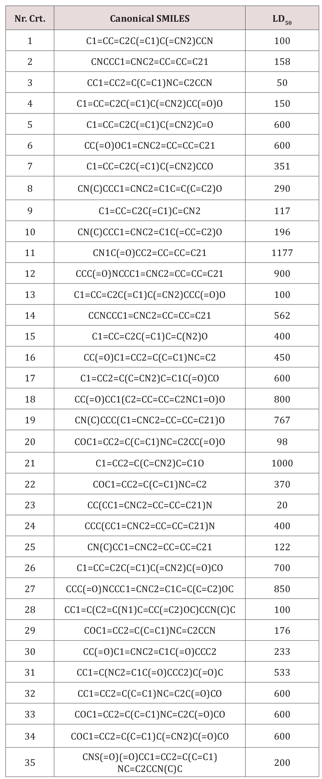
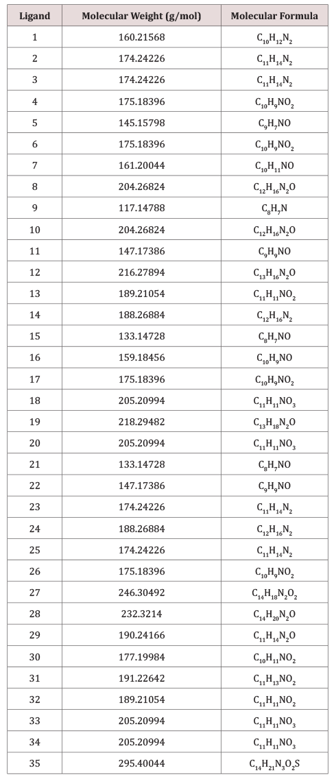
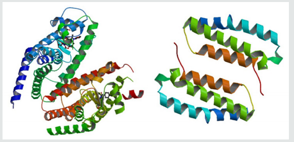
Serotonin, 5-hydroxytryptamine, represents a class of monoamine neurontransmitters, all of which having a chemical template comprised of a basic amino group separated from an aromatic nucleus by a two-carbon aliphatic chain. In this paper we present a docking study on serotonin targeting the proteins 3ADX and 2YX8, respectively, performed by AutoDock Vina. A set of thirtyfive serotonins, downloaded from PubChem, was modeled, within the hypermolecule strategy; the predicted activity was LD50 and prediction was done on similarity clusters with the leaders chosen as the best docked ligands on the Peroxisome proliferatoractivated receptor gamma. It was concluded that LD50 of the studied serotonins is not directly influenced by their binding energies to the target proteins.

Keywords: Serotonin; Docking; Binding affinity; 3ADX; 2YX8; Hypermolecule; LD50; QSAR

Introduction

Serotonin is a neurotransmitter involved in a wide variety of behaviors, including feeding and body-weight regulation, social hierarchies, aggression and suicidality, obsessive compulsive disorder, alcoholism, anxiety, and affective disorders [1-3]. Peroxisome proliferator-activated receptors are ligandactivated transcription factors that regulate genes important in cell differentiation and various metabolic processes, especially lipid and glucose homeostasis [4].

Receptor activity-modifying proteins (RAMPs) are a class of proteins that interact with and modulate the activities of several Class B G Protein-Coupled Receptors including the receptors for secretin, calcitonin (CT) and glucagon, RAMPs complex with Sumatriptan molecule [5].

A method is described to dock a serotonin into a binding site in a peroxisome proliferator-activated receptor gamma on the basis of the complementarity of the inter-molecular atomic contacts. Docking is performed by maximization of a complementarity function that is dependent on atomic contact surface area and the chemical properties of the contacting atoms [6].

Serotonin is metabolized in cells of the brain, gastrointestinal tract, liver, lungs and platelets; it exerts its effect on endothelial cells through multiple receptors [7].

A pharmacophore model can be established either in a ligand based manner, by superposing a set of active molecules with low binding energy [8] and extracting common chemical features that are essential for their bioactivity, or in a structure-based manner, by probing possible interaction points between the protein target and serotonins [9,10].

In our previous work [11], we performed a QSAR study on a set of serotonins, by the similarity cluster prediction approach, proposed by TOPO Group Cluj. In this paper, a docking study, performed to identify the geometric description of the pharmacophore in the interaction of this class of ligands with the peroxisome proliferatoractivated receptor gamma, is reported. We developed a new approach by creating similarity clusters using as leaders those ligands showing the best scores in the docking test.

Docking Study

A set of 35 derivatives of serotonin were taken from PubChem Database [12] and were divided into a training set (25 molecules) and a test set (10 molecules, in Italics), taken with the lowest docking energy (Table 1). The property chosen for modeling was LD50 (on rat, intraperitoneal route administered).

The molecular docking study was carried out to explore the binding mode of serotonin derivatives (Table 2) within the binding energy of peroxisome proliferator-activated receptor gamma 3ADX and Receptor activity-modifying protein 1 2YX8.

Table 1: Serotonin molecular structures (in SMILES code) and LD50 (taken from PubChem).

Lupinepublishers-openaccess-Drug-Designing-Intellectual-Properties

Table 2: List of Molar Mass, Molecular Formulas and Torsions of the Ligands.

Lupinepublishers-openaccess-Drug-Designing-Intellectual-Properties

Figure 1:The proteins (Peroxisome proliferator-activated receptor gamma left, Receptor activity-modifying protein 1 right) (RCSB PDB CODE: 3ADX, 2YX8).

Lupinepublishers-openaccess-Drug-Designing-Intellectual-Properties

Molecular graphics laboratory (MGL) tools and AutoDock4.2 was downloaded from www.scripps.edu [14]. We employed the Lamarckian genetic algorithm (LGA) for ligand conformational searching [15]. AutoDock Tools was used for creating PDBQT files from traditional PDB files. The grid menu is toggled [16]; after loading protein. pdbqt the map files were selected directly with setting up the grid points appropriate for the searching of ligand within the active site of the protein molecule. This way the grid parameter files are created with setting up the map files directly. The docking parameter files were completed by using the Lamarckian genetic algorithm [17].

(Figure 2) illustrates the final Lamarckian genetic algorithm docked state: binding energy of ligands with the active site of Peroxisome proliferator-activated receptor gamma (3ADX). Docking energies lie in the range: -7.0 and -5.1kcal/mol. It seems that the higher the number of rotatable bonds, the higher the ligand–enzyme affinity is.

The distribution of the docking energy (Figure 2) may be attributed to the differences in position of the functional groups within the studied compounds. Interaction of the ligand with the protein can be seen in (Figure 3).

Read More Lupine Publishers Journal of Drug Designing & Intellectual Properties  Please Click on Below Link: https://lupine-publishers-drug-designing.blogspot.com/
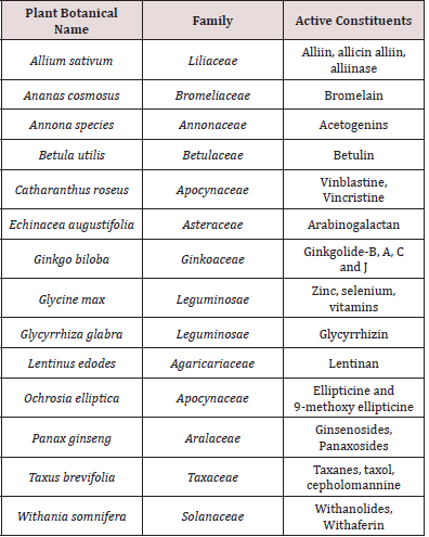
https://fashion-technology-lupine-publishers.blogspot.com/

Wednesday, 19 October 2022

Lupine Publishers| An in vivo study for the effect of Citrus reticulata (Rutaceae) fruit peels extracts on the onset of toxicity of Cerastes cerastes venom in Albino mice

 Lupine Publishers| Journal of Drug Designing & Intellectual Properties


Abstract

Venom of Cerastes cerastes has been extracted and its toxicity was investigated in the presence of aqueous and methanolic extracts of Citrus reticulata (Rutaceae) fruit peels. The decline in the mean survival time of the male albino swiss mice were used to deduce the venom property in the presence and absence of aqueous and methanolic extracts of Citrus reticulata (Rutaceae) fruit peels. The aqueous and methanolic extracts of Citrus reticulata (Rutaceae) fruit Peels significantly decrease the mean survival time compared to the venom alone. From these results it was evident that the toxicity of Cerastes cerastes venomis increased significantly in the presence of Citrus reticulata in a dose dependent manner

Keywords: Citrus reticulata; Cerastes cerastes; Venom; Toxicity

Introduction

Snakebites are severe socio-medical difficulty that lead to morbid and fatal affect on victims in Libya and other North African countries [1,2]. Immediate antivenom treatment is crucial and vital to avoid morbidity and mortality [3]. The oxidative trauma condition, which result from snake bite envenomation is another measurement of kidney destruction and severe renal failure [4], connected with the antioxidant defense system, that might be subject for treatment by antioxidant therapy [5]. ROS (Reactive oxygen species) are engaged in many inflammatory reactions, thus influencing the physiology of the cells and participate a significant function in the pathological conditions [6]. As have been free radical, ROS are involved in harming cellular components, and they play an important function in venom induced toxicity, as reported among envenomed mice [7]. Ascorbic acid is an antioxidant that has been reported to have useful effects on a number of cancer types [8,9] and could be concerned in alleviation of Reactive oxygen species cellular damage, produced during exposure to toxins, metabolism and carcinogens [10]. In addition to augmentation of protease inhibitor effects concerned in preventing organ efficient injure [11,12]. Citrus reticulata (Rutaceae) is commonly known as narangi or santra (orange). It is a small spiny tree with thick top of slim branches, extensively grown in Egypt, Tunisia and Libya [13]. Mandarin is a collection name for this class of orange with thin, loose peel. The name ‘tangerine might be applied as an interchange name to the entire group, but in trade, it is usually limited to the types with red-orange skin. The fruit has aphrodisiac, laxative, tonic and astringent properties [14,15]. It is also used to alleviate vomiting [16,17]. The fruit peel controls the skin moisture, rough and softens hard skin and possess a cleaning effect on oily skin [18]. Chemical composition of the volatile oil of the fruit peels of this species has been reported [19-23]. The effects of the volatile oil of C. reticulata has been studied against Saccharomyces cerevisiae [24], pathogenic fungi, Paenibacillus larvae, Schistosoma mansoni, Aspergillus flavus , and other microorganisms [25-30]. Very recently, the volatile oil of C. reticulata also demonstrates anticancer activity [31-33]. The main aim of the current study is to investigate the effects of Citrus reticulata (Rutaceae) fruit peels extracts on the toxicity of Cerastes cerastes venom in albino mice.

Materials and Methods

Collection of plant material and preparation of aqueous extract

The oranges were bought from a shop in Tripoli (February 2019), and the Citrus reticulata was identified and authenticated by a botanist. Orange rinds were peeled off carefully with the help of a sharp razor blade, and each rind sample was cut into smaller pieces and 30g mass of the sample was taken. The sample was initially rinsed with distilled water, and the fresh peels (30g) were added to 30ml hot distilled water. In addition, another 30g of the fresh peels were macerated in cold 99% methanol for three hours at room temperature (28-30 °C), the mixture was then filtered under vacuum and the filtrate was stored at 4 °C and used when appropriate [34].

Experimental models

Albino mice (Swiss type) of either sex weighing approximately 18–28g (2 to 2.6 month old) were utilized for investigational purpose. They were kept in cages made from polypropylene in airconditioned room with the temperature retained at 25±2 °C, and twelve hours sporadicing dark and light cycles. The mice were supplied with drinking water ad libitum and an adequate diet during the study. The authorization for the experimental procedures was obtained from the Animal Ethics Committee.

Venoms

Cerastes cerastes venom was extracted by means of physical stimulation and was gained in liquid forms, from the Faculty of Science, Zoology Department, University of Tripoli (Libya) and kept at –20 °C until utilize. A 7.5μl aliquot from the venoms was added to eight hundreds microliter of normal saline. A dosage of hundred microliter (100 nanogram) was administered to the male Swiss Albino mice.

Acute toxicity study

Acute toxicity was commonly performed to determine the LD50 value in experimental animals. The intend of doing acute toxicity study is to establish the therapeutic index of a methanolic and aqueous extracts of Citrus reticulate and to guarantee the in-vivo safety. The acute toxicity experiment was done in mice, in which all animals were overnight fasted prior to treatment and given food one hour after aqueous and methanolic extracts administration, with the period observation of common behavior at 0.5, 1, 8, 12 and 24 hours. The number of animals that died after taken the extracts was monitored daily for 7 days [35,36].

Intoxication of venom by Citrus reticulata extracts The animals (albino mice) used in this study were divided to ten groups, each of them is of six mice (male or female). Five groups were used to investigate the aqueous extracts, while the other were used for methanolic extract. The first group received only hundred microliter (hundred microgram of total protein) of the Cerastes cerastes venom (LD99 5μg/kg). Groups 2 to 4 were used as treatment groups and given an equivalent amount of the Cerastes cerastes venom with 50μl, 100μl and 200μl of aqueous Citrus reticulate extracts intraperitoneally (30g/30ml), respectively. Group 5 was given 100μl of the Cerastes cerastes venom and polyvalent anti-snake venom (ASV) was bought from India from Haffkine Bio-Pharmaceuticals Company. The number of death was recorded within twenty-four hours. Similar experiments were repeated in the same manner with the methanolic extract using groups 6 to 10.

Statistical analysis

The difference among various control group and treated groups were analyzed using ANOVA method of one-way. The obtained results were dealt with using unpaired Student’s test. All results were articulates as the mean±SEM of the number of experiments performed, with P value less than 0.05 showing significant difference among groups.

Results and discussion

Acute toxicity study

With the growing amount of research about naringin as a component of the orange and its potential utilize within the pharmacological and food industries, illuminating its toxicological outline becomes increasingly significant. In the present study, the Citrus reticulata extracts were found to be safe up to 200mg/kg orally. This present study is compared with other previous studies in which an oral single dose of 16g/kg of naringin did not produce acute oral toxicity in rats [37].

Acute toxicity of Cerastes cerastes venom and its reaction with aqueous and methanolic Citrus reticulata extracts and antivenom The Cerastes cerastes venom at the dose five micrograms per kilogram (LD99) produces 100% mortality in mice. The aqueous and methanolic Citrus reticulata extracts significantly decrease the mean survival times by 3, 5 and 6 times for 50, 100 and 200µl (30g /30mL), respectively when compared with the venom alone which was 3.1±0.3 hours. ASV was established to be efficient and showing mean survival of 2-days for 5-mice and absolute survival of one mouse. The Cerastes cerastes toxins contain of cardiotoxin, neurotoxin, proteins and enzymes. The victim may die from respiratory troubles which is the main cause of death. Assisted ventilation and ASV can save life in a lot of cases [38-40].

It has been reported that the citrus species contain glycosides and flavonones in huge amounts, and they play a main function in treating a range of pathological conditions. Hesperidin and naringein, are the major components of the citrus fruits. Intestinal microorganism are able to convert naringin into naringenin (an aglycone part). They established to have metal chelating effect, antioxidant, antidiabetic, antiviral, antiallergic, antiestrogenic, antimicrobial, ischemic heart disease adipolytic activity, anti-inflammatory, antiobesity, hypoxia, anti-cancer and hepatoprotective activity. Because of all these pharmacological action, both naringenin and naringin are assumed to be useful as a food supplement [41-47]. The accelerated death could be related to the interactions of Citrus reticulata components (which were mainly polyphenolic components) with snake venom which is not consistent with the previous studies reporting that secondary metabolites polyphenol are competent to inhibit PLA2 [48]. In the literature, it has been reported that naringin which is a flavonoid that is contained in grapefruit and recognized for its various biochemical activities and pharmacological effects on a secretory phospholipase A (sPLA2 ) of Crotalus durissus cascavella, is concerned in the releasing of arachidonic acid in phospholipid membranes [48]. sPLA2 was incubated with naringin in a ratio of 1:1 mole at 37 °C and a distinct decrease in the ultraviolet absorption signal and a changes of the circular dichroism spectra suggesting a significant effect of PLA2 structure and function [48]. The obtained results are for the whole extract of Citrus reticulate and not for naringin or naringenin and this could be explained for the lack of association between pharmacological and enzymatic activities in which the chemical modification of some amino acids induced by naringin, in particular aromatic amino acids and histidines, affected the toxin’s ability to interact with the pharmacological receptor, but did not lead to eliminate of this function. Our results and those described by Cardoso et al. expressed that enzymatic activity of sPLA2 is not crucial for pharmacological activities of this sPLA2 which was isolated from C. d. cascavella venom [49].

Conclusion

The present study confirmed that the aqueous extract of peeled Citrus reticulate accelerate the onset of toxicity of Cerastes cerastes venomis in a dose-dependent effect.

Read More About Lupine Publishers Journal of Drug Designing & Intellectual Properties Please Click on Below Link: https://lupine-publishers-drug-designing.blogspot.com/