Showing posts with label MADOHC. Show all posts

Wednesday, 27 September 2023

Lupine Publishers | Children’s Mouthwash; Commercial Product or Oral Health Guarantor

 Lupine Publishers | Journal of Dentistry and Oral Health Care


Opinion

Nowadays, when we enter a pharmacy, we come across a variety of healthcare products that come to the market every day. Types of masks, gels, detergents and etc., which may look identical in name and application but claim to be totally different and each has its own user extent. Some of these products are commercial in nature, and some are not so important in maintaining health, and in fact, one’s health is not dependent on them. Perhaps that’s why each of us never consumed some of these health and beauty products, and we do not even know them at all. If the product does not directly interact with our health, we do not need to carefully look at the need to how to consume them and do not curious about them, but when it comes to products that claim to protect our child’s health, the matter becomes more quite critical and worrying. Mouthwash is one of these products which are found at all pharmacies. So many oral health officials have been able to highlight the need to use mouthwashes among people. With the training and information provided in this area, people should use mouthwashes in addition to toothpaste and dental floss. But what is the child’s mouthwash, and do the children really need them to use? Do these products guarantee the health of their teeth or is it more a commercial product to fill the pockets of some of the companies?

Mouthwashes have different types. Some have pharmaceutical uses and should be used in certain cases only with physicians’ prescriptions. Mouthwashes that are on the market for the public use contains fluoride which prevents tooth decay. Many pharmaceutical companies have produced and marketed mouthwashes for children to facilitate the use of mouthwashes for children. These mouthwashes have been manufactured considering the least risk of swallowing fluoride. The proper taste of these mouthwashes is a feature that encourages children to use them. Mouthwash is one of the complementary methods of oral home care. These mouthwashes provide oral and dental care along with tooth brushing and dental floss, but they should never be replaced by each other. In other words, application of none of them alone has the significant effect. Before planning to buy the product, if parents tend to use pediatric mouthwashes, they should have a consultation with the pediatric dentist so that the dentist can select the better of the most appropriate one and the least harmful mouthwash. However, they should be used according to the instructions; these instructions vary from product to product and depending on the content and concentration of fluoride, the application may be different.

In other words, it should be noted that some types of mouthwashes are highly recommended by most dentists in the routine oral care program. This is due to the ease and speed of its use, and its effectiveness. In general, along with toothbrushes and dental flosses, many types of mouthwashes are also produced, each of which has its own interests in the beauty and health of teeth. These mouthwashes can be prescribed by the dentist or can be purchased OTC from the pharmacy. Nonetheless, alcohol-free mouthwash is a product that depends on the individual’s needs. The choice of the mouthwash that meets personal needs is very important. Some mouthwashes on the market contain alcohol, in particular, Ethanol, which can cause burning sensation, unpleasant taste, and dryness in the mouth. It is not recommended for children at all because the burning and spicy tastes force the child to stop using the kid’s mouthwash for future use. Consequently, if the parents intend to choose an appropriate mouthwash, first they should not choose any kinds of mouthwashes, and secondly, it is advisable to have a consultation with the pediatric dentist so that they don’t become bewilderment when choosing the proper mouthwash. To finish this point of view, for some time, fluoridecontaining mouthwash has been commonly used in children. But, in my opinion, some kids do not need to use them at all. That is, if fluoride is adequately contained in fluoride-containing toothpaste, in drinking water, or even the consumption of foods such as seafood or tea which contains this material, then, there is usually no need for fluoride mouthwashes for children.

Read More About Lupine Publishers Journal of Dentistry and Oral Health Care Please Click on the Below Link: https://lupine-dentistry-oral-health-care.blogspot.com/


Wednesday, 2 August 2023

Lupine Publishers | Signs of Tooth Eruption in Infants

 Lupine Publishers | Journal of Dentistry and Oral Health Care


Abstract

Teething is one of the challenges of medical science in Infancy period. Many studies show that teeth eruptions might have a lot of symptoms, including bad mood, skin rashes on the cheeks and around the mouth, increasing saliva secretion, swollen gums, Sucking a finger and etc. These signs and symptoms are mild in some infants, and in others may be severe; it is difficult for parents to tolerate these conditions. The peak of the severity of teething-related symptoms is when the dental buds have grown sufficiently in the gum and try to exit from the swollen gingiva.

Keywords: Teething; Saliva Secretion; Bad Mood; Skin Rash; Swollen Gum; Dental Buds

Introduction

As important as it is, tooth eruption is a matter of difficulty for both parents and children. Note that maintaining the health of teeth and gums in the child is the basis of the health of his teeth and gums for whole life. Teeth eruption is one of the most important stages of the growth, and through this stage, the baby will be able to have chewable foods. Commonly, there is a timetable for each primary tooth to erupt in the mouth. Signs of infant’s teeth eruption include disturbances in sleep and night-time wakening, mouth-watering, redness of cheeks, chewing fingers and objects, swollen gums, child malaise, bad temper, and the appearance of tooth buds [1-3]. In this article, we refer to any of these cases.

Schedule the Growth of Different Teeth in the Baby

Generally, new teeth grow in pairs. First, we see the eruption of the two lower primary central incisors teeth, and about a month later, the two upper primary central incisors teeth would erupt. Of course, in some cases, the four lower primary anterior teeth might erupt at first, and then we see the eruption of the upper primary anterior teeth or vice versa. In general, the eruptions of the primary teeth are scheduled to be as follows: [4]

a) At 6 months of age: lower primary central incisors teeth

b) At 8 months of age: upper primary central incisors teeth

c) At 10 months of age: upper and lower primary lateral teeth

d) At 14 months of age: first primary molars teeth

e) At 18 months of age: primary canines’ teeth

f) At age 2: second primary molars.

Signs and Symptoms

Given that the primary teeth are much smaller when they come out of the gum, and the process of tooth eruption is different in children, the symptoms that can usually be expected are:

a) Distracted Dreams: In some children, teething might be a painful process that can awake the baby at night. Therefore, if the baby suddenly wakes up at night, it may be due to her teeth. At this time, the best thing that mothers can do is taking her to the cradle and calm down.

b) Excessive Mouthwatering: Increasing saliva secretion can be one of the symptoms of the eruption of a new tooth. This is, of course, one of the natural stages of the baby’s teeth growth, so too much water in the mouth is not always a sign of baby’s teething. There is no way to detect the association between excessive saliva secretion and the eruption of new teeth. In a baby who is teething, his chin is often wet. Excessive saliva can cause injury and irritation of the baby’s face, so mothers should dry the baby’s face and mouth with a soft, delicate cloth. To protect the skin of the baby’s face, they can consult with their physician.

c) Swollen Gums: In some cases, before germinating of the tooth, the growth site in the gingiva of the infant becomes red, swollen and bruised. Sometimes, the rise of a tooth below the gum causes a bulge so that if parents can persuade her child to open the mouth enough, they can see the whitish shadow of the tooth under the swollen gingiva.

d) Chewing Things: Children like to take everything in their mouths, but if the baby too much does that, she may be teething. The pressure that unerupted teeth from below have on the gums, make the pain to be relieved by applying pressure from the opposite direction. Hence, most infants who are teething are willing to bite on different objects. Mothers should try to give them a teether rings to make them a little quiet. Of course, chewing on different things can also be the innate response of the baby to the strange sense of the mouth.

e) Sucking a Finger: In addition to biting on things during teething, it may be accompanied by sucking a finger. The baby can suck and bites for a few hours during the day. By doing this, he also tries to eliminate the pruritus of the gums. The joy of this action helps the baby to eliminate the pain and pressure of the teeth. Therefore, a mother may find that her baby calms down by chewing his fingers. She should try to keep her baby’s hands clean so no microorganisms can enter the baby’s mouth.

f) Change in Eating Habits: Wounds and gum’s swelling can make sucking painful for the baby. If the baby is hungry but runs away from feeding by mother’s breast or bottle, he may be teething. In this case, babies who eat solid foods tend to breastfeed or feed on the bottle, because the spoon annoys their inflamed gums. Some other babies also do quite the opposite of doing this, that is, they eat more because the bilateral pressure gives them a good feeling. On the other side, babies who still feed on the breast or bottle may eagerly start feeding at the beginning but quickly refrain from eating, because the sucking action puts very uncomfortable pressure on the gums and ear canals.

g) Baby’s Temper Tantrum: Primary tooth eruption (the outward movement of the tooth in the bone and gum) is usually done in a manner so that this action takes place more often overnight compared to the daytime, consequently, the baby will be more restless at night. The pain of teething can cause her sensitivity and irritability, and make him constantly cry.

h) Acne:In some children, teeth eruption may be accompanied with symptoms such as acne. Of course, this symptom is not definitive like fever, and it may have other causes. Skin hypersensitivities are more likely in children due to delicateness and vulnerability of their skin, and occasionally these acnes are signs of gastrointestinal symptoms.

i) Pulling the Ears: Holding, gripping or even pulling the ears, although sometimes indicative of ear infections, it can also be a sign of tooth eruption; in this situation, the pain that occurs in the jaw could be a transferred pain to the ears.

j) Cough: In some cases, when children are teething, they start coughing at that period of time.

Two Misconceptions about Signs of Teething

Many still believe that a child can have a fever during teething 5]] (even healthcare providers!), but on the contrary, some believe the temperatures above 102 degrees Fahrenheit are not related to teething [6]. If fever exists in this period, it is a mere accident and must be caused by other factors [7] . However, if we compare the temperature of the baby’s body who is teething with a child who does not, his body temperature may be slightly high, but this increase is not important enough to be called fever. Hence, why do many babies with teeth eruption have a fever? What is the explanation for that? Another misconception is that teething causing diarrhea [8,9]. Severe diarrhea or constipation is not associated with teeth eruption, and these two events should normally not be accompanied with each other. Although fever and diarrhea are not so much related to the teething, interestingly these signs are seen in some children with the onset of teeth eruption. The concurrency of diarrhea, fever and tooth eruption has two main causes: first, around the age of 4-6 months, when the teeth are emerging, the child’s immune system gradually become independent of the mother reduces the body’s resistance, and therefore this causes symptoms such as fever and diarrhea. Secondly, at this time, children take everything in their mouths to relieve the itching of the gums and discomfort that they develop during teething, but these objects may be contaminated to microbes [10], hence, there is the expectation of diarrhea in infants. Consequently, the emerging of teeth itself does not cause severe diarrhea. On the other hand, at this time, the safety of the immune system from the mother to the infant will diminish, [11] and the child’s body must build the safety components. Therefore, the reduction of maternal immunity which is transferred from the mother to the child can be a reason for fever or infection, [11] which is only a concurrency with teething; and the tooth eruption alone, do not cause this symptom.

Final Words

To summarize the article, the eruption of the teeth is a natural occurrence that occurs without acute and severe problems. It is a physiological phenomenon that will be associated with the other physiological phenomena such as increased saliva in the child, gums swelling, biting on any objects, disturbance in night sleepless, changes in eating habits, and so on.,. The tooth eruption does not pose a problem for the child. Some people mistakenly think that when the baby wants to have a new tooth, he is prone to having a fever, severe diarrhea or even constipation. The growth of teeth will never be accompanied by high fever. The teeth eruptions are different in newborns, but in general, most of them have first teeth at six months of age. Noteworthy that some of the symptoms of teething are similar to those of some diseases, which should be contacted with a pediatric pediatrician in the event of worsening of the symptoms.


Read More About Lupine Publishers Journal of Dentistry and Oral Health Care Please Click on the Below Link: https://lupine-dentistry-oral-health-care.blogspot.com/


Wednesday, 7 June 2023

Lupine Publishers | Maurice Roy’s Protest

 Lupine Publishers | Journal of Modern Approaches in Dentistry and Oral Health Care


Opinion

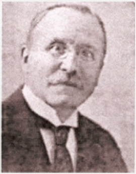
Maurice Roy was born on 24 December 1866. At 14, he entered a dental surgery as an apprentice technician. In 1884, he enrolled at the Dental School of Paris where he was awarded his diploma in 1886. He became a tenured teacher in 1900. He passed his baccalaureate. He then started medical studies. In order to meet his own needs, he worked as a dentist. He was exempted from military service because of his myopia; he was able to devote himself to writing his thesis he sustained in 1894. When the Dreyfus Affair broke out, at the cries of Zola in J’Accuse! published in 1898, he signed the petition for the rehabilitation of the captain. Having contributed towards the creation of FDI World Dental Federation in 1901, he became a member of its executive body in 1904, then its vice-president in 1911. He was re-elected to the post in 1936. In 1902 he founded the French Dentists Cooperative Society (Cooperative Society of Dentists of France), which supplied equipments to the practitioners of the country. In 1914 he created the relief committee for the wounded to the jaws and the face (Relief Committee of the wounded of the jaws and the face). He dedicated himself: he established surgical and prosthetic rehabilitation protocols that were used by other centers. In 1915 he created the Fraternal aid for French and Belgian war victims dentists (Fraternal help to French dentists and Belgian victims of war). In 1923 he took the lead of The Odontology magazine. Roy made it a key player in dentistry of that time. Every themes were subjectively studied. In 1928 he left the public welfare [1-3].

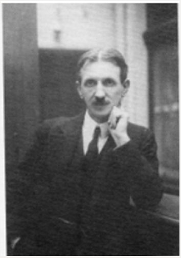
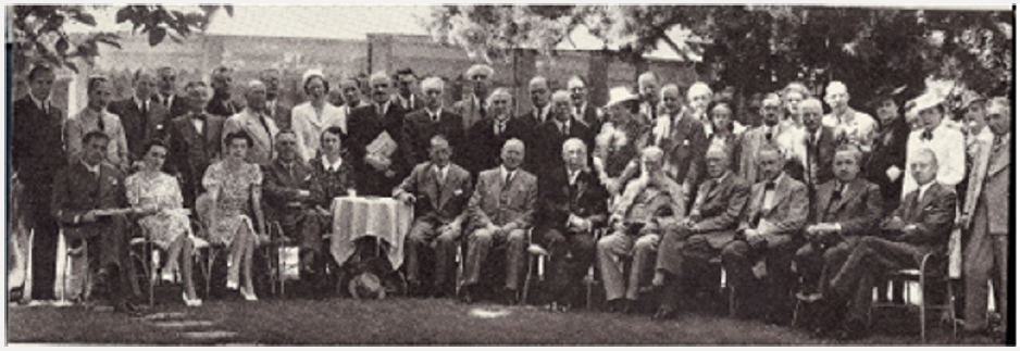
On 28 July 1933, Roy made the executive body pass a motion during FDI World Dental Congress in Edinburgh after Georges Villain, his president and friend, gave a thundering speech to denounce Nazi crimes and to help Jewish German dentists, that stated that: “The executive body of FDI, that duly gathered in session in Edinburgh on 28 July 1933 only to consider the protection of rights obtained by dentists from all over the world thanks to the diplomas granted by the competent authorities of their respective countries [4], declared that no matter of race, of religion or of politics under no circumstances must limit the liberty and the practising of our duly qualified colleagues. Likewise, no restriction which would lead them to breaches of their moral and professional obligations must be imposed on them.” Having made everything to help their colleagues although unsuccessfully the committee passed this motion unanimously excluding Germany who chose to withdraw from FDI. During the 1936 congress of Vienna, Roy received the Miller Prize for his work: the highest distinction that was awarded by the 33-member countries of the executive body including Germany. He received the Legion of Honor this same year. As they were shouted down during the 1938 congress, the Germans were absent from the 1939 congress of Zürich [5]. While the country was occupied, Roy hid aviators and Resistance fighters in his house. While he was the head teacher of the Dental School of Paris, he prohibited the wearing of the yellow star there, this could have caused him an arrestation from the Pétainist font. During a congress in Paris, he left the inaugural session just as the Germans appeared. That provoked a tremendous outcry. While the demands of the Germans were getting insistent, he stopped practicing his dental surgery in 1942, but kept his positions at the Dental School of Paris, at The Odontology magazine and at the Cooperative until his death [6,7]. His convictions caused him insulting articles in the Occupation newspapers: I’m everywhere. Because he was suspected by the Gestapo, he received only routine visits. Maurice Roy died on 5 January 1947 (Figures 1-6).

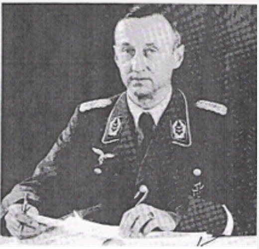
Figure 1: Maurice Roy (Roy Family, 2011).

Lupinepublishers-openaccess-dentistry-oralhealth

Figure 2: Georges Villain (1881-1938), ©BIUM, 2008

Lupinepublishers-openaccess-dentistry-oralhealth

Figure 3: Dental School of Paris - 57, rue Rochechouart (public domain).

Lupinepublishers-openaccess-dentistry-oralhealth

Figure 4: Congress of the Fédération Dentaire Internationale in Zurich, in 1939 (Ennis, 1967). Maurice Roy is in the 1st rank, 2nd on the right. There is no German dentist in this meeting.

Lupinepublishers-openaccess-dentistry-oralhealth

Figure 5: Reichszahnärzteführer Dr. Ernst Stück (1893 – 1974) (public domain).

Lupinepublishers-openaccess-dentistry-oralhealth

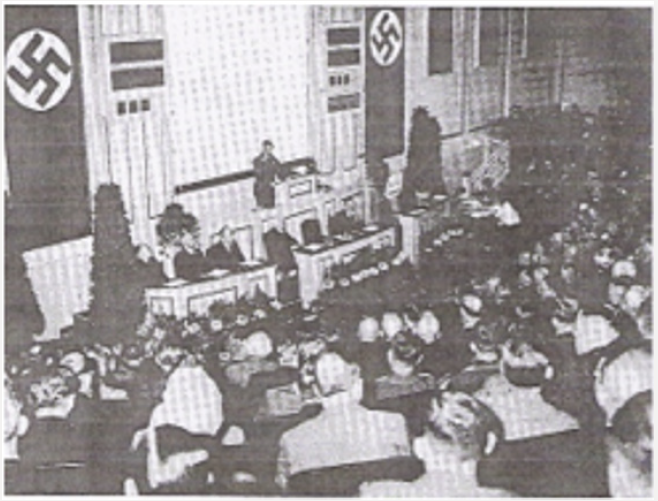
Figure 6: VIIth congress of the German dentists directed by Ernst Stück in 1935, in Berlin (public domain).

Lupinepublishers-openaccess-dentistry-oralhealth


Monday, 24 April 2023

Lupine Publishers| Atypical Manifestation of a Tooth Crack

Lupine Publishers| Journal of  Dentistry and Oral Health Care

Abstract

A patient presented with severe pain in the area of teeth #2 and #31. Prior to seeking dental care, the pain had been so severe together with other clinical manifestations that the patient sought medical attention for a heart attack. After elimination of the occurrence of a m.i., the pain was thought to be dental in origin and dental care was sought. No carious lesions or endodontic problems were found upon radiographic examination. However, careful oral examination revealed the presence of a crack transcending tooth #31. Following tooth extraction and immediate implant placement, no further pain was experienced by the patient.

Introduction

Splits or fractures are the third most common cause of tooth loss in industrialized countries, primarily affecting maxillary molars and premolars, and mandibular molars [1]. However, most clinicians would agree that tooth cracks are difficult to diagnose [2- 4]. Although pain on biting is often considered the most reliable diagnosis for a tooth with a visible crack [5], the most common symptom of a cracked tooth is pain to cold [3]. Further, pain and other symptoms associated with tooth cracks can manifest in different ways, often in areas remote from the actual crack [1]. Interestingly, it has been reported that pain associated with tooth cracks are less likely in teeth with stained cracks or exposed roots, or in non-Hispanic whites [4]. This paper discusses the atypical pain caused by a root fracture in a molar tooth and which manifested in unexpected and misleading ways.

Patient Consultation

A 50 y.o. male patient in good general health presented with pain in the area of teeth #2 and #31. The patient was a dentist and reported that he had experienced severe pain on the left side of his face, with pain extending down his left arm together with heart palpitations and elevated blood pressure. Suspecting a M.I., the patient sought medical care but a visit to the Emergency Room together with blood tests indicated that the patient was not experiencing a heart attack. The pain was ascribed to dental issues on the right side of the face. However, the patient reported no pain on biting or sensitivity to cold.

Clinical Examination

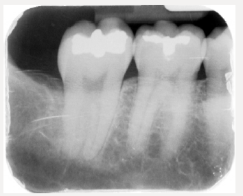
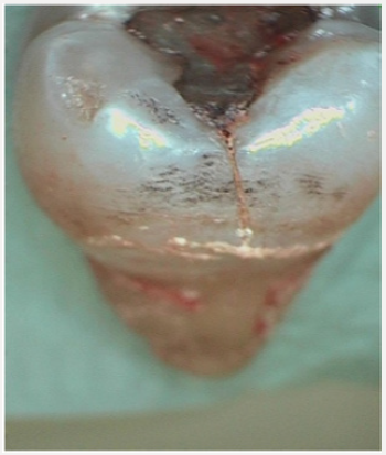
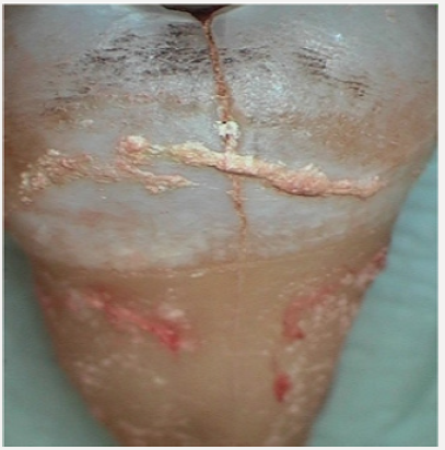
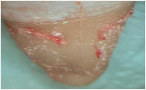
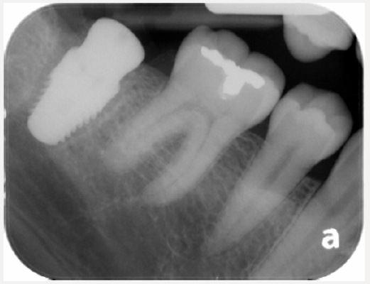
Radiographic examination of the patient, (Figure 1) indicated no carious lesions, sepsis or other issues with his dentition. However, careful intra-oral examination of the teeth indicated the presence of a crack on the distal surface of Tooth #31, (Figure 2). The crack appeared to have initiated from an apparently defective amalgam restoration, (Figure 3), and descended vertically to the apex of the tooth, (Figure 4). This conclusion appears to be justified because the literature indicates that a major predisposing factor to tooth fracture are mesio-occluso-distal restorations with mandibular first molar teeth being particularly affected [6]. Interestingly, the presence of calculus was noted in the buccal enamel and there are indications that this deposit caused widening of the enamel crack. No defects or lesions were noted in any other maxillary or mandibular teeth.

Figure 1: Radiograph of patient’s teeth.

Lupinepublishers-openaccess-dentistry-oralhealth

Figure 2: Intra-oral photograph of affected tooth.

Lupinepublishers-openaccess-dentistry-oralhealth

Figure 3: Intra-oral photograph of the affected tooth.

Lupinepublishers-openaccess-dentistry-oralhealth

Figure 4: Crack on the distal aspect of tooth #2.

Lupinepublishers-openaccess-dentistry-oralhealth

Treatment Regimen

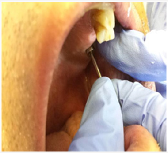
In consultation with the patient, it was decided that the affected tooth could not be salvaged, and it was extracted under local anesthesia. Because the extraction site was infection-free, the periodontal condition was satisfactory, and the bone was solid and well-vascularized, an immediate implant was placed, (Figure 5).

Figure 5: Immediate implant placed following extraction.

Lupinepublishers-openaccess-dentistry-oralhealth

Prognosis

After extraction of tooth #31 and resolution of local anesthesia, the pain was immediately eliminated. Satisfactory healing and osseo-integration are projected for the affected area.

Conclusion

Cracks in teeth weaken them and pain from cracked teeth can be felt in areas remoted from the affected tooth. What was unusual in the present case is the pain was experienced on the other side of the mouth from the affected tooth and intensified to the point that the patient suspected a myocardial infarction. Although tooth crack-induced pain often can be experienced in areas remote from the affected tooth, the existence of pain, so severe that it mimicked a heart attack, and occurring on the other side of the mouth was unusual. This particular case reinforces the need for clinicians to carefully examine all facets of the dentition of patients presenting with oral pain. Cracks in enamel do occur and, as in the present case, may originate from a defective restoration. However, crack progression to the tooth apex and sudden, sharp onset of pain is unusual. Even more unexpected is that the pain was experienced on the opposite jaw to the affected tooth.

Read More About Lupine Publishers Journal of Dentistry and Oral Health Care Please Click on Below Link
https://lupine-dentistry-oral-health-care.blogspot.com/

Wednesday, 19 April 2023

Lupine Publishers| The Age Old Misnomer: Oral Pyogenic Granuloma–A Case Report

 Lupine Publishers| Journal of Dentistry and Oral Health Care


Abstract

An essential diagnostic challenge often faced by oral physicians is diagnosing soft tissue enlargements of the oral cavity. The fundamental reason being there are a diverse group of pathologic processes that can produce such lesions. Any intra-oral enlargement seen may represent a variation of normal anatomic structures, developmental anomalies, inflammation, cysts or even a neoplasm. Confined to these are the group of reactive hyperplasias of the oral cavity, which develop in response to a chronic, recurring tissue injury that stimulates an exuberant or excessive tissue repair response. Pyogenic granuloma comes under as one of the most common entities responsible for causing soft tissue enlargements.

Keywords: Pyogenic Granuloma; Oral Cavity; Inflammatory Hyperplasia; Misnomer

Introduction

Pyogenic granuloma is one of the inflammatory hyperplasia seen in the oral cavity [1]. It is not associated with pus as its name suggests and histologically it resembles an angiomatous lesion rather than a granulomatous lesion [2]. Thus, the term is a misnomer and in reality arises in response to various stimuli such as low-grade local irritation, traumatic injury or hormonal factors [1,2]. Typically, it presents as an exuberant, red painless mass that easily bleeds, ulcerates and grows rapidly and is frequently seen on the gingiva [3]. Surgical excision with linear closure allows histologic examination of the tissue. It also has the lowest rate of recurrence and is therefore the treatment of choice [4]. Hereby, we present a case of a 21-year-old male patient who presented with a growth on the gingiva and bleeding in upper front teeth region.

Case Report

A 21-year-old male patient reported with a growth behind the teeth in the upper front tooth region. The growth had first appeared two months back and had been slow growing to attain the present size. The patient gave a history of mild intermittent pain which aggravated on chewing food and was concerned about the compromised esthetics. His medical, dental and family histories were non-contributory. He was an avid pan and gutkha chewers since three years. On examination an exophytic growth on the palatal aspect of upper right canine between the right canine and first premolar was seen. The growth was irregular in shape, about one cm in size, smooth and lobulated (Figure 1A). It was pedunculated, soft in consistency and there was bleeding on provocation (Figure 1B). Based on the clinical examination we came to a provisional diagnosis of pyogenic granuloma. An excisional biopsy was carried out under local anesthesia (Figure 1C) and the report confirmed the same (Figure 1D). The patient was recalled after one, three and six months and it showed no recurrence of the growth.

Discussion

In 1844, Hullihen [5] described the first case of pyogenic granuloma in English literature. In 1897, pyogenic granuloma in man was described as “botryomycosis hominis.” Hartzell [6] in 1904 is credited with giving the current term of “pyogenic granuloma” or “granuloma pyogenicum.” It was also called a Crocker and Hartzell’s disease [6]. Angelopoulos histologically described it as “hemangiomatous granuloma” due to the presence of numerous blood vessels and the inflammatory nature of the lesion [7]. Cawson et al. [8] in dermatologic literature have described it as “granuloma telangiectacticum” due to the presence of numerous blood vessels seen in histological sections. They described two forms of pyogenic granulomas, the lobular capillary hemangioma (LCH) and the non-lobular capillary hemangioma (non-LCH) [2]. The exact etiopathogenesis remains unknown, although contributory factors include trauma, inflammation and infectious agents. Female sex hormones may also play a role as the condition occurs at increased frequency in pregnant women and in those who use oral contraceptive pills. It is believed that trauma and female sex hormones enhance expression of angiogenic factors such as basic fibroblast growth factor (bFGF) and vascular endothelial growth factor (VEGF) which will lead to evolution of the pyogenic granuloma. Medications such as isotretinoin, acitretin, cyclosporine, lamivudine, docetaxel, imatinib, and indinavir may also be contributing factors. Infections such as caused by herpes simplex type-I and Epstein-Barr virus have also been incriminated [4]. Oral pyogenic granulomas occur in all age groups, children to older adult, but are more frequently encountered in females in their second decade due to the increased levels of circulating hormones estrogen and progesterone [2]. Pyogenic granuloma of the oral cavity appears as an elevated, smooth or exophytic, sessile or pedunculated growth covered with red hemorrhagic and compressible erythematous papules, which appear lobulated and warty showing ulcerations and covered by yellow fibrinous membrane [9]. The color varies from red, reddish purple to pink depending on the vascularity of the growth. The gingiva, especially the marginal gingivais affected more than the alveolar part [10,11]. Besides the gingiva it is also noticed on the lips, tongue or buccal mucosa, affecting the maxilla more than the mandible, the anterior region than the posterior with the buccal surfaces being affected more than the lingual surfaces. The size varies from a few millimeters to several centimeters and it is usually slow growing, asymptomatic, painless growth, but at times it grows rapidly [9,10].

Differential diagnosis of pyogenic granuloma includes peripheral giant cell granuloma, peripheral ossifying fibroma, fibroma, peripheral odontogenic fibroma, hemangioma, conventional granulation tissue, hyperplastic gingival inflammation, Kaposi’s sarcoma, bacillary angiomatosis, angiosarcoma, and non Hodgkin’s lymphoma [12]. Radiographic findings are usually absent. However, Angelopoulos [7] concluded that in some cases long standing gingival pyogenic granulomas caused localized alveolar bone resorption [12]. Histologically, the lesion appears as a lobular proliferation of capillaries with each lobule containing a central feeder vessel surrounded by aggregates of capillaries [4]. The lobules are separated by a fibro-myxoid stroma. In those lesions that are undergoing regression, there may be extensive fibrosis [4,13]. For gingival lesions, excising the lesion down to the periosteum and scaling adjacent teeth to remove any calculus and plaque that may be a source of continuing irritation is recommended. Although surgical excision is the considered the treatment of choice, management of pyogenic granuloma depends on the severity of symptoms [3,14]. If the lesion is small, painless and free of bleeding, clinical observation and follow up are advised. Other treatment modalities include laser surgery, electrodessication. Injection of absolute ethanol, sodium tetradecylsulfate (sclerotherapy) and corticosteroids have also been tried with successful results in cases with recurrent lesions [15]. The prognosis is usually excellent, and the lesion usually does not recur unless inadequately removed. The recurrence rate is higher for pyogenic granulomas removed during pregnancy. Other possible reasons for recurrence include; incomplete excision, failure to remove etiologic factors, or due to re-injury to the area, making follow up necessary [14,16].

Conclusion

Despite the fact that pyogenic granuloma is a non-neoplastic growth in the oral cavity; a proper diagnosis, timely prevention and appropriate management are of utmost importance, careful diagnosis is essential to differentiate this lesion from other vascular lesions. Surgical excision of the growth, along with curettage should be done to prevent recurrences of this common lesion. And though the term pyogenic granuloma is still the used terminology of choice, it is a well-known fact that it is not associated with pus and histologically it resembles an angiomatous lesion rather than granulomatous lesion. Thus, it indicates that despite the term “pyogenic granuloma” being a misnomer, sometimes the good old quote - “Change is the only constant” gets a reality check as clearly time wants this terminology to stay in the literature books despite it lacking any accuracy at all.

Read More About Lupine Publishers Journal of Dentistry and Oral Health Care Please Click on Below Link
https://lupine-dentistry-oral-health-care.blogspot.com/

Monday, 20 February 2023

Lupine Publishers| How to Prepare Children with Autism to Visit a Dentist?

 Lupine Publishers| Journal of Dentistry and Oral Health Care


Opinion

One of the most common problems that parents with children with autism have is to take them to a doctor’s or dentist’s office. Even if the office environment is friendly and the dentist or doctor or their assistants are kind to the child, children who have autism do not like others to come close to them, touch them, and when they are alone, someone is in contact with their bodies. When children’s physical environment is invaded, they react to this behavior and do not cooperate with the other person, and sometimes they even become aggressive. It should be said that in many cases, these children are torn apart. There are many problems when a child visits a dentist’s office. For example, special smells, a tray of dental instruments, and devices and tools may pose a threat to the peaceful world of these children. For a child with autism spectrum disorder, a dental office is full of insecurity and, consequently, is an unpleasant place [1,2].

Preparation by Parents

Here are some simple solutions that parents can do to help their children with autism when they plan to go to the dentist’s office. Make a medical bag ready for your child; it is best to let him choose this bag by himself. Meanwhile, give him times to get acquainted with the tools inside the bag so that to find out what they are. After spending some time, show him what these tools are used for, and first try them on your own, siblings or someone else. The next step is to run the show with your child, pretending to be a doctor and treat him with these tools. Now ask your child to change his place with you, and let him try this tool on you, his sister or brother or someone else. You can also use a puppet or a stuffed doll or even your own pet to display these tools. Explain to your child what each device is used for, and let him access the toys toolbox and play with them freely. When friends, neighbors, and relatives come to your home, let them play with this gadget with your child, and then tell them to change their role with your child. Remember, if your child can find a playmate to practice with these tools, he will be great of help. If possible, give him a medical phone or more realistic tools to practice with them; however, in this case, you need to make sure your child is monitored and is safe. Give him a prize after each real meeting or show-up meeting with a dentist, or returning from the medical laboratory. Finally, play with your doctor’s true name when your child feels comfortable in the “Physician and Patient” game. In addition, along with this medicine toolbox, you can read books about meeting with the dentist too [3].

How to Take an Autistic Child to a Dental Office?

It can be difficult for every child to visit a dentist, but for a child with autism, referral to the dentist is very scary. Here are some tips for parents that will make this condition easier for their autistic children [4-6].

Before the Appointment

a. First Method: Try to get an appointment from dental centers that have been established for people with disabilities. These centers experience the treatment of disabled children and usually know what to do to make your child feel relaxed and happy during treatment. If your child needs something special at a specific time, please inform the center before visiting. You can also tell them about your child’s interests so that the dental center’s staff can talk to him about his interests while he is being treated [7].

b. Second Method: Consider the appointment for a day when your child is not busy. In order to minimize the stress on your child, it is advisable to rest well before going to the dentist. So, consider a day to visit that your child has not done much. This will reduce her stress and cry. In order to provide him with the ideal condition, it is advisable for the dentist to have free time before and after the visit so that he can cope with the situation well [8].

c. Third Method: Try to tell stories about the dentists for your child. Dentists are usually a frightening person for autistic children. So, if they know what happens when they are treated, their fear will be reduced.

d. Fourth Method: Talk to him about the fears your child may face. The mouth is a sensitive part of the body, and for some autistic children, this part has a special sensitivity. As a result, your child may have many concerns. Therefore, confirm his emotions and trust him; for example, you can tell him”It’s quite common for you to be afraid, so many people are afraid of a dentist. But I know this doctor. He always tries to be very careful and do his work the best. I know you do not want to go to dentistry, but the dental practitioner takes care of your teeth to make sure that they are healthy. We have to go there, but you may not love him. “

e. Fifth Method: Plan for a stimulus activity. For example: watching a movie, visiting his favorite park and even buying a small toy are things that can make your child happy. Before you go to the dentist, choose this activity and be sure it will be very helpful. Try telling your child about this encouraging activity, because he has hope for reaching that. For example: If your child has a lot of interest in the ribbons, tell him: “After dentistry, we go shopping and you can buy two ribbons.” As a result, when your child is in fear, he will think about his ribbons during the treatment. If you think that your child will be tired after dentistry, try to consider small activities. Do not use food as encouragement, because after the treatment, he cannot eat for a while.

f. Sixth Method: Give him food before leaving. Your child can not eat anything for a while after dental procedures. A hungry child is a child who easily cries and gets tired. Make sure your child has eaten her food, and then brush her teeth without any hurry [10-12].

When Visiting the Dentist

a. Method 1: Inform your child that you are ready to go to the dentist. Your child should brush his teeth before leaving the house. Let him bring something like a toy or any other device to the dentist office which makes him more comfortable. Do not hurry him because it can cause stress in your child. Try telling him the plan a few hours before you are leaving. This way, if needed, he can finish doing his work without any anxiety.

b. Method 2: Be extremely patient and kind. If your child is stressful, he will behave strangely because he actually fights emotionally. Be gentle and consider that it will not be easy for your child too. Try to do things in your car that your child is interested in. Play his favorite music, talk with him about your interests; or if he is interested in singing, try to sing a song together. You should know that your child needs to be assured about the condition; may ask you questions repeatedly, so give him a peaceful answer.

c. Method 3: Talk with the dental center staffs, and ask for help for the comfort and relaxation of your child. Perhaps they are able to create a situation in which your child can have a more pleasant visit to the dentist.

d. Method 4: Thinking of not being beside him, may cause this image that you are leaving him which may develop more fear and stress. Make assure your child that you would be in the waiting room if he will need you. If your child is getting disturbed by being away from you, ask the dentist to let you stay in the room with him.

e. Method 5: Encourage and admire her after the meeting. Tell her she has done this job very well, and keep on the encouraging activity you have been considering. This will make him feel better about doing anything. If your child has had bad conditions during the treatment (crying, screaming, etc.), you should tell him: “It’s not the courage to not be afraid, the courage is to face it, and you did it very well. Although the dentistry is scary and difficult, you did it “. Tell him you are proud of him [13].

Characteristics of Children with Autism

These children have certain behavioral characteristics, some of which include:

Impairment of speaking or not speaking at all, and repeating words and sentences spoken by people around him

i. Susceptible to restlessness and discomfort due to problems in the sensory process, anxiety, fears, and difficulty of communicating

ii. Exaggerated reactions to smells, voices, special tastes and other sensory incitements

iii. Excessive use of body movements to calm himself up, such as shaking hands and waving hands

iv. Failure to respond when calling his name so that the child seems to be deaf

v. Express the discomfort of breaking up routines and habits, and reacting to changes

vi. Restlessness and agility, the need to play and touch objects, and anything around

vii. Avoiding eye and physical contact

viii. Problems in social skills

ix. Obsessive-compulsive and adherence to them

x. Lack of effective self-perception of his feelings and others

xi. The difficulty in understanding security and risk aversion.

xii. Performing repetitive games for consecutive hours

Also, these children prefer to play alone and have little fancy imagination. Some may also have symptoms of hyperactivity and early angry [14].

Oral Condition in Children with Autism

Children with autism often have no differences with normal children in the structures of the teeth, but as these children tend to eat soft and sweet foods, they sometimes have muscle weakness around their mouth. As a result, the reduction in the efficiency of the chewing is observed, and even sometimes they tend to keep the mouthfuls long in their mouth, which increases the rate of caries in these children. Also, due to the inability to brush correctly, and the lack of proper cooperation with parents, caries, and gingivitis are more common in these children [15].

Read More About Lupine Publishers Journal of Dentistry and Oral Health Care Please Click on Below Link
https://lupine-dentistry-oral-health-care.blogspot.com/

Saturday, 4 February 2023

Lupine Publishers | Open Tray Impression Technique Using the Direct Pick-Up Coping: A Case Report

 Lupine Publishers | Journal of Dentistry and Oral Health Care


Abstract

Dental implants have become a quite successful method for restoration of fully and partially edentulous patients. Impression procedure becomes exacting in implantology as compared to fixed partial denture because of lack of periodontal ligament fibers in implants. Traditionally, there are two different implant impression techniques for transferring the impression copings from the implant to the impression. The transfer technique uses tapered copings and a closed tray to make an impression. Conversely, the pick-up impression uses square copings and an open tray (a tray with an opening), allowing the coronal ends of the impression coping screw to be exposed. This article describes the Pick-up implant impression procedure that is inexpensive, clean, and easy to perform with materials commonly found in a restorative dental practice and deals with its advantages and limitations.

Keywords: Implant Impression; Implant Direct Impression; Open Tray - Pick Up Impression Coping

Introduction

The success of implant-supported restorations relies on how well the hard and soft tissue information is transferred to the laboratory [1,2]. The critical aspect is to record the threedimensional orientation of the implant as it is present intraorally, other than reproducing fine surface detail for successful implant prosthodontic treatment [3-5]. The development of impression techniques to accurately record implant position has become more complicated and challenging. Several impression techniques have been suggested to obtain a master cast that will ensure the passive fit of prosthesis on implants [6]. In implant dentistry, the two traditional ways of making an impression are the closed tray and the open tray technique. Both impression techniques have their advantages and disadvantages [7]. A recent systematic review revealed that the open tray impression technique is more accurate than the closed tray impression technique [8,9]. It has been shown that the pickup type impression coping is the more accurate type of impression as errors occur on removal and replacement of the transfer type impression copings, especially in the occlusion-gingival direction [10,11]. Characteristics of the Pickup type impression coping are that they are removed from the mouth together with the set impression. They require access to the retaining screw to allow release of the screw prior to removal of the impression coping-impression assembly, the analogues are attached to the impression copings while they are embedded in the impression tray. A custom tray with access to the impression coping screws is required [12].

Clinical Case

Open Tray Impression Procedure

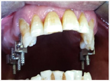
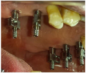
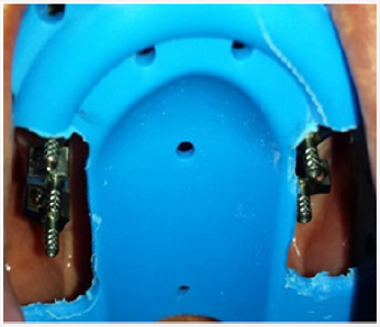
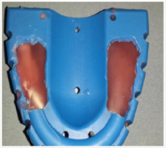
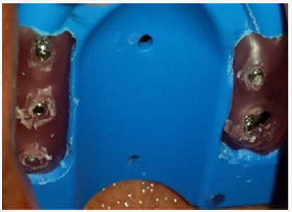
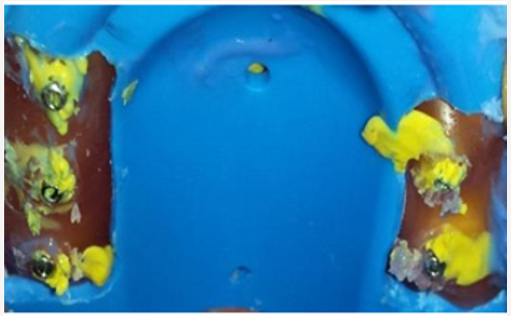
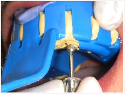
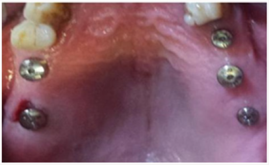
A patient consulted the Department of Fixed Prosthodontics at the Dental Clinic of Monastir to replace his bilateral terminal maxillary edentulism by implant-supported fixed prostheses . The patient had a sufficient and rectilinear mouth opening. We decided to make him five maxillary implants. To record the spatial position of the implants, we have chosen “Open tray Impression technique “using the direct pick-up copings . In the first time, the healing screws were removed (Figure 1). Then, the square impression copings were placed into the implants (Figures 2 & 3). The guide screws were tightened using the screwdriver. Retroalveolar X-rays were taken along the long axis of the implant to ensure that the impression copings were seated completely into the hex of the implants (Figure 1). The tray was perforated in the regions where implants were placed to provide access for the pick-up copings (Figure 4). The impression tray was coated with manufacturer recommended impression adhesive 5 minutes before each impression was made. Tray adhesive was applied thinly and evenly over the inner surface of each tray and extended approximately 3mm onto the outer surface of the tray along periphery. The adhesive was allowed to dry for 15 minutes before impression (Figure 4). The tray was removed from the mouth and two sections of boxing wax (Dentsply Intl) were adapted over the open-ings in the tray and sealed to the tray using a hot instrument (Figures 5 & 6).

Figure 1: Removing of healing screws.

Lupinepublishers-openaccess-dentistry-oralhealth

Figure 2: Impression copings were connected to each implant after removing of healing screws.

Lupinepublishers-openaccess-dentistry-oralhealth

Figure 3: Impression copings were connected to each implant after removing of healing screws.

Lupinepublishers-openaccess-dentistry-oralhealth

Figure 4: Intra oral view of pick up impression copings through the impression tray.

Lupinepublishers-openaccess-dentistry-oralhealth

Figure 5: two sections of boxing wax (Dentsply Intl) were adapted over the open-ings in the tray.

Lupinepublishers-openaccess-dentistry-oralhealth

Figure 6: two sections of boxing wax (Dentsply Intl) were adapted over the open-ings in the tray.

Lupinepublishers-openaccess-dentistry-oralhealth

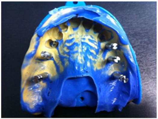
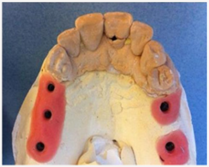
Other open-tray impression protocols recommend wiping off the excess impression material that extrudes through the hole in the tray with a gloved finger or injecting impression plaster through the hole(s) in the tray to fill the remaining void. This may be messy to accomplish. Additionally, the powder on some gloves may inhibit the polymerization of vinyl polysiloxane (VPS) impression materials Wax placed over the hole in the tray prevents contact of the impression material with the gloves [13]. The heavy consistency polyvinylsiloxane impression material was loaded inside the impression tray and light consistency polyvinylsiloxane impression material was meticulously syringed around the impression copings to ensure complete coverage of the copings (Figure 7). The screwdriver was used to loosen the guide screw within the impression post (Figure 8). The impression was taken out from the patient’s mouth. The corresponding analog was selected and the impression post was placed into the implant analog and the guide screw was tightened by using the screwdriver (Figure 9). The healing abutments were replaced immediately to prevent soft tissue collapse over the implant (Figure 10). Before screwing in, implant analog should be placed in line with the grooves of impression coping to achieve accurate passively fitting prosthesis. The impression was now ready to be used to create a model (Figure11).

Figure 7: Open tray Impression.

Lupinepublishers-openaccess-dentistry-oralhealth

Figure 8: The copings screws are unscrewed to be removed along with the impression.

Lupinepublishers-openaccess-dentistry-oralhealth

Figure 9: Impression with coping analog assembly.

Lupinepublishers-openaccess-dentistry-oralhealth

Figure 10: The healing abutments were replaced immediately.

Lupinepublishers-openaccess-dentistry-oralhealth

Figure 11: Occlusal view of hard and soft tissue cast recovered from impression.

Lupinepublishers-openaccess-dentistry-oralhealth

Discussion

In direct technique, the impression coping lies within the impression. Advantages of direct technique are minimization of dimensional changes in impression upon removal from patient’s mouth, no need to screw in coping again as it comes along with the impression and useful in patients with angulated implants [14]. When impressions are required for multi implant restorations the precision of the impression is even more critical. This is because frameworks will be constructed from the master cast and mis-fit in the frameworks can lead to stress applied to the implants on screwing down the framework [12]. For situations in which there were three or fewer implants, most studies showed no difference between the pick-up and transfer techniques, whereas for situations in which there were 4 or more implants, more studies showed more accurate impressions with the pick-up technique (open tray) than the transfer technique (closed tray) [6]. Fourteen studies compared the accuracy of pick-up and transfer impression techniques [15,16]. Of the 14 studies, five showed more accurate impressions with the non-splinted pick-up technique [17], two with the transfer technique, and three showed no difference between them [8,18].

Closed tray (transfer, indirect) technique using tapered impression copings is an easy and simple technique ideal for single implant impressions and multiple parallel implants in patients with limited mouth opening [14]. When there is limited mouth opening they can be used as there may not be sufficient space for access to the screws retaining pick up type impression copings with the impression in place and in patients with an exaggerated gag reflex, when the impression has to be removed as quickly as possible [12]. The clinical situations which indicate the use of the closed tray technique are when the patient has limited inter arch space, tendency to gag, or if it is too difficult to access an implant in the posterior region of the mouth [10].

Polyether and VPS were the recommended materials for the implant impressions. Results indicated that the 2-step VPS impression was significantly less accurate than the 1-step putty and light-body VPS combination impression, the mediumbody VPS monophase impression, and the medium-body polyether monophase impression [5].

Conclusion

Several impression techniques have been advocated for implant impressions to obtain a definitive cast. Different impression techniques have some advantages and some limitations, but selection of technique depends upon operator choice and various clinical situations. Open tray (pick-up, direct) technique using square impression copings is more accurate method usually preferred in multiple implants with different angulations.

Read More About Lupine Publishers Journal of Dentistry and Oral Health Care Please Click on Below Link
https://lupine-dentistry-oral-health-care.blogspot.com/

Monday, 9 January 2023

Lupine Publishers| Open Tray Impression Technique Using the Direct Pick-Up Coping: A Case Report

 Lupine Publishers| Journal of Dentistry and Oral Health Care


Abstract

Dental implants have become a quite successful method for restoration of fully and partially edentulous patients. Impression procedure becomes exacting in implantology as compared to fixed partial denture because of lack of periodontal ligament fibers in implants. Traditionally, there are two different implant impression techniques for transferring the impression copings from the implant to the impression. The transfer technique uses tapered copings and a closed tray to make an impression. Conversely, the pick-up impression uses square copings and an open tray (a tray with an opening), allowing the coronal ends of the impression coping screw to be exposed. This article describes the Pick-up implant impression procedure that is inexpensive, clean, and easy to perform with materials commonly found in a restorative dental practice and deals with its advantages and limitations.

Keywords: Implant Impression; Implant Direct Impression; Open Tray - Pick Up Impression Coping

Introduction

The success of implant-supported restorations relies on how well the hard and soft tissue information is transferred to the laboratory [1,2]. The critical aspect is to record the threedimensional orientation of the implant as it is present intraorally, other than reproducing fine surface detail for successful implant prosthodontic treatment [3-5]. The development of impression techniques to accurately record implant position has become more complicated and challenging. Several impression techniques have been suggested to obtain a master cast that will ensure the passive fit of prosthesis on implants [6]. In implant dentistry, the two traditional ways of making an impression are the closed tray and the open tray technique. Both impression techniques have their advantages and disadvantages [7]. A recent systematic review revealed that the open tray impression technique is more accurate than the closed tray impression technique [8,9]. It has been shown that the pickup type impression coping is the more accurate type of impression as errors occur on removal and replacement of the transfer type impression copings, especially in the occlusion-gingival direction [10,11]. Characteristics of the Pickup type impression coping are that they are removed from the mouth together with the set impression. They require access to the retaining screw to allow release of the screw prior to removal of the impression coping-impression assembly, the analogues are attached to the impression copings while they are embedded in the impression tray. A custom tray with access to the impression coping screws is required [12].

Clinical Case

Open Tray Impression Procedure

A patient consulted the Department of Fixed Prosthodontics at the Dental Clinic of Monastir to replace his bilateral terminal maxillary edentulism by implant-supported fixed prostheses . The patient had a sufficient and rectilinear mouth opening. We decided to make him five maxillary implants. To record the spatial position of the implants, we have chosen “Open tray Impression technique “using the direct pick-up copings . In the first time, the healing screws were removed (Figure 1). Then, the square impression copings were placed into the implants (Figures 2 & 3). The guide screws were tightened using the screwdriver. Retroalveolar X-rays were taken along the long axis of the implant to ensure that the impression copings were seated completely into the hex of the implants (Figure 1). The tray was perforated in the regions where implants were placed to provide access for the pick-up copings (Figure 4). The impression tray was coated with manufacturer recommended impression adhesive 5 minutes before each impression was made. Tray adhesive was applied thinly and evenly over the inner surface of each tray and extended approximately 3mm onto the outer surface of the tray along periphery. The adhesive was allowed to dry for 15 minutes before impression (Figure 4). The tray was removed from the mouth and two sections of boxing wax (Dentsply Intl) were adapted over the open-ings in the tray and sealed to the tray using a hot instrument (Figures 5 & 6).

Figure 1: Removing of healing screws.

Lupinepublishers-openaccess-dentistry-oralhealth

Figure 2: Impression copings were connected to each implant after removing of healing screws.

Lupinepublishers-openaccess-dentistry-oralhealth

Figure 3: Impression copings were connected to each implant after removing of healing screws.

Lupinepublishers-openaccess-dentistry-oralhealth

Figure 4: Intra oral view of pick up impression copings through the impression tray.

Lupinepublishers-openaccess-dentistry-oralhealth

Figure 5: two sections of boxing wax (Dentsply Intl) were adapted over the open-ings in the tray.

Lupinepublishers-openaccess-dentistry-oralhealth

Figure 6: two sections of boxing wax (Dentsply Intl) were adapted over the open-ings in the tray.

Lupinepublishers-openaccess-dentistry-oralhealth

Other open-tray impression protocols recommend wiping off the excess impression material that extrudes through the hole in the tray with a gloved finger or injecting impression plaster through the hole(s) in the tray to fill the remaining void. This may be messy to accomplish. Additionally, the powder on some gloves may inhibit the polymerization of vinyl polysiloxane (VPS) impression materials Wax placed over the hole in the tray prevents contact of the impression material with the gloves [13]. The heavy consistency polyvinylsiloxane impression material was loaded inside the impression tray and light consistency polyvinylsiloxane impression material was meticulously syringed around the impression copings to ensure complete coverage of the copings (Figure 7). 

Read More About Lupine Publishers Journal of Dentistry and Oral Health Care Please Click on Below Link: https://lupine-dentistry-oral-health-care.blogspot.com/

Tuesday, 29 November 2022

Lupine Publishers| Malocclusion and Changes in Orofacial Motricity in Children Patients

 Lupine Publishers| Journal of Dentistry and Oral Health Care


Abstract

Introduction: Orofacial motricity is related to the functional and structural aspects of the orofacial and cervical regions, including the functions of the stomatognathic system. There is evidence on the possibility of alterations of the orofacial myofunctional and malocclusions.

Objective: to determine the frequency orofacial motricity alterations in children with malocclusion and also to verify the possible existence of association between these variables.

Methods: A cross-sectional study of the secondary data of children patients undergoing an extension program at the Federal University of Pernambuco, city of Recife, Brazil. Those of both sexes, aged between six and nine years, according to the inclusion criteria and attended between the years 2016 and 2017, in the actions of the Speech and Hearing and Dentistry team.

Results: Sample of 44 children, both gender, 56.8% male, with a mean age of 7 ± 08 years old. The frequence of orofacial motricity alterations reached 61.4%; 70.5% had deleterious oral habits and 54.5% had non-ergonomic postural habits. There was a significant association between the presence of orofacial motricity alterations and Angle’s Class II malocclusions, presence of anterior open bite and posterior crossbite (p <0.05). Also between non-ergonomic postural habits and posterior crossbite (p<0.05).

Conclusion: Changes in orofacial motricity were observed in more than half of the children’s patients, and there was an association with some investigated malocclusions.

Keywords: Malocclusion; Mastication; Child Health Services; Orofacial Motricity; Harmful Oral Habits

Introduction

Malocclusion is a developmental disorder of the facial skeleton that affects different groups of muscles, such as facial expression, mastication and tongue muscles. It has a multifactorial origin which means that it can be caused by congenital, hereditary or local factors that have repercussions on the development of dental arches [1]. In addition to the problems related to the function, which are originated from morphological alterations, another relevant aspect that is also involved is the facial aesthetic, which causes psychological distress in those individuals who are affected with this condition [2]. According to data from the World Health Organization (WHO), malocclusion is the third complication of oral problems, following the top two which are tooth decay and periodontal disease. Furthermore, according to the National Research in oral health, SB Brazil 2010, nearly 25% of the children by age 5 had one type of malocclusion, also the numbers keep increasing among older children. Thus, malocclusion represents a serious public health problem due to its high prevalence and negative impacts on the quality of life of those individuals who are affected with it [3].

Orofacial Motricity (OM) is related to the functional and structural aspects of the orofacial and cervical regions, including the functions of the stomatognathic system, such as suction, swallowing, chewing, breathing and articulation [4]. Due to the anatomical complexity of these muscular structures and because they are so closely connected, any alteration could cause disturbances, in both Speech Therapy and Dentistry areas, and those sequelae can affect functional and esthetic elements [5]. Studies have shown a high frequency of malocclusions and alterations in children which are related to orofacial motricity problems. These changes could be particularly associated with the presence of deleterious oral habits; which represents one of the most important risk factors for the development of malocclusion and cause damage to the phonoarticulatory organs, changing not only the functionality of mastication, but also speech and swallowing [6]. Therefore, despite the scientific evidence regarding the association between malocclusion and alterations in orofacial motricity, the amount of interdisciplinary studies on the subject is not sufficient. Consequently, it is very difficult to have a full understanding and specific approaches when treating the patients who have those problems. This study aims to determine the frequency of alterations in orofacial motricity of children with malocclusion, by verifying the possible association between these variables.

Methods

This research is linked to the activities of an Extension Program at the Federal University of Pernambuco, “Smiling in Family” (SOFA) city of Recife, northeast of Brazil, with interdisciplinary approaches to improve oral health for the children patients and their families (Dentistry, Law, Medicine, Occupational Therapy, Psychology, Social Assistance and Speech-Language). This study was approved by the Ethics in Research Committee of the Federal University of Pernambuco (CAAE 77741417.0.0000.5208). All the children patients that participated in the study were authorized by the parents and ∕ or guardians through the signing of the consent form. For this study, 103 charts of children patients were taken in consideration. The age of the children ranged between six and nine, both genders were included. Those children attended the extension program from the second semester of 2016 until the first semester of 2017 (only regard the interdisciplinary actions between Speech Therapy and Dentistry). The inclusion criteria for the sample were defined according to the age, from six to nine years old, mixed dentition period, when the first permanent molars was erupted and the presence of any type of malocclusion. The records of children with neurological impairment, congenital defects or undergoing orthodontic or functional orthopedic treatment were excluded before the first evaluation.

The information was recorded during the anamnesis and physical examination of the children. The variables included in this study were: age, gender, disorders related to breathing, chewing, swallowing and phonation; the presence and classification of deleterious oral habits; presence and type of posture habit, presence and classification of malocclusions. Changes in orofacial motricity included morphological changes, decreased tension and altered movement of orofacial structures, and possible functional repercussions. The morphological aspects of face, lips, tongue, cheeks and occlusion, tension and mobility of the lips, tongue and cheeks were evaluated through clinical observation; counterresistance tests with disposable wooden spatula and gloved finger to check the tension; mouth-to-smile movements, inflation and contraction of cheeks, tongue protrusion and retraction, and tongue movement towards the four cardinal points to verify mobility [7]. Problems related to breathing, chewing, swallowing, phonation, presence of deleterious oral habits and posture habits were considered. Changes in the occlusal normal pattern were considered according to the following orientations: anteroposterior, transverse and vertical. Patients were classified according to Angle criteria for the anteroposterior relationship between permanent first molars: Class I, Class II division 1, Class II division 2 and Class III. The presence of crossbite in the anterior regions (when it affected one or more anterior teeth) or posterior (when present in this region) was investigated. In this case, it was subdivided into bilateral (when present on right and left sides) or unilateral (when involving only one side). Regarding the vertical changes, it was established as a parameter of normality a vertical overpass not more than 50%, which means that the upper permanent incisors should cover only half of the clinical crown of the lower permanent incisors. When the superior incisors covered greater than 50% of the inferior incisors it was categorized as overbite, on the other hand, the absence of vertical overpass it was categorized as open bite [8].

The gathered information was tabulated in the EXCEL worksheet. The data was analyzed descriptively through absolute frequency and percentage. It was applied the Pearson’s Chi-square statistical test in order to evaluate the hypothesis of a significant association between two variables. When the investigated condition was not verified by using the chi-square test, it was used the Fisher’s exact test. The margin of error used in the statistical test decisions was 5% and the statistical program used to obtain the statistical calculations was the SPSS (Statistical Package for the Social Sciences) version 23.

Results

For this study, 103 charts of children patients were taken in consideration, but after the analysis, only 44 (42.7%) of those were selected. The sample was comprised of 44 infants of both genders, even though the number of male children represented 25 (56.8%) of the participants, also the average of age was 7 ± 8 years. All children that participated in the actions of the Extension Program had malocclusions. According to Angle’s Classification, the frequency of children with malocclusion Class I was 22 (50%) of the subjects. Also, among the other types of vertical, transverse, and sagittal malocclusions, it was noticed that bilateral posterior crossbite was presented by 11(25.0%) individuals of the sample, as shown in Table 1. This also presents a frequency of 27 (61,4%) for alterations in Orofacial Motricity. Table 2 shows the distribution of patients according to changes in functional performance, presence of deleterious and postural oral habits. The data in this table show that the changes in respiratory pattern were more frequent among children with malocclusion 12 (27.3%), 31 (70.5%) with deleterious oral habits, especially digital sucking 9 (20, 5%) and 24 (54.5%) with postural habits.

Table 1: Distribution of assessed children patients, according to classification of malocclusion and presence of orofacial motricity alteration.
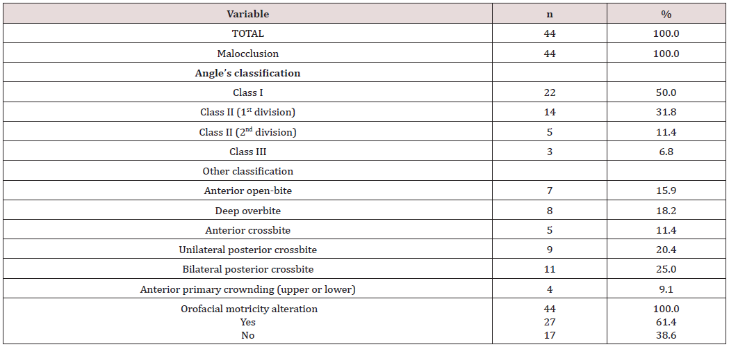
Table 2: Distribution of assessed children patients, according to changes in functional performance, deleterious oral habits and postural habits.
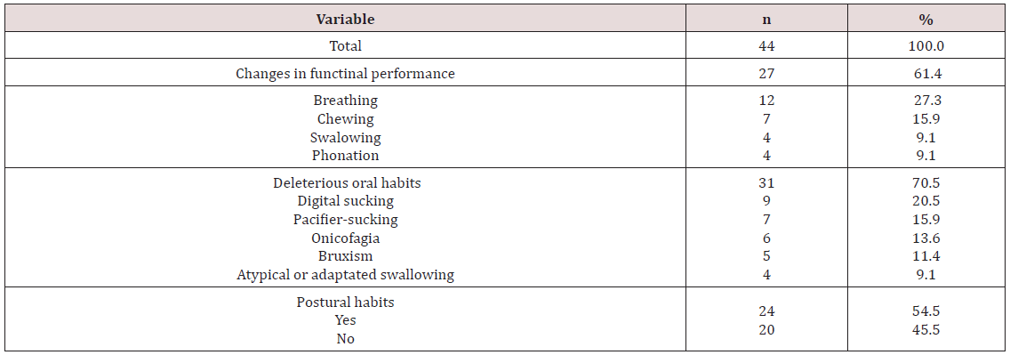
Table 3: Distribution of assessed children patients, according to malocclusion and orofacial motricity alteration.
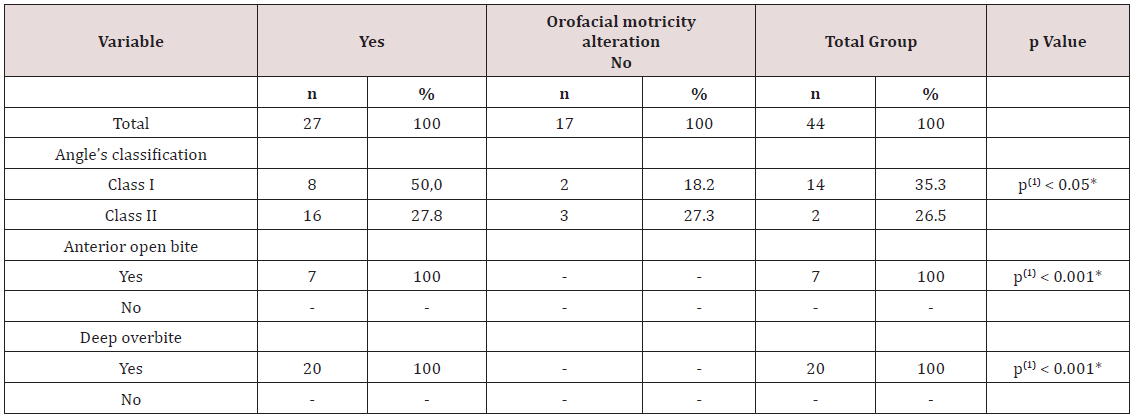
Those last ones included, in descending order: the interposition of the lower lip and/or the tongue between the arches 12 (27.3%), protraction of the mandible 7 (15.9%) and support of the hand in the mandible 5 (11.3 %). Table 3 shows the significant changes (p <0.05) between the presence of alteration in the orofacial motricity and the type of malocclusion. It demonstrates a significant association between the presence of alterations in orofacial motricity and Angle’s Class II malocclusions (when organized as Division I and II), anterior open bite and posterior crossbite (p <0.05), by grouping unilateral and bilateral posterior crossbite for this purpose. Despite the fact that all children with Angle’s Class III malocclusion presented alteration in orofacial motricity, the test could not be established due to the low frequency of these cases, which were found only in 3 patients. Other possible associations were also tested, then it was seen a significant association between the presence of postural habits and the posterior crossbite; followed by the groups of unilateral and bilateral crossbite (p <0.05). The study did not find any significant association regarding the age or gender of the participants (p> 0.05).

Discussion

It has been very difficult to compare this study with others available in the literature. This might have happened due to the lack of studies regarding this content, mainly with the corroboration of both sciences, Dentistry and Speech Therapy, because of their differentiated methodology and approaches. This interaction allows understanding better the structures and dynamism of stomatognathic system as well as the guidelines for future researches. The methodology adopted in this research was based on a secondary data. The information gathered during the actions of the Extension Program was filed in order to be used as data for future projects. It is important to emphasize that, in this specific situation, one of the goals of the Extension Program is to do research, thus the data collected during the activities were already with this purpose. Moreover, the children’s parents signed a Term of Free and Clarified Consent authorizing to use the information for future researches. The investigated sample was a representation of the appointments in the dental clinic, which represents almost half of the patients who were treated in the period considered for this research. It was possible to determine the frequency of orofacial motricity of those who had patients with malocclusions, seeking for a possible association between the variables. On the other hand, it is not possible to compare the children with or without malocclusion, because one of the inclusion criteria was to have any type of malocclusion [9].

Regarding the distribution of malocclusion in the children during the mixed dentition period, the authors recognize this period as the one with important dentoalveolar changes; thus great part of the occlusal problems can be perpetuated in the permanent dentition if no therapeutic conduct is performed as soon as possible, especially in situations of crossbite and open bite [10]. An epidemiological study developed in the city of Lins, State of São Paulo compared the distribution of these problems according to Angle’s classification. The sample was comprised of 734 individuals by age 12. The goal of this research was to evaluate the prevalence of malocclusions as well as the severity and the need of treatment [11]. The authors observed similar results in descending order of Class I, II and III malocclusions. They also found a higher percentage of Class I (55.92%) and Class II (42.86%), with lower Class III (1.22%) as the present study.

When considering other classifications, the cross-bite has a higher frequency. A study [12] demonstrated the importance of early treatment of this common malocclusion in children’s patients. The authors established that is necessary an adequate differential diagnosis regarding its etiology, skeletal, dental or functional impairment. This study also reinforces the high prevalence of unilateral posterior crossbite compared to bilateral; however this characteristic was not seen in this current research. Changes in orofacial motricity were found in the majority of the infantile patients of this study. However, it should be considered that the sample only included children with malocclusions, which could lead to a greater predisposition of disorders mainly in the specialty of Speech Therapy. A similar result was seen in other study, but with lower percentage. For this study, in the city of Santa Maria, Rio Grande do Sul, the authors investigated the presence of speechlanguage disorders in 262 schoolers aged from four to six years old. They also found a higher frequency of alterations in orofacial motricity (31.3%), followed by alterations in speech (21.37%) and language (4.58%) [13].

These alterations can interfere in different ways, for example, the respiratory function can be altered by functional or organic factors. Thus, when the nasal breathing becomes mixed (nasal and oral) or predominantly oral, it becomes anti-physiological, which generates different compensatory changes. The presence of inappropriate postural habits, associated with oral breathing can lead to changes in growth and development of the face and teeth which are in formation. These changes lead to a change in the children’s occlusion pattern and also interfere in the phonetic system [14]. The oral habits are strongly related to the presence of malocclusions, it means that they are a potential factor in the development of alterations of the structures and functions of the Stomatognathic System, depending on the intensity, frequency and duration of the stimulus, besides the genetic predisposition of the individual. In a literature review [15] regarding this content, the open bite was observed as the most associated malocclusion. This condition happens due to mechanical interposition between the upper and lower dental arches, usually by sucking a pacifier or finger. In this current study, postural habit was more frequent in children who grew up as oral breathers, so when the oral habits persist during the child’s development it may provide a pattern of oral breathing, as was found in other studies [6,16].

According to the consulted literature [17]. Regarding the deleterious oral habits, it was found a lower prevalence of digital sucking habits when compared to pacifiers; fact not observed in the present study. It was also noticed a controversy regarding the possibility of similar or greater damages caused by digital suction due to the difficulty of removing this habit. Despite scientific evidence of the association between malocclusion and changes in orofacial motricity, the number of interdisciplinary studies regarding this content is not enough, which explains the difficulty of understanding and having a consistent approach in terms of integral health care to these problems. Even with few articles regarding this content, most of those studies shown a significant improvement in relation to the treatment as well as in the patients’ quality of life, if an early diagnosis and approach occur in the children. However, it is necessary to keep studying this content, not only seeking for an approach with methodological rigor but also comprising a more representative sample of the population so that the results can provide more conclusive information about this subject.

Conclusion

Changes in orofacial motricity were observed in more than half of the subjects, it was also noticed that oral breathing is the most frequent condition. It reinforces the need to integrate actions between Dentistry and Speech Therapy.

Read More About Lupine Publishers Journal of  Dentistry and Oral Health Care Please Click on Below Link: https://lupine-dentistry-oral-health-care.blogspot.com/