Showing posts with label Open access Journal of Orthopedics. Show all posts

Tuesday, 20 September 2022

Lupine Publishers| Primary Cemented Hemiarthroplasty in Unstable Inter- Trochanteric Fracture of Femur in Elderly

 Lupine Publishers| Journal of Orthopedics and Sports Medicine


Abstract

Introduction: Unstable Intertrochanteric fractures are Complex fractures which become more difficult when it’s associated with age related osteoporosis. Internal fixation methods like Dynamic hip screws and proximal femoral nails have lot of complications which adds to the morbidity and mortality of the patient.

Material and Methods: 23 patients aged more than 60 years presenting with unstable Intertrochanteric Femur fracture were operated with primary cemented bipolar Hemiarthroplasty Along with Greater Trochanter and Lesser Trochanter reconstruction with Steel wires or non-absorbable sutures.

Result: The average Harris Hip Score (HHS) at final follow up was 85.24 with 9 excellent, 7 good, 3 fair and 2 poor results.

Conclusion: Hemiarthroplasty in unstable Intertrochanteric fractures is better than other methods of internal fixation in elderly patients as it has less complications and the major advantage of bipolar Hemiarthroplasty is early mobilization and less mechanical complications.

Keywords: Bipolar; Hemiarthroplasty; unstable Intertrochanteric fracture; osteoporosis

Introduction

Around 45% of all hip fractures are Intertrochanteric fractures (IT fractures) [1] and the incidences of hip fractures are still higher in elderly. With increase in life expectancy, there is an increase in population of elderly patients. Osteoporosis which is very common in elderly, is a major cause of fractures following trivial trauma. About 35-40% of Intertrochanteric fractures are unstable [2]. The treatment of stable IT fractures is osteosynthesis with good results but there has been a wide consensus regarding the best treatment modality for unstable Intertrochanteric fractures in elderly patients. The major problem with these fractures is difficulty in obtaining acceptable reduction, poor bone quality and high rates of morbidity and mortality due to various factors especially prolonged recumbency. The primary goal of treatment is stable fixation and early mobilization. The treatment of these fractures evolved from conservative treatment with the skeletal traction to operative procedures like fixed angle blade plates, sliding hip screws and intramedullary devices. [3] Hemiarthroplasty and Total Joint Replacement is mainly used for femoral neck fractures and as a salvage for failed pinning and other complication of primary surgical procedure [4]. osteosynthesis in unstable Intertrochanteric fractures with osteoporosis has failure rate between 4-16.5%. [5] When internal fixation is adopted, the range of general complications such as Deep Venous Thrombosis (DVT), Pulmonary embolism, Pneumonia, bed sores etc. ranges from 22- 50%. [6-7] The primary aim of this study is to analyze the role of primary cemented Hemiarthroplasty in unstable Intertrochanteric fracture of femur in elderly patients.

Material and Methods

The study was done prospectively on 23 patients aged more than 60 years presenting with unstable Intertrochanteric Femur fracture. One of the cases died on 8th month postoperatively and one case was lost to follow up after 4th month postoperatively. These 2 cases were excluded and only 21 cases were included in the study. The fractures were classified according to AO/OTA and Evan’s classification and the fractures with AO/OTA type 31-A2.2 and 31-A2.3 and Evan’s type 3 and 4 were included in the study. Only the patients who were independently mobile before sustaining injury were included in the study were included. Written informed consent was taken from all the patients. The patients who were unfit for surgical procedure or polytrauma patients, open or pathological fractures, old or malunited fractures were excluded from the study. The patients who had any significant rheumatological, neurological or psychiatric disorders were also excluded from the study.

Surgical Technique

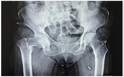
Figure 1: Preoperative X-ray.
Lupinepublishers-openaccess-orthopedics-sports-medicine

Primary cemented bipolar Hemiarthroplasty was done using posterolateral approach in lateral position. The joint capsule opened using inverted ‘T’ capsulotomy incision and then the fracture pattern assessed. The femoral head was then extracted by cutting the head of femur from neck, at sub capital region. Then the Greater Trochanter (GT) and Lesser Trochanter (LT) were temporarily reconstructed according to their comminution and displacement. Then excessive part of neck trimmed, and femoral canal prepared with reamers and broaches. Trial stem was then inserted taking care of appropriate version of prosthesis, by taking the long axis of leg and transcondylar axis of lower end of femur as reference. In severely comminuted fractures, especially in comminuted LT fractures, exact prosthesis height determination was difficult. So, both the knees were brought together, and height of prosthesis was marked. Second generation cementing technique and cement restrictor was used in all the cases. Appropriate prosthesis size selected, and the prosthesis stem was sunk till the previously marked point to avoid limb length discrepancy. Definitive reconstruction of GT and LT was then done using SS wires by Tension Band Wiring and/or encirclage technique. In cases of severe comminution Ethi bond sutures were used to keep Trochanter in place. There were a few cases where LT was severely comminuted, then it was left as it is within the soft tissue and the calcar was made using cement mantle. The sleeves of Gluteus Medius and vastus medialis anchored to GT using bone drills and wires. Short external rotators were also attached. The joint was then reduced; range of motion and stability was checked. Wound is then closed in layers over suction drain. Patients were allowed to sit on 1st postoperative day onwards and standing with support was allowed on 2nd postoperative day. Walking with support was allowed on 3rd –4th postoperative day and walking without support was encouraged and allowed as tolerated by patient. Follow up was done on 2nd week, 4th week, 3 months, 6 months, 1 year and 2 year. On each follow up clinic-radiological evaluation was done and Harris Hip Score (HHS) used for assessment. HHS less than 70 is graded as poor outcome, 70-79 as fair, 80-89 as good and 90-100 as excellent outcome. (Figures 1-3).


Result

Prospective study on 21 patients operated for unstable osteoporotic Intertrochanteric fractures with primary Hemiarthroplasty. The average age of the patients was 70 years and it included 14 male and 7 female patients. The duration of days since injury to presentation ranged between 1 to 13 days, average 4.33 days. The fracture pattern was of AO/OTA type A2.2 and A2.3 and Evan’s type 3 and 4. The average duration of surgery was 89.76 minutes and average intraoperative blood loss was 426.19ml. Postoperatively patient was made to bear weight on 1st day to maximum on 8th post-operative day (POD), average 3.52 days. Walking without support was started on 28th to 62nd day. There were 8 patients which were not able to walk without support. Five of them used stick to walk with limp, 2 used walkers and 1 patient refused to walk at all and was wheelchair bound and had poor result. Remaining 13 patients walked well without any support. The average hospital stay was 9.48 days with maximum of 25 days in a patient who developed bed sore on 13th POD and was advised to stay in hospital for proper nursing care and dressing. There was 1 patient who developed superficial Surgical Site Infection (SSI) which was managed conservatively with antibiotics and dressing and one patient who developed deep SSI on 6th week POD for which revision surgery had to be done with implant removal and re-implantation. There were 5 cases that had limb length discrepancies, 1 had limb lengthening and 4 had shortening which was managed with shoe raise but still continued to walk with limp and used walker/ stick. The average Harris Hip Score (HHS) at 3weeks was 74.29 and at final follow up average HHS was 85.24 with 9 excellent, 7 good, 3 fair and 2 poor results. All the patients were followed up for at least 1 year at maximum for 3 years 5 months; average follow up period was of 26.1 months.

Discussion

Management of Intertrochanteric fractures has always been a challenge. Every case is different, and no single surgical approach is applicable for all the patients. There are a number of surgeries proposed for Intertrochanteric fractures, either osteosynthesis by plates like Dynamic Hip Screws (DHS), intramedullary nails like Proximal Femoral Nail (PFN) etc. or replacement surgeries like Hemiarthroplasty or total hip replacement surgery. DHS and PFN are excellent implants which give very promising results, but all these also have high chances of morbidity due to mechanical failure and prolonged recumbence leading to conditions like pneumonia, bed sores, DVT etc. Since Intertrochanteric fractures are more common in elderly and these old patients are still more prone for such complication. Another challenge in elderly patients is Osteoporosis. Due to severe depletion of bone stock, the fixation is weak, poor screw hold, higher chances of cut outs and implant failure. Osteosynthesis with implants like DHS is easier and has better results with simple 2 parts Intertrochanteric fractures rather than comminuted unstable Intertrochanteric fractures. To avoid such complications in unstable Intertrochanteric fractures in elderly a good alternative is replacement of femoral head and neck with prosthesis, i.e. Hemiarthroplasty. In Intertrochanteric fractures doing Hemiarthroplasty is surgically more difficult as we have to repair Greater Trochanter (GT) and Lesser Trochanter (LT) also with steel wires or non-absorbable sutures but the surgery is very rewarding. In the above study we have done Hemiarthroplasty with Bipolar prosthesis with reconstruction of GT and LT with steel wires or non-absorbable sutures in 21 cases of unstable Intertrochanteric fractures in elderly patients. We had to make calcar with cement in a few cases where repair of LT was not possible.

The average age of the study patients was 70 years (ranging from 61-91 years). The patients presented after an average of 4.33 days after injury (ranging from 1-13 days). Postoperatively the patients were made to bear weight on average 3.52 days after surgery and walking with support allowed from 4th, 5th day onwards, with maximum delay of 28 days in one patient and another one patient refused to walk at all. Majority of the patients started walking after about 4 weeks, with exception of 8 cases which continued to use support for walking. Due to early mobilization of the patient there were very few complications and the hospital stay of the patients also drastically reduced. The average hospital stay was 9.48 days ranging from 7 days to maximum of 25 days in one patient. Study done by Thakur et al. [3] had an average age of study patients of 80.7 years and average duration of injury to surgery was 3 days. The patients were mobile with walker on average 2.6 days and average duration of hospital stay was 17.5 days. Another study done Ahmed Emory et al. [8] had average hospital stay period of study patients of 8.78 days and the mean time interval between injury and operation was 2.9 days. Out of 41 patients, 27 cases were able to ambulate independently using walker and 13 cases need assistance along with walker. Study done by Sachet et al. [9] has Harris Hip Score (HHS) at final follow up 84.8 +- 9.72 (58-97) with 10 excellent, 15 good, 7 fairs. 2 poor and 1 failed result. Choy et al. [10] in his study had mean HHS at last follow-up 80.6+- 9.3, with excellent in 8, good in 19, fair in 9 and poor in 4 out of total 40 cases, i.e. better than fair results in 67.5% cases. In our study final mean HHS was 85.24. out of total 21 cases, 9 had excellent results, 7 good, 3 fair and 2 poor results, i.e. 76.19% had better than fair results. In our study one patient had developed bed sore on 13th post-operative day (POD), which healed with nursing care and dressing. This patient refused to walk and had poor results. There was one patient who developed superficial surgical site infection (SSI) which was managed conservatively with dressing and antibiotics and one patient developed deep SSI on 6th week POD for which revision surgery was done with implant removal and reimplantation. This patient was a known diabetic with improper control of blood sugar. There were no cases of dislocation or loosening. Similar study done by Nikunkj et al. [1] on 28 patients of Intertrochanteric fractures had 1 patient with superficial SSI, 1 patient with deep SSI, 1 case of acetabular erosion, 4 patients of nonunion GT and 2 patients with GT cerclage wire breakage. There was 1 patient with periprosthetic fracture 6 months after surgery. Overall, he had mean HHS 84.8 +- 9.72 at final follow up. Another study done by KV Puttakemparaju et al. [11] on 20 cases had 1 case of deep SSI for which implant removal was done, 1 case of grade 1 bed sore which healed with dressings and antibiotics. There was no dislocation, rotational deformities or subsidence of stem. Gashi et al. [12] in his study compared outcome of primary cemented bipolar Hemiarthroplasty with DHS in elderly patients with unstable Intertrochanteric fracture and concluded that early mobilization was significantly better in Hemiarthroplasty than DHS. General and mechanical complications were more common in DHS group. At final follow up mortality rate didn’t differ in between the two groups but mean HHS was better in Hemiarthroplasty group. Comparative study done by Mohamad Emmi et al. [13] concluded that HHS (86+-9 vs. 75+-7.6), range of flexion (105+- 11o vs. 90 +- 17o), external rotation (35 +-7o vs. 20+- 7o) were significantly higher in bipolar group compared to DHS group (P<0.05). However, there were no significant differences in pain severity in both the groups. Yee Suk Kim et al. [14] in a comparative study found that elderly patients with AO type A2 Intertrochanteric fractures, patients treated with Hemiarthroplasty were able to perform early ambulation. However, no significant difference in operative time, amount of postoperative transfusion, clinical results, hospital stay, and radiological failure rate was observed between bipolar Hemiarthroplasty and compression hip screw fixation groups. Study done by Sameer Ajit Mansukhani et al. [15] on DHS, Cemented bipolar Hemiarthroplasty and PFN for treatment of unstable Intertrochanteric fractures found that mean intraoperative blood was significantly less with PFN as compared with other two, whereas there was no significant difference in mean hospital stay and intraoperative time of all the three groups. There were more early complications seen in DHS group as compared to PFN and Hemiarthroplasty. Late complications such as femoral head AVN and implant cut outs were higher in PFN group whereas chances of hip dislocation were higher in Hemiarthroplasty patients. Mortality at 12 months was highest after bipolar Hemiarthroplasty and least after DHS. Reoperation rates were highest for PFN as compared to other two groups. On functional assessment unaided walking was best with DHS whereas other parameters didn’t have any significant difference. He concluded that Treatment of unstable Intertrochanteric fracture of femur is a matter open to debate. Intertrochanteric fractures of elderly must be treated with considering the age of the patient, mental status, bone quality, and the type of fracture. It is certain that the main objective is to prevent the possible complications by early mobilization and to help the patient to return to their daily life. Kayali et al. [16] in their study found that cone Hemiarthroplasty can be an alternative treatment for unstable Intertrochanteric fractures in elderly patients so as to achieve earlier mobilization. The limitations of our study were that the sample size was small and there was no comparison of bipolar Hemiarthroplasty with osteosynthesis surgeries like DHS and PFN. The study period was not too long and so long-term complications like hip osteoarthritis, loosening, protrusion, stem failure etc. cannot be assessed.

Conclusion

Bipolar Hemiarthroplasty is a reasonably good alternative in unstable Intertrochanteric fractures in elderly patients who have severe osteoporosis. The major advantage of bipolar Hemiarthroplasty is early weight bearing and less short and midterm complications. Internal fixation is a preferred especially in young patients with stable Intertrochanteric fractures but have high complication rates in elderly with unstable fractures due to prolonged Recumbency and severe osteoporosis.

Read More About Lupine Publishers Journal of Orthopedics and Sports Medicine Please Click on Below Link: https://lupinepublishers-orthopedics.blogspot.com/

Tuesday, 9 August 2022

Lupine Publishers| Rehabilitation Following UCL Repair with Internal Brace

 Lupine Publishers| Journal of Orthopedics and Sports Medicine


Introduction

Elbow injuries in the overhead athlete, particularly baseball pitchers, continue to increase in frequency because of extreme repetitive valgus stress [1-4]. This repetitive stress results in ulnar collateral ligament (UCL) insufficiency which produces elbow pain, medial joint laxity, and an inability to throw.16 Pitchers are the most injured players in Major League Baseball and elbow injuries account for 22-26% of pitching injuries [1,5,6]. The risk factors related to sustaining a pitching related UCL injury are pitching when fatigued, a high pitch volume, improper mechanics, and repetitive throwing at maximal effort in the youth player and throwing a high number of pitches at peak velocity in the professional athlete [7]. The current preferred surgical treatment for most UCL tears that fail conservative management is a reconstruction using one of several autogenous grafts. [8] Extensive follow-up data on UCL reconstructions with a minimum 2-year follow-up shows that just 83% of the athletes undergoing reconstruction were able to return to the same level of play or higher and that on average return to competition took 11.6 months [9].

However, recent technological advances have sparked renewed interest in repair of the UCL augmented by an internal brace (Internal Brace; Arthrex Inc) in a search for a surgical option that would allow a faster recovery than what is typical following UCL reconstruction. Repair of the UCL with internal brace is a direct repair of the native ligament with a spanning tape dipped in collagen (Internal Brace) anchored on each end of the UCL [10]. (Figure 1) Two 3.5mm Swive Locks spanned with a 2mm piece of Fiber Tape (Arthrex, Inc. 1370 Creekside Blvd., Naples, FL, 34108) and size 0 nonabsorbable sutures are used to repair the native ligament back to its anatomic origin and insertion ensuring that tension of the Fiber Tape matches that of the native UCL during range of motion (ROM). A UCL repair with internal brace is reserved for use in cases of partial or complete tears at the origin or insertion of the UCL with good ligament tissue and low-grade mid-substance partial UCL tears [10]. In patients with chronic, attritional damage to the UCL and associated loss of elbow joint stability, reconstruction remains the most appropriate surgical intervention [8,9,11,12]. The decision to perform a surgical repair of the UCL, rather than a reconstruction, can only be made intra-operatively from direct visual assessment of the UCL.


Rehabilitation Guidelines

Rehabilitation after UCL repair with internal brace surgery is accomplished via a sequential and progressive 5 phased approach, designed to return the athlete to their previous level or higher as quickly and safely as possible [13-15] (Table 1). Initially rehabilitation interventions are designed to minimize the effects of immobilization, facilitate early healing of the UCL, re-establishing pain-free ROM, reduce pain and inflammation, and retard muscular atrophy. Early limited passive elbow/forearm ROM exercises and grade I/II joint mobilizations are incorporated in conjunction to neuromodulate pain, promote articular cartilage nutrition and aide in the synthesis, alignment, and organization of collagen tissue [16-19], [20-25]. Local modalites, including Cryotherapy, electrical stimulation and Class IV deep tissue laser are used to control pain, inflammation, speed healing of the incision and increase nitrous oxide in the healing tissue [26]. Pain free, submaximal isometrics are used to initiate muscle activation and retard atrophy for all planes of elbow, forearm, wrist and shoulder movements. Shoulder external rotation (ER) and internal rotation (IR) isometrics are performed with caution and must be completely pain free. Rhythmic stabilization and neuromuscular control drills for shoulder, elbow and wrist along with seated scapular and postural exercises are also introduced early in the rehabilitation process.

The controlled mobility phase runs for a total of 3 weeks starting at the second week after surgery and focuses on a stepped restoration in elbow ROM (outlined in Table 1), improved muscular strength/endurance, and normalizing joint arthrokinematics. Active-assisted, active, and passive ROM exercises, as well as more aggressive joint mobilizations, are all incorporated for the elbow, forearm and wrist with the primary goal to achieve full elbow extension and minimize the risk of developing an elbow flexion contracture [26-29]. Elbow flexion contractures are the most common postoperative complication following elbow surgery and must be diligently avoided. At any sign of flexion contracture, we find using a low load–long duration (LLLD) stretch in conjunction with joint mobilization and stretching to be extremely beneficial for regaining full elbow extension. A light resistance exercise band (Theraband CLX Performance Health, 1245 Home Ave, Akron, OH 44310) is applied to the wrist and used to place a LLLD stretch on the anterior elbow structures for 12-15 minutes, for a total of 60 minutes a day. (Figure 2) Strengthening exercises at this point are performed beginning with concentric and progressing to eccentric muscle contractions with the focus placed on a comprehensive strengthening program for the throwing athlete, such as the Thrower’s Ten Program [30,31].

The intermediate phase is from postoperative week 6 to 8 and emphasizes the maintenance of joint mobility, improving muscular strength, endurance, neuromuscular control of the elbow complex, and continuing with a functional progression of activity. Stretching, flexibility and mobilizations are used to maintain full motion with a particular focus on elbow extension and forearm pronation flexibility. At 4 weeks the athlete is progressed to the advanced thrower’s 10 program to place greater demands on the posterior shoulder and scapular muscles [32]. Neuromuscular control manual resistance exercises are incorporated for both the shoulder and elbow, proprioceptive neuromuscular facilitation, rhythmic stabilizations, and slow reversal hold techniques. 2-handed plyometrics are introduced 6 weeks following surgery progressing to 1-hand exercises 2 weeks later. The fourth phase of UCL repair rehabilitation is the advanced phase which runs from weeks 9 to 14 and is specifically designed to increase strength, power, endurance, and neuromuscular control to prepare for a return to sports using strengthening activities that emphasize high speed, eccentric contractions, and plyometrics. Elbow flexion exercises here emphasize high speed eccentric control training elbow deceleration. Weight machine exercises are begun 10 weeks after surgery and include, seated chest press, seated rowing, and front latissimus dorsi pull-downs. A hitting program is permitted at week 10 and an interval throwing program 11 weeks after surgery if the athlete meets the objective criteria for throwing. Reinold et al. [33] provides the best description of sports specific interval programs. Pitchers generally are able to advance to throwing off of a mound 8 weeks after they begin a throwing program.

The return to activity phase is the last part of the process and emphasizes a proper dynamic warm-up, continued exercise loads and managing the progression back to unrestricted activity and competitive throwing [34,35]. The general time frame to return to play following a UCL repair with internal brace is approximately 5 months. Functional testing can aide the return to play decision process. We use the prone ball drop test, developed by the senior author (KEW) which utilizes a 1kg (2 pound) plyoball with the patient prone, shoulder abducted to 90˚, and elbow extended. The patient is instructed to perform as many ball drops and catches as possible in a 30 second timeframe, comparing successful cathese bilaterally seeking a goal of 110% for the throwing side (Figure 3). At our center, 350 UCL repairs with internal brace have been performed. Of these, 1-year follow-up data is available for 79 throwers, showing 98% of the 1-year follow ups returned to their pre-injury level of activity.

Summary

The UCL is a frequently injured in overhead athletes and these injuries continue to climb in number in youth athletes. Surgical repair of the UCL with internal brace is a viable option in athletes who meet specific findings at the time of surgery. The rehabilitation of this unique surgical procedure has been presented based on our experience treating in excess of 350 athletes over the past 3 years. The average time required for an athlete to return to participation in our cohort is 7 months which is approximately 5 months less than average return to play times after UCL reconstruction surgery. Long-term results of this surgery and rehabilitation program are still needed but our initial experience is extremely promising.

Read More about Lupine Publishers Journal of Orthopedics and Sports Medicine Please Click on below Link: https://diabetes-obesity-lupine-publishers.blogspot.com/

Monday, 10 January 2022

Lupine Publishers| Two Trajectorieess a Promise of Reform and Ashaaram Pattern what are we Missing?

 Lupine Publishers| Journal of Orthopedics and Sports Medicine


Abstract

The working of social and economic institutions, inequality is also a product of the way political institutions work. Government has known to serve the interest of economics elites and this most often works to the detriment of common people. This refers to the article ‘’Two trajectorieess’’ A Promise of reform and Ashaaram Pattern, what others do. A molar leadership deficit, our political leaders choose not to speak on certain issues. But they are forgetting that they have a molar responsibility towards domestic violence testing among divorced women begins privately manage pharmaceutical intuition associated to slums occupied area in Pune University, India. The horrific incidence in kathua and unnao has shown.com the ugliest side of the country’s politician. The right procedure is not followed for the enquiry of the domestic violence and his contention by government police. The term intimate partner violence is often used synonymously with domestic abuse or domestic violence, but it specifically refers to violence occurring within a couple relationships (i.e., marriage, non-cohabitating intimate partners). To these, the World Health Organization (WHO) adds controlling behaviours as a form of abuse. Intimate partner violence has been observed in opposite and same-sex relationships, and in the former instance by both men against women and women against men.

The high risk of experiencing domestic violence among divorced women in pharmacy institution in India who associated in slum occupied underscores the need for effective, instructional-based and standard behaviour culturally primary prevention. To inform such domestic violence primary prevention strategies for this population, author herein aimed to identify correlates of domestic violence testing in divorce women. Utilizing a cross-sectional design, potential correlates of domestic violence experience were explored among a geographically-clustered random sample of speak up few known women recently-divorce women residing in private pharmacy institution in slums in Pune, India. In domestic violence testing was associated with less educational attainment by the participant’s, less satisfaction of the job with the respect they earn, if not given’’ at the time of college hours, poorer living of life style, unhealthy food and conflict skills, and greater acknowledgement of domestic violence occurrence in college and friends. These connection suggest strategies that could be incorporated into future domestic violence early prevention interventions for this vulnerable population (i.e. promoting completion of formal education of men alongside women, migration causes of institution harassment, abuse language, more personal, skills, and challenging norms surrounding domestic violence add or before inspect the pharmacy institution.

Introduction

Women who have experienced intimate violence disease are at greater risk for physical and mental health problems including posttraumatic stress disorder (PTSD) and dependency. Most women get a severe stroke and the impact of all may compound these costs. Researchers have reported that women with these experiences are more difficult to treat; many do not access treatment and those who do, frequently do not stay because of difficulty maintaining helping relationships. However, these women’s perspective has not been previously studied in close monitoring in privately manage pharmaceutical institution [1,2].The purpose of this study is to describe the experience of seeking help for divorcées women dependency by Indian pharmaceutical institution to avoid the present violence. Women with dependency with a history of depression want help however the health and social services do not always recognize their calls for help or their symptoms of distress. Dependency thickens and stiffens walls, which can inhibit quality of life and will power, confidence.

Perception of alcohol use among well–Educated employees of pharmacy institution before and after the office hour in slums Pune Suburban has conflict. Little is known about how the overall employment conditions in a country impact the likelihood of employment of privately running out pharmacy institution in Pune University India [3,4]. Correlation of intimate domestic violence has aware of physical, sexual, psychological abuse, and control perpetrated against an intimate condition, is reported and prevalent globally accepted. Approximately one third of divorcee women reporting physical and/or *abuse by their head of the department during their office hour. Not only is an abuse word a violation of human rights that often results in physical injury. Divorcee women who experience domestic violence have higher odds of depression (measure face reading , body sacred, appearance, lower standards of dress, anxiety and other mental health disorders [5,6] true principal health, burn out stages and gynaecologic morbidity, rashes, sores, or lesions in the mouth or nose, or under the skin among other chronic disease states which is not known. Additionally, their children suffer from greater morbidity and mortality. In India, although national estimates suggest decreasing frequency, one in three women still report having been abused by their head of the department during their lifetime. Further, this figure is likely an underestimate of the abuse women suffer post-marriage with little hesitation, as it did not survey violence perpetration by the in-law or other members who believes traditional cultural draws. Divorcee women who reside in privately manage pharmacy institution in pune university India’s slums are among those at greatest risk of domestic violence.

While the disparate figures between slum- and non-slum occupied communities may be in sum art factual due to shameinduced underreporting in higher income communities, factors that drive increased domestic violence perpetration and compel women to remain in abusive relationships are likely disproportionately greater in slum communities. Women in slum communities may be more likely to experience domestic violence because their reporting officers and related to inadequate finances, crowding, and poor sanitation, discrimination, and subordination, reside in communities where normalization and acceptance of Domestic violence is greater, alcohol use is greater, perception has not known, have weakened support systems that do not allow them to develop and exercise positive coping mechanisms, no longer yoga knowledge, weaken immunity profile [7]. Further, in Pune slum communities, at the time of appointment, many women transition from unclear to join the intuitions (no appointment letter produce to employee) and newly enter the slums from surrounding rural areas; thus, the differences in upbringing within the couple may also influence illegal expectations and prompt conflict. Further, divorcee women residing in slums may be more likely to stay in abusive relationships because of poorer knowledge, skill and physiological and mental unawareness to support services, NGO’s, organisation head dependency systems, stronger perceptions of hopelessness and surrender, and residence in environments where domestic violence and other forms of violence occurs with frequency and acceptance. The risk imposed by these factors is compounded by local sanctions that encourage divorcee women to weaken ties with natal family members and their community post-marriage, that limit the time the couple spends together alone to develop their relationship both pre- and post-marriage, and external pressure on the couple (i.e. fertility].

Thus, domestic violence prevention for women residing in slum communities requires a culturally-educated, communityeducated approach that recognizes the structural factors of slum environments pharmaceutical institutions that shape domestic violence risk. Further, given the high domestic violence burden and limited and saturated support resources, focus in resourcelimited sittings should be on primary prevention. National evidence suggests that almost two-thirds of divorcee women who report domestic violence, state the abuse had begun within the first two years of job, underscoring the need for such prevention efforts to occur pre- or immediately post-marriage. To date, few studies have examined drisk factors for domestic violence experience among women residing in slum communities in India. T hose who have, identified the following risk factors: age, low educational attainment of self and spouse, young age of marriage, having a legal versus illegal, additional dowry request from marital family, employment, employment status, residence in a joint family, renting versus owning one’s residence, fewer class rooms in the institution and shared rooms, accepting attitudes toward women beating, shorter duration of marriage, and women alcohol dependency cannot ignore. And although causal directionality could not be established, one cross-sectional study among slum-dwelling women found participation in social groups and vocational training to be associated with domestic violence experience, perhaps because participation challenges social norms. Of note, none of these studies specifically examined correlates of violence in early stage, critical for primary domestic violence prevention. As part of formative work that led to the development of an intervention for the primary prevention of domestic violence for newly-wed couples residing in Pune slums, we aimed to explore correlates of domestic violence experience among recently-married women.

Methods

This study was considering it is fundamental characteristics and importance of present situation and instruction at all levels of our educational systems, from pre-primary to graduate.

Study Design

The study was conducted in Pune university pharmacy institutions, the second largest city in the western state of Maharashtra, India. According to most recent data from world university ranking. The study employed a cross-sectional design, wherein semi-structured interviews were conducted during the academic year 2016-2018. Interviews were conducted one-on-one in privately by trained female study staff in Marathi. Participant recruitment and enrolment. To be eligible for the study, participants had to be: a divorcee woman over 18 years of age; recently divorce; in a first marriage; in a second marriage.

Data Collection

Sample has collected to speak up methodology and data was selected using a muster name and item semi-structured questionnaire administered one-on-one in private by a trained female study team member.

Participant and study team safety

The study protocol was developed using the guidance of the AICTE and safety recommendations for research on violence against women.

Discussion

This study is the first to report correlates of domestic violence experience in early marriage among women residing in slums linkage private pharmaceutical institutions in pune university, India. We identified nine key potential domestic violence correlates: Teachers are not allowed to give physical punishment to the students. If seen strict action will be taken. During assembly nobody will be allowed to enter the school, the teacher should stand behind the respective classes. No P.F will be deducted from teacher salary i-e compulsory. Defence of unqualified pharmaceutical confidentiality that must be stop. Mobile phone should be kept in the office while signing the attendance register and collect it while leaving the school. Performed such other duties as may be not prescribed. No personal work is allowed during school time. Fourth Saturday of the month is holiday. Poor transportation facility.

Acknowledgment

This study has been guided by under supervision and guidance of Renowned Laboratory Scientist Respected Dr. Ramesh Paranjape’ Retd. Director and Scientist ‘G’ National AIDS Research Institute India. I express my sincere gratitude towards Respected Sir’ for motivation and being great knowledge source for this research. This work was supported by Award Number R.S.S.M./447/2017 Rastriya Samta Swatantra Manch. The content is solely the responsibility of the authors and does not necessarily represent the official views of the Rastriya Samta Swatantra Manch. Thanks go to grammar teacher who reviewed the manuscript grammar.

Read More About Lupine Publishers Journal of Orthopedics and Sports Medicine Please Click on Below Link: https://lupinepublishers-orthopedics.blogspot.com

Monday, 11 October 2021

Lupine Publishers| A Rare Case of Fracture Dislocation of Hip: Is There a Pip kin's Type 5?

 Lupine Publishers| Orthopedics and Sports Medicine


Abstract

Fractures of the femoral head associated with neck fracture and posterior dislocation of the hip are uncommon. Pip kin and later Brumback classified these complex fracture dislocations. However in today's era patient have more atypical injury pattern. We encountered a case which has never been described in literature and cannot be fit into any previously described classification system. We present the radiological details and management.

Keywords: Femoral Head Fracture; Trochanter Fracture; Posterior Dislocation; Acetabular Fracture; Sciatic Injury; Pipkin's Types; Total Hip Replacement

Introduction

Modern day accidents not only produce unpredictable but at times unclassifiable injuries. Fractures of the femoral head associated with posterior dislocation of the hip are uncommon as such [1]. Pip kin was the one who sub classified Epstein-Thomas type V fracture-dislocations into four additional subtypes which is still most widely followed [2]. Later Brumback et al. further classified femoral head fractures emphasizing hip stability, with type "B" injuries being unstable [3]. The paucity of these cases is the main constraint to make an algorithm of management. Nevertheless the results depend on early anatomical reduction with the potential threat of osteonecrosis irrespective of approach [4,5]. We hereby describe a case of posterior dislocation of hip which is novel and never been described before. The purpose is to make the fraternity aware of such a case which is unclassifiable even though the management protocol may not be grossly different.

Case

A 52 year old migratory laborer had an accident at civil construction site where he fell down from a height of 20 feet landing first on knees (in kneeling position). He presented to emergency department with severe pain in right groin, and inability to move the left lower limb. On examination the limb was shortened, externally rotated and a bony mass felt in the loin which did not move with the movement of distal thigh. There was severe tingling and decreased sensation in front and lateral aspect of leg and patient was unable to dorsiflex his ankle and toes. His pulsations were normal but he had had transient hypotension with tachycardia which corrected with initial resuscitation. First X ray showed fracture about right hip (Figure 1) mostly a trochanteric fracture but a careful evaluation showed incongruity of head and a "vacant" on super lateral acetabulum. A CT scan was followed wherein the fracture was better delineated. A posterior dislocation of hip with fracture about the trochanter was seen. The head was also fractured and the infra foveal part could be seen lying the acetabulum. A fracture line also ran through the posterior wall of acetabulum which was not more than 25% of the wall and also was undisplaced (Figure 2a- 2c). The patient was admitted and limb placed in Thomas splint and prepared for next morning. No attempts to reduce the dislocation were made.

Under general anesthesia, the patient was positioned laterally and a Moore's posterior approach was taken guided by dislocation and wall fracture. The gluteus maximus had a huge rent and external rotators were torn, the head was seen indenting the sciatic nerve (bowstring effect) which was contused but intact (Figure 3a). The neck was ostetomised with help of saw at the appropriate level and the calcar was reconstructed using encirclage wiring (Figure 3b). Ethibond was used to suture the greater trochanter. Next 2 temporary K wires were used in the ace tabular walls and reaming done and cement less cup size 52 was fixed augmented with 2 screws (Figure 3c). The K wires were removed after the cup was found stable. The femur was prepared and uncemeneted corail stem size 11 was inserted and size 36x0 femoral head was found stable. C arm was used to confirm the placement of implants (Figure 3d). All soft tissues were closed in best possible way, the hip was stable (Figure 3e). An abductor brace was applied post operatively. Postoperatively day one faradic stimulation was started and patient was allowed side tuning. Check X ray was done which showed a reasonable reconstruction (Figure 4). The patient was allowed sitting with non weight bearing mobilization from day 3 after the pain had subsided. 3 weeks after surgery the patient was discharged when he went back to his home state and never came back for follow up.

Discussion

Femoral head fractures in combination with posterior dislocation of the hip are rare presentation. Since first reported by Birkett in 1869 only a small number of cases have been reported world widen [6]. Epstein et al.'s found about 10% in their series. It was Pip kin who subs classified these fractures and various other classifications have been proposed by several authors who claim to be improved version [5, 7]. Pipkin's classification remains most popular and widely accepted. They divide them into 4 types with type 3 having associated ace tabular fractures and type 4 having associated neck fractures. Our case is unique with fracture in the trochanteric area. The trochanteric area has never been described. The ace tabular fracture is also an addition actually a combination pattern of Pipkin's 3 and 4. We believe that there should be a type 5 including a both neck and ace tabular fracture in association of head fracture and a subtype T wherein fracture configuration is in trochanteric region instead of classical neck.

The cases of type 3 or 4 are very sporadic for any protocol to be made. In younger individual attempts to fix the neck fragment, often augmented with vascularised fibular graft has been made in past but long term follow up is lacking. Osteonecrosis is a complication and surgeons have tried different approaches with varying results [8,9]. In our case closed reduction was not possible and moreover the impeding ischemia to sciatic pushed.com for an early operative intervention. Taking into consideration- a difficult fracture pattern, age, urgency of surgery, peripheral set up hospital and an invincible avascular necrosis; we thought a Total hip arthroplasty (THA) was an appropriate solution than open reduction. Yet the challenges of choosing the right implant from the whole Pandora box of prosthesis implants remained. In surgery, the aim was to stick to principles of bone preservation like calcar reconstruction and thus avoiding the distal fixation stems which were reserved for future revision if a situation arose. A constrained hip would have more advantages in this circumstance of soft tissue rent, but the large head option of 36 was stable enough in our case. We admit management to be debatable with expertise hands. Final outcome does depend on return of sciatic function and proper occupational therapy since he was a laborer. A draw back in our report remains that despite best attempts to persuade the patient to visit nearest ortho clinic; he never turned up but telephonically did tell to having a over the counter sitting job in the village and satisfied with his hip.

Conclusion

The case highlights a rare injury and attempts to give an expansion to the present Pipkin’s classification. Even though the demerit of this report is a lacking follow up X ray and clinical picture, the initial presentation is worth to be noted among the practioners and scholars.

Read More About Lupine Publishers Journal of Orthopedics and Sports Medicine  Please Click on Below Link: https://lupinepublishers-orthopedics.blogspot.com/

Monday, 30 August 2021

Lupine Publishers| Incidence of Patella Alta in Anterior Knee Pain- Assessment with Patellar Height Ratio's

 Lupine Publishers| Orthopedics and Sports Medicine Open Access Journal (OSMOAJ)


Abstract

Background: Altered patellar alignment is associated with anterior knee pain and multitude of conditions that affect the patellofemoral joint. The study aim was to report the incidence of patella altaand patella baja in indian population with anterior knee pain and to investigate whether the normal limits of the patellar height ratios are applicable in indian population.

Methods: Lateral radiographs of 528 knees with anterior knee pain were performed. Patellar heights were measured to calculate the Insall-Salvati (IS), Insall-Salvati Modified (MIS), Blackburne-Peel (BP), Caton Deschamps (CD) ratios.

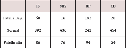
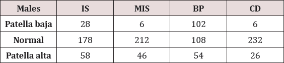
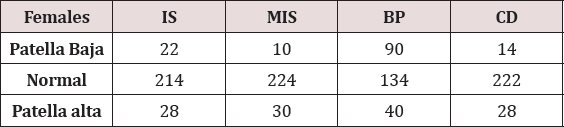
Results: The overall mean LT/LP ratio was 1.03 (SD 0.37) with incidence of 16.2% (86/528) for patella alta with Insall-Salvati. Mean MIS was found to be 1.68 (SD 0.018) and incidence of patella alta as per MIS ratio was 14.3% (76/528). Blackburne-Peel and Caton-Deschamps ratios revealed mean ratios of 0.88 (SD 0.15) and .99 (SD 0.16) and the incidence of patella alta 17.8% with BP ratio and 10.2% with CD ratio respectively.

Conclusion: Incidence of patella alta was found to be 17.8% with BP ratio, 16.2% with IS ratio, 14.3% with MIS ratio and 10.2% with CD ratio. The BP and IS ratio were the most sensitive with MIS ratio being the more specific one and CD ratio the least sensitive.

Keywords: Insall-salvati ratio; Modified insall-salvati; Blackburne-peel; Caton deschamps; Patellar alignment

Introduction

Anterior knee pain remains one of the commonest musculoskeletal complaint with patients, and evaluation of patellar height is done commonly as many conditions are associated with abnormal patellofemoral relationship. The lever arm function of the patella plays an important role in knee extensor mechanism and improves the quadriceps strength by 30-50% [1]. The joint reaction force of the patellofemoral joint varies with the patellar height. A high riding patella or patella alta is associated with patellofemoral malalignment and a reduced patellofemoral contact often leading to patellofemoral pain or instability [2-3]. A low riding patella or patella baja is often associated with limited knee range of motion, patellofemoral arthritis and Osgood Schlatter disease [4]. Numerous methods of patellar height have been described in the literature: Blumensaat [5] Insal-Salvati [6], modified Insal-Salvati [7], Blackburne-peel [8], Caton-Deschamps [9], DeCarvalho [7] and Koshino [10]. These ratios are based on the ratio of the patellar length to the reference point from the tibia. A very few studies have compared the different indices for the patellar height analysis regarding their reproducibility and reliability.

The study objective was to analyse the commonest methods for measuring the patellar height in patients with anterior knee pain and the aim of this study was to report the incidence of patella alta and patella baja and investigate whether the patellar height ratios have significant variations in adult indian population in which sitting on the ground, kneeling, and squatting is common.

Materials and methods

528 lateral x-rays of the knee (212 male and178 females) were collected between the time period August 2017 to February 2018 from patients with anterior knee pain, with difficulty or pain on squatting or sitting. Patients with underlying knee pathologies, knee deformities and knee surgery were excluded. Institutional review board approval was obtained prior to conducting the study. Lateral radiographs of the knee set in 30° of flexion were taken with assistance of a goniometer. 30° of flexion of knee results in better visualization of the tibial insertion. Radiographs were perpendicular to the film and centered on the joint at a distance of 100cm.

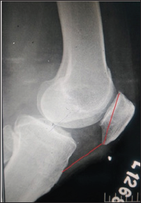
Figure 1: IS.

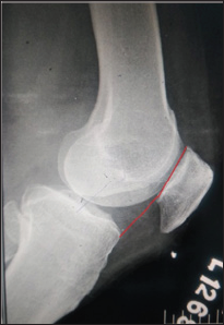
Figure 2: CD.

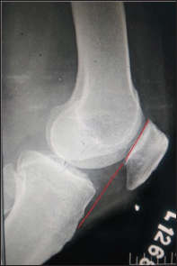
The measurements were made with the digital imaging software (Radiant diacom viewer) by a single experienced physician. The patella length was measured from the superior articular pole to the inferior non articular pole (Figure 1). The distance from the origin of the patellar tendon at the inferior pole of patella to its insertion on the tibial tubercle was taken as the patellar tendon length. The Insall-Salvati index is calculated as ratio of LT/LP, where LT is the length of the patellar tendon and LP is the patella length (Figure 1). The Blackburne-Peel index (Figure 2) is calculated as PP/PG where PP is the perpendicular height of the distal part of the joint surface of the patella to a line projected anteriorly to the surface of the tibial plateau and PG is the length of the articular surface of patella. The ratio PTG/PG, in which PTG is the distance from the lower edge of the articular surface of the patella to the anterosuperior angle of the tibia, and PG is the length of the articular surface of the patella is calculated as Caton-Deschamps index (Figure 3). The Modified Insall-Salvati index consists of the ratio PT/PG, wherein PT is the distance from the lower edge of the joint surface of the patella to the insertion of the posterior or deep surface of the patellar tendon in the tibial tubercle, and PG is the length of the joint surface of the patella (Figure 4).

Figure 3: BP.

Figure 4: MIS.

Results

Table 1.

The overall mean LT/LP ratio was 1.02 (SD 0.37). Comparison between genders revealed that the mean LT/LP ratio was higher in males than females with a mean of 1.04 (SD 0.29) and 1.01 (SD 0.46), respectively (Table 1 & 2). Using criteria of defining abnormal patellar position (1.00±20%) based on Insall's study, the overall incidence was 16.2% (86/528) for patella alta (Tables 3 & 4). The mean PT/PG ratio was 1.68 (SD 0.018). The MIS ratio was higher in males with mean of 1.70(SD 0.29) than females with a mean of 1.66 (SD 0.29). The incidences of patella alta as per MIS ratio were 14.3% (76/528). Blackburne -Peel and Caton- Deschamps ratios revealed mean ratios of 0.88 (SD 0.15) and .99 (SD 0.16). Males were found to have a higher mean in both the ratios compared to the females (BP ratio: 0.89, SD 0.12 and CD ratio: 1.0, SD 0.18 in males and BP ratio: 0.88, SD 0.05 and CD ratio: 0.98, SD 0.06 in females). The incidence of patella alta were 17.8% and 36% with BP ratio and CD ratio respectively.

Table 2.

Table 3.

Table 4.

Discussion

Vast majority of the studies in literature are often quoted with the data obtained from the Caucasian population. There are none, if at all few studies of patellar height being done in Asian population. There are morphological changes in patella of the Caucasian and the Asian population which make the patellar height ratios even more significant. The present study is an observational study done with aim of assessing the patellar alignment in anterior knee pain patients.

The position of the patella has an important role on patellofemoral function. Abnormalities in patella position have thus been associated with anterior knee pain and many extensor mechanism disorders. While many techniques have been developed to measure patellar position such as the Blacburne's ratio, the Insall Salvati ratio still remains one of the most popular, largely because it is easy to use and remember [7,8] .Despite its popularity, recent studies have suggested that the current normal ranges should be extended, as the ratios may not be applied to ethnicities outside western regions [11,12].

The Insall-Salvati (IS) method uses the length of the patellar ligament in relation to the length of the patella6. The patellar morphology and morphological differences in the anterior tuberosity of the tibia (ATT) directly affect the measurements made using this method. Grelsamer and Meadows [7] developed the modified Insall-Salvati (ISM) method based on the length of the joint surface. Difficulty in identifying this parameter is considered to be the main measurement bias. The Modified Insall -Salvati ratio has its advantages over the IS ratio that it can efficiently find out the patella alta in patients with small patellar articular surface which is not taken into account in IS ratio (Figure 5). Digital radiography seems not to present greater details for this anatomical reference. The Blackburne-Peel (BP) method exchanges the reference point of the ATT for the joint surface of the tibial plateau, while keeping the joint surface of the patella. Although Berg et al. [13] found that this was the most accurate and reproducible method in conjunction with the IS index, and Seil et al. [13] ranked it as the second most reproducible method in conjunction with the IS index, we did not obtain similar results in our analysis, such that it was only better than the ISM index. Lack of definition of the reference line of the tibial plateau, such as which condyle to use as the reference, or whether this line runs parallel to the joint surface or perpendicular to the long axis of the tibia, contributes towards lower concordance with this method. The method of Caton et al. [2], which uses the joint surface of the patella and the angle of the tibial plateau as references, also presents difficulty regarding identification of the joint surface, as well as presenting a certain amount of variability in defining the angle of the tibial plateau. Despite these factors, this method was the one that showed greatest concordance in the study by Seil et al. [13].

Read More About Lupine Publishers Orthopedics and Sports Medicine Open Access Journal (OSMOAJ)  Please Click on Below Link: https://lupinepublishers-orthopedics.blogspot.com/