Lupine Publishers | Journal of Gynaecology and Women's Healthcare
Abstract
Background Adenomyosis is a frequent gynecological disease of unknown etiology causing menstrual pain disorders and pelvic congestion, that definite histopathological based diagnosis of adenomyosis relies on the presence of endometrial glands and stroma within the myometrial tissue, junctional zone assessed sonographically could elucidate the nature of disease progressive pathological changes.
Aim: To investigate the value and usefulness of Junctional Zone indices in suspicion and diagnosis of adenomyosis disease in correlation to histopathological findings
Methodology: A clinical research trial conducted on 200 research study subjects scheduled for hysterectomy procedure due to abnormal uterine bleeding and/or dysmenorrhea unresponsive to medical treatment performed at Ain Shams University maternity hospital from January 2018 till March 2019, all research study subjects have undergone two‐ and three‐dimensional transvaginal sonography before the day of surgery.
Results: There was statistically significant difference between research groups (Adenomyosis of the inner myometrium, Serrated junctional zone, Linear junctional zone,) as regards 2 D features anechoic lacunae, asymmetric corpus myometrium ,myometrial cysts, fan shaped shadowing, mean number of 2D features (p values <0.001), concerning 3 D features Mean JZ max, Mean JZ diff, JZ interruption, Sub endometrial lines and buds, mean number of 3D features (p values <0.001).
Conclusion: Junctional zone changes could be denoting early phases of adenomyosis disease development furthermore the 3D sonographic features implemented were considered and shown to be more valuable in elucidating the pathological changes confirmed by histopathological examination.
Keywords: Adenomyosis; Sonographic; Endometrial lining; Myometrium
Introduction
Adenomyosis is a frequent gynecological disease of unknown etiology causing menstrual pain disorders and pelvic congestion ,being heterogeneous in nature from being focal or simple to severe or diffuse affecting the whole myometrial tissue mass .Sonographic features revealed by 2D and 3D sonography are considered elucidating tools aiding in the diagnosis of the clinical condition however research interests are rising to correlate the sonographic criteria to histopathological and clinical criteria of adenomyosis [1,2]. Junctional zone representing an interface between the endometrial lining and myometrium is considered a cornerstone sonographic tool when properly applied in 3D and 2D sonography technologies to elucidate the severity and distribution patterns of adenomyosis [3,4].
Interestingly prior research studies have revealed and displayed that the pathophysiological origin of adenomyosis starts by repeated traumas to the junctional zone, making the anatomical and histological boundaries between the endometrium and myometrial tissues weaker causing subsequent progression of the disease by successive menstrual cycles [5,6]. On the other hand, it is crucial to mention that definite histopathological based diagnosis of adenomyosis relies on the presence of endometrial glands and stroma within the myometrial tissue. on the contrary other researchers have mentioned that endometrial‐sub endometrial myometrial zone disruptive issues observed by imaging techniques e.g. as junctional zone thickening, disruption or infiltration, should be considered a separate category than adenomyosis. Interestingly pathologists were not capable to clearly discriminate between the junctional zone and the outer myometrial layer further more by light microscopic examination, the muscle fibers directions of the inner and outer myometrial zones could show variability [7,8].
Aim
To elucidate and investigate the value and usefulness of Junctional Zone indices in suspicion and diagnosis of adenomyosis disease in correlation to histopathological findings using 2D and 3D transvaginal sonographic imaging
Methodology
A clinical research trial conducted on 200 research study subjects scheduled for hysterectomy procedure due to abnormal uterine bleeding and/or dysmenorrhea unresponsive to medical treatment performed at Ain Shams University maternity hospital from January 2018 till March 2019, all research study subjects have undergone two‐ and three‐dimensional transvaginal sonography before the day of surgery. Junctional zone maximum thickness, junctional zone maximum irregularity and sonographic features of adenomyosis have been compared with the junctional zone histopathology described as follows
a) Adenomyosis within the inner myometrium, ≥2mm myometrial invasion without contact to the basal endometrial layer,
b) Serrated junctional zone, >3mm myometrial invasion with contact to the basal endometrial layer or
c) Linear junctional zone, no or marginal myometrial invasion ≤3mm with contact to the basal endometrial layer.
Exclusive research criteria was as follows Uterine volume above 300mL due to fibroids, the existence of four or more fibroids, malignant disease, sonographic examinations involved the usage of Voluson E8 Expert machine using a multifrequency trans vaginal probe (6‐12MHz) sonographic maximum thickness measurement of the anterior and posterior uterine walls and existence of any myometrial lesions (fibroids and adenomyosis characteristics) have been recorded defined and measured. Adenomyosis features implemented were as follows. Heterogeneity, myometrial cystic lesions (>1mm), anechoic lacunae (<1mm), shadowing being fan shaped and asymmetry of myometrial corpus. JZ was evaluated in all three planes (sagittal, transversal and coronal) and measured as the maximum (JZmax) and minimum (JZmin) JZ in each wall. Anterior and posterior uterine walls have been measured using the sagittal or transverse planes and the fundal and two lateral walls were measured using the coronal plane. The walls with the largest junctional zone maximum thickness and junctional zone difference measurements have been implemented to determine peak JZ irregularities.
Uterine specimen histopathological examination
The uterus was divided into two halves immediately after performance of hysterectomy procedure fixed using formalin before histopathological examination with a slice thickness of 1‐1.5cm all over the uterus. Slices were examined in the beginning macroscopically. Followed by detailed by microscopic examination comparison with normal myometrial tissue was performed with macro and microscopic examination.
Statistical analysis
Research data were collected, revised, coded and entered to the Statistical Package for Social Science (IBM SPSS) version 23. The quantitative research data were presented as mean, standard deviations and ranges while qualitative variables were presented as number and percentages. The comparison between groups regarding qualitative data was done by using Chi-square test. Also the comparison between more than two independent groups with quantitative data and parametric distribution was done by using One Way ANOVA. The confidence interval was set to 95% and the margin of error accepted was set to 5%. So, the p-value was considered significant at the level of < 0.05.
Results
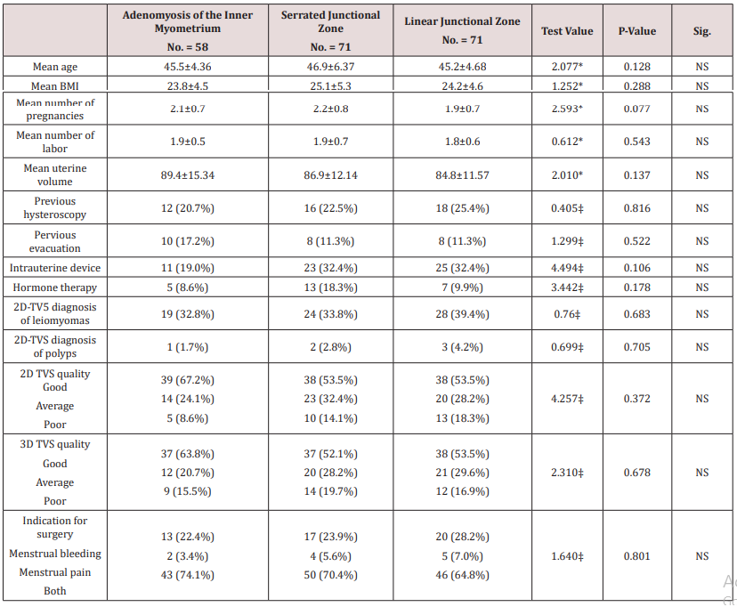
Table 1 reveals and displays demographic research data of the three research groups categorized according to presence of adenomyosis within inner myometrium (n=58), serrated junctional zone (n=71), linear junctional zone (n=71)in which there was no statistical significant difference as regards mean age, BMI, number of pregnancies, number of labor, mean uterine volume ,previous hysteroscopy ,previous evacuation ,intrauterine device ,hormone therapy, 2D-TV5 diagnosis of leiomyoma’s, 2D-TVS diagnosis of polyps, 2D TVS quality, 3D TVS quality, Indication for surgery (p values=0.128,0.288, 0.077, 0.543, 0.137, 0.816, 0.522, 0.106, 0.178, 0.683, 0.705, 0.372, 0.678, 0.801 consecutively).
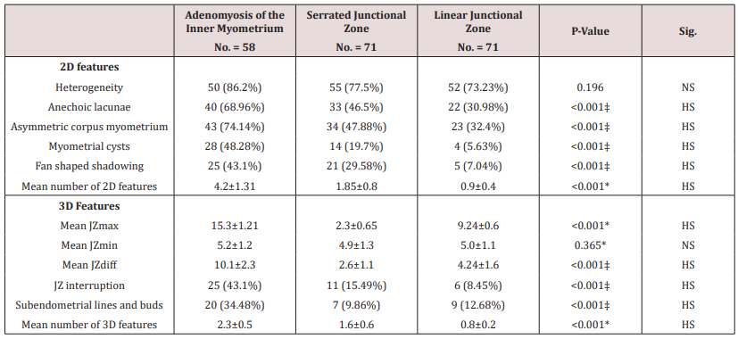
Table 2 reveals and displays that there was statistically significant difference between research groups (Adenomyosis of the inner myometrium, Serrated junctional zone, Linear junctional zone,) as regards 2 D features anechoic lacunae, asymmetric corpus myometrium ,myometrial cysts, fan shaped shadowing, mean number of 2D features(p values <0.001),concerning 3 D features Mean JZ max, mean JZ diff, JZ interruption, sub endometrial lines and buds, mean number of 3D features (p values<0.001).
Discussion
Adenomyosis a frequent uterine pathology that is usually diagnosed in a definitive manner after hysterectomy reveal the requirement and necessity to understand its imaging features from early to late stages of pathological development that could aid in enhancement of management protocols for those category of cases permitting early definitive management that could prevent progression of the pathological disease process [9] Adenomyosis is a pathological insult that could represent in focal or diffuse manner leading to pelvic congestion issues and uterine bleeding disorders affecting the woman’s quality of life. Implementing advanced 3D sonographic technology with junctional Zone measurements could enhance the clinical diagnosis however still that requires extensive research efforts to elucidate the normal anatomical and histological changes of junctional zone in correlation to adenomyosis disease [10,11].
The current research study recruited 200 research study subjects undergoing hysterectomy procedures due to uterine bleeding issues unresponsive to conservative measures and all research study subjects had gone through extensive and meticulous 2D and 3d sonographic assessment before the hysterectomy procedure the research team of investigators have revealed an displayed the following research results in which there was statistically significant difference between research groups (Adenomyosis of the inner myometrium, Serrated junctional zone, Linear junctional zone) as regards 2D features anechoic lacunae, Asymmetric corpus myometrium ,Myometrial cysts, fan shaped shadowing ,mean number of 2D features(p values <0.001),concerning 3D features Mean JZ max, Mean JZ diff ,JZ interruption, Sub endometrial lines and buds, mean number of 3D features (p values<0.001.
In a prior research study have revealed and displayed that, a high percentage of cases having severe menstrual Bleeding issues and/or dysmenorrhea scheduled for a hysterectomy or had considerable junctional zone changes [12].Another research team of investigators have shown that around one‐third of cases having adenomyosis within the inner myometrial zone have junctional zone serration [13,14]. Interestingly in harmony with the current research study findings a prior research team of investigators revealed a strong statistical correlation between junctional zone changes and abnormal uterine bleeding disorders [1,4,12]. Even though it was displayed by other research studies contradicting to the current research findings that junctional zone serration can be a normal histological finding due to physiological changes in multigravida cases [3,9,10].
Furthermore, another research study similar to the current research study in approach and methodology have shown that 3D transvaginal sonography observed a statistically significant correlation between junctional zone increased thickness and irregularities and adenomyosis disease within the inner myometrial zone [8,14]. Another interesting issue observed and mentioned from previous research study findings that there is raised rates of false‐positive sonographic diagnosis of adenomyosis disease using 2D than with 3D‐ trans vaginal sonography, particularly within cases showing junctional zone serration those research findings could be justified by the fact that 3D‐ trans vaginal sonography is superior in elucidating junctional zone measurements precisely than 2D trans vaginal sonography. 7 However prior research groups have revealed that there should be a discrimination between junctional zone diseases and abnormalities and adenomyosis however junctional zone changes could reflect adenomyosis at early phases of pathological course of development [11].
Another prior research study have implemented histopathological based categorization using 3mm endometrial invasion depth as a cutoff value to denote junctional zone abnormalities furthermore junctional zone thickening could be a pathological responsiveness to the endometrial invasiveness consequently thickening of junctional zone usually co-occurs with interruption of junctional zone integrity, sub endometrial sonographic lines and budding [5,9,13].
Conclusion and Recommendations
The current research study reveals and displays that the junctional zone changes could be denoting early phases of adenomyosis disease development furthermore the 3D sonographic features implemented were considered and shown to be more valuable in elucidating the pathological changes confirmed by histopathological examination. However, the future research studies are required to be multicentric in fashion putting in consideration racial and ethnic differences and the presence of various uterine volumes to aid in future development of clinical imaging guidelines to suspect and diagnose adenomyosis at early stages enhancing the level of clinical care offered for those categories of cases.
Read More about Lupine Publishers Journal of Gynecology Please Click on Below Link:
https://lupinepublishers-gynecology.blogspot.com/


No comments:
Post a Comment
Note: only a member of this blog may post a comment.