Wednesday 31 August 2022

Lupine Publishers | Risk Assessment of Type 2 Diabetes Among Population of Rawalpindi Pakistan

 Lupine Publishers | Journal of Nursing & Health Care


Abstract

Objective: To determine the risk for developing type 2 diabetes among population of Rawalpindi city.

Subjects & Methods: A cross-sectional descriptive study was conducted among 90 healthy attendants of the patients admitted in wards of Holy Family Hospital, Rawalpindi to assess their risk for developing type 2 diabetes. Data was gathered through consecutive sampling. All study participants knew they did not have any type of diabetes and they were not receiving anti-diabetic drugs. The data was collected during 2 weeks in July 2018 by means of structured questionnaire. The data was analyzed by using SPSS version 25.0

Results: Of the total 90 study subjects, 57 were females. Mean age of respondents was 44.6±5.78 years. About 10% respondents had Body Mass Index (BMI) greater than 35kg/m2 and had very high risk of developing type 2 diabetes within next 10 years. Daily consumption of fruits and vegetables seemed to have statistically insignificant relationship (P>0.20) with reduction in risk for type 2 diabetes. Only 01 respondent out of those physically active at work was at high risk for type 2 diabetes. About 27.8% respondents had positive family history. Risk of developing type 2 diabetes was insignificantly associated with gender (P>0.94). Overall, only 6 respondents predominantly females 55-64 years old had high to very high risk of developing type 2 diabetes.

Conclusion: Majority of the study participants had low to slightly elevated risk of developing type 2 diabetes. This risk can better be eliminated by lifestyle modification.

Keywords: Type 2 Diabetes; Risk Assessment; Body Mass Index; Family History

Introduction

Type 2 Diabetes is a chronic metabolic disorder showing enormously raised prevalence worldwide. Number of people affected by this epidemic is expected to double in next decade [1]. This disease is incurable. However, various treatment modalities endorsed are lifestyle modifications, reducing obesity, intake of oral hypoglycemic drugs and insulin sensitizers [1]. Type 2 Diabetes is among the greatest public health threats associated with drastic escalation of its incidence globally [2]. Type 2 Diabetes ought to investigate in overweight adults of any age with history of one or more risk factors [3]. This disease is attributed to amalgamation of numerous environmental and genetic risk factors [3]. However, investigation of type 2 diabetes should commence at age of 45 among people not having relevant risk factors [4].
Type 2 diabetes is found to be prevalent in certain races like African Americans, Hispanics and Native Americans who are more susceptible to diabetes than Caucasians [5] Even victims are unaware of their disease due to mildness of associated symptoms [5]. WHO has regarded population of developing countries as more prone to develop type 2 diabetes [6]? According to International Diabetes Federation Report 2015, 415 million people are suffering from this disease globally and about 642 million people are expected to be victimized by 20406. Proportion of diabetics is likely to double in near future primarily due to increased life expectancy and urbanization irrespective of the prevalence of obesity [7]. However, there is likelihood to arrest this increase by lifestyle modifications [8]. A systematic analytical study carried out among Pakistani population in 2015 showed 11.8% people with type 2 diabetes and situation was expected to be grave with passage of time [9]. The present study is intended to assess the risk factors for type 2 diabetes among Pakistani population specifically of Rawalpindi city to identify the risk factors for this disease. This research will provide useful information to our policy makers for strategic planning in this concern.

Subjects and Methods

A cross-sectional descriptive study was carried out among healthy attendants of the patients admitted in wards of Holy Family Hospital, Rawalpindi to determine their risk of developing type 2 diabetes. Information was gathered from 90 attendants not suffering from type 2 diabetes through consecutive sampling during two weeks in July 2018. Confirmed diagnosed cases of type-II diabetes were excluded from this study. The information was collected from study participants by using Type 2 diabetes risk assessment form designed by Prof. Jaakko, Department of Public Health, University of Helsinki. Data was collected from study participants pertinent to their demographic profile, BMI, waist circumference, physical activity, dietary habits, family history and relevant health profile. Waist circumference was measured by means of inch tape and weight was measured by weight machine. Data was analyzed by using SPSS version 25.0. Frequency and percentage were calculated for all variables. Gender based risk of type 2 diabetes was statistically confirmed by applying Fisher’s Exact test. Statistical association of type 2 diabetes risk with regular consumption of fruits and vegetables was verified by chi-square test. P-value≤0.05 was taken as significant.

Results

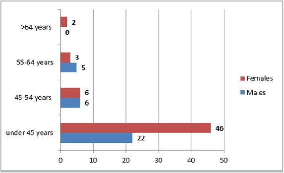
Figure 1: Age wise gender distribution of study participants (n=90).

lupinepublishers-openaccess-nursing-journal

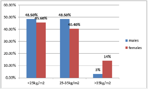
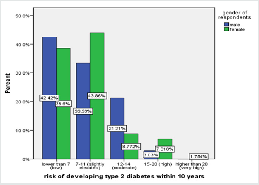
Majority of study participants (63.33%) were females. Mean age of respondents was 44.6±5.78 years. Most of the males (48.5%) were observed to have waist circumference less than 94cm while majority of female respondents (43.9%) had 80-88cm waist circumference. About 75.6% study subjects were under 45 years of age and only 2.2% respondents were above 64 years old. Age based gender distribution of the study subjects is shown in Figure 1. Fruits, vegetables, and berries were consumed every day by 44.4% of study subjects. Moreover, daily consumption of fruits and vegetables revealed statistically non-significant relationship (P>0.20) with reduction in risk of type 2 diabetes. Most of the females in comparison with males had BMI greater than 35kg/ m2 as depicted below in Figure 2. Out of 9 respondents with BMI>35kg/m2 only 3 were determined to be at high to very high risk (Risk score 15-20) of developing type 2 diabetes. Out of 90 study participants, 31.6% females had waist circumference more than 88cm while only 18.2% males had waist circumference more than 102cm. About 42% of females and 67% of males were found to have 30 minutes of daily physical activity at work. Only 1 study subject out of those engaged in daily physical activity at work was found to be at high risk of developing type 2 diabetes within 10 years as depicted below in Table 1. Only 16.7% respondents were taking medication for hypertension regularly while only 12.2% participants gave history of hyperglycemia during their lifetime out of which 54.5% were males. According to type 2 diabetes risk assessment scale designed by Prof. Jaakko, risk of developing type 2 diabetes within next 10 years among study subjects is illustrated below in Figure 3. However, risk of developing type 2 diabetes was found to be insignificantly associated with gender as depicted below in Table 2. 27.8% respondents had their immediate family members suffering from type 2 diabetes while 37.8% subjects had no relevant family history.

Figure 2: Gender Based BMI of study participants.

lupinepublishers-openaccess-nursing-journal

Figure 3: Gender based risk of developing type 2 Diabetes within 10 years.

lupinepublishers-openaccess-nursing-journal

Table 1: Risk of developing type 2 Diabetes in relation to physical activity (n=90).

lupinepublishers-openaccess-nursing-journal

Table 2: Association of Gender with risk of developing type 2 diabetes within 10 years.

lupinepublishers-openaccess-nursing-journal

Discussion

The prevalence of diabetes mellitus is estimated to rise from 2.8% in 2000 to 4.4% in 2030. This means that about 366 million world population will be diabetic by the end of next 10 years [10]. This prevalence is more likely to grow exponentially in third world countries [11]. In response to the current scenario, implementation of primordial preventive measures to control this modern epidemic is the need of time. In present study, low risk of developing type 2 diabetes within next 10 years was found among 36 (40%) study subjects whereas high to very high risk was found among only 6 (7%) respondents out of which 5 were females. Another Asian study carried out in 2016 on 150 urban slum developers using Finnish Diabetes risk score showed 11.3% people at high to very high risk of developing type 2 diabetes within next 10 years [12]. The reason for low risk in current study might be inadequate sample size (90). Real picture could better be achieved by conduction of research on more individuals. The current study showed high risk (1%) of developing type 2 diabetes among 5% of study subjects found to be physically inactive and were taking medication for high blood pressure. Similarly, an Indian study showed about 11% risk of developing type 2 diabetes among 53.3% study subjects found to be physically inactive [12]. Both research showed proportionate relationship of lack of physical activity/exercise with risk of developing type 2 diabetes. Although social and print media is playing marvelous role in getting our people aware of diverse physical activities in accordance with their circumstances and workplace but apart from physical fitness people should also be sensitized specifically for their wellbeing. This study concluded high to very high risk of developing type 2 diabetes among 7% respondents with their positive family history, while a study by [13] among population of Bangladesh revealed 47.7% of participants with positive family history [13]. A similar Brazilian research carried out in 2013 reflected that 47% of respondents had positive family history of type 2 diabetes [14]. Contrary to international research, current study is depicting raised percentage of positive family history among Pakistani population. This factor can better be scrutinized by conduction of research on large number of individuals.

About 14% female study subjects in current study had BMI>35kg/m2. Another research revealed higher risk of type 2 diabetes among females due to greater tendency of putting weight among them. This feature is significantly attributed to variations in sex hormones in addition to genetics, familial tendency, and lifestyle [15]. There is need to do rigorous research on this aspect across countries by eliminating confounders to determine scientific association of obesity with hormones. According to our study, 5 females out of 6 were determined to be at high to very high risk of developing diabetes. Contrary to these results, an international study concluded the high risk for type 2 diabetes among males [16]. The reason for this variation might be the racial difference and social set up. However, these factors need in depth insight to reach the accurate conclusion. The present study revealed that consumption of fruits and vegetables is insignificantly associated with protection from risk of developing type 2 diabetes (P>0.20). Likewise, a study by [17] portrayed insignificant association (P>0.20) in this regard [17]. This aspect entails detailed elaboration of associated factors like intake of fatty foods and protein diet apart from consumption of fruits and vegetables. Our results showed that only 15 respondents were regularly taking medication for high blood pressure out of which 5 subjects were at high risk of developing type 2 diabetes within next 10 years. On the other hand, a prospective study carried out among hospitalized patients of Bulgaria revealed higher risk for developing type 2 diabetes among hypertensive patients [18]. As current study is cross-sectional descriptive and carried out among non-diabetics, this might be the reason for less individuals at high risk for developing type 2 diabetes within next 10 years.

Conclusion and Recommendation

High risk of developing type 2 diabetes was found among females 55-64 years of age with very high BMI, no physical activity and specifically with diabetic history of immediate family members. Considering study results, risk of developing type 2 diabetes in the community can be minimized by increased physical activity and weight reduction However, these study subjects should regularly be followed up for development of type 2 diabetes.

Read More About Lupine Publishers Journal of Nursing & Health Care Journal Please Click on Below Link:
https://lupinepublishers-nursing-healthcare.blogspot.com/

Tuesday 30 August 2022

Lupine Publishers| Plastic in Brick Application

 Lupine Publishers| Journal of Civil Engineering and its Architecture


Abstract

This paper outlines the utilization of municipal plastic waste (MPW) in construction industries. Both the MPW and the construction industries are increasing rapidly and world’s recycling rate of either Municipal Solid Waste (MSW) or MPW specifically is still low. Production of bricks is non-eco-friendly and a waste generating process because of the greenhouse gases released. Utilizing MPW as construction materials especially in production of bricks is one of a promising step towards a sustainable resources and waste management. Plastic waste can substitute either partially or completely one or more of the materials in brick production. Further research based on recent research and a better understanding in utilization of plastic waste in brick is needed to produce a high durability and quality of bricks as well as to achieve the optimum balance in all aspects especially in terms of cost and functionality.

Introduction

Urbanization caused a vast and rapid growth of construction industries which requires a lot of building materials that utilizes natural resources either in their production plant or as the materials itself. More recently the world concern about the demands for construction materials and the rate production of plastic that increases swiftly every year [1]. In turn, both industries contribute in increasing the MSW. Since the rate of production is projected to double the value in every 10 years, a more sustainable and safer way is needed to be taken into action [2]. Banning or minimizing plastic usage is not practicable to solve the problem as it is nearly impossible for different sectors to run efficiently without plastic. Mining of natural resources on the other hand is an energy waste process as only 900 million tons of raw materials is produced from 6000 million tons of waste generated [3,4]. They may be differ in constitution of raw materials but posses the same in contributing to various environmental threats. Hence, utilizing plastic waste in brick production can solve both the MPW and demands for construction materials. Previous studies showed the possibility of using plastic waste in bricks application but the bricks produced are still lacking of durability as a safe construction materials. The aim of this paper is to review the application of plastic waste in bricks.

Review of Research on Application of Plastic Waste in Bricks

The table layout the use of plastic in manufacturing bricks. Plastic waste has been utilized in various extend producing various properties of bricks and the properties of bricks produced has been assessed following standards provided Table 1.

Table 1: Application of plastic in brick

Lupinepublishers-openaccess-journals-Civil-engineering-Architechture

Discussion

Previous studies showed the possibility of using plastic as binder with the aid of catalyst through depolymerization of PET to replace cement. Extensive studies of PET as aggregate is more common in compared to other plastic waste. From the table, it showed a significant decrease in compressive strength is observed for more than 50% replacement of binder with PET waste. In fact that PET is thermoplastic, poor bond is created between the matrix and the aggregates and over stress may result in debonding of bricks and structural failure [21]. Increasing the amount of PET increases the softening point of the bricks produced. However, if the amount of plastic waste which acts as binder is too low, the residual unfilled voids will increase and in turn, increasing the water absorption as well as decreasing its compressive strength. When plastic waste is used as a mold, it can be clearly seen that it can withstand a high compressive strength in compared to using it as binder. However, lacking of compaction of plastic waste in plastic waste bottle may result in entrapped air that will contribute to a low compressive strength to the structure. A slight amount of entrapped air is enough to disrupt the structure of the bricks. Incorporating plastic waste as an aggregate is more common in compared to other methods. A few other materials are added to increase the durability of the bricks produced. Using an optimum amount of plastic waste as an aggregate generally shows an acceptable compressive strength. Expanded PS has a flexural properties that is important for buildings built on a problematic soils as the incorporation of expanded PS reduces the cracks appearance on building structure. Normal bricks do not posses this properties. In addition, bricks that are incorporated with expanded PS as aggregate are lighter and it can retain its strength due to its less permeable properties that helps to inhibit hydration. When PVC is used as aggregates, a low compressive strength can be observed and cracks were rapidly observed around the PVC particles because of the modulus mismatch as PVC posses a lower elastic modulus in compared to the cement. The durability of plastic bricks depend on the amount of plastic incorporated in the mixture. An extensive reviews for the study of waste in construction has been highlighted [21,22]. With the right formulation, plastic waste can be a great substitution for raw materials extracted from natural origin.

Conclusion

A few conclusions can be drawn based on the review of studies on application of plastic in bricks;

a) A variety of plastic waste has been used in many ways in bricks production. The compressive strength of the bricks produced comply the standard outlined, which is more than the acceptable range outlined.

b) A suitable proportion between plastic waste and other materials used need to be optimized to meet the standard outlined for manufacturing of bricks. Further research and development is needed to improve the quality and durability of plastic bricks.

Read More About Lupine Publishers Journal of Civil Engineering and its Architecture Please Click on below Link: https://lupinepublishers-civilengineering.blogspot.com/

Monday 29 August 2022

Lupine Publishers| A Standard Pediatric Dental Clinic

 Lupine Publishers| Journal of Dentistry and Oral Health Care


After 22 years of experience in pediatric dentistry, as well as working in various pediatric clinics and collecting materials over the years a standard pediatric dentistry clinic can be designed and launched. Therefore, a standard design and decoration of a clinic will have a significant impact on the success of the clinic.

Dentistry decoration is very important and influential in the morale of patients, especially in children; and it should be designed in such a way to give patients a sense of relaxation and comfort. In order to be successful in designing a dental office environment, you need to know enough about the science of color and lighting design and the correct layout of the necessary equipment in the office space, so you can design a stylish and functional decor for dentistry, especially for children. In the design of the interior decoration of the office, you have to master the internal and external environment of the building, the principles of lighting, and the proper layout of medical equipment, so that you can make the right design using the correct and integrated design.

Remember to use the rules of the architecture of the day in the world as much as possible to design interior decoration so that with the help of these sciences and laws you can design a clinic that is very special and relaxing. Choose the effects of colors in terms of color and design that make patients feel more comfortable. Nowadays, dentists use a lot of ways to attract customers. One of them is to put a staff member to advertise their work and explain the services and benefits they offer as well as someone who invites you to visit different parts of the clinic like a tour to explain the various functions of each space. Many modern dental clinics are equipped with amusement and recreational facilities, specially designed children’s reception areas and even relaxation spaces. Adaptive space design elements such as ceiling height, doors, woodwork s, lighting, and painting must all be carefully designed to create a favorable atmosphere for referrals patients.

Clinic Spaces

Have you ever thought that what kind of spaces should be used by a standard dental clinic? In other words, what specific rooms do you have in your clinic? Generally, each pediatric dental clinic should be equipped with eleven completely separate spaces.

a) Receptions area and File archive

b) Waiting room for parents

c) Management room

d) Play room

e) Dentist’s private office

f) Examination room

g) Treatment room

h) Radiography room

i) Central Sterilization center

j) Dental lab

k) Supporting department

Reception area

The admission section is the first space that visitors enter, so it can be of great importance to design. Because patients will judge and view details of the entire project when they enter the reception area. Therefore, the design of this space should be such that it attracts them. In general, the admission section should convey the sense of inviting and welcoming. For the office clerk, who is responsible for accepting patients and archiving file an appropriate place must be considered for her. The admissions section should be such that the patients sit on one side and the office staffs stay on the other side. At the same time, visitors can talk to the staff at the reception desk and fill in the necessary forms.

Waiting room for parents

The second part should be considered for patients who come to the dentist’s office. Pediatric patients are better off staying in their waiting room before the treatment begins. Many modern dental clinics are equipped with a laptop and mobile phone charging stations at the coffee shop, which they can take advantage of these during their children’s treatment. Some clinics also have foot massage devices, as well as a place to show new oral and dental products, all of which are an example of this new facility. In some clinics, there is a special area to use for the aquarium. The installation of a special shelf for a variety of magazines and books can give parents entertainment. It is better to have separate bathrooms for children and adults in the corner of the hall.

Management room

In the design of the management room, the goals are determined by types of decorations and designs. The management room should represent the personality, authority, ability of a good manager. In fact, this decoration tells the patient that the manager has his own rules. When entering the room, the patient must understand that it can be trusted by the manager, regardless of the type of contact, the appearance of the manager, the arrangement of the furniture and any details about it.

In addition to the type of office arrangement, one should also be sensitive to the colors used. Never use fancy colors for interior design. Colors with a cream range and a combination of cream colors with white and light brown are the most suitable colors for the interior design of office chairs. The interior lighting of the room is also very important. It’s better to use modern lighting to make the management room decorations more attractive.

Play room

When designing a dental clinic for children, by installing posters of popular children’s characters on the wall or putting toys in the waiting room, you would encourage them to come to the clinic. If your clinic has a great deal of space, with a number of small play tables, you can create an environment where children can draw and play together. The use of creative children’s designs and bright and cheerful colors in the interior decoration of the dentistry takes the child’s mind out of the atmosphere of the dental space. For the kid’s waiting room, you can use children’s cartoon images and characters, in the form of posters on the walls; or have a shelf of full of toys to play. In this environment, children will be away from the noisy environment and adults’ complaints; and they will be more comfortable with their peers in the room, with no fears and concerns. Installing DVDs or TVs to play animations is another important thing that can entertain children. If the waiting room is large enough, considering a playground is a very interesting and tempting idea to attract children.

Dentist’s private office

The private office is becoming increasingly important in design with the belief that it is important for the dentist to have “private” areas to rest there during the day for mental breaks. It is better to have a comfortable sofa, or even beyond that a recliner chair equipped with a massage device in the room so that remove fatigue from the dentist’s body. Large windows always give e sense of peaceful atmosphere and make the psychological break more relaxed and smooth. Consider a private closet and bathroom to accompany the office.

Examination room

It is designed for initial patient examinations. Of course, in most clinics, this room has been integrated into the treatment room due to lack of space. But for a pediatric dental clinic, the separation of such a room is essential for children, because for the initial examination it is not necessary for the child to go to the treatment room and have too much anxiety. It should be noted that this is the first place a child gets familiar with the dentist. In addition, this room should create a feeling of a happiness and childish atmosphere in the child. In the examination room, we have to try to create a stressfree space where the child would feel more comfortable. A dental environment should be a happy and comfortable place for children. Therefore, the principle design of this room is very important for the initial impact on the child.

The examination room and treatment room to some extent in terms of instrumental layout and decoration arrangement should be different. Having options such as colored dresses or dresses with children’s favorite cartoon characters, masks and colored gloves in this room, will have an impressive effect. It would be more impressive if pediatric dental chairs are characterized by different special childish designs.

The walls of the examination room should be in bright colors and decorated with special posters for kids. Near the dental chair, there should be oral hygiene kits including baby doll toothbrush, children’s toothpaste, and a dent form. On the dental chair, devices such as kid’s dental turbines, kid’s handpieces with a rubber tip on it, and small monitors to be installed. By doing this strategy, you can prepare a good background for children to familiar with the environment and some of the dental instruments.

Treatment room

The next space includes the patient treatment ward. The dental chair in the treatment room is a device that the patient undergoes for the duration of the treatment while at the same time tolerates dental panic and anxiety. A dental unit with a variety of instruments, such as the turbine, handpieces, suction, lamp, etc. may be daunting for children who are going to the dental office for the first time. For a pediatric dental clinic, this room is one of the most important parts of the clinic. This room is a place where the child should not get anxious and stressed when arriving. Therefore, it must be designed and installed in such a way that the patient feels calm and comfortable. The atmosphere inside the treatment room should be cheerful and childish. Dental units with pediatric designs should be replaced by the conventional units. The color of the room and the units should be cheerful and relaxing. In the decoration and design of this room, there must be a special creativity that upon the arrival, the child does not have the feeling to be taken to the dentist’s office.

Instead, he will have the feeling that he has entered into a friendly and childish environment or imaginary space. To reduce the stress on children, place a dental chair in front of your office window. You can bring mental relaxation to your treatment room if you follow these principles:

1) If the office room is on the ground floor and adjacent to the courtyard, you can use full-glass windows: and plant all kinds of greens and flowers in the garden opposite these windows.

2) If the room is on the floor of a building with its window opening to the leaves of the trees, you can give a Stunning and tranquil view to patient’s eyes on the dental unit.

3) But in some cases, your office may be in the basement of a building and the room where the unit is to be installed does not have a window; in this case, you can hang a beautiful, relaxing painting in front of the dental chair

You can even have an aquarium with different fish in front of your unit. Various studies have shown that water and slow motion of fish can be effective in reducing anxiety and stress in patients. As another step, you can put headphones on your unit for the patient, and play light and relaxed music; or, let the children’s music spread through the speakers with a gentle tone of voice. Sometimes the installation of a television in front of the unit can also be effective because the patient’s mind focuses on other programs and images. And their minds divert the issue of pain and fear from the dental environment to other issues.

Radiography room

Regardless on which wall the x-ray machines are placed, make sure that the equipment and chair are positioned to allow ease of access to both left and right sides of the mouth. Many offices plan for a panographic radiographic machine even if they do not currently have one. The installation of a suitable air suction device in the radiographic room is mandatory. The radiograph door must have a lead of 2 millimeters and be fitted with appropriate grips and hinges for the door. Today, many dental offices make use of digital radiography.

Central sterilization room (CSR)

To control the infection, this is the area that provides the facilities for washing the clothes in the same environment so that employees do not have to carry clothes with them to wash them home or out of the health environment. This is also a special room to sterilize all instruments used daily. This section includes a washing room, an ultrasonic machine, an oil machine, a pack unit, an autoclave unit and other necessary pieces of equipment.

Dental laboratory

You can design the lab outside the range and in the hallway or separate it with one in the treatment room. If a laboratory space is included in the clinic design, it is better to place this room at the very end of the clinic space away from the treatment room and playroom. This will reduce the sound emission from the lab, which is a factor in exacerbating stress in children and parents. Some dentists prefer to use a lab in the city

Supporting department

The final part of the clinic space is including a staff break room, a changing room, a small kitchen for staffs and a warehouse of materials and equipment.

Tips for choosing color and light for a dental clinic

Can you imagine the world around you without any color? Each color has a special physical and emotional effect on the viewer, and people have different reactions to different colors. Color, light, and water can help you create a clear and vivid environment in the office. First of all, you have to pay attention to what color you are interested in? Do you prefer to use gentle colors for walls and units or use energy colors that inspire a happy mood in you and your patients?

Undoubtedly, the use of happy colors is very important for designing the baby’s waiting room. In choosing your color scheme, you need to be careful about matching colors. The use of bright colors like light blue or cream makes the office space wider. The use of mirrors can also help to make the office larger. Mild colors can also affect the patient’s inner peace.

You can also use a colorful background for the waiting room and the treatment room, and only consider the degree of darkness and brightness of the color in these two rooms. It is better to use the same color for the waiting room and the treatment room. In this situation, patients who got familiar with the reception room before entering the treatment room will get familiar with the new environment without any stress.

Avoid using absolute white or yellow light, to create a light like daylight by combining these two colors by installing suitable lamps. Choosing the tooth color for all types of composites and ceramics is easier and more accurate in these light conditions. Also, the use of natural herbs brings a refreshing effect to your clinic and helps to stimulate relaxation. Colors that are commonly used in the design of the clinic environment are bright colors. White is at the top of these colors and is almost the first color that reminds you of a Treatment room. The use of white color in this space cannot be simply attributed to its conventionality. The white color is a color that reflects the light well, while at the same time special beauty. This color is also very effective in making people feel relaxed. So, the white will be the first color to be offered in the decoration design of the clinic.

Read More About Lupine Publishers Journal of Dentistry and Oral Health Care Please Click on below Link: https://lupine-dentistry-oral-health-care.blogspot.com/
Saturday 27 August 2022

Lupine Publishers | Types of Headaches and Causes

 Lupine Publishers | Journal of Clinical & Community Medicine


Mini review

There are three main categories of headaches and they are classified as primary and secondary.

Primary headaches: Englobe the commonest types of headaches such as migraines, tension headaches, cervicogenic, cluster headaches, sinus related headaches and hormone related headaches.

Secondary headaches: Are more severe and usually of malignant cause.

Tertiary headaches: Are cranial neuralgias.

Primary headaches

A. Migraines: They are the result of abnormal electric signals within the brain and may come with or without auras (preceeding signs). Presentation-wise they tend to be unilateral, of frontotemporal distribution, may be described as pulsatile or throbbing, with an increasing intensity and can last from a few hours to a few days. Common associated symptoms are nausea, vomiting, photosensitivity and phonophobia. The underlying causes of migraines are unknown and are believed to be a mix of environmental and genetic factors. The associated triggers can be psychological, dietary and environmental [1].

B. Tension headaches: T.H are usually of a muscular origin, can be cured with over the counter analgesics. They tend to be of bilateral presentation and presenting band-like around the head, usually the frontal aspect of the head and no noted associated signs and symptoms (can vary from a person to another), mild to moderate intensity and are not aggravated by physical activity. Tension headaches are the most common type of primary headache [2].

C. Cluster headaches: C.H are a group of idiopathic headaches and are associated with trigeminal neuralgia however they can also be associated with genetics, tobacco and hypothalamus disorders but the exact cause is unknown. They usually present unilaterally (fronto-temporal) and peri-orbital pains. The increase in intensity tends to be quick and can be severe. They can last up to a few hours and commonly associated symptoms are usually linked to the trigeminal distribution (3 branches, and tend to affect the ophthalmic branch, V1, sensory), or even horner’s like (ptosis, miosis, anhidrosis) or facial nerve. They tend to affect men in their late 20s though women and children can get them as well. They are rather rare.

D. Hormonal headaches: H.H are triggered by hormonal imbalances or changes within the body. They can be associated with low oestrogen concentration, usually at the beginning of the menstrual cycle or withdrawal of hormone therapy (Figure 1).

Figure 1: Frontal acute subarachnoid hemorrhage.

lupinepublishers-clinical-community-medicine

Secondary Headaches or cervicogenic Headaches

They considered «red flags» headaches and are usually linked to underlying pathologies such as haemorrhages (intracranial, subdural or other) giant cell arteritis, internal carotid dissection, aneurysm, meningitis, tumour and some GIT conditions.

For example, sinus headaches can be considered secondary due to increased pressure or infection within the sinuses.

Other pathologies associated to secondary headaches are: IC haematoma (due to whiplash?), vascular disorders (stroke, haemorrhage, hypertension, arteriovenous malformation), non-vascular intra cranial disorders (CSF, ICP pressure issues), pulmonary (obstructive sleep apnea, hyperventilation), Neurologic (post-seizure, cranial neuralgia, brain abscess, hydrocephalus, herpes zooster, optic neuritis), substance withdrawal, infection (meningitis, encephalitis, HIV,..), disorders of homeostasis (hypoxia, eclampsia), cervicogenic, TMJ, psychiatric disorder, Renal (renal insufficiency, dialysis in the case of first use syndrome), Gynae (pregnancy, dysmenorrhea), iatrogenic, neoplastic causes.

They tend to be screened with the following rules:

a. Systemic symptoms presentation (constitutional or neoplastic)

b. Abnormal neurological symptoms

c. Insidious or new-onset, over 40 years old and rapid progression in intensity or sudden (thunderclap), in the AM presenting in the occipital area

d. High blood pressure

e. No previous headache history

Tertiary headaches

Cranial neuralgias such as trigeminal neuralgia (sensory 3 branches and motor mandibular), glossopharyngeal neuralgia, occipital neuralgia, herpes zooster in CSP/TSP, MS, constant pain caused by compression/irritation/distortion of cranial nerves or upper cervical roots by structural lesions.

Read More About Lupine Publishers Journal of Clinical & Community Medicine Please Click on below Link:
https://journalofclinicalcommunitymedicine.blogspot.com/
Friday 26 August 2022

Lupine Publishers | Detection and Analysis of Pathogenic Bacteria from Raw and Pasteurized Milk

 Lupine Publishers | Journal of Complementary & Alternative Medicine


Abstract

Milk is a proper and complete healthy diet for all individuals. Milk contains all the basic nutrients such as carbon, calcium, and rich water content which facilities bacterial contamination if hygienic conditions are not properly maintained. Different types of bacteria such as gram-positive and gram-negative can contaminate milk in many ways such as use of unclean utensils, storage tanks, and other factors which includes diseased animals, or/and animals suffering from mastitis. Most commonly isolated organisms from raw milk samples are E. coli, Shigella, Staphylococcus aureus, Klebsiella, and spores forming Bacillus species which can cause serious enteric illness if contaminated consumed. Pasteurization process extensively reduces the risk of contamination and makes the milk safer for drinking. In this study, we have collected 24 samples in total of raw milk (n=12) and pasteurized milk (n=12). Further, we processed the milk samples on different agars. For raw milk, we used Nutrient agar, Mannitol salt agar, and Eosin Methylene blue agar whereas for pasteurized milk we have used dye reduction test. Raw milk indicated high contamination as compared to the milk which is pasteurized. Even some locally available pasteurized milk has detected contaminating agents. As the results indicated that raw milk is highly contaminated so for the safety of individuals and the population, it is suggested that we should use pasteurized milk in our daily life. Furthermore, we must apply some safety precautions including maintenance of tanks, storage temperature, and sterilization of utensils in which milk is kept.

Introduction

Milk is extremely healthy food for all individuals. Milk of cattle, buffalo, goat, and sheep contains the same but different concentrations of a chemical constituent AU-Hahn et al. [1]. A vast variety of products are made from milk such as cheese, yogurt, butter, cream, and other dairy food. The nutritional complex of milk is extremely multifaceted. It contains all the main molecules which are required for the body and bone development. One cup of whole cow’s milk contains calories, proteins, 88% water, carbohydrates, sugar, and fiber Langiano E et al. [2]. All these compounds make the milk, an excellent source of high-quality food for all age groups Elgohary et al. [3]. A carbohydrate makes up around 5% of milk usually in the form of lactose sugar which some individuals cannot digest Mohammad-Mehdi Soltan et al. [4]. In milk, some vitamins are also present such as vitamin B12, vitamin D, and calcium, riboflavin, phosphorus. Clean milk is drawn from a healthy cow that has a low microbial load and good for drinking but when milk is stored at a low temperature, it can be spoiled quickly and contaminated with bacteria. We can say unpasteurized milk acts as the best medium for microbial development Uddin et al. [5].
Raw milk comes from cows, goats in farms which is free from microorganisms, but the treatment process of milk can make it harmful or unhealthy Boycheva S et al. [6]. Raw milk is contaminated with various sources such as a closed milking system, improper bulk tanks, unhygienic storage equipment, and unclean processing area and other factors Mungai EA et al [7]. Various types of bacterial species can contaminate milk in different ways which includes fecal contaminated organisms, gram-positive, and gram-negative etc. Oliver et al. [8].
Pasteurization is a process in which raw milk is treated at high temperatures to reduce or eliminate the bacteria from milk and make the milk safer for consumers Desmasures N et al. [9]. Pasteurized milk is considered to be safe for a person because the heat treatment process kills the bacteria and other organisms such as salmonella, Campylobacter, E. coli, etc Yoti et al. [10]. Pasteurized milk does not reduce the nutritive value of milk. Basically, in the pasteurization process, heating of milk and quick cooling leads to the elimination of certain bacteria. For best results, pasteurized milk heated at a high temperature of 145 degrees Fahrenheit for 30 minutes. This method is also called batch-pasteurization, which is not commonly available Banwart GJ et al. [11].
There is another method is the Ultra-Heat Treatment method which involves very high temperatures and less time such as milk is treated with high heat 280 degrees Fahrenheit for 1 to 2 seconds Campos MRH et al. [12]. This temperature extended shelf life for up to 9 months. Another method is flash pasteurization, in which a liquid milk sample is heated for 15 minutes at 161.6 degrees Fahrenheit Chatterjee SN et al. [13]. This method is also known as high-temperature short time.
In the early 20th century, there were no proper instructions for temperature and heating time to get rid of the bacteria from milk. But in 1943, HTST (high-temperature short time) came in use Carvalho PLN et al. [14]. In this method, 72 °C is used for 15 seconds while in batch pasteurization, 63 °C is used for 30 minutes to maintain the thermal death time (the temperature which is required for complete killing of bacteria) Desmasures N et al. [15].
Another important and commercialized method of pasteurization is low temperature-short time (LTST). In this method, the temperature is used for the killing of bacteria at 145 F for 30 minutes Devi et al. [16]. It is also known as vat pasteurization. This method takes a thousandth second to treat milk, so it is called millisecond technology (MST).
Milk contamination occurs from various factors such as diseased animals which are suffered from mastitis (an animal disease), from improper cleaning of milking equipment and milk tanks in which milk is collected, stored, and transported Deshmukh AM et al. [17]. The most common cause of raw milk contamination is soiled teats, udders, and tails of animals. Milk is an enrichment medium for the development of microbes Eberhart RJ et al. [18]. During the transportation of milk at normal temperature, the microbes multiply and decline the quality of milk. This study was conducted to detect and analyze the pathogenic microbes among different collected samples of raw and pasteurized milk Ekici K et al. [19].

Material and Methods

Sample collection

We have collected 24 milk samples to conduct this study. Out of which, twelve (12) raw milk samples were collected from local dairy shops, and twelve (12) pasteurized milk samples were collected of different brands from the supermarket.

After the collection, the sterility of the samples was checked.
After drawing a 1ml sample each from different bottles by placing them at refrigeration temperature. The quality of these samples was checked according to the following steps:
1. All samples were collected aseptically.
2. All the 12 samples of raw and pasteurized milk had streaked on N.A, EMB, MSA plates.
3. Plates were incubated at 37c for 24 hours.
4. The next day plates were observed, and biochemical testing had performed for further identification.

Biochemical testing

Biochemical testing was performed to differentiate bacterial species. Bacterial species were differing from each other due to some changes in carbohydrates, protein, fats metabolism, production of certain enzymes, etc., Vlaemynck G et al. [20]. There are various types of biochemical test which were performed for various bacterial species such as gram -ve isolates, IMVIC was performed.

Imvic (Indole Methyl Red Voges Prokeur Citrate)

Indole test: The indole test is mainly done for the detection of the tryptophan enzyme in which tryptophan broth was incubated with tested organisms. It was incubated at 37 °C for 24 hours. After incubation, Kovac’s reagent was added to check the presence of the enzyme. If the tryptophan enzyme is present red ring is observed at the top of the layer of broth (cherry red color represents indole present).
Methyl Red: This is a biochemical test that is used to detect the formation of different acids by microorganisms such as acetic acid and formic acid by the fermentation of sugars Beerens H et al. [21]. Testing isolates were inoculated in Clark’s broth and incubated at 37 °C for 24 hours. After incubation red color was observed by the addition of methyl red which indicated production or fermentation of sugars. If acid produces the color of broth change into red after adding indicator whereas the organism does not produce acid the color of broth remains yellow Pathogen Safety Data Sheets and Risk Assessment 2014 [22].
Voges Proskauer Test (VP): This test is mainly used for the determination of organisms that produces acetyl methyl carbinol as a result of glucose fermentation. Tested organisms were inoculated in Clark’s broth and incubated at 37 °C for 24 hours. After incubation 1ml of broth culture was transferred in a tube, in which 0.6ml of 5% alpha naphthol followed by 0.2ml of 40% KOH were added. A positive test indicates the development of red color after 10 to 15 minutes. A negative test indicates no coloration Banwart, GJ et al. [11].

Citrate Test: The citrate test was done to detect the ability to consume citrate by an organism as the only source of carbon and energy. Tested organisms were picked from isolated pure culture and streaked on citrate agar slant and incubated at 37 °C for 24 hours. After incubation, the color of the slant was observed. When organisms metabolize the citrate the color of the slant turns into bright blue. If organisms do not utilize the citrate the color of the slant remains green.
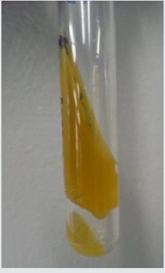
TSI (Triple Sugar Iron Test): TSI stands for a triple sugar iron test. This analysis was performed to check the capability of a microorganism to produce hydrogen sulfide by the fermentation of available sugars. This test is made for the enteric bacteria which includes Shigella and salmonella species. This test indicates that tested organisms ferment sugars such as glucose, sucrose, and lactose that showed the production of by-products. If slant and butt both turned yellow indicate that an acidic condition was developed, and all the sugars were fermented. If alkaline slant and acidic butt show only glucose fermentation while both butt and slant show alkaline that indicates that no sugar fermentation has occurred. The blackening of the medium was indicated by the production of hydrogen gas. Carbon dioxide gas production is indicated by the cracks and bubbles formation.
Urease Test: Urease test is performed to detect the breakdown of urea by the action of an enzyme urease done by microorganisms. Streaked the isolated colony on urea slant and incubated at 37 °C for 24hours than observed the color change of slant. If urease enzyme present urea breakdown into ammonia and slant turned pink in color. If organisms were not produced urease enzymes the color of slant remains the same light orange.
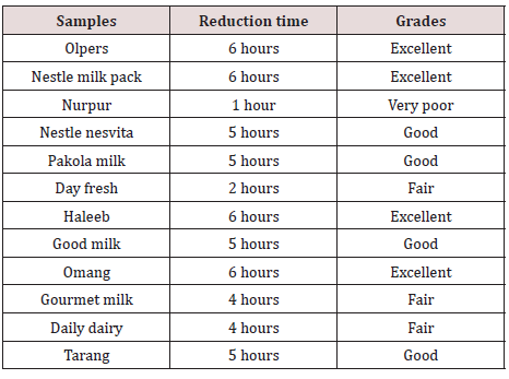
Dye reduction test (methylene blue): This test was used to estimate the microbial load of pasteurized and packed milk (Table 1).

Table 1: Grading of milk according to the time in which dye decolorize.

Lupinepublishers-openaccess-complementary-alternative-medicine-journal

Results

Bacterial Identification in raw milk samples

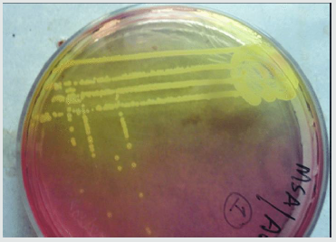
In our study different types of bacteria were identified from the raw milk samples. According to these results, the most common isolates in raw milk samples were staphylococcus aureus, Salmonella, and Bacillus. Other enteric organisms were also found like E. coli, Klebsiella spp, and Proteus spp. We performed bacterial identification on N.A, EMB, and MSA for all 12 raw milk samples. The following results were obtained that are mentioned in Table 2. These organisms are further identified on the basis of colonial and biochemical characteristics as mentioned in Table 3.

Table 2: Bacterial identification on N.A, EMB and MSA.

Lupinepublishers-openaccess-complementary-alternative-medicine-journal

Table 3: Colonial and biochemical characteristics.

Lupinepublishers-openaccess-complementary-alternative-medicine-journal

Methylene blue reduction test

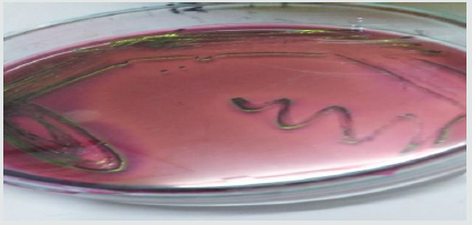
No colonial growth found on the plates of EMB, N.A, MSA. Samples were proceeded to perform M.B.R. The results obtained are mentioned in Table 4 & Figure 1A-1E.

Table 4: Qualitative analysis of Milk Sample by Methylene blue reduction test.

Lupinepublishers-openaccess-complementary-alternative-medicine-journal

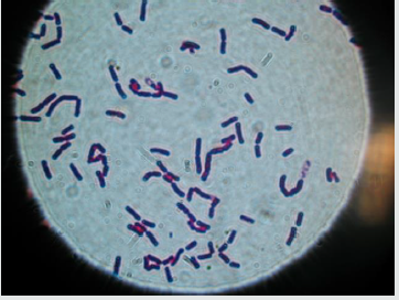
Figure 1: Colonial identification of raw milk samples.

Lupinepublishers-openaccess-complementary-alternative-medicine-journal

Figure 1A: Staph. aureus on MSA.

Lupinepublishers-openaccess-complementary-alternative-medicine-journal

Figure 1B: E. coli on EMB.

Lupinepublishers-openaccess-complementary-alternative-medicine-journal

Figure 1C: Microscopy of Bacillus Cereus.

Lupinepublishers-openaccess-complementary-alternative-medicine-journal

Figure 1D: TSI Result of E. coli.

Lupinepublishers-openaccess-complementary-alternative-medicine-journal

Figure 1E: IMVIC Result of E. coli.

Lupinepublishers-openaccess-complementary-alternative-medicine-journal

Discussion

Milk is the best food for all age groups with all requirements which is necessary for individual growth and development. Different types of milk are available for the consumer to consume according to their taste and desire Karmali MA et al. [23]. Milk is composed of all basic nutrients such as calcium, phosphorus, vitamins, and water content Potter M et al. [24]. The composition of milk is considered as complete nutrition. The high amount of nutrients in milk supports the growth of microorganisms and causes spoilage of milk. High water content in milk is the basic factor for bacteria to grow Hill C et al. [25].
Raw milk is unheated (untreated milk) are readily available in the markets. Which is highly contaminated with pathogenic bacteria. Bacteria are present in the environment that’s why it contaminates the milk and its products easily Czaplicki A et al. [26]. Milk becomes contaminated with different ways such as fecally contaminated water when added in the milk, unsterile utensils, improper bulk tanks, soil, and major and most dangerous factor is that unhealthy condition of milking man because hygiene of personal is very important if caretaker of cows and the milking man was not properly clean in ordered to wash their hands and if suffer from any diseases so organisms should be transferred and contaminate the milk another factor is a diseased animal Hijiumi et al, 2011. Animal health is also very important because cow suffer from mastitis and organisms enter into milk and causes spoilage Ekici k et al. [17].
According to different studies, untreated milk is not healthy for human use until it is not properly boiled. But sometimes after boiling, microorganism remains present in the milk and causes enteric diseases such as diarrhea, vomiting, etc Oliver sp et al. [21]. All these effects and health issues of raw milk increases the value of pasteurized milk. Pasteurized milk is of better quality and long shelf life than raw milk and no health issues are measure from pasteurized milk Hill C et al. [23]. Pakistan is the biggest milkproducing country in the world. Milk contains major nutrients that support human health and makes the bones strong and prevent osteoporosis, but these nutrients also support the growth of bacteria which causes contamination. According to studies typhoid fever is very common in young children.
In this study we collected 24, out of which 12 were raw milk samples from local dairy shops of Karachi city and 12 were branded and local pasteurized milk samples such as Olpers, Haleeb, Nestle Milk Pack, Nurpur, Nestle Nesvita, Pakola, Day fresh, Good milk, Omang, Gourmet milk, Daily Dairy, and Trang. This study showed that raw milk is highly contaminated with soil, unhealthy water, and a high number of pathogenic bacteria. Whereas no growth was observed in pasteurized milk.
The outcome shows that the numbers of aerobic spores are readily available in milk samples because the reported concentration of aerobic spores in grass and maize vary from 10 to >105 per g. When spores present in soil and animal feed, they form high numbers of spores these spores excreted in feces. Bacillus species are rich in the environment and therefore naturally present in the soil. The proportion of milk samples that showed activist for S. aureus, the major pathogen-related with diseased cow condition worldwide. All types of bacteria which are present in milk samples are diseases causing but some of them are not related to foodborne diseases such as Klebsiella and Proteus these organisms cause pneumonia and urinary tract infections, respectively.
In this study the presence of microorganisms in the pasteurized milk was less than raw milk, the Methylene blue reductase test was performed for the detection of the quality of milk sample, pasteurized milk samples including Olpers, nestle milk pack, Haleeb and Omang showed the excellent quality of milk with 6hrs, Pakola, good milk, Trang and nestle Nesvita indicated good value of milk with 5 hours reduction time while Nurpur, the day fresh, daily dairy and gourmet milk showed poor quality and their reduction time was 1hr to 2hr. The implementation of good sanitation practices and applications of HACCP (hazard analysis of critical control points) will improve the quality of milk and also done by increasing the temperature and timing of the heating process will help to reduce the growth of microorganisms Armstrong C et al. [27].
A study of S. aureus in raw and pasteurized milk was conducted in the Reconcavo area of the state Bahria, Brazil. In this study, a total of 70 samples were studied out of which 50 samples of raw milk and 20 samples of pasteurized milk were taken. Out of 50 samples of raw milk, 34 showed the contamination of S. aureus while out of 20 samples of pasteurized milk 6 samples contaminated with S. aureus with percentages of 68% and 30% respectively. This contamination showed that both types of milk were health hazardous. In Dhaka city Bangladesh, a study for the characterization of pathogenic bacteria in raw and pasteurized milk had conducted. This study showed raw milk is highly contaminated with fecal bacteria known as Shigella, Klebsiella E. coli, whereas pasteurized milk was slightly contaminated with different types of bacteria such as Bacillus app Uddin et al. [5].
Pasteurization quality will be increased by increasing the heating time because milk becomes contaminated by different types of microorganisms that might be reduced by plenty of milking and transport measurement. The large number of Escherichia coli in milk samples showed that fecal contamination in milk and diseased condition of cows. Proper time and temperature in the pasteurization process must be maintained to eliminate pathogens and other contamination also some immediate actions for raw milk safety should be taken because individuals may suffer from illnesses. Due to the use of raw milk because of their unawareness.

Conclusion

e general public because of the possible presence of pathogenic bacteria. Internationally, outbreaks of foodborne sickness have been attributed to unprocessed milk. This can be reduced by using boiled water for cleaning all utensils and equipment. It is also preferred that detergents or disinfectants should be dissolved in boiled water for cleaning of all utensils and equipment which immediately helps to reduce milk contaminates. For pasteurized milk or locally available brands of milk, one should make sure that their processing areas are cleaned and proper which provides, ultra-high temperature to decrease bacterial contamination in milk.

Read More About Lupine Publishers Journal of Complementary & Alternative Medicine Please Click on below Link:
https://lupine-journal-complementary-medicine.blogspot.com/

Thursday 25 August 2022

Lupine Publishers| Chrono-Nutrition for Pregnant Women: Metabolic and Pediatric Insights

 Lupine Publishers | Journal of Pediatrics and Neonatology


Abstract

Pregnant women undergo metabolic adaptations and challenges that predispose them to a variety of complications. The objective of this article was to highlight the importance of eating timing and especially limited evening eating on optimizing pregnancy status and pediatric health and welfare. Because of limited evening and nocturnal metabolic capacity to handle nutrients (e.g., glucose), pregnant women are recommended to take smaller and earlier evening meals and instead distribute their main food meals over the course of morning and day hours. Such a food intake regimen would considerably reduce diabetes and obesity risks and improve upcoming lactation performance and pediatric health and welfare.

Keywords: Eating Timing; Pregnant Woman; Chrono-Nutrition; Pediatric Health

Philosophy

Chrono-nutrition is emerging as a new research area with growing interest (1). Chrono-nutrition deals with optimizing eating timing. The goal of chrono-nutrition is to synchronize eating patterns with circadian physiological rhythms of human metabolism (2,3). This synchrony aims to reduce risks from a multitude of metabolic disorders (e.g., diabetes, obesity, cancer) and improve human health and welfare. Basically, human glucose tolerance is known to decline during evening hours and overnight. This means that cells are not as much capable to assimilate and metabolize nutrients in the evening as are they in the morning (3). In other words, if large evening meals are taken, because of incomplete and inefficient nutrient utilization by different cells, nutrient overload will occur that will in turn predispose the body to a variety of metabolic complications, such as diabetes, obesity, and metabolic syndrome.
Pregnant women undergo physiological adaptations and are already prone to metabolic challenges (e.g., diabetes and obesity) (2,3). The above complications are, thus, very likely to happen in pregnant women. As such, making the evening meal smaller and taking it earlier (not later overnight) would help the pregnant body better cope with the metabolic challenges. As another complementary strategy, pregnant women are recommended to take several small evening meals made of mainly fruits and vegetables instead of starch and fats. This practice would be predicted to improve insulin action and prevent obesity. Improving evening nutrient and hormonal metabolism by evening exercise has also been advised (4). The goal has been to optimize circadian timing of eating and exercise towards optimal lifestyle. Optimizing meal properties is a new research area with many unanswered questions. Future research is required to address different aspects of this promising field of study.

Conclusion

Smaller and earlier evening meals made of mainly fruits and vegetables instead of starch and fat are recommended for pregnant women to help improve intermediary metabolism and reduce metabolic complications. These effects will result in improved lactation physiology and pediatric health and welfare.

Acknowledgments

Thanks to Ferdows Pars Agricultural and Livestock Holding Co., (Tehran, Iran) for supporting the author’s programs of optimizing science education in the third millennium.

Read More About Lupine Publishers Journal of Pediatrics and Neonatology Please Click on below Link:
https://lupinepublishers-pedaitrics.blogspot.com/
Wednesday 24 August 2022

Lupine Publishers | Characterization of Olive Oils Used as Medium in Canned Fish

 Lupine Publishers | Journal of Food & Nutrition


Abstract

Background: The assessment of the quality extra virgin olive oils used as liquid medium in canned fish together with the levels of phenolic compounds is very important in order to evaluate if the higher price of the extra virgin olive oil in canned fish in comparison with fish in olive oil is justified.

Objective: The aim of the study was to identify and quantify the phenolic compounds in processed food such as canned fish in order to assess the added value of extra virgin olive oil used as a liquid medium in canned fish. The authenticity of olive oils used as liquid medium in canned fish was assessed by determination of sterol composition, fatty acid composition and content of stigmastadiens.

Materials and Methods: Various fish products in olive oil or extra virgin olive oil were sampled randomly from different manufactures on the Slovenian market. The content of oil was first separated from fish and then immediately analyzed for sterol composition and concentrations, fatty acid composition, stigmastadiens levels, acidity, peroxide number and phenolic compounds composition and content.

Conclusion: Oil in canned fish products corresponded to the declared labelling on the packaging. However, the levels of determined phenolic compounds were much lower and the composition of phenolic compounds was different compared to the usual extra virgin olive oil from Slovenian producer.

Introduction

Fish products are often sold together with vegetable oil such as olive oil. Lately, the market also offers fish products with extra virgin olive oil. Olive oil which contains 98-99% triacylglycerols and a high proportion of stable oleic acid can provide combined with fish meat the recommended ratio between n-3 and n-6 fatty acids [1-3]. Olive oil can also represent a significant source of antioxidants such as vitamin E and a source of various phenolic compounds that are not present in other foodstuff [4, 5]. Antioxidants such as phenolic compounds in olive oil can not only have a protective role on human health [6], but they can also protect the important compounds such as unsaturated fatty acids against oxidation [7]. However, due to the high costs of olive oil production, the frauds such as adding cheaper oils to olive oil can be found. Moreover, products such as fish products in olive oil, where fish meat may cover the specific taste of olive oil, can often be the target of frauds. Due to the increased possibility of frauds, the market norms for olive oil compared to other oils are very strict. However, olive oils sold together with fish meat are not addressed. According to Commission Regulation (EEC) No. 2568/91 and Commission Implementing Regulation (EU) No. 1348/2013 the sterol composition, fatty acid composition and the determination of stigmastadiens in olive oil are important indicators for olive oil authenticity. Overall, the assessment of the quality extra virgin olive oils used as liquid medium in canned fish is very important in order to evaluate if the higher price of the extra virgin olive oil in canned fish in comparison with fish in olive oil is justified. While the phenolic compounds from olive oils have an important protective role on human health and prevent oils from oxidation, the main aim of the study was to identify and quantify the phenolic compounds in processed food such as canned fish in order to assess the added value of extra virgin olive oil used as a liquid medium in canned fish. In addition, the authenticity of olive oils used as liquid medium in canned fish available on the Slovenian market was assessed by the determination of sterols composition and content, fatty acid composition and the levels of stigmastadiens. As far as we are concerned this is the unique study that involves the determination of phenolic compounds in olive oil from processed food available on the Slovenian market by high performance equipment. The results obtained in this study will help the testing olive oil laboratories to better evaluate the quality and potential frauds of extra virgin olive oil added to processed food such as canned fish.

Materials and Methods

Various canned fish products (n=16) containing olive oils and extra virgin olive oil used as liquid medium were collected randomly in the city of Koper in January, February of 2016 and in August and September 2018. Canned tuna in olive oil, canned tuna in extra virgin olive oil, canned mackerel in extra virgin olive oil from different manufacturers were bought in different supermarkets in 2016 and 2018. Each type of canned fish products available containing extra virgin olive oil as a liquid medium from different manufactures were sampled. The content of oil was first separated from fish and then immediately analysed for the selected compounds. Sterols composition and the sterols content in olive oil from canned fish was determined by thin layer chromatography and capillary gas chromatography with flame ionization detector (GC-FID) [8]. The fatty acid composition of olive oil was determined by in situ transesterification with GC-FID [9]. The content of stigmastadienes in olive oils samples were determined by the column and capillary gas chromatography [10]. The determination of acidity and peroxide number was carried out according to the standard methods [11,12]. The phenolic composition of olive oils was determined by high performance liquid chromatography - mass spectrometry (HPLC-ESI-QqQ) [13,14] and the unknown compound present in high levels was additionally identified by HPLC-ESI-qTOF [15].

Materials and Methods

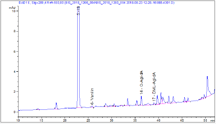
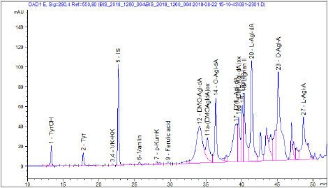
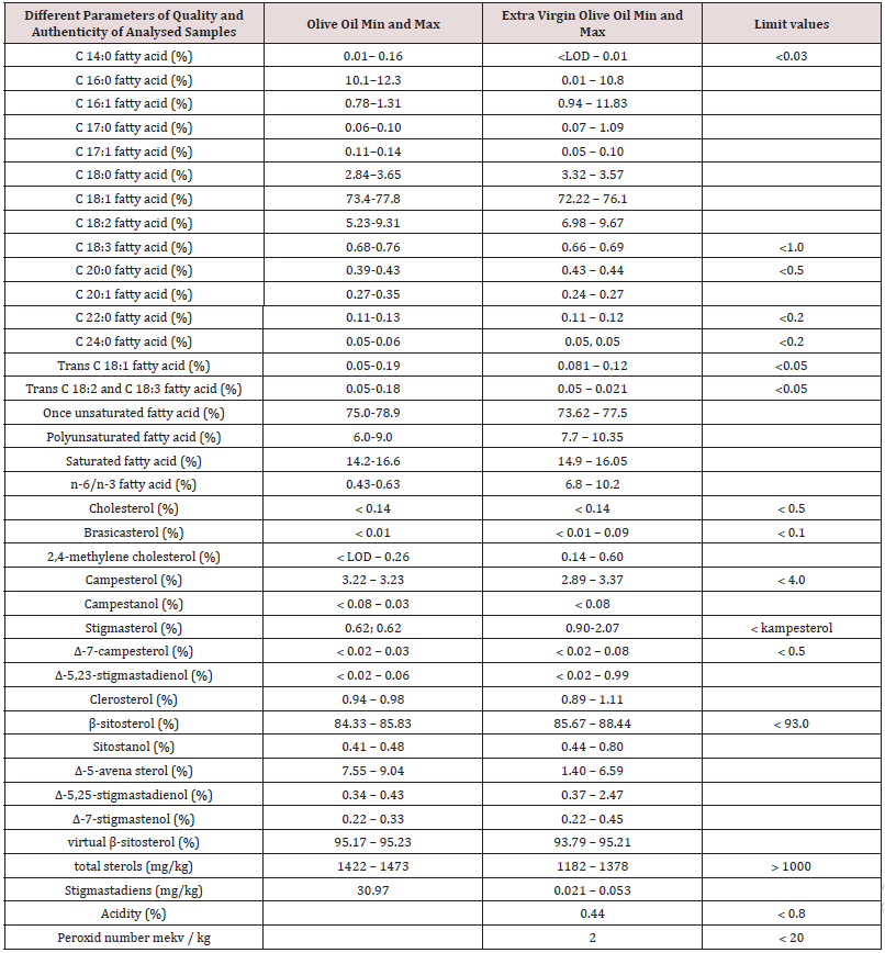
Table 1 shows that the fatty acid composition of the samples does not correspond to the typical fatty acid composition of olive or extra virgin olive oil. The differences might be due to migration of fatty acid from fish into the oil, heat treatment of fishery products in olive oil or the addition of refined oil. The exceeded values of trans fatty acids could be due to the heat treatment of canned fishery products in olive oil or the addition of refined oil. Likewise, from the table 1 is shown that sterol composition is not typical for olive oil. This is most likely due to sterol migration from fish to vegetable olive oil. The content of stigmastadienes is elevated only in olive oils that are not declared as extra virgin olive oils. Therefore, this confirms that the extra virgin olive oil in analyzed samples is properly declared and that stigmastadiens determination could be the right method to distinguish the refined oil from extra virgin olive oil in canned fish samples. Moreover, quality parameters as free fatty acid content and peroxide number of declared extra virgin olive oils corresponds to the limits determined for the category of extra virgin olive oil, therefore, below or equal to 0.8% (acid limit for the extra virgin olive oil category) and below or equal to 20 mekv / kg (the peroxide number limit for the extra virgin category of olive oil). The content of total phenolic compounds in oils from canned fish were approximately 100 times lower compared to the levels find in declared extra virgin olive oil from Slovenian producer (Table 2, [16]). As was expected the content of phenolic compounds was the lowest in refined olive oil. However, the levels of total phenolic compounds in extra virgin olive oils were only slightly higher (Table 2). The levels of the end oxidation compound tyrosol and hydrohytyrosol of the main phenolic compounds in olive oil secoiridoids were low. This suggests that olive oils were low in phenolic compounds previous the heat treatment. From figures 1-3 the differences in composition of phenolic compounds of refined olive oil from canned tuna (Figure 1), extra virgin olive oil from canned tuna (Figure 2) and extra virgin olive oil from Slovenian producer (Figure 3). Extra virgin and refined olive oils added to fish products had significantly lower content of oleuropein and ligstroside derivatives, that are highly appreciated antioxidants in the quality extra virgin olive oils. In the Figure 2 the high unusual peak for typical extra virgin olive oils was observed. With HPLCqTOF was determined that the unusual pick was represented by the compound with molecular formula C10H12O4 could be ferulic acid derivate or hydroxytyrosol acetate.

Figure 1: The chromatogram determined by DAD at 280 nm in a sample of refined olive oil added to canned fish.

upinepublishers-openaccess-Food-Nutrition

Figure 2: The chromatogram determined by DAD at 280 nm in a sample of extra virgin olive oil added to canned fish.

upinepublishers-openaccess-Food-Nutrition

Figure 3: The chromatogram determined by DAD at 280 nm in a sample of extra virgin olive oil from Slovenia producer.

upinepublishers-openaccess-Food-Nutrition

Table 1: Different parameters of quality and authenticity of analysed samples such as fatty acid composition of olive oil, determination of sterols, stigmastadiens, acidity and peroxide number.

upinepublishers-openaccess-Food-Nutrition

Table 2: Total phenolic compounds, tyrosol and hydroxytyrosol content in analysed samples.

upinepublishers-openaccess-Food-Nutrition

Conclusion

Even though by the quality parameters it is clear that oil in canned fish products corresponds to the declared labelling on the packaging, the levels of determined phenolic compounds were much lower than it is expected for extra virgin olive oils. In addition, the composition of phenolic compounds was different compared to the usual extra virgin olive oil from Slovenian producer.

Acknowledgement

This research was partially sponsored by Post‐doctoral project from University of Primorska untitled “Development of analytical methods for determining the potencies and the quality of fish products in olive oil for the purpose of ensuring the nutritional security of the population”. The author gratefully acknowledges the European Commission for funding the InnoRenew CoE project (Grant Agreement #739574) under the Horizon 2020 Widespread- Teaming program. The authors are also greatful to Saša Volk and dr. Vasilj Valenčič who perfromed the analysis of fatty acids compostion, sterol composition and concentartions, peroxide number and acidity of the olive oil samples involved in this study.

Read More About Lupine Publishers Journal of Food & Nutrition Please Click on Below Link:
https://lupine-publishers-food-and-nutrition.blogspot.com/
Tuesday 23 August 2022

Lupine Publishers| 3rd Nerve Palsy After Microsurgical Clipping of Basilar Top Aneurysm

 Lupine Publishers| Journal of Neurology and Brain Disorders


Abstract

Postoperative oculomotor nerve palsy is a rare complication that occurs in the setting of micro¬surgical aneurysm clipping. While a number of theories have been postulated to explain the development of postoperative oculomotor nerve palsies, the underlying pathophysiology of such complications still remain to be elucidated. In this report, we present a case of postoperative isolated ipsilateral oculomotor nerve palsy after clipping of basilar tip aneurysm which we believe may be attributed to periperative oculomotor nerve manipulation related neuropraxia.

Keywords:Oculomotor Nerve Palsy; Basilar Apex Aneurysm; Postoperative

Introduction

Oculomotor nerve is the third cranial nerve that enters the orbit through the superior orbital fissure and controls muscles that drive most movements of the eye and raise the eyelid. Oculomotor nerve is derived from the basal plate of the embryonic midbrain. Cranial nerves IV and VI also participate in the control of eye movement. Oculomotor nerve palsy is an eye condition resulting from damage to the third cranial nerve or a branch there of. As the name suggests, oculomotor nerve supplies the major¬ity of the muscles that control eye movements. Therefore, damages to oculomotor nerve will render affected individuals unable to move his or her eye normally. Unilateral oculomotor nerve palsy is often encountered in the setting of ipsilateral aneurysms located at the Posteri¬or Communicating Artery (PcomA), Internal Carotid Artery (ICA) or their junction (PcomA/ICA). There have been some reports on ocu¬lomotor nerve palsy as a result of aneurysms in basilar tip, anterior artery or anterior communi¬cating artery. In the present report, we describe a case of basilar apex aneurysm presenting with ipsilateral postoperative oculo¬motor nerve palsy that is thought to be attrib¬uted to peroperative oculomotor nerve handling followed by neuropraxia.

Case Report

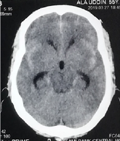
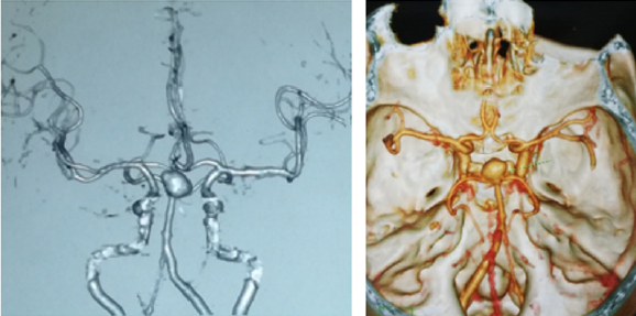
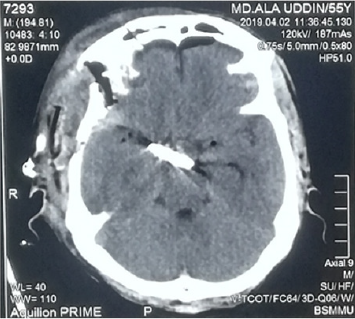
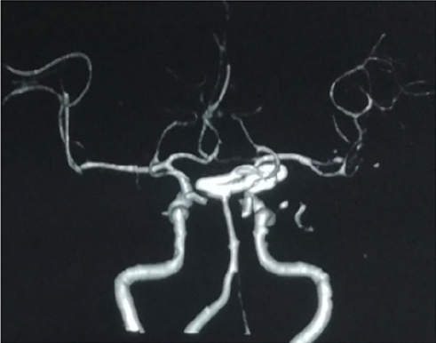
A 55years old hypertension male with no other significant past medical history or contributing family history complained about sudden severe headache for one hour followed by several episodes of vomiting and unconsciousness for 05 hours on admission at our hospital. Physical examination results were within nor¬mal limits except for considerable neck stiffness, positive Kernig sign and Brudzinski’s sign. Computed Tomography (CT) scan demonstrated diffuse subarachnoid hemorrhage, which was especially concentrated in ambient, sylvian fissure, interpeduncular and suprasellar cisterns with intraventricular extensions and mild triventricular hydrocephalus (Figure1). Computed tomography of the head show¬ing diffuse subarachnoid hemorrhage especially concentrated in ambient, sylvian fissure, interpeduncular and suprasellar cisterns with intraventricular extensions and mild triventricular hydrocephalus. CT Angiography (CTA) revealed a large saccular basilar tip aneurysm (approximately 10.7×9.5×10.5mm) (Figures 2A & 2B). The aneurysm was micro surgically clipped (Figures 4) via the right Orbitozygomatic approach (Figures 3A & 3B). Postoperatively the patient developed complete right third nerve palsy characterized by the presence of dilated pupil, ptosis and downward deviation and abduction of the eyeball. CT scan revealed no postoperative intracranial hematoma and the surgical clip was in the proper location. However, compression of the basal cisterns and assessment of the ventricular system were noted (Figure 5). Postoperative CTA showed no existence of another aneurysm or vasospasm (Figure 6). The patient was discharged 02 weeks after the surgery. At this point, his mydriasis and eye lid drooping were still present to a lesser extent. At one-month follow-up, the patient’s pupil was slightly contracted and reflexed reluctantly to direct and indirect light stimulation.

Figure 1: Computed tomography of the head show¬ing diffuse subarachnoid hemorrhage especially concentrated in ambient, sylvian fissure, interpeduncular and suprasellar cisterns with intraventricular extensions and mild triventricular hydrocephalus.

Lupinepublishers-online-journal-of-neurology-and-brain-disorders

Figure 2A & 2B: Computed tomography angiography show¬ing a large saccular basilar tip aneurysm (approximately 10.7×9.5×10.5mm). The arrow indicates the aneurysm.

Lupinepublishers-online-journal-of-neurology-and-brain-disorders

Figure 3A & 3B: Right sided Orbitozygomatic craniotomy (one piece), extradural anterior clinoidectomy & clipping of basilar apex aneurysm through trans-sylvian approach.

.
Lupinepublishers-online-journal-of-neurology-and-brain-disorders

Figure 6: Postoperative CTA showed no existence of another aneurysm or vasospasm and the surgical clip was in the proper location.

Lupinepublishers-online-journal-of-neurology-and-brain-disorders

Figure 7: On postoperative day two, the patient developed a complete right third nerve palsy.


Discussion

There are many proposed hypotheses pertain¬ing to the etiology of oculomotor nerve palsy before and after aneurysm surgery. Direct com¬pression by the aneurysm is the most classic and common cause. Other causes include direct injury to the third nerve intraoperatively [1-6], microvascular ischemia [7-9], focal hemato¬ma formation [3], vasospasm [8-12], anomaly of the vessels along the oculomotor nerve [10], elevated intracranial pressure and herniation, compression by intracranial structures other than aneurysms [7], and undetermined ori-gins [11]. In the present case, compression by aneurysm may not be possible because of the anatomi¬cal distance between the two entities. Postoperative CT, CTA decrease the chances of hematoma formation, vasospasm and elevated intracranial pressure. Although we cannot rule out the possibility of ischemic injury to the oculomotor nerve. Small vessel ischemic injury to oculomotor nerve usu¬ally exhibits pupilsparing [9] whereas our patient demonstrated a blown pupil. So, we believe that, probably from coarse dissection of the cavernous sinus dura and as well as direct anterior clinoidectomy or from heat of the low power drill may be responsible for the 3rd nerve palsy of our patient.

Conclusion

The novel use of intramuscular cerebrolysin and citicoline in a patient with kernicterus was safe and effective.

The Neuroreparative Effects of Cerebrolysin have Been Attributed to [3-5]:

a) Inhibition of apoptosis.

b) Improving synaptic plasticity and induction of neurogenesis.

c) Augmenting the proliferation, differentiation, and migration of adult.

d) subventricular zone neural progenitor stem cells, contributing to neurogenesis.

e) Induction of stem-cell proliferation in the brain.

Citicoline (cytidine diphosphate choline) is a mononucleotide made of ribose, pyrophosphate, cytosine and choline is a watersoluble naturally occurring substance that is generally grouped with the B vitamins. It is also considered a form of the essential nutrient choline. It is a safe substance with generally minor side effects which may include digestive intolerance after oral administration [6]. An accumulating research evidence suggests that citicoline is endowed with interesting pharmacological properties that can make it useful in the treatment of various disorders that has no universally accepted effective treatment including neurological conditions such as Parkinson’s disease, brain ischemia, hemorrhagic stroke, Alzheimer’s disease; and ocular condition such as glaucoma, nonarteritic ischemic neuropathy and amblyopia [6].

The Neuro-Protective Effects of Citicoline Were Attributed to the Followings [6]:

a) Preservation of cardiolipin and sphingomyelin

b) Preservation of arachidonic acid content of phosphatidylcholine and

c) phosphatidylethanolamine.

d) Partial restoration of phosphatidylcholine levels.

e) Stimulation of glutathione synthesis and glutathione reductase activity.

f) Reduction of phospholipase A2 activity.

g) Increasing glucose metabolism in the brain.

h) Increasing cerebral blood flow.

Reducing oxidative stress and preventing excessive inflammatory response in the brain by inhibiting the release of free fatty acids and reducing blood brain barrier breakdown. Enhances cellular communication by increasing the availability of neurotransmitters, including acetylcholine, norepinephrine, and dopamine. Lowering increased glutamate concentrations and increasing the decreased ATP concentrations induced by ischemia. Citicoline increases dopamine receptor densities, and therefore could improve memory impairment resulting from poor environmental conditions. Citicoline could also improve focus and mental energy and could be useful in the treatment of attention deficit disorder. Citicoline has also been shown to improve visual function in patients with glaucoma [7].

Conclusion

The novel use of intramuscular cerebrolysin and citicoline in a patient with kernicterus was safe and effective.

Read More About Lupine Publishers Journal of Neurology and Brain Disorders Please Click on below Link: https://brain-disorders-lupine-publishers.blogspot.com/