Tuesday 30 November 2021

Lupine Publishers| Chemically-Induced Peptic Ulcer: Gastroprotective Effects of Peach Fruit

 Lupine Publishers| Journal of Gastroenterology and Hepatology


Abstract

Background: Peptic ulcer, an inflammatory breach in the gastric and duodenal mucosa, is caused by hyper secretion of gastric acid and defective mucosal protective elements, affecting approximately 4 million people each year. The currently available antiulcer drugs are reported to have severe side effects. Hence, there is a need to develop more effective and less toxic alternative therapeutic agents from natural sources. The current study was conducted to investigate the therapeutic potential of Prunus persica Linn. Batsch fruit (Peach) against chemically-induced gastric and duodenal ulcers.

Material and Methodology: 70% aqueous methanolic crude extract of Prunus persica (Pp.Cr) was prepared. Gastroprotective effects were evaluated by oral administration of Pp.Cr at the doses of 100, 300, 500 mg/kg for ten days. Normal control and intoxicated groups were treated with distilled water. Bismuth subsalicylate (88 mg/kg) was administered to the standard control group. On 10th day, all animals were intoxicated with ethanol (5 ml/kg) except normal control group, and one hour after intoxication, all animals were sacrificed, stomach isolated; and pH and acidity of gastric contents, ulcer index and percentage protection were calculated. One stomach from each group was analyzed histo pathologically. Duodenal protective effects were assessed by administering Pp.Cr (100, 300, 500 mg/kg) for fourteen days. The standard control group was treated with cimetidine (100 mg/kg). On 14th day, cysteamine (600 mg/kg) was administered in two divided doses at an interval of four hours to experimental animals to induce duodenal ulcer. All animals were dissected out 48 hours after the administration of first dose of cysteamine. Duodenum was isolated and analyzed for severity score. Histopathological examination of one duodenum from each group was performed.

Results: In gastric ulcer model, Pp.Cr was found to increase pH and decrease the total acidity of gastric contents, and decrease the ulcer index, in dose-dependent manner at the doses of 100, 300 and 500 mg/kg. In duodenal ulcer model, Pp.Cr was observed to decrease the severity score and ulcer index, again in dose-dependent manner.

Conclusion: The results of current study demonstrate the gastric and duodenal protective effects of the crude extract of Prunus persica (Pp.Cr), corroborating their folkloric use in the prevention and treatment of peptic ulcer.

Keywords: Ulcer; Duodenum; Prunus persica; Gastroprotective; Gastric Ph; Ulcer Index

Introduction

Peptic ulcer, an inflammatory breach in the lining of GIT, results from hyper secretion of gastric acid or defects in mucosal protective factors [1]. The most frequent sites of ulcer formation are stomach and proximal portion of duodenum and the ratio of duodenal to gastric ulcers is 4:1 [2]. Peptic ulcer is caused by various noxious factors; such as Helicobacter pylori infection, NSAIDs, alcohol intake, smoking and stress. 60% of gastric and up to 90% of duodenal ulcers develop due to the H. pylori infection [3]. Peptic ulcer can be treated through different approaches, based on chemically synthesized drugs; i.e. reduction of gastric acid secretion (proton pump inhibitors, H2 receptors antagonists, anticholinergics, prostaglandin analogues), neutralization of gastric acid secretion (antacids), protection of GIT mucosa (sucralfate, bismuth subsalicylate) and use of antimicrobial drugs like amoxicillin, clarithromycin, tetracycline and metronidazole [4]. No doubt, the available drugs have reduced the incidence and prevalence of peptic ulcer but they are associated with severe side effects; such as impotence, arrhythmias, hyperplasia, gynaecomastia, and hematopoietic changes [5]. Therefore, there is a constant need to explore the new possible therapies with lesser side effects to reduce or treat the peptic ulcer problems and plants are the best alternatives which are being used from the ancient times. World Health Organization (WHO) has estimated that about 75% of the world’s population rely on herbal medicines for the prevention and treatment of various diseases because of their fewer side effects and low cost. Approx. 80% of population in Pakistan is using indigenous plants for the treatment of various diseases [6]. Prunus persica used in the present study is a tree or large shrub having tremendous medicinal importance.

Different parts of plant; i.e. leaves, flowers, fruits, kernels of fruit and seeds have traditional medicinal uses and extensively studied. The plant is used for the treatment of whooping cough, chronic bronchitis and gastritis. The plant has demulcent, antiscorbutic, aphrodisiac, anti-pyretic and anthelmintic activities. Leaves have demulcent, expectorant, vermicidal, anthelmintic, insecticidal, sedative and diuretic properties. Leaf paste is used externally to kill worms in wounds and fungal infections. The flowers are used as laxative and diuretic, oil extracted from seeds, is used for the treatment of piles, stomach troubles of children and earache and used as an abortifacient. Peach kernels are used for the treatment of menstrual disorders, blood disorders, coughs and rheumatism. The bark is used to treat the leprosy and jaundice [7]. Prunus persica has been reported to possess Antiallergic [8], Antibacterial [9], Anti-inflammatory [10], Antiphotoaging [11], Prokinetic [12] and Antioxidant properties [13]. Hence, indigenous medicinal plant; Prunus persica, was selected on the basis of its traditional use as antiulcer agent. The objective of the current study was to investigate the antiulcerogenic potential of the crude extract of Prunus persica fruit by using chemically-induced peptic ulcer models; i.e. ethanol induced-gastric ulceration and cysteamineinduced duodenal ulceration.

Research Methodology

Material and Equipment

All chemicals used in experiments were of analytical grade including; Bismuth subsalicylate, Cimetidine, Cysteamine hydrochloride, Ethanol, Formalin, Ketamine and Xylazine. Equipment used were centrifuge machine, digital weighing balance, oven, pH meter, refrigerator, rotary evaporator and vortex mixer.

Phytochemical Section

Collection of Plant Material: The frsesh fruit of Prunus persica (Peach) was purchased from local market of Bahawalpur, Pakistan. After collection, plant was identified by authentic botanist; Mr. Abdul Hamid, department of Life Sciences, the Islamia University of Bahawalpur, Pakistan. Dried plant specimen was deposited in Herbarium of Pharmacology research laboratory, department of Pharmacy and Alternative Medicine, the Islamia University of Bahawalpur, Pakistan. Voucher number PP-FT-05-15-92 was issued for future reference.

Preparation of the Crude Extract: After removal of seeds, fresh fruit of Prunus persica was cut into small pieces and soaked in 70 % aqueous methanol for three days with occasional shaking at room temperature. Then the soaked material was filtered first through muslin cloth and then through filter paper. The process of soaking and filtration was repeated twice. After third filtration, residue was discarded. Filtrate was subjected to evaporation using rotary evaporator to remove solvent under reduced temperature (50°C) and pressure. The obtained crude extract was then labelled as Pp.Cr and weighed to calculate percentage yield. The extract was stored in freezer for future use.

Phytochemical Analysis of Pp.Cr: The crude extract of Prunus persica (Pp.Cr) was subjected to phytochemical analysis for the detection of secondary metabolites; i.e. alkaloids, carbohydrates, coumarins, flavonoids, glycosides, phenols, phlobatannins, proteins, quinones, saponins, tannins and terpenes.

Pharmacological Section

Experimental Animals: Wistar albino rats (150-250 g) and Swiss albino mice (18-30 g) of either sex were housed in animal house of Pharmacology Research Laboratory, department of Pharmacy, faculty of Pharmacy and Alternative Medicine, the Islamia University of Bahawalpur. Animals were kept in polycarbonate cages (47×34×18 cm3) under standard conditions of temperature (25±2°C), humidity (60±5 %) and 12 h/12 h light and dark cycle. Animals were provided with standard pellet diet and tap water ad libitum and allowed to acclimatize for at least one week before the commencement of experiment. The study protocols and procedures were approved by the Institutional Research Ethics Committee of department of Pharmacy and Alternative Medicine, Islamia University of Bahawalpur; i.e. Pharmacy Research Ethics Committee (PREC) and the reference No. 40-2015/PREC; dated 02.09.2015 was issued for future reference.

Experimental Models: Peptic ulcer was induced by intragastric administration of ethanol (gastric ulcer) and cysteamine hydrochloride (duodenal ulcer) to evaluate the antiulcer potential of Pp.Cr.

a) Ethanol-Induced Gastric Ulcer: Gastric ulcer was induced by oral administration of ethanol (5ml/kg) in wistar albino rats, according to the method described by Ahmed [14]. Animals were divided into six groups, each comprises of six animals. Normal control group and intoxicated groups were treated with distilled water (5 ml/kg, p.o) for 10 days. Treatment groups received different doses (100, 300 and 500 mg/kg) of Pp.Cr and the standard control group was treated with bismuth subsalicylate (88 mg/ kg) for 10 days. On 10th day, one hour after respective treatment to 24 hour fasted rats, ethanol (5 ml/kg) was administered orally to all the animals except the normal control group. One hour after administration of ethanol, all animals were anaesthetized with ketamine (50 mg/kg)/Xylazine (5 mg/kg) combination and sacrificed. The stomach was isolated from each animal and gastric contents were collected in centrifuge tubes.

i) Analysis of Gastric Contents for Ph and Total Acidity

Gastric contents were centrifuged at 1000 rpm for 10 minutes. pH of supernatant was noted using pH meter. 1ml of supernatant liquid was taken in conical flask and diluted with distilled water to make the volume up to 10 ml. Two or three drops of phenolphthalein were added in this solution and titrated with 0.01N NaOH until the appearance of pink colour. The volume of 0.01N NaOH used corresponds to total acidity which is expressed as mEq/L/100g of gastric contents. Total acidity was calculated using following formula: Acidity (meq/L/100g) = (Vol. of NaOH × Normality × 100)/0.1

ii) Scoring of Ulcer

0 = No ulcers

1 = Changes on superficial layer of the mucosa without congestion

2 = Necrotic changes on half the mucosal thickness

3 = Necrotic changes on more than 2/3rd of the mucosal thickness

4 = Complete mucosal destruction with hemorrhage

iii) Determination of Ulcer Index and % Protection

The Ulcer Index (UI) was calculated using following formula:

UI = Un + Us + Up × 10−1

Un = average number of ulcers per animal

Us = average severity score

Up = percentage of animals with ulcers

% Protection was calculated using following formula:

% Protection= UI Intoxicated-UI treated/UI Intoxicated × 100 [16].

b) Cysteamine-Induced Duodenal Ulcer: Duodenal ulcer was induced by oral administration of cysteamine hydrochloride (600 mg/kg) in two divided doses at an interval of 4 hours in wistar albino rats, according to the method described by Santos with slight modifications [17]. Animals were divided into six group, each comprising of six animals. Normal control and intoxicated groups were treated with distilled water (5ml/kg) for 14 days. Treatment groups received different doses (100, 300 and 500mg/kg) of Pp.Cr, the standard control group was treated with cimetidine (100 mg/ kg) for 14 days. On 14th day, one hour after respective treatment, cysteamine hydrochloride (600 mg/kg) was administered orally in two doses at an interval of 4 hours to all animals except the normal control group. Twenty four hours fasted animals were dissected out 48 hours after the first dose of cysteamine hydrochloride. Duodenum from each animal was isolated and analyzed for severity score.

i) Scoring of Ulcer: Duodenum from each animal was cut open along the antimesenteric side and washed with normal saline. Ulcer scoring was done according to severity of ulcer with the help of magnifying glass.

0 = No ulcer

1 = Superficial lesions on duodenal muco

2 = Deep ulcer or transmural ulcer

3 = Perforated or penetrated ulcer

ii) Determination of Ulcer Index and % Protection: The Ulcer Index (UI) was calculated using following formula:

UI= Average of severity score + (ratio of ulcer positive/total animal in each group × 2)

% Protection was calculated using following formula:

% Protection= UI intoxicated-UI treated/ UI intoxicated × 100 [18]

c) Histopathological Examination

The isolated stomach and duodenum were preserved in 10% formalin. Tissues were sectioned at 5 μm, stained with Haematoxylin and Eosin and analyzed under light microscope to observe histopathological changes; e.g. oedema, inflammation, infiltration and erosion in stomach and duodenum [13].

d) Acute Toxicity Test

Acute toxicity assay was performed according to the guidelines of OECD (Organization of Economic Co-operation and Development). Swiss albino mice weighing 18-30g were divided into 5 groups each consisting of 5 animals. Mice were fasted overnight before the study, however food was provided to the animals during the study. Normal control group received distilled water (10 ml/kg) orally whereas treatment groups received different doses; i.e. 500, 1000, 5000 and 10,000 mg/kg of Pp.Cr. Following parameters were observed: alertness, grooming, writhing reflex, corneal reflex, convulsions, lacrimation, salivation, urination, sweating, hyperactivity, touch response, pain response, gripping strength and righting reflex at 0.5, 1, 2, 4, 6, 12, 24, 48, 72 hours, 7 and 14th day and mortality, if any, was noted after 48 hours [19].

Statistical Analysis

The interpretation of the results was done by expressing the data as Mean + SEM. Results of treatment groups were compared with intoxicated group using one way Analysis of Variance (ANOVA). All the data was analyzed using Graph Pad Prism software version 5. The results were considered to be significant at p < 0.05 level.

Results

Phytochemical Analysis

Medicinal plants are backbone of traditional system of medicine and are enriched in phytochemical constituents which serve as lead compounds in drug discovery and design. In the current study, 70% aqueous methanolic extract of Prunus persica (Pp.Cr) was prepared and phytochemical analysis was performed that confirms the presence of alkaloids, carbohydrates, flavonoids, glycosides, phenols, proteins, tannins and terpenes.

Pharmacological Section

Acute Toxicity Studies

It was observed that the 70% methanolic crude extract of the plant P. persica was safe up to the dose of 10 g/kg.

Gastroprotective Effects of Pp.Cr against Ethanol-Induced Gastric Ulceration

a) Effects of Pp.Cr on Ph of Gastric Contents

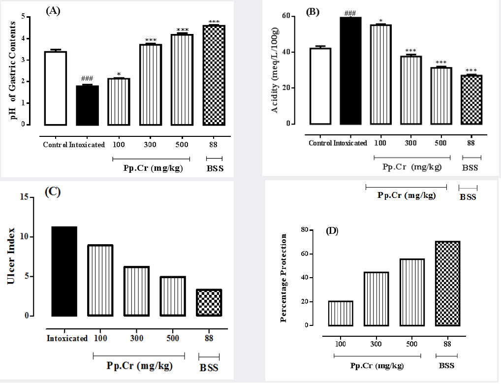
It was observed that Intoxicated group showed highly significant (p<0.001) reduction in pH of gastric contents as compared to normal control group. The pH of gastric contents in bismuth subsalicylate treated group was highest which was statistically highly significant (p<0.001) when compared with the intoxicated group. Pretreatment with 100, 300 and 500 mg/ kg of Pp.Cr produced highly significant (p<0.001) increase in pH dose dependently (as shown in Figure 1A) in comparison with intoxicated group. The effects of Pp.Cr are summarized in Table 1.

Figure 1: The graphs showing the effects of the crude extract of Prunus persica (Pp.Cr) alongwith bismuth subsalicylate (BSS), on (A) pH of gastric contents, (B) Acidity, (C) Ulcer index and (D) percent gastric protection in ethanol-induced gastric ulceration in rats.
lupinepublishers-openaccess-journal-gastroenterology-hepatology

Table 1: The effects of the crude extract of Prunus persica (Pp.Cr) alongwith bismuth subsalicylate, on pH, total acidity of gastric contents, ulcer index and % protection in ethanol-induced gastric ulceration in rats.

lupinepublishers-openaccess-journal-gastroenterology-hepatology

The values are expressed as mean ± SEM of six animals in each group and the results are analyzed using one way ANOVA. The values of treated groups are compared with those of intoxicated group. P values are considered as significant (*) if p<0.05 and highly significant (***) if p<0.001. The values of intoxicated group are also compared with normal control group and considered highly significant (###) if p<0.001.

b) Effects Pp.Cr on Total Acidity of Gastric Contents

Oral administration of the crude extract of Prunus persica (Pp. Cr), at the doses of 100, 300 and 500 mg/kg, as well as bismuth subsalicylate (88 mg/kg) for ten days showed the dose-dependent reduction in total acidity of gastric contents (Table 1). The rise in total acidity in intoxicated group was highly significant (p<0.001) as compared to normal control group. BSS treated group exhibited significant (p<0.001) reduction in total acidity as compared to the intoxicated group. Pp.Cr 100 mg/kg significantly (p<0.05) reduced the total acidity when compared with intoxicated group. The total acidity values for Pp.Cr treated groups at the doses of 300 mg/kg and 500 mg/kg showed highly significant reduction in total acidity as compared to intoxicated group; i.e. P<0.001 as depicted in Figure 1B. The results Pp.Cr (500 mg/kg) were comparable with BSS treated group.

c) Effects of Pp.Cr on Ulcer Index

The crude extract of Prunus persica (Pp.Cr) was observed to decrease the ulcer index in dose- dependent fashion as summarized in Table 1. The value of ulcer index in intoxicated group was 11.21 while it was 3.3 in BSS treated group. The values of ulcer indices in Pp.Cr treated groups were found to be; 8.93, 6.20 and 4.95 at the doses of 100, 300 and 500 mg/kg respectively. The results are elaborated in Figure 1C.

d) Effects of Pp.Cr on Gastric Percent Protection

The percentage protection shown by Pp.Cr after 10 days of the administration was 20.33%, 44.69%, and 55.84% at the doses of 100, 300 and 500 mg/kg as shown in Table 1. The gastric percent protection at different doses of Pp.Cr that is dose-dependent (Figure 1D). The standard control group showed the highest gastroprotective effects; i.e. 70.56%.

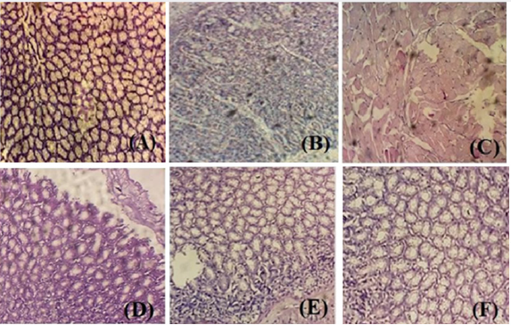
Figure 2: The effects of different doses of Pp.Cr and bismuth subsalicylate on ethanol-induced histopathogical alterations (A) normal control, (B) intoxicated (ethanol 5 ml/kg), (C) Pp.Cr 100 mg/kg + intoxication, (D) Pp.Cr 300 mg/kg + intoxication, (E) Pp.Cr 500 mg/kg + intoxication and (F) BSS 88 mg/kg + intoxication
lupinepublishers-openaccess-journal-gastroenterology-hepatology

Histopathological examination of stomach from one animal of each group was performed to confirm the ulcer formation and to evaluate the gastroptrotective effects of different doses of Pp.Cr. No gastric mucosal damage was observed in normal control group (Figure 2A). Intoxicated group showed the typical characteristics of ethanol-induced ulcer; i.e. gastric mucosal damage, edema and leucocytes infiltration of the mucosal layer (Figure 2B). Histological examination indicated that pretreatment with different doses 100, 300 and 500 mg/kg (Figures 2C-2E) of Pp.Cr and bismuth subsalicylate (88 mg/kg, Figure 2F) showed the gastroprotective effects as evidenced by reduction in mucosal damage and edema. Pp.Cr was found to prevent the mucosal damage significantly

Protective Effects of Pp.Cr against Cysteamine-Induced Duodenal Ulceration

Effects of Pp.Cr on Severity Score of Duodenal Ulcer

Table 2: The effects of the crude extract of Prunus persica (Pp.Cr) along with cimetidine on severity score, ulcer index and duodenal percent protection in cysteamine-induced duodenal ulceration in rats.

lupinepublishers-openaccess-journal-gastroenterology-hepatology

The values are expressed as mean ± SEM of six animals in each group and the results are analyzed using one way ANOVA. The values of treated groups are compared with those of intoxicated group. P-values are considered as significant (*) if p<0.05, more significant (**) if p <0.01 and highly significant (***) if p<0.001.

The effects of Pp.Cr on severity index of duodenal ulcer are tabulated in Table 2. The mean value of severity score in intoxicated group, treated with cysteamine, was calculated as 2.5±0.22. Cimetidine (100 mg/kg), an antisecretory agent, selected as the standard drug for the study, showed highly significant (p<0.001) reduction in severity score when compared with intoxicated group. The effects of Pp.Cr (100 mg/kg) on severity score were insignificant (P>0.05) when compared with intoxicated group. Pp.Cr (300 mg/kg) was able to reduce severity score significantly (p<0.05) as compared with intoxicated group. Pp.Cr (500 mg/kg) showed more significant (p<0.01) reduction in severity score as shown in Figure 3A.

Effects of Pp.Cr on ulcer index

Cysteamine produced the inflammatory changes and hemorrhagic spots in duodenum. No duodenal mucosal damage was observed in normal control group. The crude extract of Prunus persica (Pp.Cr) decreased the ulcer index in dose-dependent fashion as elaborated in Table 2. The value of ulcer index in intoxicated group was calculated as 4.5; whereas it was 1.5 in cimetidine treated group. The effects of Pp.Cr on ulcer indices in rats were found to be; 3.83, 2.99 and 2.16 at the doses of 100, 300 and 500 mg/kg respectively. The effects of Pp.Cr (500 mg/kg) on ulcer index were comparable with cimetidine treated group as depicted in Figure 3B.

Figure 3: graphs showing the effects of the crude extract Prunus persica (Pp.Cr) along with cimetidine (CIM), on (A) severity score, (B) Ulcer index and (C) percent protection in cysteamine-induced duodenal ulceration in rats.

lupinepublishers-openaccess-journal-gastroenterology-hepatology

Effects of Pp.Cr on Duodenal Percent Protection

Oral administration of Pp.Cr showed the protection against cysteamine-induced duodenal ulcer. The percentage protection observed at the dose of 100 mg/kg was 14.88%. The percentage protection was calculated as 33.55% at the dose of 300 mg/kg. Similarly, at the dose of 500 mg/kg percentage protection was found to be 52% as depicted in Figure 3C. The standard drug cimetidine showed significant antiulcer effects with percentage protection of 66.66% (Table 2). The antiulcerogenic potential of Pp.Cr at the dose of 500 mg/kg was comparable with that of standard control group.

Effects of Pp.Cr on Histopathological Parameters

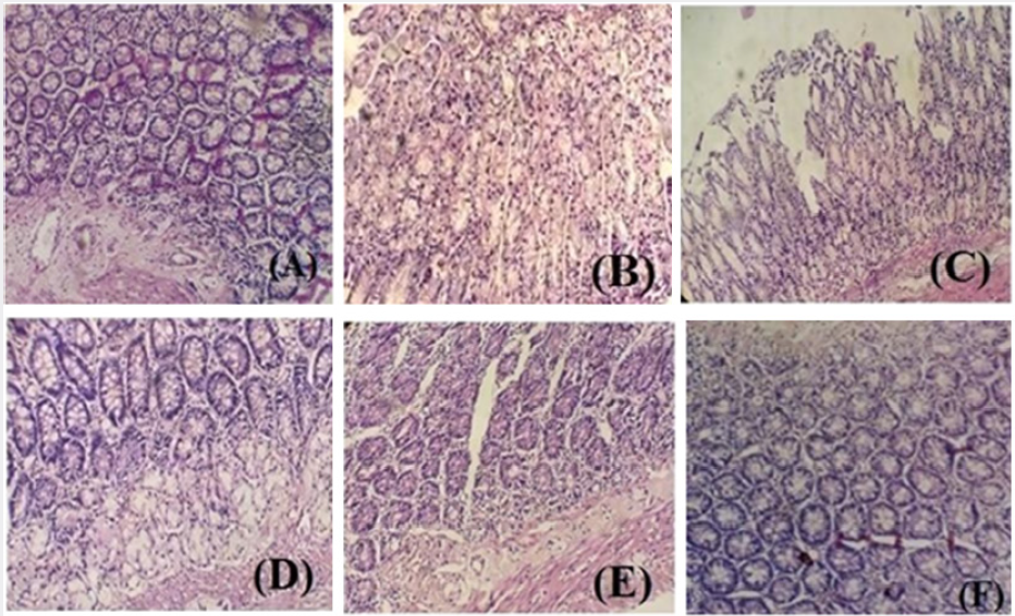
No mucosal damage was observed in normal control group (Figure 4A). Intoxicated group showed inflammation and hemorrhagic spots in duodenal mucosa (Figure 4B). Histological examination indicated that pretreatment with different doses (100, 300 and 500 mg/kg) of crude extract of Pp.Cr and cimetidine (100mg/kg) showed the antiulcer effects as evidenced by reduction in inflammation and edema. Pretreatment with cimetidine showed the remarkable protection against cysteamine-induced duodenal ulcer (Figure 4F). Pp.Cr prevented the mucosal damage significantly (Figures 4C-4F).

Figure 4: The effects of different doses of Pp.Cr and cimetidine on cysteamine-induced histopathogical alterations (A) normal control, (B) intoxicated (cysteamine hydrochloride 600 mg/kg), (C) Pp.Cr 100 mg/kg + intoxication, (D) Pp.Cr 300mg/kg + intoxication, (E) Pp.Cr 500 mg/kg + intoxication and (F) CIM 100 mg/kg + intoxication.
lupinepublishers-openaccess-journal-gastroenterology-hepatology

Discussion

Ethanol has been reported to enhance the gastric acid secretion and reduce the blood flow, leading to microvascular injuries. It also causes the necrosis in the gastric mucosa, reducing the secretion of bicarbonates and gastric mucus [20]. Ethanol has also been found to induce mucosal cell surface damage and increases the permeability of mucosal capillaries, resulting in mucosal hemorrhage. These effects then allow the gastric acid back diffusion hence; exacerbate the mucosal damage already caused by ethanol [21]. So any substance that decreased the acid secretion or protects the mucosal layer could be gastroprotective. Standard drug, bismuth subsalicylate, showed the gastroprotective effects by forming a protective coating over gastric mucosa especially ulcerated sites to protect it from hydrolytic action of acid and pepsin. It also exerted the cytoprotective action by stimulating the prostaglandin biosynthesis, mucus and bicarbonate secretion [22]. The crude extract of Prunus persica (Pp.Cr) showed the presence of alkaloids, phenols and flavonoids. Alkaloids have been reported to possess the antiulcerogenic activity because of their ability to reduce gastric acid secretion, increase the pH of gastric contents and protect the mucous membrane [23]. Phenols have been reported to possess protective and therapeutic potential for peptic ulcer by up-regulating tissue growth factors and prostaglandins, down-regulating anti-angiogenic factors, enhancing endothelial nitric oxide synthase-derived NO, suppressing oxidative mucosal damage, amplifying antioxidant performance, antacid, and antisecretory activity and increasing endogenous mucosal defensive agents [24]. Flavonoids have been found to prevent the formation of free radicals and reduce injuries caused by oxidative stress [25]. Furthermore, Pp.Cr showed the presence of tannins which have been reported to produce layer over gut lining by precipitating micro proteins at ulcerative site, thus inhibit GIT secretions and also provide protection to gastrointestinal mucosa from irritants [26]. Tannins have also been found to resist the action of proteolytic enzymes produced by Helicobacter pylori [27].

Phytochemical analysis of Pp.Cr showed the presence of glycosides which have been found to reduce the expression of TNF-α [28]. Hence, the antiulcerative could be attributed to these secondary metabolites. H. pylori infection of the antral mucosa stimulates the release of gastrin by inhibiting the secretion of somatostatin. Gastrin has ability to enhance maximal acid secretory capacity. The administration of the crude extract of Prunus persica (Pp.Cr) was found to increase the pH of gastric contents, indicating that the extract could act as antisecretory agent because gastric acid back diffusion was one of major factors involved in the pathogenesis of ethanol- induced gastric ulcers. However, another possible mechanism of action could be its cytoprotective action; i.e. enhancement of the protective elements of the gastric mucosa; e.g. mucus and bicarbonate secretions in the same way as of bismuth subsalicylate, the standard drug. Exposure of gastric mucosa to necrotizing agents (e.g. ethanol) reduces these mucus and bicarbonate secretions, making gastric mucosa more susceptible to damaging action of gastric acid and pepsin. Ethanol also causes an increase in intracellular level of calcium, resulting in stimulation of gastric secretions [29]. The tannins present in the Pp.Cr could be the reason behind the decreased mucosal damage. The crude extract of Pp.Cr could also exert gastroprotective effects through cytoprotective action as ethanol has been reported to exert its direct toxic effects on gastric mucosa by reducing the secretion of bicarbonate ions and mucus. Ethanol has also been found to cause the gastric mucosal damage by producing free radicals leading to increased lipid peroxidation [30]. The P. persica plant extract could also have antioxidant potential that prevented the mucosal damage.

As peptic ulcer encompasses both gastric and duodenal ulcer, antiulcerogenic potential of the crude extract (Pp.Cr) was also assessed against cysteamine-induced duodenal ulcer alongwith ethanol-induced gastric ulcer. The cysteaime-induced duodenal ulcer model is a widely used and reproducible model of peptic ulcer because location, histopathology and pathophysiology of this chemically-induced ulcer are same as that of duodenal ulcer in humans [30]. Cysteamine has been studied to increase the gastrin level and thus increase the gastric acid secretory output. Cysteamine has also been reported to reduce the bicarbonate secretion and alkaline mucus secretion from the duodenal Brunner’s gland. Moreover, cysteamine-induced delayed gastric emptying causes the accumulation of gastric secretions in stomach. Due to the absence of peristaltic movement, these secretions do not mix with gastric contents. When these concentrated secretions enter the duodenum, where bicarbonate secretion is already reduced by cysteamine, they promote the ulcer formation [30]. The preventive effects of Pp.Cr in cysteaime-induced duodenal ulcer could be due to the presence of various secondary metabolites such as alkaloids, glycosides and tannins. The scavenging effects of flavonoids and phenols present in the Pp.Cr may also the reason of protective outcomes in duodenal ulcer. The histopathological studies of both stomach and duodenum further confirm the results.

Read More About Lupine Publishers Journal of Gastroenterology and Hepatology Please Click on Below Link:  https://currenttrendsingastroenterology.blogspot.com/

Lupine Publishers| Modeling the Temperature of the Evacuation Chamber with Artificial Neural Networks

 Lupine Publishers| Journal of Robotics & Mechanical Engineering


Abstract

This investigation approaches the artificial neural networks applied to the ore drying process in carbonate-ammonia leaching. To carry out this research, the main variables that characterize the process were identified. Besides, it was collected the data that comprise a whole month of facility´s operation. Furthermore, it was developed a regression analysis backwards, step by step, which allowed to determine that the linear correlation coefficient did not reach values higher than 0,62. In addition, it was pinpointed a two layered feed - forward back propagation neural network to model the temperature. Thins one reached the correlation coefficient values of 0,97 during its training and 0,95 in validation, as well as 0,87 in its generalization.

Keywords: Artificial Neuronal Network; Regression; Feed-Forward Backpropagation; Mineral Drying

Introduction

In a global context, nowadays, modern control systems play a fundamental role when developing solutions to issues or problems presented in domestic and industrial applications. The main contributions of modern control systems at industrial level contribute to technological innovation, profitability and maintainability of the controlled processes. Within the advanced control strategies under investigation to automate complex processes are: adaptive control, predictive control based on models, robust control, and intelligent control, among others. Intelligent control relies on several techniques such as: fuzzy logic, evolutionary algorithms, and artificial neural networks. Artificial neural networks can be used effectively and accurately for modeling systems with complex dynamics, especially for nonlinear processes that vary over time. The growing interest in neural networks is due to its great versatility and the continuous advance in network training algorithms and hardware [1-4]. The nickel producing companies have continuous processes of great complexity that require automation to achieve a greater efficiency in their productions. In the process of ore preparation, it is important to maintain a temperature control at the outlet of the dryer evacuation chamber, in order to obtain the mineral drying with an established humidity level of 4 to 5,5 %. It must also be ensured that the temperature at the outlet of the electrofilters is above the dew point temperature; to prevent the deterioration of electrofilters, which leads to high economic losses, from accelerating considerably. The inefficiencies in the control of the outlet temperature of the dryer evacuation chamber in the ore preparation process are taken as a research problem and as an objective to obtain an artificial neural model for the outlet temperature on the basis of the main input variables, using Matlab as a calculation tool.

Materials and Methods

Description of the Mineral Drying Process

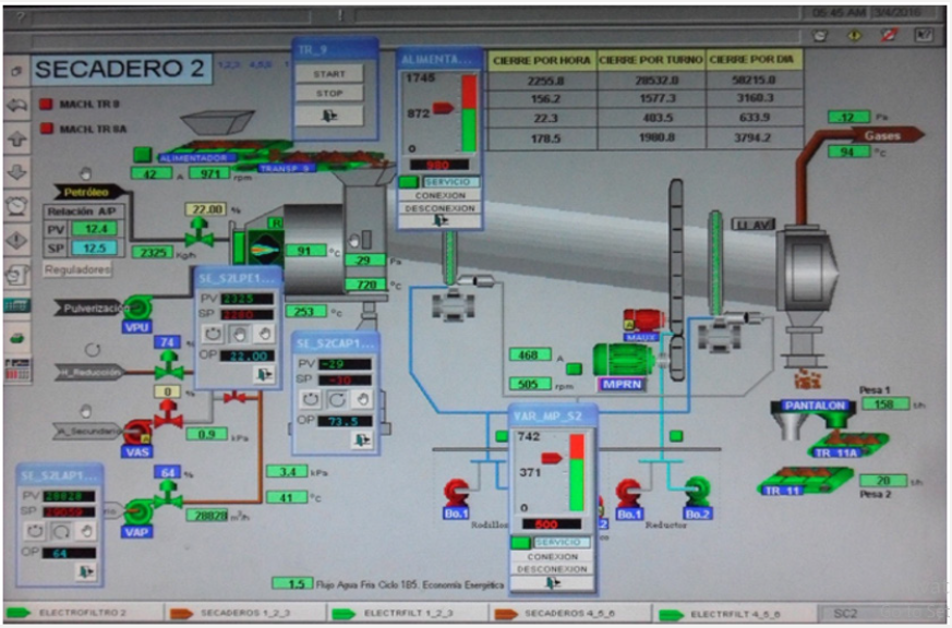
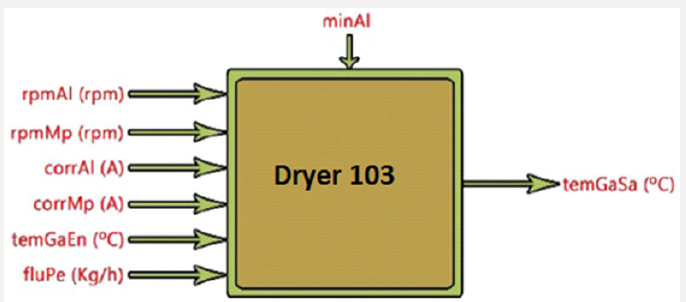
The drying of the ore is carried out in elongated cylinders formed by a combustion chamber where the hot gases that dry the ore are produced, and by the cylinder where the ore will receive the drying process. These drums (Figure 1) have in their interior lifting elements that are responsible for allowing the transfer of heat between the hot gas and the mineral, in addition the dryer drum has a motor system coupled to the body of this which allows it to rotate on its axis. The dryer drum externally rests on two wheels that has two pairs of roller. Internally the dryer is formed near the combustion chamber by guides or baffles welded to the body of the drum that are the ones that direct the mineral towards the outside of the cylindrical part of the drum [5]. The mineral dryer is a complex physical-mathematical modeling object with a large number of input and output parameters which are in a complex interdependence (Figure 2).

Figure 1: Schematic diagram of the dryer.

Lupinepublishers-openaccess-robotics-mechanical-engineering-journal

Figure 2: Structural diagram of the mineral drying process.

Lupinepublishers-openaccess-robotics-mechanical-engineering-journal

The Input Parameters in the Process are:

a) rpmAl - Feed motor speed [rpm].

b) rpmMp - Speed of the main motor [rpm].

c) corrAl - Feed motor power [A].

d) corrMp - Power of the main motor [A].

e) temGaEn -Temperature in incoming gases [ºC] (coming from the Reduction Furnaces Plant).

f) fluPe - Oil flow at the burner inlet [kg/h].

The Output Parameter is:

a) temGaSa - Oulet gas temperature [ºC].

In addition to the input and output parameters, it is important to highlight a specific disturbance of this process that influences it, which is: minAl - Mineral fed to the dryer. It is known that there are other parameters that are involved in the drying process of the ore and that in turn influence the temperature of the exhaust gases in the evacuation chamber (granulometry in the entrance mineral, humidity of the entrance mineral, exact amount of mineral fed to the dryer), but due to the process itself, they are not registered. Due to the automation existing in the process, the values of the process parameters are sensed by the instrument corresponding to each of them and the signal is sent to the computer located in the process control office. The data obtained along 1 month of operation, were recorded every 240s and processed with the Stat graphics Plus V 5.1 software.

Artificial Neural Networks

The determination of the type of artificial neural network, the number of layers and the number of neurons in each layer that best characterize the process of ore drying process was carried out through a trial and error process that plays with the number of neurons and the maximum permissible error. Through Matlab’s Toolbox (nnstart), the performance of artificial neural models was evaluated by using the mean square error and the correlation coefficient between the real values and those obtained by the network [6]. The objective was to provide the network with an adequate number of neurons in the hidden layer to learn about the characteristics of the possible relationships between the sample data. Through the trial and error process, it was identified the feedforward back propagation structure that provided better results. The proposed network consists of two layers: a hidden layer and an output layer. The output layer will only have one unit, which will indicate the value of the oulet gas temperature associated with each input vector presented to the network. The hidden layer will have a variable number of neurons.

Results and Discussion

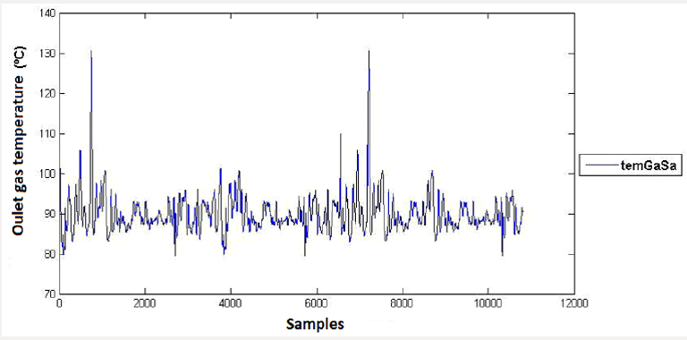
Figure 3 shows the behavior of the exhaust gas temperature in the evacuation chamber, between its minimum and maximum values of 79,59 and 130,51°C, respectively, for the month of work. Once the database was analyzed, the sample functions that evaluate the measures of central tendency and dispersion of the sample were determined through a descriptive statistical analysis (Table 1). The mathematical model that best represents the relationship between the variables analyzed. Table 2 shows the regression analysis for the output pulp density, where a 0,7correlation coefficient is observed.

Figure 3: Control chart for the dependent variable.

Lupinepublishers-openaccess-robotics-mechanical-engineering-journal

Table 1: Summary of the sample´s descriptive statistical analysis for one month.

Lupinepublishers-openaccess-robotics-mechanical-engineering-journal

Table 2: Regression analysis summary.

Lupinepublishers-openaccess-robotics-mechanical-engineering-journal

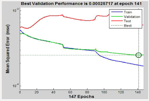
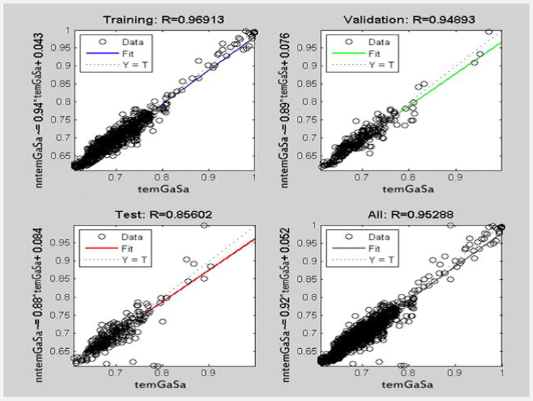
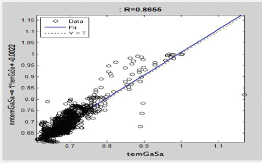
Figure 4 shows the training behavior of the network for the learning process, observing the training, validation and test curves, which converge to the iterations for an error of 0,00026. Figure 5 shows the behavior of the correlation coefficients for the training, validation, testing and adjustment of the artificial neuron network (it is assumed as an artificial neuronal model for the oulet gas temperature in the ore drying process “nntemGaSa” and the real temperature “temGaSa”). Figure 6 shows the generalization of the network with 1767 data not presented during training, where a 0,87correlation coefficient is observed.

Figure 4: Behavior of training and validation of the neural network.

Lupinepublishers-openaccess-robotics-mechanical-engineering-journal

Figure 5: Correlation coefficients of the neural network.

Lupinepublishers-openaccess-robotics-mechanical-engineering-journal

Figure 6: Network Generalization.

Lupinepublishers-openaccess-robotics-mechanical-engineering-journal

Conclusion

The capacity of the feed-forward back propagation network for the simulation of pulp sedimentation processes in the industry was demonstrated. The structure that best characterizes the behavior of the temperature in the exhaust gases of the evacuation chamber is characterized by two layers with 50 neurons in the hidden layer and one in the output layer, with the Levenberg Marquart learning method (trainlm), and the log-sigmoidal (logsig) and sigmoidal hyperbolic tangent (tansig).

Read More About Lupine Publishers Journal of Robotics and Mechanical Engineering Please Click on Below Link:  https://robotics-engineering-lupine-journal.blogspot.com/


Monday 29 November 2021

Lupine Publishers| The Role Effectiveness of Physical Activities on Mental Disorders in Students with Low Mobility Using General Health Questionnaire (Ghq)

 Lupine Publishers| Journal Of  Orthopedics and Sports Medicine


Abstract

Today, the emergence and growth of mental disorders are more noticeable in the student population due to the changes in health conditions of the world. Therefore, the purpose of this study was examining the role effectiveness of physical activities on mental disorders in students with low mobility using General Health Questionnaire (GHQ). The study was a causal-comparative research and has been conducted through the field method. The instrument was included the General Health Questionnaire-28 (GHQ-28). The statistical sample was included 430 male and female physical education and non-physical education students. The collected data was analyzed by descriptive and inferential statistical methods and analytical tests. The results showed that there was a significant difference between athlete and non-athlete students in all variable components of this study in two groups (P<0.05). It seems that regular physical activity can play an important role in students’ physical and mental health and students who participated in fitness and exercise programs have reported that their attitude and efficiency had a good state. Therefore, the increase of motivation through culture and the promotion of public awareness about the benefits of physical activity can be considered as one of the options for the development of the student sport.

Keywords: Anxiety; Social Function; Physical Problems; Depression; Physical Activities

Introduction

Today, the poor movement that has been created as a result of tremendous improvements in technology and mechanization of life and different jobs affects lifestyle in human societies. These very serious changes have had profound effects on human relationships at different levels of family and relationships with friends [1]. If we look at human’s physical construction, we will realize that human need naturally different kinds of physical activities for his/her entire development and evolution and the lack of adequate mobility and the lack of proper physical activities will disrupt different body systems. This great development in terms of health and well-being is one of the most important problems of today’s human [2]. This is very important for students, especially for students who are studying in a Master’s degree. Students should have a physical and mental health due to their role in social, cultural, and economic structures and their preparation to participate in social activities [3]. Today, the World Health Organization (WHO) considers a multidimensional concept for the concept of health multi-dimension and knows it as a positive concept based on the interests of individual and social sources and physical abilities. The WHO expresses that health is the welfare state and mental, physical, and social well-being and it is not only the lack of disease or body defects [4]. According to the definition of this organization, Health have physical, social, mental, emotional, and spiritual components. All five components have the interaction with each other for a healthy and strong person, so that if we give up one of them, we will be out of balance and our health will suffer in all domains [5, 6]. Therefore, dimensions of this definition (mental, physical, and social wellbeing and not merely the lack of disease) have the interaction with each other and these dimensions cannot be distinguished with a clear boundary [7]. There are indicators for the physical health and social welfare in many countries that those are reviewed and revised every few years, but the complexity and difficulty of definition often leads to neglect and ignore this in mental health [8]. The lack of mental health causes that a person suffers pains and physical and emotional symptoms such as isolation, headache, worry, anxiety, difficulty in falling asleep, and daily dysfunction [9]. Studies show that one of every two people will be prone to depression, so studies about improvement of mental health and its related factors are the most important studies in psychology [10]. In past decade’s huge changes were in the industrial world and considerably surround the lifestyle of many human communities. This has caused people to reduce physical activities, and have problems and diseases such as obesity, muscle weakness, cardiovascular and respiratory disease [11].

However, due to developments on the world health situation there is less risks to face this kind of problems. The considerable problem is emergence and growth of mental disorders .Purpose of mental health is the certain aspects such as human intelligence, mind and thought. According to Kameo someone has mental health, that who has no symptoms of anxiety and disability, able to establish communication with others and able to deal with the pressures of life [12]. We can found that the need for research in field of mental health and the survey of the effective factors on it is necessary with a look at the implications of urban lifestyle, apartment house, and the mechanization of societies in recent years and its negative effects especially on individuals’ mental health in a society and unresolved challenges [13]. The participation in sport activities as a scientific approach can increase individuals’ happiness and mental health. The various studies have shown that sport activities have psychological, emotional, and social benefits in addition to physical benefits, for example, the reduction of anxiety and depression, the increasing of sleep duration and better social relationships have been reported in different studies [14]. If a human has not enough calm to draw lost physical and personal resources and exposed to these severe psychological pressures repeatedly, the process of destruction will begin and then he/she will be sensitive to the disease especially psychosomatic diseases. Therefore, a human finds him/herself in anxiety without justification, fatigue, depression or feelings of dissatisfaction, and aimless, and he/she will hurt to relationships between individuals in the same way. One of the ways that psychologists introduce for the prevention and treatment of mental health problems is the role of physical activities in mental health, because the industrialization of societies and the reduction of physical activity in individuals have been revealed the need of sport more than ever and an extensive attention has been paid to exercise especially the role of exercise in psychological issues.

Research evidence show some environmental controversies that affect students’ mental health such as the desire for different social entertainments against willingness to study, the desire for physical superiority against organ limitations, the need to the lessons development against the feel of incompetence, the fear of personality expression against the desire for self-esteem, and job selection [15,17]. According to researchers’ idea who has mental health is a person that is distant from anxiety and disability symptoms and she/he can establish a constructive relationship with others, and is able to cope with life stresses [18]. On the other hand, conducted studies in the field of psychology and exercise have shown that exercise and physical activities are one of the effective methods for the prevention and treatment of mental illnesses [16,19]. Scientific evidences have shown that the participation in physical activities and the increasing of cardiovascular fitness are considered as an important factor in the improvement of mental health and mood. Nine factors are important in the creation of happiness and mental health that the participation in physical activities and exercise is one of the most important factors studied psychological and physical indexes in women that they concluded that aerobic activities and trainings had many benefits for behaviour and mood. Also, they stated that physical activities s were associated with the reduction of stress, tension, and the increasing of self-esteem [19].

Mental health is very importance for students in every community, because they are the future of any society. Mental disorders can lead to academic failure or the dropout. Students due to the specific conditions of students such as far away from family, getting into large and stressful collections, Economic problems and Lack of sufficient income, high volume courses, and intense competitions are prone to lose their mental health. They need an appropriate intervention for coping with such stressful situations. Regular physical activity at a moderate level course is one of strategies that scholars and researchers recommend to maintain and promote mental health [11,12] mental health (physical symptoms, anxiety, depression and dysfunction) of students in individual and team athletes with non-athletic and found out the amount of social dysfunction of team sports athletes than individual athletes and non-athletic and depression in non-athletes was more than two other groups. Also comparing each of the subscales between boys and girls athletes found that boys mean depression was more than girls [20].

Research results from Wang et al shows that any form of physical activity can protect and provide mental health. Sport in creating positive change, satisfaction with their sense of competence and efficiency play important role that are the component of mental health [14]. In his study concluded that following physical exercises, characters such as anxiety, depression and self-esteem varies to improvement that without regard to types of exercise activities that classified as aerobic have the most effects in terms of physiological and psychological.

Anxiety and depression are considered as trends and common mental disorders. So that Ghaffari (1384) studies results shows that non-athletes depression is less than professional and nonprofessional athletes. Studies suggests that physical activity and exercise has effect in promoting mental health, reducing depression,increasing welfare, mental and social health, self-confidence, self-belief and self-discovery. This study has compared male and female athlete and non-athlete students’ mental health, since many researchers have emphasized the role of physical activity and exercise as an instrument for the prevention of diseases and mental disorders and due to the existence of significant differences in men and women’s physiological characteristics. According to the findings of this study and differences of effectiveness in students due to studied different factors, we hope that can provide more effective and coherent programs for more participation in sports activities of different groups with an attention to these differences to improve students’ dimensions of mental health and the reduction of mental and psychological stresses in addition to the improvement of their physical abilities with the participation in sports activities.

Methods

This descriptive-analysis study was conducted on the causalcomparative method. The instruments of this study were included Goldberg’s General Health Questionnaire-28 (GHQ-28) and a demographic questionnaire that its validity and reliability have been proven in different studies. Goldberg’s General Health Questionnaire measures four categories of non-psychotic disorders such as somatic symptoms, anxiety and insomnia, social dysfunction and depression. Likert’s scoring method (0 to 3) was used for each case. The scoring criteria were the intensity of the evaluated symptoms and a higher score indicated a high intensity of the symptoms. According to this, a person with a score of 23 and lower score was considered as a healthy person and with a score of 24 and higher score was considered as suspected person who had a disorder. The statistical population of this study was all male and female physical education and non-physical education students in Universities Azad Islamic Branch of Gilan province. The statistical sample of this study was included 430 students that they were selected by randomized multi-stage cluster sampling. Athlete students were individuals who participated at least 3 times a week in sports programs and activities and non-athlete students were individuals who in this study did not have any physical activity or who their activities were limited to daily routine of life and education. The collected data were classified by descriptive statistical methods and were analyzed by dependent T-test. The SPSS software (version 21) was used for data analysis (α≤0.05).

Results

According to the results of the statistical analysis of this study, the status of general health indicators among physical education and sport sciences students was in the range of 19.68% that 27.11% was abnormal and 72.89% was normal in terms of general health status. Also, the results of statistical analyzes on the general health status among non-physical education students was in the range of 8.21%) that 42.17% was statistically abnormal and 57.83% was a good status. The male subjects’ age mean was 26 ± 3 and female subjects’ age mean was 24±7 in this study. The subjects’ age distribution was 63.17% in the age range of 20-30 years and 36.83% in the age range of over 30 years. 48.72% of physical education and sport sciences students were male students and 51.28% of them were female students. 56.11% of non-physical education students were male students and 43.89% of them were female students. The results in (Tables 1-3) showed that there was a significant difference between physical education and nonphysical education students (P<0.05).

Table 1: The results of independent T-test in the relationship with female Students’ General health subscales.

Table 2: The results of independent T-test in the relationship with male Students’ General health subscales.

Table 3: The results of independent T-test in the relationship with physical education and non-physical education Students’ mental health subscales.

Discussion

the purpose of this study was examine the role effectiveness of physical activities on mental disorders in students with low mobility using General Health Questionnaire (GHQ).The results of this study showed that there was a significant difference between physical education and non-physical education teachers in mental health (P<0.05). It means that physical education teachers had a higher mental health score in comparison with non-physical education teachers. This result is consistent with the results of Hamer Rass and Hayes, Anonymous, Narimani, Ahmadi and Pirhayti and Nariman’s study [21-23].

Undoubtedly, the establishment of a healthy and happy society depends on the physical and mental health of the members in the society and needs to efforts of healthy, efficient, and thoughtful human forces. In this regard, the human and specialist resources in education that often have acceptable scientific and practical knowledge and abilities need to be able to overcome threatening factors of general health by the management of their organizations. Otherwise, the reduction of their capability will be inevitable. In this regard, the highest harm will be for students who are trained and the educational goals will encounter with serious problems. On the other hand, teachers will be aware of harmful problems and resources in their profession with the cognition of these factors and will use coping methods to deal with its undesirable effects.

The results of this study are consistent with the results of Bakhshalipour et al that they examined the effect of aerobic training on non-athlete postmenopausal women’s mental health. On the other hand, the difference in mean of mental health in the comparison of male and female athlete and non-athlete students did not show a significant difference in any of the subscales, but it was significant in all subscales with the comparison of male athlete and non-athlete students. So that male athlete students’ mental health had better condition than the male non-athlete students. This results is consistent with the results of study. They stated that male athlete students who participate regularly in physical activities such as walking, jogging, swimming, aerobics, or playing in sports teams can be able to do boring tasks in a longer period than inactive male students that this can be due to adaptive responses in a body as a result of regular exercise. There is a lot of evidence about the benefits of regular physical activity for general health. Studies show that intense and regular exercise is associated with less stress and anxiety. Also, people who participate in fitness and exercise programs report that their attitudes and efficiency are better at work; for example, they make fewer mistakes. It seems that exercise can play an important role in students’ physical and mental health due to all subjects are students who study at master’s level and they do many research works, and modern life conditions that have physical abnormalities such as back pain, joint damage, and diseases such as obesity and cardiovascular diseases. Therefore, the increasing of motivation through culture and the promotion of general knowledge about the benefits of physical activity can be considered as one of the options for the improvement of student sport. A program that may not have been seriously considered so far. According to the results of these findings, it seems that one of the factors that cause student athletes have better mental health than non-athletic students is their participating in healthy activities such as sport activities. Tuckers research results Showed, that physical fitness significantly reduces mental disorders in people.

Conclusion

Generally, Athletes that, participate different sports field based on personal interests and tastes the exposure groups and sports cooperation and cooperation with them and enjoy the new friendships. This may be cause removing such feelings and dissociable and helps create collective spirit, while non-athletic students deprived this opportunity.

Read More About Lupine Publishers Journal of Orthopedics and Sports Medicine Please Click on Below Link: https://lupinepublishers-orthopedics.blogspot.com

Wednesday 24 November 2021

Have a Blissful Thanksgiving!!!


On this Thanksgiving I wish you Happiness, Beautiful Moments, The warmth of your Family, Healthy Life, Love and Safety. Look around you and be thankful for what you have and appreciate life. We are very thankful for the tremendous support from our Honorable Editors and beloved Authors who supported our Organization in every aspect till now and hope it would continue still the same .

And may you get to experience glimpses of the divine and spread smiles, love, support to everyone you meet. Wishing everyone a Blessed Thanksgiving Day from our Lupine Publishers family

Sunday 21 November 2021

Lupine Publishers| Associated Risk Factors in Pre-diabetes and Type 2 Diabetes in Saudi Community

 Lupine Publishers| Journal of Diabetes and Obesity


Abstract

Background and Objective: The prevalence and incidence of type 2 diabetes mellitus (T2DM) are increasing worldwide. Pre diabetes is a high-risk state for the development of diabetes and its associated complications. This study aims to determine the associated risk factors among T2DM and pre diabetes patients among adult Saudi population.

Methods: For the present study, we analyzed participants who are older than 20 years old and had undergone a blood test to assess HbA1c. A total of 1095 were selected to be enrolled for the present study. All patients were from the population of the Primary health and Diabetic Centres at King Fahad Armed Forces Hospital. Participants were defined as having T2DM according to self-report, clinical reports, use of anti diabetic agents and HbA1c (≥6.5). Non T2DM participants were divided into normoglycemic or pre diabetic group as follows: HbA1c < 5.7, (normoglycemic) or HbA1c 5.7-6.4 (pre diabetes). Laboratory assessments included HbA1c, lipids, creatinine and urinary micro albumin.

Main results: Of the 1095 participants analyzed, 796 were women (72.7%). Age was 45.1±11.1 and BMI was 30.7±5.7. Hypertension had been diagnosed in 415 (38.2%) participants. Blood measurements revealed the following values: creatinine 68.2±22.0umol/L , Urine micro albumin (g/min) 55.4±200.3, total cholesterol levels 4.9±1.0mmol/L, high density lipoprotein 1.3±0.3mmol/L, triglyceride levels 1.5±0.7 and low density lipoprotein 3.0±0.9mmol/L. Of the overall 1095 analyzed participants, pre diabetes was present in 362(33.1%), 368(33.6%) were classified as T2DM and 365 (33.3%) as normoglycemic. When comparing pre diabetic with normoglycemic and T2DM population, pre diabetic subjects were more likely to have hypertension and higher triglyceride than normoglycemic but less than T2DM subjects. In addition, pre diabetic patients compared with T2DM ones had higher levels of low density lipoprotein and high density lipoprotein. Logistic regression analysis showed no significant association of any of the co variables with normoglycemic subjects in front of the pre diabetic reference group, whereas the odds of being in the diabetic group gets multiplied by 7.56 for each unitary increase in male gender (p< 0.0001, OR: 7.56, 95% CI 3.16-18.23). Also, individuals with hypertension had higher odds of being in the DM group than in the prediabetic (p<0 .0001, OR: 6.06, 95% CI 3.25- 11.28). Age of subjects had lower odds of being in the DM group than in the pre diabetic (p<0 .0001, OR: 0.85, 95% CI (0.82-0.89).

Conclusion: This study found the major clinical differences between pre diabetic and T2DM Patients were the higher hypertension and hypertriglyceridenia in the T2DM patients. Clearly, despite the small sample size, this study has posed important public health issues that require immediate attention from the health authority. Unless immediate steps are taken to contain the increasing prevalence of obesity, diabetes, pre diabetes, the health care costs for chronic diseases will pose an enormous financial burden to the country

Keywords: Type 2 Diabetes; Pre diabetes; Risk factors

Abbreviations: T2DM: Type 2 Diabetes Mellitus; IFG: Impaired Fasting Glucose; BMI: Body Mass Index; HTN: Hypertension; AER: Albumin Excretion Rate; DN: Diabetic Nephropathy; OR: Odds Ratio; CI: Confidence Interval; I-IFG: Isolated Impaired Fasting Glucose

Introduction

Diabetes mellitus is a major cause of excess mortality and morbidity. The prevalence and incidence of type 2 diabetes mellitus (T2DM) are increasing worldwide [1]. T2DM patients have a higher risk of developing microvascular and macrovascular disease than the general population. The occurrence of these complications depends largely on the degree of glycemic control as well as on the adequate control of cardiovascular risk factors [2-5]. In Saudi Arabia, primary epidemiological diabetes features are not different. The diabetes mellitus prevalence among adult Saudi population has reached 23.7%, a percentage being the highest across the globe [6,7]. Statistics regarding the increasing trend of diabetes and pre diabetes in the world have also been observed in Saudi Arabia. As per the WHO country profile 2016, 14.4% of Saudi population has diabetes, while prevalence in males is 14.7% [8]. In 2015, the prevalence of pre diabetics was found to be 9.0% in Jeddah with 9.4% in men, while for diabetes, it was 12.1% with 12.9% adult male population suffering from it [9]. Another study conducted in Saudi population revealed that the diabetes prevalence in their study was found to be 25.4%, while impaired fasting glucose (IFG) was 25.5%. The strongest risk factors were age > 45 years, high triglycerides levels, and hypertension [10].

Pre diabetes is a high-risk state for the development of diabetes and its associated complications [11-13].

Recent data have shown that in developed countries, such as the Unites States and the United Kingdom, more than one-third of adults have pre diabetes, but most of these individuals are unaware they have the condition [14-16]. Once detected, pre diabetes needs to be acknowledged with a treatment plan to prevent or slow the transition to diabetic [17,18]. Treatment of pre diabetes is associated with delay of the onset of diabetes [19]. Detection and treatment of pre diabetes is therefore a fundamental strategy in diabetes prevention [11].

Current recommendations for pre diabetes screening by the American Diabetes Association focus nearly exclusively on adults who are overweight or obese as defined by body mass index (BMI) until the patient meets the age-oriented screening at 45 years [11]. Further, the recently released recommendation from the US Preventive Services Task Force regarding screening for abnormal glucose levels and T2DM limits screening to individuals who are overweight or obese [20]. This focus on obese or overweight individuals, although obesity and pre diabetes have shown trends of increasing prevalence. United States Preventive Services Task Force has recommended screening of diabetes in adults devoid of precise symptoms and in individuals with BP higher than 135/80mmHg [21]. This study aims to determine the associated risk factors among T2DM and pre diabetes patients among adult Saudi population.

Methods

For the present study, we analyzed participants who are older than 20 years old and had undergone a blood test to assess HbA1c. A total of 1095 were selected to be enrolled for the present study. All patients were from the population of the Primary health and Diabetic Centers at King Fahad Armed Forces Hospital. Participants were defined as having T2DM according to self-report, clinical reports, use of anti diabetic agents and HbA1c (≥6.5) [11]. Non T2DM participants were divided into normoglycemic or pre diabetic group as follows: HbA1c<5.7, (normoglycemic) or HbA1c 5.7-6.4 (pre diabetes) [11]. 362 subjects were found to be pre diabetic. Almost similar number of normoglyceic and T2DM subjects was selected to be analyzed for comparison. All data were collected by personal interview and on the basis of a review of electronic medical data. Weight (kg) and height (cm) were measured by physician and nurse interviewers and recorded. Overweight and obesity were defined as BMI 25-29.9 and ≥30.0kg/m2 respectively [22]. Blood Pressure readings were within a gap of 15 minutes using a mercury sphygmomanometer by palpation and auscultation method in right arm in sitting position. Two readings were taken 15 min apart and the average of both the readings was taken for analysis. Hypertension (HTN) was also diagnosed based on anti HTN medications or having a prescription of antihypertensive drugs and were classified as Hypertensive irrespective of their current blood pressure reading or if the blood pressure was greater than 140/90 mmHg i.e. systolic BP more than 140 and diastolic BP more than 90 mm of Hg – Report of the American College of Cardiology/American Heart Association Task Force on Clinical Practice Guidelines [23]. Laboratory assessments included HbA1c, lipids, creatinine and urinary micro albumin. HbA1c was expressed as percentage. High performance liquid chromatography was used. Fasting serum lipids were measured on a sample of blood after fasting for 14 hours. We used the enzymatic method for determining the cholesterol and trigylcerides levels. Diabetic nephropathy (DN) was assessed by measurement of mean albumin excretion rate (AER) on timed, overnight urine collections. We use a polyclonal radioimmunoassay for albumin measurement. DN is defined as an albumin excretion rate of >20g/min in a timed or a 24hr urine collection which is an equivalent to >30 mg/g creatinine in a random spot sample.

Statistical Analysis

Univariate analysis of demographic and clinical laboratory was accomplished using one-way analysis of variance (ANOVA) with posy hoc analysis between variables, to estimate the significance of different between groups where appropriate. Chi square (X2) test were used for categorical data comparison. The adjusted odds ratio (OR) with a 95% confidence interval (CI) was calculated. In order to evaluate the adjusted association of aforementioned factors on being normoglycemic or diabetic in relation to the pre diabetes group, a multinomial logistic regression model was fit, in which the categorical dependent variable was normoglycemia, pre diabetes or T2DM(with pre diabetes as the reference category), and significant variables in bivariate analyses were included as explanatory variables. Despite of the ordinal nature of the dependent variable, ordered logistic regression was not adjusted because the aim of the study was not the association of factors with a latent degree of diabetes but the differential profile of pre diabetes in front of normoglicemia and diabetes. As all the participants were the same age, adjusting for age was not applied. All statistical analyses were performed using SPSS Version 22.0. The difference between groups was considered significant when P<0.05.

Results

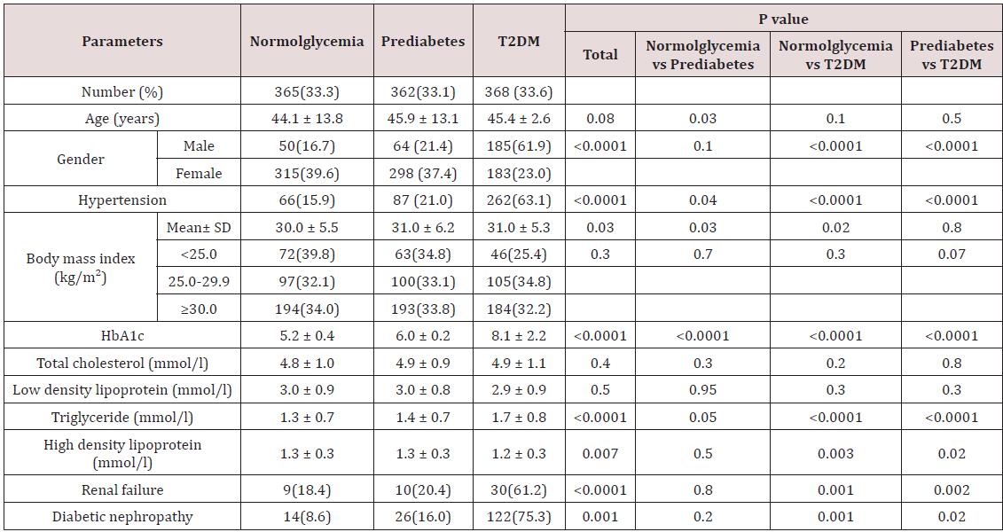
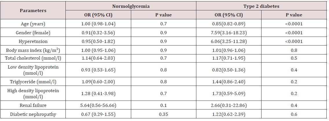
Of the 1095 participants analyzed, 796 were women (72.7%). Age was 45.1±11.1 and BMI was 30.7±5.7. Hypertension had been diagnosed in 415 (38.2%) participants. Blood measurements revealed the following values: creatinine 68.2±22.0umol/L, Urine microalbumin (g/min) 55.4±200.3, total cholesterol levels 4.9±1.0mmol/L, high density lipoprotein 1.3±0.3mmol/L, triglyceride levels 1.5±0.7 and low density lipoprotein 3.0 ±0.9mmol/L. Of the overall 1095 analyzed participants, pre diabetes was present in 362(33.1%), 368(33.6%) were classified as T2DM and 365 (33.3%) as normoglycemic. Table 1 shows the clinical characteristics and laboratory data of the three groups according to the predefined glycemic status. When comparing pre diabetic with normoglycemic and T2DM population, pre diabetic subjects were more likely to have hypertension and higher triglyceride than normoglycemic but less than T2DM subjects. In addition, prediabetic patients compared with T2DM ones had higher levels of low density lipoprotein and high density lipoprotein. In Table 2, logistic regression analysis showed no significant association of any of the covariables with normoglycemic subjects in front of the pre diabetic reference group, whereas the odds of being in the diabetic group gets multiplied by 7.56 for each unitary increase in male gender (p<0.0001, OR: 7.56, 95% CI 3.16-18.23). Also, individuals with hypertension had higher odds of being in the DM group than in the pre diabetic (p<0 .0001, OR: 6.06, 95% CI 3.25-11.28). Age of subjects had lower odds of being in the DM group than in the pre diabetic (p<0 .0001, OR: 0.85, 95% CI (0.82-0.89).

Table 1: Characteristics of patients with Normoglycemia, prediabetes and type 2 diabetes mellitus.

lupinepublishers-openaccess-journal-diabetes-obesity

Data are means ± SD or number (%)

Table 2: Multinomial logistic regression results according to glycemic status. Association with clinical characteristics among prediabetic as a reference category.

lupinepublishers-openaccess-journal-diabetes-obesity

Discussion

This study showed that multiple risk factors are related to T2DM, but not to the pre diabetes group, including age, female gender and HTN. Generalization to all population could not be due to regionalized characteristics. In addition, it does not evaluate the healthcare services offered in our city. The size of our sample and the cross section type of the study should be of consideration.

T2DM is a major health concern worldwide and is increasing in parallel with the obesity epidemic [24]. Prevalence of T2DM has increased dramatically with 1 million people reported to have been diagnosed with T2DM in 1994, increasing to 382 million by 2013, and with prediction of 592 million by 2035 [25]. Given that both genetic and environmental factors contribute to T2DM progression, it has been proposed that amongst increasing globalization, Asian ethnicities including Saudi Arabia have been unable to adapt to food and lifestyle related aspects of westernized culture [26]. Hence when matched for the same gender, age, and body weight, those with Asian ethnicity appear to have a greater risk of poor metabolic health than Caucasian counterparts including Europeans people [27]. This increased risk for T2DM has been reported in both Asians and Saudi Arabia [6-10,28].

Currently, the population with pre-diabetes has reached approximately 318 million around the world, accounting for 6.7% of the total number of adults. About 69.2% of the prediabetes population lives in low or middle-income countries [29]. Understanding pre diabetes may be crucial to reducing the global T2DM epidemic and is defined either by the presence of isolated impaired fasting glucose (I-IFG); or isolated impaired glucose tolerance (I-IGT); or both IFG and IGT. To maintain glucose homeostasis greater secretion of insulin is required from the pancreatic cells, and hence hyperinsulinemia develops. Prolonged hyperinsulinemia and/or fatty pancreas may in turn lead to the dysfunction of pancreatic cells, resulting in impaired insulin secretion [30]. Decreased insulin secretion and concomitant increased blood glucose levels consequently also lead to the reduced uptake of glucose by skeletal muscle, thereby enhancing muscle insulin resistance [31]. IFG, determined from fasting plasma glucose, occurs as a result of poor glucose regulation, resulting in raised blood glucose even after an overnight fast, while IGT is due to an individual being unable to respond to glucose consumed as part of a meal, resulting in increased postprandial blood glucose [11]. More recently, prediabetes has also been identified by mildly elevated HbA1c [32,33].

The younger age of T2DM in our cohort is consistent with that seen among other groups such as the Australians, the American Indian and Alaska natives [34-36]. Age of subjects had lower odds of being in the DM group than in the pre diabetic (p<0 .0001, OR: 0.85, 95% CI (0.82-0.89) in concordance with earlier reports [37,38]. Odds of being in the diabetic group gets multiplied by 7.56 for each unitary increase in male gender (p< 0.0001, OR: 7.56, 95% CI 3.16- 18.23). As seen in this study, majority of the female participants were either overweight (59.6%) or obese (78.6%). The reason for such an observation has not been completely elucidated but is proposed to be associated with obesity which is highly prevalent in the populations worldwide. Since obesity is closely linked to increased insulin resistance and decreased insulin sensitivity and higher risk of diabetes, arresting the obesity pandemic among our population should be a priority [39-41]. Special, culturally oriented community-based intervention programs need to be implemented. The frequency of pre diabetes in 27.2% of the female cases out of the total cohort in this study was six times higher than other, estimated to be 4.2% in 2006 [42,43]. Due to our small sample size, this is inconclusive and needs to be verified by extending our study to more of our communities. Nevertheless, our findings warrant special attention from the health authorities since although HbA1c is not as sensitive as IGT test, it has consistently been shown to be a good predictor of increased risk for cardiovascular diseases and T2DM in many populations around the world [44,45].

Previous cross-sectional studies have reported that multiple risk factors are related to pre-diabetes, Such as increased age, overweight, obesity, blood pressure, and dyslipidemia [37,46,47]. More importantly, impaired glucose tolerance was found to be an independent risk factor for cardiovascular disease, the hazard ratio of death was 2.22 (95% CI = 1.08–4.58), and arterial stiffness and pathological changes in the arterial intima occurred in the stage of IGT [48]. The participants in our study with pre-diabetes had higher BMI, more frequent HTN, higher triglyceride, frequent renal failure and DN than those without pre-diabetes but lower than participants with T2DM. logistic regression analysis showed no significant association of any of the covariables with normoglycemic subjects in front of the pre diabetic reference group, whereas the odds of being in the diabetic group gets multiplied by 7.56 for each unitary increase in male gender. Also, individuals with hypertension had higher odds of being in the DM group than in the pre diabetic. Age of subjects had lower odds of being in the DM group than in the pre diabetic which was consistent with earlier studies [37,38].

Previous studies have reported that overweight and obesity were the mainly factors contributing to insulin resistance, and insulin resistance was the basis of diabetes and other chronic diseases [49,50]. In the present study, BMI was significantly higher in the pre diabetes than the normal groups, p=0.03. When BMI was classified into three types. The total numbers of overweight and obese people in the pre-diabetes and normal groups were 293 and 291, respectively (the total number were 362 and 365, respectively), and there were statistically non significant differences in being overweight or obese between the pre-diabetes and normal groups (OR = 1.02, 95% CI = 0.86–1.21, p=0.8). Increasing evidence suggests that the excess body fat in overweight/obese people might lead to increased degradation of fat, which resulted in the production of large amounts of free fatty acids (FFAs). When the level of FFAs was higher in blood, the capacity of liver tissue for insulin-mediated glucose uptake and utilization was lower, so the blood glucose level was high in circulation [51]. In other words, high FFAs in the blood were one of the important pathogenic factors of obesity caused by insulin resistance [52]. The fact that BMI categories was not a significant factor in our study is the cohort mean BMI was in the obesity range, p=0.3. However, the mean BMI was significantly different between the studied groups, p=0.03.

A high level of triglycerides was not significantly associated as a risk factor for developing pre-diabetes and T2DM (OR = 1.09, 95% CI = (0.60-2.00), P=0.8, 1.44(0.86-2.40),P=0.2) respectively. High level of triglycerides could increase the fat deposition in muscle, liver, and pancreas, and it could damage the function of mitochondria and induce oxidative stress which, in turn, could cause insulin resistance, but also lead to impaired islet B cell function [53]. Some studies suggested an interrelation between hyper triglyceridemia and insulin resistance and that they promote each other’s development [54,55]. In concordance with our result, in some epidemiological studies, for instance, the Framingham Heart Study, hyper triglyceridemia was more prevalent in type 2 diabetes mellitus patients than in the normal population, suggesting that hyper triglyceridemia is a causal factor of type 2 diabetes mellitus [56]. However, this paper was a cross-sectional study, thus it was impossible to determine the causal relationship between hyper triglyceridemia and pre-diabetes and T2DM.

Hypertension was found to be a risk factor for T2DM but not for the pre diabetes group in our study (OR = 6.06, 95% CI =3.25- 11.28, p<0.0001, OR = 0.95, 95% CI = 0.50-1.82, p=0.9) respectively. A possible mechanism is that the activity of angiotensin II is increased in the circulatory system of patient with hypertension. Angiotensin II activates renin-angiotensin-aldosterone system and affects the function of the pancreatic islets, resulting in islet fibrosis and reduced synthesis of insulin, and ultimately leading to insulin resistance [57,58]. Insulin resistance can also aggravate the condition of hypertension. Directly or indirectly through the activity of renin-angiotensin-aldosterone system, insulin promotes renal tubular to reabsorb Na+ and water, leading to the increased blood volume and cardiac output; this is considered as one of reasons for the development of hypertension [59]. Interactions between abnormal glucose tolerance, hypertension, and dyslipidemia could impair endothelial cell and result in atherosclerosis or other cardiovascular complications. Therefore, the management of daily diet of people with pre-diabetes and the monitoring of body weight, blood lipids, and blood pressure is very important.

Results of our investigation must be interpreted in light of some limitations such as the cross-sectional design, which does not let to establish any causal relation with respect to prediabetic state and only provides mere associations. Moreover, the classification of glycemic state was based on HbA1c, instead of its combination with a glucose tolerance test. Then, it is expected that the lack of glucose tolerance test data leads to a suboptimal estimation of glycemic state because normoglycemic group may include some individuals with impaired glucose tolerance that should have been included in pre diabetic group. Considering the goal population, a larger cohort would have probably provided a greater power of the statistical analyses.

Conclusion

This study found the major clinical differences between pre diabetic and T2DM patients were the higher hypertension and hyper triglyceridenia in the T2DM patients. Clearly, despite the small sample size, this study has posed important public health issues that require immediate attention from the health authority. Unless immediate steps are taken to contain the increasing prevalence of obesity, diabetes, pre diabetes, the health care costs for chronic diseases will pose an enormous financial burden to the country.

Conclusion

Use a plant based protein blend diet pea - lowers levels of hunger hormone, ghrelin. Quinoa -chock full of anti-inflammatory compounds called flavonoids. Hemp - contains 20 amino acids, healthy omega fats and fiber (including 9 the body cannot make on its own). Coconut - packed full of healthy saturated fats that go straight to the liver for a quick energy boost. Monk fruit - contains powerful antioxidants called mogrosides. Cinnamon - clinically proven to support healthy blood sugar levels AND healthy triglyceride levels. Vanilla Bean - loaded with minerals like magnesium, potassium, and calcium. Vanilla also has mood-boosting and energy enhancing effects on body. Zero alcohol use.

disease”.

Read More About Lupine Publishers Journal of Diabetes and Obesity Please Click on Below Link: https://diabetes-obesity-lupine-publishers.blogspot.com/