Saturday 29 May 2021

Lupine Publishers | The Aging Process and Time

 Lupine Publishers | Journal of Forensic & Genetic Science


Abstract

In the common sense view, time is the cause of aging. But common sense knows nothing about either one or the other; to the extent that common sense confuses age, aging and old age, breaking all the rules of terminological precision.

Introduction

The concept of Aging

First of all, let's see why aging is a consequence rather than a cause., through three examples:

    a) The gradual staining of teeth does not result from aging; instead of that, it's a prodrome of aging.

    b) Wrinkles are not the result of aging; they appear when cells have exhausted their ability of scissiparity. We can say that wrinkles are one of causes of aging among others (inter alia)

    « Wrinkles (observed reality) « ⇒ aging (concept) »

    c) Osteoarthritis is not the result of aging; it results rather from antecedent factors. In fact, aging is a consequence of osteoarthritis:

    « Osteoarthritis (observed reality » ⇒ « aging (concept) »

    Wrinkles and osteoarthritis are syndromes of aging

    Medicine draws a distinction between chronological age, which is expressed with time units, and biological age, which depends on the physical health of an individual, and which is expressed in units of estimated time. For non living things, such as artifacts, we have introduced the expression "technical age".

The Chronological Age

Age, that is chronological age, is a concept of time. It is roughly evaluable, but not observable: no medical expert can accurately guess the exact age of an individual. Strictly speaking, age is a concept corresponding to "what separates birth from today": the relevant information, expressed as a number of years, months, and days, is very poor. Age of all things, alive or not, increases at the same rate. A person who has been around for a great many years is considered aged, something that does not inform in any way as to his or her physical or mental health. The verb "to age" has a double meaning: to become aged (age increase) and to become old (deterioration of physical condition). Early in 2014, a Frenchman almost 103 years old beat his personal cycling record by travelling 26 km in one hour: this was the feat of a man who was "advanced in years" - Cicero would say "grandis natu" Gaffiot [1]- but not old.

The Biological Age

Aging, that is the increase in biological age, is a gradual and unavoidable degradation of the physical condition; biological age concerns the physical condition at a given point in the process of aging. This is a reality which is observable, but which is difficult to evaluate. Indeed, there are countless criteria for assessing aging and old age, and the selection of one of them is necessarily arbitrary and incomplete anyway: the information contained by reality is extremely rich.

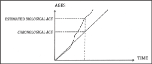
Figure 1: Comparing the estimated biological age and the
Lupinepublishers-openaccess-Forensic-Genetics

The Figure 1 is an imaginary example: the chronological age curve of an individual is a straight line, while the estimated biological age curve is erratic. On a given date, when the biological age exceeds the chronological age, an individual looks older than the average (Figure 1).

The Technical Age

The "technical age" takes into account the material aging, the wear, the maintenance, as well as the planned obsolescence or not. « Artifact: from Latin « artis factum », made from art. ».

The Aging of Bacteria

Bacteria are prokaryotic unicellular organisms whose genome consists of DNA with one chromosome. The statistical modeling of their development can help.com to understand aging. Bacteria reproduce by splitting through scissiparity one mother turns into two daughters, « ... which in their turn become mothers, each giving two. » daughters, etc. The population doubles with each generation; the exponential increase leads to a mathematical modeling such as:

1 → 2 → 4 → 8 → 16 → 32 → 64 → 128 ... 2 Exp (n)

    We start with one bacterium: 1 is written: 2 Exp(0)

    The first generation gives 2 bacteria, written: 2 Exp (1)

    The second generation gives 4 bacteria, written: 2 Exp(2)

    At the nth generation we have N = 2 Exp (n) bacteria

In a homogeneous medium, the generations reproduce at approximately the same rate "μ" (Greek letterμ); therefore, the number "n" of generations is approximately:

n & μ t

where "t" is the time indicated by the laboratory chronometer after "n” duplications. The number of bacteria finally reads:

    N # 2Exp (μ t) (1)

    The proliferation rate "μ” has three causes:

    a) Endogenous factors, specific to the bacterium. They depend on its heredity that is its genotype. One factor is the virulence, defined as the ability to multiply: Escherichia coli are able to divide every 20 minutes. Mbovis (Mycobacterium bovis) has a slower generation rate, but it is just as pathogenic. Benet [2].

    b) Exogenous factors, specific to the environmental surrounding: hygrometry, light, temperature, presence of sugar or nitrogen, gravity; for example, salmonella typhimurium is three times more virulent in microgravity.

    c) The potentiation of endogenous and exogenous factors, one making the other more effective in some way.

    Genotype + surrounding + potentiationμ

Temperature is an exogenous factor that is easy to control, and bacteria are very sensitive to temperature: between 20°C and 25°C, a population of Salmonella will double every hour, and within 10 hours, their number will be multiplied by 1000. When the temperature is lowered, the energy intake is reduced and their development is slowed down due to lack of homeothermy: "μ” decreases. Below4°C, the deactivation sets in, and in liquid nitrogen, at c.-196 C (c.77°K), μ = 0.

    Homeothermy: Internal regulation of temperature, controlled by the hypothalamus. Dinosaur slacked this function, as do many modern animals, including reptiles.

    These experiments have a double interpretation:

    a) The rate of proliferation "μ" contains energy components (glucose, heat), hygrometric, gravitational, and chemical components, and possible stochastic factors.

    b) Time "t" is not the cause of their development. A "substitution rule” provides a theoretical confirmation: the angle of terrestrial rotation "α” is such as

    α =ωt

    where "ω” is terrestrial rotation speed. It leads to

    t = α / ω

    In equation (1) we replace”t” by "α / ω”, and we obtain

    N= 2 exp (μ α / ω) (2)

Equation (2) proves that the number of bacteria does not depend on time. Time has no impact on bacteria aging. Work done in the USA has observed non-pathogenic bacteria which are deactivated after about hundred transitions, through exhaustion of their genetic ability of scissi parity prescribed by the genome: this is the clinical death of the line, by completion of a genetic program, without temporal impact. At the 100th generation issued from a primary bacteria, the theoretical population reaches c. 2100 bacteria at time "t100" measured when the duplications, observed with a microscope, stop: then μ = 100 / t100 is the average duplication speed.

From t = 100/n, which is the life expectancy of a bacteria line, and from t100/100, which is the average lifespan of a bacterium, it appears that a favourable environment, by activating vitality (that is the factor "μ”), reduces the life expectancy of a population of bacteria and the average lifespan of a bacterium. Professor Valter Longo, director of the USC School of Gerontology (CA), discovered that bacteria deprived of sugar could double their lifespan. Interesting conclusions can be drawn for human health.

The Aging of Cells

L. Robert refers to the activity of biological clocks in the cell aging process. But the rhythms which are observed are irregular and imprecise, and they depend on the environment: therefore they do not have the reliability and accuracy that a clock should have. The metaphor of the biological arrow of time which is supposed to orient cell development is inappropriate; moreover L. Robert describes interactions between the extracellular matrix and the cell: the cell and the matrix have their own modalities of aging which modify the program sequence of the ECM synthesis and its action on the cell as well Klein. [3] This action, which partly determines mitosis (cell division), finally exhausts its abilities: the author recalls the work done in vitro by L. Hayflick (in the USA in the early 1960s), which has shown a limit of 50 to 60 duplications of the cell population: the analogy with a biological arrow of time is obviously disqualified.

The innocuousness of time is clear from Hayflick conclusions: the life expectancy of a cell depends on the rate of duplication (rate of transtability). Transtability is the ability of a cell, which is unstable like any system, to divide into two cells, themselves unstable, whereupon each new cell then divides in its turn: cytoplasmic division is caused by the quest to reach a steady state which is never in fact achieved. The weaker the transtability, the slower the aging; therefore the aging of a cell can be reduced by acting on main causes of imbalance: stressors and genetic weaknesses. The longevity of a cell is determined by the length of its telomere, which is a segment located at the ends of a chromosome. The telomere gets shorter as the number of cell divisions increases, and also due to stressors. This segment, which is protected by an enzyme (telomerase protects the integrity of the genome), controls the start of mitosis. The telophase, or terminal phase of mitosis, consists in the splitting of the cell nucleus into two nuclei, followed by cell division. Unlike non-pathogenic cells, cancer cells are able to subdivide indefinitely: on a human scale, they do not age, because telomerase is hyperactive inside tumoral cells. A cell does not age because of a biological arrow of time, but because of a genetic and environmental mitogen induction: mitosis is induced by the gene, the environment, and a possible potentiation of these two causes.

The Causes of Aging

Time is not the cause of aging in bacteria and cells. But can this refutation of the confusion between time and aging be generalized to all systems? The innate and the acquired are active components of the aging of any system, living or not: even before it is completed, any building or human-made system begins an aging process: what is innate in a bridge is its architecture, its structure, and the quality of the work and materials; what is acquired derives from functional and climatic stressors, maintenance, and repairs. The anonymous concept of aging is replaced by a more detailed analysis, involving complex engineering and a suitable maintenance protocol.

The Millau Bridge was designed by the English architect Norman Foster for an approximate lifespan of 120 years, which corresponds to 120 terrestrial revolutions. In comparison, the pyramids of Giza are still standing after over 4000 years. They are not simple assemblages of stones: their interior layout includes chambers, passages, and complex anti-intrusion devices. Aging is the normal outcome of the development of living things, regardless of their complexity (ontogeny in humans); it corresponds to a systematic evolution of their state towards different states, resulting from degradation of all their parts, and everything that links and orders these parts. The transtable process. In Latin, trans means beyond (inCaesar); transabeo means go across, go beyond in Virgilius. Gaffiot In the Dictionnaire Larousse 1923, trans means beyond, through. The Dictionnaire Quillet 1929 defines prefix Trans: transition from one state to another.

What is innate in a living being consists essentially of its genetic heritage. The genetic program controls its transtable faculties, as it does inside bacteria and cells. Similar systems age differently from each other, and of course, this would not be the case if time was the cause of their aging. What is acquired is the lifestyle, i.e., the interaction with the rest of the Universe. So a lack of medicalization and hygiene, superimposed on endemic nutritional deficiencies, which generate a range of different pathologies, can reduce life expectancy by a factor of 3 or 4, bringing.com back to the standard of living of the Eneolithic; as shown by the destitution of a billion or so of our fellow humans in the early twenty first century. A report by the Haut Comite de la Sante. Publique [4] gave, for the French male population in 1996, the proportions of deaths due to certain kinds of behaviour relative to the total number of deaths due to all causes: accident 9%, alcohol 13%, tobacco 21% (HCP). The gradual reduction of the gap in average lifespan between men and women which has been observed in recent years is explained by increased smoking and alcohol consumption by women, from adolescence and even pre-adolescence. Data collected in the late 1980s in India showed an inversion of the average lifetimes of men and women: 45 years for men and 43 years for women. (OMS) This reversed gap was explained by the fact that men ate first, while women and children shared the leftovers; in addition, the situation was worsened by smoking among women .The potentiation of reciprocal action between the innate and the acquired constitutes an additional category of active causes of aging (allergies, stressors from different sources, e.g., physical.

Eneolithic: end of Neolithic (3000-2500 BC).

Aging and Organic Degradation

Public access to caves occupied by humans in the paleolithic broke the precarious environmental and atmospheric equilibrium, and triggered a rapid deterioration of petroglyphs by oxidation of pigments and mildew, as observed in the Lascaux caves. In some Egyptian sanctuaries, the same causes brought about the same effects on the magnificent polychromatic frescoes which were found in their original state just before the sanctuaries were opened to crowds of visitors. Museum curators do not consider time to be the cause of aging in the works of art they look after. Light (mainly ultraviolet radiation), temperature, and humidity (and in particular, changes in temperature and humidity), air pollution, inappropriate handling, and specific micro-predators are acknowledged in museum conservation as major aging factors for many materials (wood, leather, paper, textiles, pigments). This is why museums protect their artworks by exposing them only to dim light and ensuring proper ventilation. Very focused efforts are thus made to fight against aging due to physical and chemical stressors, not against the ghostly intervention of time.

The Self Organisation

Belousov's oscillating chemical reactions involve the selforganisation of dissipative structures (Prigogine) [5]. It turns out that these striking experiments are dependent on the necessary supply of fresh reagent: this energy intake modifies the physical state and the organization of the structures. Without it, the reaction would stop. The energy is the sole cause of the observable phenomenon of self-organisation:

No energyno self - organization

Therefore, there is no chemical arrow of time in selforganization.

The Biological Arrow Of Time

Spontaneous and extended pulsations of a myocardiac fragment in a glucose solute proceed with the same atemporal protocol; namely, we observe a transtability of the physical state of the fragment, with consumption of energy in the form of sugar. Time is powerless once again:

No energyno pulsation

The cardiac pulsations are perfectly observable: a pulsation is not a concept, and the confusion between pulsations and time is obviously a mistake. In addition, the biological rhythms do not comply with the accuracy and regularity requirements of a clock. Therefore, we may talk about biorhythm, but in no way about chronobiology: the biological arrow of time is an inappropriate metaphor.

The Paradox of Aging

These results confirm that aging is not related to time. However, time can be expressed in relation to symptoms of aging of any system, including ourselves:

« Symptoms of aging (observable reality) >⇒» « time goes by »

We conclude with an astonishing paradox, which is summarized by the aphorism:

We don't age because time goes by, but time goes by because we age [6].

Read More About Lupine Publishers Journal of Forensic Sciences Please Click on Below Link:
https://forensic-journal-lupine-publishers.blogspot.com/

Friday 28 May 2021

Lupine Publishers | Molecular Typing Of Capsular Polysaccharides of Staphylococcus Aureus Isolated From Cases of Bovine Mastitis by PCR

 Lupine Publishers | Journal of Dairy & Veterinary Sciences

Abstract

Forty five Staphylococcus aureus isolated from cases of bovine mastitis were subjected to Molecular typing by Polymerase chain reaction to determine their capsular polysaccaharide type. Of the 45 isolates, 33 were confirmed to carry a cap5 locus and cap8 locus was detected in remaining 12 isolates. To the best of our knowledge this is the first report of capsular polysaccharide typing of S.aureus isolates from India

Keywords: Staphylococcus aureus; Polymerase chain reaction and capsular polysaccaharide type

Introduction

S. aureus produces a variety of extracellular and cell wall associated components which are involved in the pathogenesis of bovine, ovine and caprine mastitis [1]. S. aureus strains produce capsular polysaccharide (CP) in-vivo [2] or under defined culture conditions [3]. Although capsule production of staphylococci was first recognized in 1930 [4], the prevalence of encapsulation among S. aureus has been appreciated only recently. Eleven capsular polysaccharide serotypes have been proposed on the basis of agglutinating reactivity with adsorbed rabbit antiserum and precipitation in double immuno diffusion [5,6]. Of these capsular serotypes 5 and 8 are the most predominant serotypes in human and animal S. aureus infections.

Studies on the prevalence of encapsulated strains in bovines shows the considerable variability that exist in the prevalence of serotype 5 and 8 capsules among bovine mammary isolates of S. aureus from different countries (Tollerstud et al., 2000). Moreover, the presence of S. aureus in raw milk is a public health problem, because it was reported that 95% of S. aureus isolates from bovine mastitis were either CP5 or CP8 in Norway [7]. For effective control of bovine mastitis caused by S. aureus in a particular geographical location, a careful characterization of the prevalent strains in the target population is essential [6]. Studies on capsular serotyping of isolates are important for the rational design of mastitis vaccines, containing staphylococcal capsular antigens. If improved vaccines against bovine mastitis are to be generated, more studies are required to elucidate the role of these polysaccharides in the pathogenesis of bovine mastitis [7].

However, capsular serotyping employing conventional techniques fails to identify non encapsulated strains of S. aureus. Hence DNA based technique for differentiation of serotypes provide an alternative to conventional serotyping and has a potential to overcome the problems associated with the current serotyping techniques which relay on inconsistent expression of phenotypic traits [7-9]. No data regarding the prevalence of capsular serotypes of S. aureus causing bovine mastitis is available in India. The proposed study would help in understanding the prevalence of capsular serotypes of S. aureus in Puducherry, India. This data would help in formulating vaccine based strategies for control of mastitis.

Materials and Methods

Cultures used in the study. Forty five Staphylococcus aureus obtained from the milk of dairy cattle with clinical and subclinical mastitis in and around Puducherry, India and S. aureus strain Reynolds (Capsular polysaccharide type 5) and S. aureus strain Wrights (Capsular polysaccharide type 8) were used as standard reference for identification of Capsular polysaccharide types of S. aureus by PCR Identification of S. aureus isolates [10]. The S. aureus isolates were initially selected on the basis of colony appearance and a positive tube coagulase test and their identity was verified by [8] and Garrity et al. [9]. Their identity was confirmed by PCR using the primer pairs targeting the nuc gene of S. aureus. DNA extraction. Strains were grown on Luria broth 37 °C overnight. Genomic DNA was extracted with a standard phenol-chloroform procedure as described elsewhere [10]. Detection of capsular genotype by PCR. The PCR assay for typing of capsular polysaccharide S.aureus was carried out as described by Verdier et al [11]. Genomic DNA was used as a template for PCR amplification with the primers Cap5 k1 (5’-GTCAAAGATTATGTGATGCTACTGAG-3’) and Cap5 k2 (5’-ACTTCGAATATAAACTTGAATCAATGTTATACAG-3’) located in cap5k for capsular type 5 and the primers Capsule 8 k1 (5'GCCTTATGTTAGGTGATAAACC-3') and Capsule 8 k2 (5'-GGAAAAACACTATCATAGCAGG-3') located in cap8I for capsular type 8. The PCR amplification was carried out in an automated thermal cycler (Eppendorf mastercycler, Germany) according to the following programme. initial denaturation at 94 OC for 4min followed by 25 cycles of denaturation at 94 OC for 30seconds , annealing at 55 OC for 30sec and extension at 72 OC for 1min and final extension at 72 OC for 5min. The amplified products were analysed on agarose gels along with positive control, negative control and molecular size marker (100bp la).

Results and Discussion

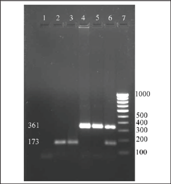
All the 45 isolates S. aureus were subjected to molecular typing using the primer pairs targeting the cap5 locus and cap8 locus of capsular polysaccharide of S. aureus. The primer pairs successfully amplified the DNA prepared from the field isolates of S. aureus as well as the DNA prepared from the reference cultures used in the study. The sizes of the amplicons were 361bp for capsular type 5 and 173bp for capsular type 8 (Figure 1). Among the 45 S. aureus isolates subjected for PCR with CP5 and CP8 primers, 33 isolates were confirmed to be CP5 strain and 12 isolates were confirmed to be CP8 strain. Out of the 45 isolates, 73.33% were found to carry the cap5 locus and 26.66 % were found to carry the cap8 locus. Poutrel et al. [1] reported that a majority of S. aureus from cases of mastitis strains belong to serotype 5 (CP5) and 8 (CP8). In their study they used monoclonal antibodies to S. aureus capsular polysaccharide types 5 and 8 to serotype the isolates by enzyme- linked immunosorbent assay and showed that 69% of 212 isolates recovered from cow's milk in France were serotype 5 (51%) and serotype 8 (18%) and 30.6% were non-typeable [12].

Figure 1: Screening field isolates of S.aureus for capsular types.
Lupinepublishers-openaccess-Dairy-Veterinaryscience

Lupinepublishers-openaccess-Dairy-Veterinaryscience

Tollersud et al. (2000) have showed the variability in prevalence of serotype 5 and 8 capsules among bovine mammary isolates of S. aureus from different countries. They performed immunoblot assay using CP5 and CP8 antibodies and isolates that consistently giving weak reactions with antibodies to CP5 and CP8 were further evaluated by immunodiffusion or ELISA. Capsular serotyping of 274 bovine mastitis isolates of S. aureus from Europe, showed that the majority of isolates from Denmark (23 out of 39 isolates), Sweden (29 out of 38 isolates) and Ireland (62 out of 101 isolates), were of serotype 8 [13]. Isolates from Iceland showed an equal distribution of serotype 5 (10 isolates), serotype 8 (13 isolates) and non-typeable isolates (11 isolates), whereas in Finland half of the isolates (32 out of 62 isolates) tested were non-typeable. Serotyping of the U.S. isolates revealed that only 42% of 362 isolates from seven different states were typeable with the available antisera and showed 27 % of the isolates were serotype 8 strains and 15 % were serotype 5 strains, but the majority (58%) of U.S. isolates were non-typeable.

Strains of S. aureus that do not react with antibodies to CP5 or CP8 are referred to as non-typeable (NT) by conventional serotyping. Karakawa et al. [12] and Lee et al. [13] reported that these NT strains also fail to react with specific antibodies to serotype 1 or 2 CP. This is one of the problems encountered in the conventional serotyping of S.aureus. Han et al. [13] reported the usefulness of monoclonal antibodies reactive with the type 5 and 8 CP in characterizing S. aureus from clinical isolates that monoclonal antibodies have been described, and has also been demonstrated. Monoclonal antibodies for CP5, CP 8 and 336 were used to characterize 107 isolates of S. aureus [14-15]. Forty six per cent of them were typed as 336, while serotype 5 and 8 accounted for 12.1% each. The rest were declared as non-typeable. However O'Brien et al. [14] and Ma et al. [15] reported that Type 336 isolates do not express capsule but do express cell surface polysaccharide or the 336 polysaccharide (336PS), which resembles S. aureus cell wall teichoic acid and hence not a true capsular type. In order to avoid the problems encountered in the conventional serotyping, a PCR method was developed by Verdier et al. [11] to detect capsular types of S. aureus. In their study using the rabbit polyclonal antibodies specific to capsular polysaccharide types 5 and 8, 81 of the 195 isolates were capsular serotype 5 (T5) (42%), 88 were capsular serotype 8 (T8) (45%), and 26 (13%) were non-typeable. A PCR method was developed to detect capsular type of S. aureus isolates since serotyping method allowed typing of only 87% of strains (169 of 195). All strains included in the study have been investigated by PCR. But PCR method allowed genotyping of 100% strains [16-18].

Their study revealed that all S. aureus clinical isolates included in the study carried either the cap5 (46% of cases) or the cap8 (54% of cases) locus by PCR method, and demonstrated that the capsular phenotype that was determined by conventional serotyping method was confirmed by PCR. However, all 336 serotype strains that reacted specifically with 336 antibodies but not with capsular polysaccharide type 5 or 8 antibodies, carried the cap8 or cap5 genes (cap8 18 and 8 cap5 isolates). This study revealed the predominate capsular polysaccharide types prevailing among the bovine S.aureus isolates was the CP5 compared to CP8. Data on the S. aureus capsular polysaccharide types will help in formulating vaccine based strategies for the effective control of bovine mastitis due to S. aureus.

Read More About Lupine Publishers Journal of Dairy & Veterinary Science Please Click on Below Link:
https://lupine-publishers-dairy-veterinary.blogspot.com/

Wednesday 26 May 2021

Lupine Publishers| Review and Hypothesis about Gout

 Lupine Publishers| Advancements in Cardiovascular Research (ACR)


Introduction

The first identification about gout as clinical entity was made by the Egyptians in the year 2640 b.C. (Schwartz 2006). For many centuries it was not unveil the mystery of the origin of the illness. Gout is the unique pathology that belongs to the human race. When uric acid is deposited in the articulate tissue produce an intense inflammation, basic element in the development of gout. Interesting is the evidence of ultra sonographic signs of monosodium urate crystalline articulate deposits in 25% of clinically asymptomatic hyperuricaemic subjects (more than 8mg/ dL) [1], and approximately 9% of the joints without clinical signs of flogosis [2]. Large epidemiological studies have now demonstrated that gout is an independent risk factor for incident coronary heart disease, [3-6] heart failure, [7] stroke [8] peripheral artery disease [9] and death cardiovascular [10,11]. But, several meta-analyses have concluded that hyperuricaemia is an independent risk factor for coronary heart disease [12,13] and also, for the development of hypertension [14,15]. The standard diagnostic goal remains the identification of the typical birefringence of crystals of uric acid under polarized light microscope in the synovial fluid and in the aspirated material from the tophi [16,17]. Hyperuricaemia with or without urate deposit (modern denomination of the formerly called gout) is currently one of the most frequent dysmetabolic diseases. [18,19].

Over the last decades, to uric acid has been attributed a possible role of cardiovascular risk prevalently the left ventricular hypertrophy for an increase in inflammatory mediators, such as tumor necrosis factor alpha and activation, and the rennin angiotensin system, increase in interstitial fibrosis of the myocardium and producing endothelial dysfunction [20,21]. Uric acid being a small molecule, is able to penetrate within the vascular wall cells, stimulating inflammatory activity leading to smooth muscle cell hypertrophy and favoring the endothelial dysfunction process [22]. An increase in arterial stiffness has also been demonstrated [23]. The antioxidant effect seems to be attenuated by the increase in its plasma concentration to turn into a pro-oxidant effect when it exceeds the 6 mg / dL level. In recent years, scientific evidence has suggested the possibility of a pathophysiological correlation between the action of xanthine oxidase and the genesis of cardiovascular damage in patients with chronic hyperuricaemia with and without uric acid deposits [24,25].

Uric Acid

Uric acid is an organic heterocyclic compound. It is the endproduct of the metabolism of purine nucleotides that are the principal constituents of cellular energy stores, such as ATP, and components of DNA and RNA. Urate ions appear as monosodium urate with a solubility limit in plasma of about 6.8 mg/dL at 37°. When the crystals precipitate by excessive solute, they are coated with polypeptide or protein molecules [26,27]. Precipitation of uric acid can occur for other factors such as the state of tissue hydration, pH, cation concentrations and the presence of proteoglycans, collagen and chondroitin sulphate. Uric acid is a molecule with an effective antioxidant action (protective: intracellular localization) and a potent pro-oxidant effect (negative impact: extracellular localization) in relation to the micro-environment in which it is located [28]. This dual role has been described as the “uric acid paradox”.

Plasma Proteins

Acid-bearing drugs bind to albumin. Those with basic function are bound to globulins. They are subdivided into three fractions: alpha, beta and gamma. The first two cover transport functions, while the third includes the immuno globulins involved in the body’s defense processes.

Synovial fluid

The synovial fluid under normal conditions is a viscous light yellow and clear. The fluid contains few proteins and cells but is rich in hyaluronic acid (about 300mg/dL) synthesized by type B synoviocytes which provides high viscosity. The larger molecules, such as immunoglobulin’s and complement, are found to be in lower concentrations, made due to the physiological function of the filter. The protein content of normal synovial fluid ranges from 25-30% of the total plasma protein content, with a very lower proportion of the higher molecular weight proteins such as alpha- 2-globulin, haptoglobin and fibrinogen (which belongs to beta globulins) because the synovial membrane is impermeable to high molecular weight proteins. Fibrin and fibrinogen are normally not present in synovium. During the inflammatory process an increase of the total proteins is observed as a consequence of the increase of the permeability of the synovial vessels.

Personal Experience

Cardiovascular

About three decades ago, working as a general practitioner and cardiologist at the “Dr. Julio Méndez” Municipal Sanatorium (Buenos Aires, Argentina), hyperuricaemia was a factor of concern and discussion about its possible action on the cardiovascular system. We had no doubts about its role in the development of joint gout but we did not have certain data about its action on the arteries. The question was whether hyperuricaemia is an independent risk factor for cardiovascular disease. We knew that the uric acid is transported by very low density lipoproteins which would make them more easily precipitated, prevalently if they find an acid medium inside the arteries. Moreover, the acidity could be caused by the depolymerization of mucopolysaccharides and the presence of bivalent cations would concurrently precipitate the circulating proteins (uric acid carriers), we thought that with the contact of uric acid with chondroitin sulfuric acid of the arteries (similar to the articulate cartilage component), could develop the inflammatory cascade. Hypertension frequently coexists and contributes with hypoxia facilitating the inflammatory action of uric acid and lipoproteins.

Therefore, uric acid would not in itself be a coronary risk factor, but would act in combination with hypertension, hyperlipidaemia and/or hyperglycaemia. During the time I drove of the Ischemic Heart Disease team, in order to know the relationship between hyperuricaemia and cardiovascular disease, 40 patients both gender was enrolled with ischemic heart disease studied with electrocardiogram, cycloergometry, echocardiogram and coronary angiography. The male group (35) had a mean age of 56 years, with a history of myocardial infarction [27] and myocardial ischemia [8], and in the female group (5) had a mean age of 64 years only with myocardial ischemia. All patients had a hyperuricaemia 10% higher than normal level for each gender. Only one patient had hyperuricaemia, the rest of the sample had two or more cardiovascular risk factors: high blood pressure (55%), hereditary history of cardiovascular disease (50%), smoking (43%), Obesity (38%), hyperlipidaemia IIa (38%), hyperlipidaemia IV (38%), diabetes mellitus (30%), gout (39%), and sedentarism (18%).

Conclusion

Apparently hyperuricaemia alone not be a risk factor but, in combination with hypertension, hyperlipidaemia and/or hyperglycaemia, could develop the inflammatory cascade acting on the endothelium. No woman had myocardial infarction or articulate gout. The level of uricaemia was not related to the severity of cardiovascular disease. Interestingly, the group older than 60 years with hyperuricaemia had greater risk. Hyperuricaemia, hypertension, hyperlipidaemia and age greater than 60 years in males would be associated with a higher severity of cardiovascular risk, and would favor the development of atherosclerosis [29].

Joints

There were also several doubts about the development of joint gout in patients with serum acid levels below 7mg/dL, and why the lack of clinical symptomatology in other patients with severe reduced glomerular filtration rate with exceptional levels of uricaemia such as 17mg/dL. Monosodium urate is deposited only if the medium is acid as occurs in the urine, and in contact with the chondroitin sulfuric acid in the joints. A possibility of producing a gouty acute attack with non-high levels of uricaemia could indicate that the uric acid introduced into the joint would not have an element that would function as a shock absorber. In the same line of thought, it was possible to arrive at the conjecture that during the acute inflammation, and for the benefit of the local vasodilatation, there could be a passage of elements that had a damping action. The question was: What are the elements that are not found in large quantities in normal synovial fluid and increase during inflammation? It was first thought of the higher molecular weight plasma proteins as the alpha and beta globulins as well as fibrinogen. An investigation was carried out in order to clarify these doubts It was decided to take samples of the synovial fluid of the knee from gouty (20) and non-gouty (20) patients. Hyaluronidase to synovial fluid was added in order to decrease its own viscosity. Once liquefied, a sample was taken to perform a protein electrophoresis. Technically it was not possible to measure fibrinogen intraarticulate. The next step was to compare the amounts of alpha, beta, and gamma proteins of the two patient groups. It was observed that in patients with acute joint gout fractions of alpha and beta globulins were always significantly lower than those of health patients. (Unpublished data) We hypothesized that patients with less alpha and beta globulins in the synovial liquid were more sensitive to developing joint gout.

Conclusion

The acute attack of gout, without a high uric acid level, could be caused by the low presence of high molecular weight proteins in the synovial fluid. If the disease is heritable, the reduction of alpha and beta proteins could be inherited. The self-limitation of the gouty attack could occur by the entry of proteins of high molecular weight, secondary to the vasodilatation produced by the acute inflammation, which would function as buffer. Other proteins and/ or substances could also be buffers such as fibrinogen.

Read More About  Advancements in Cardiovascular Research (ACR) Click on Below Link:

https://lupine-publishers-cardiovascular.blogspot.com/


Tuesday 25 May 2021

Lupine Publishers| Orthogonal Arrays and Row-Column and Block Designs for CDC Systems

 Lupine Publishers| Current Trends on Biostatistics & Biometrics (CTBB)


Abstract

In this article, block and row-column designs for genetic crosses such as Complete diallel cross system using orthogonal arrays (p2, r, p, 2), where p is prime or a power of prime and semi balanced arrays (p(p-1)/2, p, p, 2), where p is a prime or power of an odd prime, are derived. The block designs and row-column designs for Griffing’s methods A and B are found to be A-optimal and the block designs for Griffing’s methods C and D are found to be universally optimal in the sense of Kiefer. The derived block and rowcolumn designs for method A and C are new and consume minimum experimental units. According to Gupta block designs for Griffing’s methods A,B,C and D are orthogonally blocked designs. AMS classification: 62K05.

Keywords: Orthogonal Array; Semi-balanced Array; Complete diallel Cross; Row-Column Design; Optimality

Introduction

Orthogonal arrays of strength d were introduced and applied in the construction of confounded symmetrical and asymmetrical factorial designs, multifactorial designs (fractional replication) and so on Rao [1-4] Orthogonal arrays of strength 2 were found useful in the construction of other combinatorial arrangements. Bose, Shrikhande and Parker [5] used it in the disproof of Euler’s conjecture. Ray-Chaudhari and Wilson [6-7] used orthogonal arrays of strength 2 to generate resolvable balanced incomplete block designs. Rao [8] gave method of construction of semi-balanced array of strength 2. These arrays have been used in the construction of resolvable balanced incomplete block design. A complete diallel crossing system is one in which a set of p inbred lines, where p is a prime or power of a prime, is chosen and crosses are made among these lines. This procedure gives rise to a maximum of v =p2 combination. Griffing [9] gave four experimental methods:

parental line combinations, one set of F1’s hybrid and reciprocal F1’s hybrid is included (all v = p2 combination)

parents and one set of F1’s hybrid is included but reciprocal F1’s hybrid is not (v = 1/2 p(p+1) combination)

one set of F1’s hybrid and reciprocal are included but not the parents (v =p(p-1) combination) and

one set of F1’s hybrid but neither parents nor reciprocals F1’s hybrid is included (v = 1/2p(p-1). The problem of generating optimal mating designs for CDC method D has been investigated by several authors Singh, Gupta, and Parsad [10].

For CDC method A, B and C models of Griffing [9] involves the general combining ability (g ca) and specific combining ability (sca) effects of lines. Let nc denote the total number of crosses involved in CDC method A, B and C and it is desired to compare the average effects or g ca effects of lines. Generally, the experiments of these methods are conducted using either a completely randomized design (CRD) or a randomized complete block (RCB) design involving nc crosses as treatments. The number of crosses in such mating design increases rapidly with an increase in the number of lines p. Thus, if p is large adoption of CRD or an RCB design is not appropriate unless the experimental units are extremely homogeneous. It is for this reason that the use of incomplete block design as environment design is needed for CDC method A, B and C. Agarwal and Das [11] used n-ary block designs in the evaluation of balanced incomplete block designs for all the four Griffing’s [9] complete diallel cross (CDC) systems. Optimal block designs for CDC method a and b and variance balanced designs for CDC method c have been constructed by Sharma and Fanta [12,13] but their designs consume more experimental units. This call designs for CDC methods A, B and C which consume less experimental units in comparison to their designs and at the same time are A-optimal or optimal. We restrict here to the estimation of general combining ability (gca) effects only. For analysis of these designs [12-14].

I in the present paper, we are deriving block and row-column designs for complete diallel cross (CDC) system i.e. methods A, B, C and d through orthogonal arrays and semi balanced arrays. Block designs and row-column designs obtained for methods a consume minimum experimental units and are A-optimal. Block designs obtained for method C are optimal in the sense of Kiefer [15] and consume minimum experimental units but row-column designs are neither A-optimal nor optimal. Conversely block designs and row-column designs obtained for methods B are A-optimal. Block designs obtained for method D are optimal in the sense of Kiefer [15] but row-column designs are neither A-optimal nor optimal. The rest of this article is organized as follows: in section B and C we have discussed universal optimality of designs for 1-way and 2-way settings. In section 4 and 5, we give some definitions of orthogonal array, semi balanced arrays and orthogonally blocked design and relation of orthogonal; array with designs for CDC system and optimality with examples and theorems. In section 6 we give relation of semi-balanced array to CDC system along with theorem and for example.

Model and Estimation in 1-Way Heterogeneity Setting

According to Sharma and Tadesse let d be a block design for a CDC systems experiment involving p inbred lines, b blocks each of size k. This means that there are k crosses in each of the blocks of d. Further, let rdt and sdi denote the number of replications of cross t and the number of replications of the line i in different crosses, respectively, in d [ t = 1,2, …, nc; i = 1, 2, …, p]. It is not hard to see that,

Lupinepublishers-openaccess-Biostatistics-Biometrics-journal

nc = number of crosses and n = bk, the total number of observations. For estimating general combining ability (gca) effects of lines, we took the following linear model for the observations obtained from block design d.

Lupinepublishers-openaccess-Biostatistics-Biometrics-journal

where y is an n×1 vector of observations, 1n is the n×1 vector of ones, △1 is the n × p design matrix for lines and △2 is an n × b design matrix for blocks, that is, the (h,λ)th element of △1 ( respectively, of △1) is 1 if the hth observation pertains to the lth line (respectively, of block) and ois zero otherwise. μ is a general mean, g is a p × 1 vector of line parameters, Β is a b × 1 vector of block parameters and e is an n × 1 vector of residuals. It is assumed that the vector of block parameter, Β is fixed and e is normally distributed with


Lupinepublishers-openaccess-Biostatistics-Biometrics-journal

where I is the identity matrix of conformable order. Using least squares estimation theory with usual restriction Lupinepublishers-openaccess-Biostatistics-Biometrics-journal, we shall have the following reduced normal equations for the analysis of proposed design d, for estimating the general combining ability (gca) effects of lines under model (2.1).

Lupinepublishers-openaccess-Biostatistics-Biometrics-journal

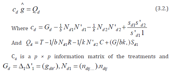
In the above expressions, Gd =△1 △′1 = (gdii´), gdii = sdi and for i ≠i´, gdii´ is the number of crosses in d in which the linesi and i´ appear together. Nd= △1△′2 = (ndij), ndij is the number of times the line i occurs in block j of d and Kd = △2△′2 is the diagonal matrix of block sizes. T = △′1 y and B = △′2 y are the vectors of lines totals and block totals of order p × 1 and b × 1, respectively for design d. A design d will be called connected if and only if rank (Cd) = p -1, or equivalently, if and only if all elementary comparison among general combining ability (gca) effects are estimable using d. We denote by D (p, b, k), the class of all such connected block design {d} with p lines, b blocks each of size k. In section 3, we will discuss Kiefer’s [15] criterion of the universal optimality of D (p, b, k).

Lupinepublishers-openaccess-Biostatistics-Biometrics-journal

Where y is an n × 1 vector of observed responses, μ is the general mean, g, Β and ã are column vectors of p general combining ability (gca) parameters, k row effects and b column effects, respectively, △1(n× p), △'2(n× k), △'3(n×b) are the corresponding design matrices, respectively and e denotes the vector of independent random errors having mean 0 and covariance matrix σ2In.LetNd1 = △1 △'2 be the p × k incidence matrix of lines vs rows and Nd2 = △1 △'3 be the p × b incidence matrix of treatments vs columns and △1△'3= 1k1b.Let rdl denote the number of times the lth cross appears in the design d, λ = 1, 2, . . . , nc and similarly sdi denote the number of times the ith line occurs in design d, i = 1, . . . p. Under (3.1), it can be shown that the reduced normal equations for estimating the gca effects of lines with usual restriction Lupinepublishers-openaccess-Biostatistics-Biometrics-journal, after eliminating the effect of rows and columns, in block design d are

Lupinepublishers-openaccess-Biostatistics-Biometrics-journal

is the number of times line i occurs in row j of d, Nd2= (ndi.t),

ni.t is the number of times the cross i occurs in column t,

sd1 is the replication vector of lines in design d,

Q is a p × 1 vector of adjusted treatments (crosses) total,

T is a p × 1 vector of treatment (line) totals,

R is a k × 1 vector of rows totals,

C is a b ×1 vector of columns totals, respectively, in design d,

G is a grand total of all observations in design d,

Now we state the following theorem of Parsad et al. [16] without proof.

Theorem: Let d* ∈ D1 (p, b, k) be a row - column design and d* ∈ D (p, b, k) be a block design for diallel crosses satisfying

(i) Trace (Cd*) = k-1b {2 k (k-1-2x) + p x (x+1)

(ii) (Cd*) = (p-1)-1k--1 b {2 k (k -1-2x) + p x (x+1)} (Ip - p-1 1p 1′p) is completely symmetric.

Where x = [2k/p], where [z]is the largest positive integer not exceeding z, Ip is an identity matrix of order p and 1p1′p is a p × p matrix of all ones. Then according to Kiefer [15], d*ɛ D1 (p, b, k) or d*∈ D (p, b, k) is universally optimal and in particular minimizes the average variance of the best linear unbiased estimator of all elementary contrasts among the gca effects. Furthermore, using d*ɛ D1 (p, b, k) or d*∈ D (p, b, k) all elementary contrasts among gca effects are estimated with variance.

Lupinepublishers-openaccess-Biostatistics-Biometrics-journal

Some Definitions

Definition 4.1: According to Bose and Bush [17], an r × N matrix A, with entries from a set ∑ of p ≥ 2 elements is called an orthogonal array of strength d, size N, r constraints and p levels if d × N sub matrix of A contains all possible × 1 column vectors with the same frequency λ. The array may be denoted by (N, r, p, d). The number λ may be called the index of the array. Clearly N = λ pd.

Definition 4.2: According to Rao [8], a (N, r, p) array is said to be a semi-balanced array of strength d if for any selection of d rows α1, α2, . . . , αd, we denote d rows by n(i1, i2, . . ., id).

(i) n (i1, i2, . . ., id) = 0 if any two ij are equal.

Lupinepublishers-openaccess-Biostatistics-Biometrics-journal

Where s represents summation over all permutation of distinct elements i1 , i2, . . ., id.

Definition 4.3: According to Gupta et al. [18], a diallel cross design to be orthogonally blocked if each line occurs in every block r/b time, where r is the constant replication number of the lines and b is the number of blocks in the design.

Relation between Orthogonal Array (p2, p+1, p, 2) and Designs for CDC System

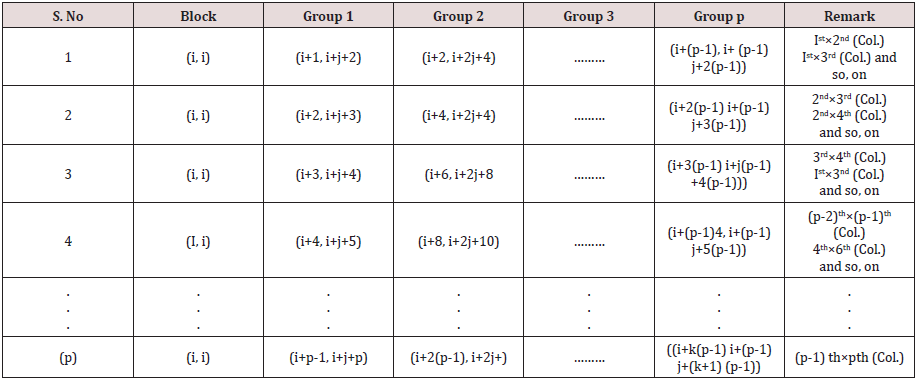
Consider an orthogonal array (p2, p+1, p, 2), where p is a prime or power of a prime. If we divide this array into p groups where each group contains p × (p+1) elements and identify the elements of each group as p lines of a diallel cross experiments. Now we perform crosses in any two columns of (p+1) constraints in first group and perform crosses among the lines appearing in the corresponding columns in (p-1) groups, we get p initial columns blocks as given below, which can be developed cyclically mod(p) to get design d1 for diallel cross experiment Griffing’ s method A with p2 distinct crosses of p parental lines consisting of p self, 1/2 p (p-1) number of F1 crosses, and the same number of reciprocal F1’s with parameter v = p2, b = p, k =p , r =1. By this procedure we obtain p(p+1)/2 designs for diallel cross experiment Griffing’s method A (Table1). Note: All column blocks will be developed cyclically mod (p).

Table 1:

Lupinepublishers-openaccess-Biostatistics-Biometrics-journal

Now considering in d1 , the cross of the type (i, j) = (j, i), i, j = 1,2, . . . p, we may obtain design d2 for Griffing’s method B with parameters v = p(p+1)/2, b =p, k =p, r1 =1,for cross of the type (i, i) and r2 =2, for cross of the type (i, j), where i, j = 1, 2, . . ., p, respectively. Considering rows as row blocks in block designs d1 and d2 we may also obtain row-column designs d3 and d4 for Griffing’s methods A and B with parameters v = p2, b = p, k =p, r =1 and v = p(p+1)/2, b =p, k =p, r1 =1,for cross of the type (i,i) and r2 =2, for cross of the type (i, j), where i, j = 1, 2, . . ., p, respectively. If we ignore the crosses of the type (i, i) in d1, where i = 1, 2, . . . p, we may obtain the block design d5 for Griffing’s method C with parameters v = p (p-1), b =p, k =p and r =1. Considering the crosses of the type (i, j) = (j, i) in d5 we may also derive design d6 for Griffing’s method D with parameters v = p (p-1)/2, b =p, k =p and r =1, where i < j = 1, 2,. . ., p. Considering rows as row blocks in block designs d5 and d6, we may also derive row-column designs d7 and d8 for Griffing’s methods C and D with parameters v = p (p-1), b =p, k =p , r =1 and v = p(p-1)/2, b =p, k =p and r =1, respectively, The designs d7 and d8 are neither optimal nor A-optimal. It is not hard to see that block and row-column designs obtained for Griffing’s methods A and C consume minimum experimental units and in theses designs every cross is replicated only once and each line occurs in every block r/b time, where r is the constant replication number of line and b is the number of blocks in the design. In block designs for Griffing’s methods B and D each line also occurs in every block r/b time. Hence according to Gupta et al. [18] these designs are orthogonally blocked. In an orthogonal design no loss of efficiency on the comparisons of interest is incurred due to blocking. A block design for which N = θ 1p 1′b is orthogonal for estimating the contrasts among gca parameters, where N denotes the line versus block incidence matrix and θ is some constant. For designs dk ∈ D (p, b, k), where k = 1, 2, 5, and 6 and designs dk ∈ D1 (p, b, k), where k = 3, 4, we have ndkij = 2, for k =1, 2,. . ., 8; i =1, 2, . . .p, j = 1, 2,. . ., p and their information matrices Cdk are as given below.

Lupinepublishers-openaccess-Biostatistics-Biometrics-journal

Where Ip is an identity matrix of order p and 1p is a unit column vector of ones. Clearly Cdk given by (5.1) is completely symmetric and Trace ( Cdk ) = 2 (p-1)2 which is not equal to the upper bound given in (5.3).

Lupinepublishers-openaccess-Biostatistics-Biometrics-journal

Hence the designs d1, d2, d3, and d4 are not optimal. The information matrix Cdk given by (5.2) is completely symmetric and Trace ( Cdk ) = 2 (p-1) (p-2) which is equal to the trace given in (5.3). Hence the designs d5 and d6 are optimal in the sense of Kiefer [15] and in particular minimizes the average variance of the best linear unbiased estimator of all elementary contrasts among the gca effects. To prove that the designs d1, d2, d3, and d4 are A- optimal, we consider the following criteria. A design d* ∈ D (p, b, k) is said to be A-optimal in D (p, b, k) if and only if Trace ((Vd*)≤Trace(Vd))

Here d* denotes designs d1, d2, d3, and d4 and d denotes designs d5 and d6. The Trace (Vd*) is equal to 1/2 and Trace (Vd*) is which is greater than 1/2.Hence designs d1, d2, d3, and d4 are an A-optimal.

Remark: The variances of the best linear unbiased estimators of elementary contrasts among gca effects are equal in A-optimal designs and also in optimal designs. It means that all the designs are variance balanced, this fact is particularly attractive to the experimenter, as it enables one to carry out the analysis of the experiment in an extremely simple manner.

Now we state the following theorems.

Theorem: The existence of an Orthogonal Array (p2, p+1, p, 2) implies the

existence of Lupinepublishers-openaccess-Biostatistics-Biometrics-journal different layouts A- optimal incomplete block designs with parameters v = p2, b = p, k =p, r =1.

(ii) existence Lupinepublishers-openaccess-Biostatistics-Biometrics-journal different layouts A- optimal incomplete designs for Griffing’s method B with parameters v = p (p+1)/2, b =p, k =p, r1 =1, for cross of the type (i, i) and r2 =2, for cross of the type (i, j), where i, j = 1, 2, . . ., p, respectively.

(iii) existence of p(p-1)/2 row-column designs for Griffing’s [9] methods A and B with parameter v = p2, b = p, k =p , r =1 and v = p (p+1)/2, b =p, k =p , r1 =1,for cross of the type (i, i) and r2 =2, for cross of the type (i, j), where i, j = 1, 2, . . ., p, respectively.

Theorem:- The existence of an Orthogonal Array (p2, p+1, p, 2) implies the existence of Lupinepublishers-openaccess-Biostatistics-Biometrics-journal different layouts optimal incomplete block designs for Griffings [9] methods C and D with parameters v = p(p-1), b =p, k =p , r =1 and v = p(p-1)/2, b =p, k =p and r =1, respectively.

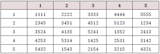
Example: Following Rao [19] we construct an orthogonal array (25,6,5,2) of rp =5, the 4 orthogonal Latin squares with bordered elements are (Table 2). This arrangement may be expressed in five groups as given below Table 3. The above arrangement is an orthogonal array (25, 6, 5, 2). From the above array we can derive the designs for the four experimental methods described by Griffing [9]. The procedure is explained below.

Table 2:

Lupinepublishers-openaccess-Biostatistics-Biometrics-journal

Table 3:

Lupinepublishers-openaccess-Biostatistics-Biometrics-journal

For Griffing methods A and B, we can take any two columns from first group and corresponding columns from other groups i.e. 2, 3, 4, and 5 and arrange them in columns and then we obtain design for methods A and B. Thus, we may obtain 14 different layouts designs for each method A and method B. We may also obtain 10 different layouts row-column designs for each of these methods A and B.

Remark: - Block and row column designs for methods A and B containing crosses with last column of group 1 are neither A-optimal nor optimal.

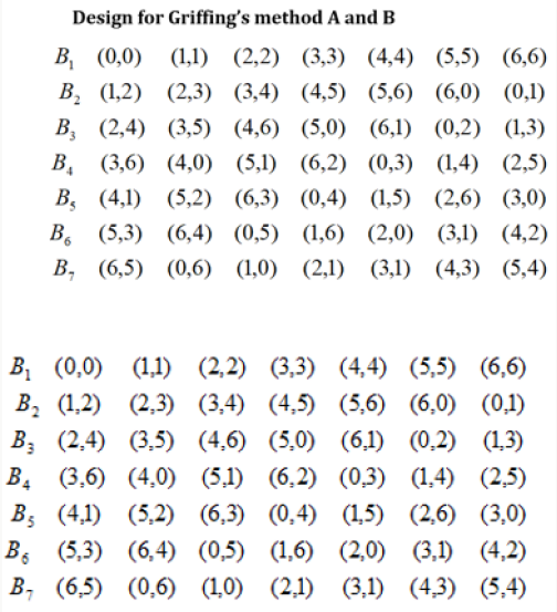
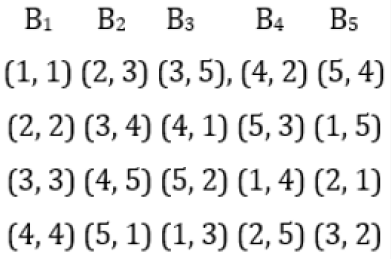
Example: Suppose we take the first two columns from first group and corresponding columns from other groups i.e. 2, 3, 4, and 5. Elements in brackets are considered cross between lines and then we obtain the following design for Griffing’s experimental methods A and B with parameters v = 25, b = 5, k =5, and r =1 and v = 15, b = 5, k =5, and r =1, respectively, with the condition that the cross (i, j) = (j, i) for method (2), where i < j = 1, 2, . . ., 5.

Figure 1 In the above design, considering rows as row blocks, we may we may obtain row-column designs for Griffing’s methods A and B respectively. From the above design we may also derive designs for methods C and D by ignoring the first column and considering (i, j) ≠ (j, i), in other columns, where i, j = 1, 2, 3, 4, and 5, with parameters v = 20, b = 5, k =5, and r =1. In Griffing’s method C design considering (i, j) = ( j, i) , where i< j = 1,2, 3, 4, and 5, we obtain a design for CDC method D with parameters v = 10, b = 5, k =5,and r =2, Thus, from the above array we may obtain 10 different layouts of designs for method C and 10 different layouts of designs for method D.

Figure 1: Design for Griffing’s Method A and B.

Lupinepublishers-openaccess-Biostatistics-Biometrics-journal

Relation between Semi-Balanced Array (p(p-1)/2, p, p, 2) and Designs for CDC System

Consider a semi-balanced (p(p-1)/2, p, p, 2), where p is an odd prime or power of odd prime. There are (p-1)/2 total sets in a semi-balanced (p(p-1)/2, p, p, 2). If we identify the elements of semibalanced array as lines of a diallel cross experiment and perform crosses in any two sets among the corresponding lines appearing in the same two sets, we get a mating design for diallel cross experiment involving p lines with v =p2crosses, each replicated once. The mating design can be converted into block design for diallel cross experiment Griffing’s methods A with parameter v = p2, b = p, k =p and r =1, considering rows as blocks. In the above design considering the cross (i, j) = (j, i) where i < j = 0, 1, 2, . . ., p, we obtain design for Griffing’s method B with parameters v = p(p+1)/2, b =p, k =p , r1 =1,for cross of the type (i, i) and r2 =2, for cross of the type (i, j), where i, j = 1, 2, . . ., p, respectively.. From block designs obtained for methods A and B we may also obtain row-column designs for Griffing’s methods A and B by considering columns as row blocks with parameters v = p2, b = p, k =p and r =1 and v = p(p+1)/2, b =p, k =p , r1 =1,for cross of the type (i,i) and r2 =2, for cross of the type (i, j), where i, j = 1, 2, . . ., p, respectively. From the above mating design deleting the first row and considering (i, j) ≠ ( j, i), where i < j = 0, 1, . . ., p we can also derive designs for methods C, with parameters with parameters v = p (p-1), b =p, k =p , r =1. Considering (i, j) = (j, i), where i, j = 0, 1, . . ., p, we obtain design for Griffing’s method D with parameters v = p(p-1)/2, b =p, k =p and r =2. Thus, using above techniques, we may obtain Lupinepublishers-openaccess-Biostatistics-Biometrics-journal different layouts designs for Griffing’s methods A, B, C and D. The information matrices of block designs and row-column designs for methods A and B are same as given in (1). So, block designs and row-column designs for methods A and B are A- optimal. Similarly, the information matrices of designs for methods C and D are the same as given in (1), so designs for methods C and D are universally optimal in the sense of Kiefer [15] and in particular minimizes the average variance of the best linear unbiased estimator of all elementary contrasts among the gca effects. These designs are orthogonally blocked. Now we state the following theorems.

Theorem 1: The existence of semi -balanced array (p(p-1), p, p, 2) implies the existence of different layouts of Lupinepublishers-openaccess-Biostatistics-Biometrics-journal A- optimal incomplete block designs and row-column designs for Griffing’s [9] methods A and B with parameters v = p2, b = p, k =p and r =1 and v = p(p+1)/2, b =p, k =p and r =1,respectively, where we consider the cross (i, j) = (j, i) for method B, where i < j = 0, 1, 2, . . ., p.

Theorem 2: The existence of Semi -Balanced Array (p (p-1), p, p, 2) implies the existence of Lupinepublishers-openaccess-Biostatistics-Biometrics-journal different lay outs of optimal incomplete block designs v = p(p-1), b =p, k =p and r =1 and v = p(p-1)/2, b=p, k=p and r=2, respectively, for Griffing’s [9] methods C and D.

Example. If p =7, the residue classes 0, 1, . . . , 6 (mod7) form a field . We write the 7 elements of GF (7) as 0, ±1, ±2, ±3 and hence the key sets are, using the formula (7.1), where

(0, 1, 2, 3, 4, 5, 6), (0, 2, 4, 6, 1, 3, 5), and (0, 3, 6, 2, 5, 1, 4) (8.1)

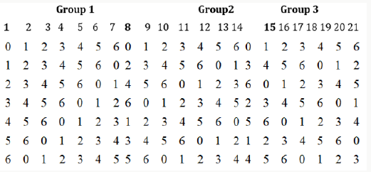
second and third vectors are obtained from the first on multiplying by 2 and 3, respectively. Writing (8.1) vertically (shown in bold numbers) and generating the other columns by the addition of elements GF (7) as indicated in (7.3). We obtain 21 columns as shown below which is divided into three groups (Figure2).

Figure 2:

Lupinepublishers-openaccess-Biostatistics-Biometrics-journal

From the semi balanced array given above, we may obtain designs for Griffing’s four experimental methods by superimposing group 2 over group 1 or group 3 over group1 or group 3 over group 2. We superimpose group 2 over group 1 and obtain following design for Griffing’s methods A and B with parameters v =49, b = 7, k =7, and r1 =1 and v = 28, b = 7, k =7, and r =1, with the condition that the cross (i, j) = (j, i), where i < j = 0, 1, 2, . . ., 6 (Figure 3).

From the above design we can derive designs for methods C, and D

Figure 3:

Lupinepublishers-openaccess-Biostatistics-Biometrics-journal

(i) By ignoring the first row and considering (i, j) ≠ (j, i) in other rows, where i < j = 0, 1, . . ., 6, for method C and

(ii) similarly ignoring first row and taking other rows and also considering (i, j) = (j, i) in other rows, where i, j = 0, 1, . . ., 6, thus we may obtain Lupinepublishers-openaccess-Biostatistics-Biometrics-journal different layouts of designs for method C and D.

Conclusion

In the present article we have given block and row-column designs for Griffing’s CDC system i.e for all methods A, B, C, and D by using orthogonal array (p2, p+1, p, 2) and semi -balanced array (p(p-1), p, p, 2). Block and row-column designs for methods A and block designs for method C consume minimum experimental units and are A-optimal and optimal, respectively. These designs are.


Read More About  Current Trends on Biostatistics & Biometrics (CTBB) Click on Below Link:

https://lupine-publishers-biostatistics.blogspot.com/


Monday 24 May 2021

Lupine Publishers| Groups 4 and 15 and Organotin Condensation Polymers for The Treatment of Cancers and Viruses

 Lupine Publishers| Modern Approaches on Material Science (MAMS)


Abstract

This short review describes the use of group 4 metallocenes, group 15 organometallics and organotin polymers in the treatment of human cancer tumors and viruses. These metal-containing polymers show good inhibition of all the main group solid tumors including pancreatic, lung, brain, breast, prostate and colon human cell lines. They also show inhibition of a variety of viruses including zika, herpes and vaccinia viruses. Synthesis of the polymers is rapid employing interfacial polymerization and commercially available reactants. They offer physicians a new class of drugs for the treatment of a variety of cancers and viruses.

Keywords: Cancer; Viruses; Interfacial polymerization; Brain cancer; Pancreatic cancer; Zika virus; Vaccinia virus; Breast cancer; Herpes virus

Introduction

Use of metal-containing agents to treat various medical problems is well known [1-22]. Here the focus is on activities to supply metalcontaining polymers for the treatment of various cancers and viruses. While we have had extensive experience with platinum and palladium polymers for the treatment of a variety of cancers, the current emphasis is on polymers formed by incorporation of groups 4 and 15 metals and organotin condensation polymers for the treatment of cancers and viruses [23-41]. These two polymer types are different with their own separate biological characterizations [26]. For instance, the platinum and palladium polymers are addition products and not stable for long times in solution. By comparison, the groups 4 metallocene and organotin and group 15 polymers are condensation polymers and exhibit good stability to over 30 weeks in solution so can be treated differently with respect to biological and physical characterizations [26-41].

Synthesis

Synthesis occurs employing interfacial polymerization [42- 46]. It is a rapid polymerization system because high-energy reactants are employed. These high-energy reactants are acid halides. A typical condensation reaction has an activation energy of about 30-40Kcal/mol whereas the activation energy for the acid halide reactions is on the order of 20Kcal/mol. The interfacial polymerization is employed industrially to synthesize aromatic polyamides (nylons) and polycarbonates so industry is familiar with the system [47,48]. These interfacial polycondensation reactions form polymer within less than one minute in decent yield. For the syntheses described here, commercially available reactants are employed allowing ready reproduction and scale-up to ton levels in a somewhat straightforward manner. Rapid stirring is employed, generally about 18,000 rpm. This allows both the rapid polymerizations to occur with an increase in interfacial contact area of over ten thousand compared to non-stirred systems, and good reproducibility. For the systems described here, the reaction vessel is a simple glass reaction vessel, one-quart Kimax emulsifying jar, fitted onto a Waring Blender. To illustrate the overall reactions, products formed for the organotin polymers have a repeat unit described as follows.

R2SnX2+X-R-Y-> -(-SnR2-R-)-

where X and Y are normally Lewis bases such as alcohols, amines, acid salts, thiols, etc. These reaction sites are often varied for a single Lewis base such as an amino acid, shown below, that has both acid and amine reactant sites. Examples of overall reaction products for each of the three condensation polymer groups are given following. Reaction between the amino acid diglycine and dimethyltin dichloride is described (Figure 1). The polymer is described as a poly (amine ester) with the organotin unit considered an organic moiety such as a methylene unit in such naming. For the Group 4 metallocenes, the reaction employing titanocene dichloride as the Lewis acid, the repeat unit for a product formed from titanocene dichloride and chelidonic acid is given (Figure 2). Finally, for reactions involving group 15 metals, the repeat unit formed from reaction between triphenylantimony dichloride and 3,5-pyridinedicarboxylic acid forming a polyester is given (Figure 3). The metal is generally located in the Lewis acid portion while the non-metal reactant is the Lewis base. In certain cases, the Lewis base portion may also contain a metal, usually iron and cobalt. The iron is present as a ferrocene while the cobalt is present as a cobaltocene [32].

Figure 1: Synthesis of organotin poly (amine esters) from reaction of diglycine and dimethyltin dichloride where R represents simple chain extension.

Lupinepublishers-openaccess-material-science-journal

Figure 2: Synthesis of polyesters from reaction with titanocene dichloride and chelidonic acid where R represents simple chain extension.

Lupinepublishers-openaccess-material-science-journal

Cancer

It was initially mistakenly assumed that these metal-containing compounds inhibited cancer by the same mechanism as the platinum-containing drugs as cisplatin and other similar platinum containing drugs [26,50]. (The platinum-containing drugs currently are employed in over 60% of the chemo drug treatments generally as one of the components.) It is now known that this is not true so that they can be coupled with the drugs described here as co-drugs that will affect inhibition of cancer through two distinct avenues. The platinum-containing drugs are quite toxic resulting in the presence of many negative side effects [26]. Our effort is to create drugs that have similar or superior ability to inhibit cancer but without the unwanted side effects. All of the metal-containing drugs operate primarily on the DNA site for inhibition of the cancer cell lines [26,50].

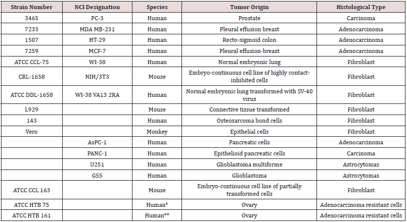
The polymers synthesized by us have shown good ability to inhibit a variety of cancer cell lines Table 1. These cell lines represent all of the major human solid tumor cell lines. These cell lines include resistant cells meaning cell lines that have shown ability to resist treatment with the traditional anticancer drugs [39] (Table 1). Inhibition depends on the metal atom present as well as the nature of the Lewis base. With respect to the metal, in general, inhibition is of the order Hf=Zr>Ti>Sn>Sb, Bi, As. Inhibition is also dependent on the specific Lewis base. A primary measure of the ability for a drug to inhibit cancer growth is the effective concentration, EC. The 50% effective concentration, EC50, is the concentration of a toxicant, drug, or antibody that induces an inhibitory response halfway between the baseline and maximum after a specified exposure time. The desired outcome is to have low EC50 values as this indicates that only a small concentration of the anti-cancer agent is needed to elicit inhibition. For the compounds described here, once inhibition begins, the slope of the dose/concentration curve is high with inhibition being total. Depending on the specific Lewis acid/base the EC50 value is typically between milligrams/mL to nanograms/mL. The metal-containing compounds are often coupled with a Lewis base that exhibits some biological activity hoping for a syngeneic effect. Drugs that have been employed as the Lewis bases include ciprofloxacin, diethylstilbestrol, cephalexin, acyclovir, thiamine, dicumarol, camphoric acid, histamine, 2-ketoglutaric acid, salicylic acid, dipicolinic acid, isomanide, glycyrrhetinic acid, phentolamine, thiodiglycolic acid. Lewis bases that themselves exhibit no ability to inhibit cancer can also exhibit good inhibition when coupled with a metal-containing moiety. These include a wide variety of diols such as ethylene glycol, Figure 4 [29,50]. Recently, water-soluble drugs possessing the metal-containing unit were synthesized [29] employing as the Lewis base poly (ethylene glycol), PEG. The resulting water-soluble polymers exhibit good inhibition of the cell lines. Figure 5 contains the reaction between titanocene dichloride and PEG forming water soluble polyethers (Figures 4 & 5).

Figure 3: Synthesis of triphenylantimony polyesters from reaction with 3,5-pyridinedicarboxylic acid where R is simple chain extension.

Lupinepublishers-openaccess-material-science-journal

Figure 4: Reaction between ethylene glycol and dibutyltin dichloride forming polyethers.

Lupinepublishers-openaccess-material-science-journal

Figure 5: Formation of water-soluble polyethers from reaction of titanocene dichloride and various poly (ethylene oxides) where R represents simple chain extension.

Lupinepublishers-openaccess-material-science-journal

Viruses

These metal-containing polymers also inhibit a variety of viruses including ones where no current drugs are available for treatment [40,41,49]. Table 2 contains viruses that have been inhibited by our metal-containing drugs including most recently the zika virus. These viruses include both DNA and RNA viruses. They include several that have been identified as possible weapons of mass destruction, namely the vaccinia virus. Three DNA viruses are effectively inhibited by the metal-containing polymers (Table 2). They are the vaccinia virus used to vaccinate humans against smallpox; herpes simplex virus 1, the virus responsible for over 45 million infections yearly in the US, comprising one of five adolescents and adults; and the varicella zoster virus, also a herpes virus and responsible for chickenpox and shingles. Thus, the metalcontaining polymers represent a possible potent approach towards inhibiting unwanted viruses (Table 2).

Table 1: Caner cell lines inhibited by metal-containing polymers described here.

Lupinepublishers-openaccess-material-science-journal

From a cancer patient with ovarian cancer that had previously been treated with cytoxan, adriamycin, 5-fluorouracil, and Fur IV. From a cancer patient with ovarian cancer that had been treated with adriamycin, cyclophosphamide, and cisplatin.

Table 2: Viruses inhibited by metal-containing polymers discussed in this report.

Lupinepublishers-openaccess-material-science-journal

Why Polymeric Drugs?

A critical question is “Why Polymeric Drugs?” What advantageousness do polymeric drugs offer [50-60]. Following briefly describes some advantages. Each of these advantages is related to the size of polymers and what such size offers. First, because of their size, polymers travel through the body, in particular the kidney and bladder, more slowly lessening organ damage allowing the organs to limit the negative effect [50,61]. Second, cancer cells are less cohesive, offering greater porosity, and are not as coherent as normal cells with relatively “rough” exteriors. This allows polymers to have a greater opportunity to be “snagged” by the cancer cells allowing them extended ability to be associated with the cancer cells resulting in a greater ability to inhibit cell growth. This scenario is described as the enhanced permeability and retention effect [50,62-64]. Third, increased size allows for a greater designing of the drug increasing its effectiveness [65-69]. This fine tuning includes attachment of “biological homing agents”. Thus, polymeric drugs offer advantageous over small molecule drugs that can be used to more effectively combat unwanted diseases compared to small molecule drugs.

Summary

Metal-containing polymers show ability to inhibit all the major solid tumor cancers as well as important viruses. They are easily synthesized and offer physicians new drugs to attack these harmful illnesses.

Read More About Lupine Publishers Modern Approaches on Material Science Click on Below Link:

https://lupine-publishers-material-science.blogspot.com/