Saturday 30 October 2021

Lupine Publishers | Pressure of Climatic Factors on Sheep Bluetongue Epidemics in Tamil Nadu

 Lupine Publishers | Journal of Dairy & Veterinary Sciences


Abstract

This research correlates the effect of climatic factors on bluetongue epidemiology in sheep of delta districts in Tamilnadu based on merteriological data collected from 2014 to 2016. There are a total of eighty two villages of delta districts were affected with Blue tongue virus (BTV). We used multiple linear regression analysis to assess the effect of climatic factors on the prevalence of bluetongue. Our results showed that maximum temperature, rain fall and relative humidity positively correlates with the number of BTV epidemic in sheep flocks. These findings suggest that the risk of BTV infections in sheep increased in warm and humid environments. Moreover, climatic factors can be used to forecast the possible epidemic in this area and other regions characterized by similar climatic conditions.

Keywords: Epidemiology; Blue Tongue Virus; Culicoides Spp; Laminitis; Coronitis; Facial Edema; Necrosis of Cardiac; Vectorial Capacity; Multiple Linear Regression Analysis Model; Semi Intensive Livestock; Antioxidant System

Introduction

Bluetongue, is caused by the bluetongue virus (BTV) and transmitted by Culicoides spp. [1,2]. Bluetongue virus was pandemic in nature with areas between 40◦N and 35◦S in the world [3,4] . Symptoms of this disease are usually characterized by laminitis, coronitis, and facial edema. In serious cases, pulmonary edema, pleural effusion and necrosis of cardiac and skeletal muscle can be observed [5].

Many researchers have evaluated the correlation between meteorological factors and the distribution of the Blue tongue virus [9,10]. However, few researchers have specifically attempted to investigate the influence of climatic factors on bluetongue epidemic. In the present study, we have investigated the effect of climatic factors on the epidemics of bluetongue, by means of multiple linear regression analysis model.We designed to assess the influence of climatic factors on the epidemiology of bluetongue in sheep in Tamilnadu, in order to provide accurate information to design the more effective and economical surveillance and control systems for bluetongue.

Materials and Methods

Sheep farming of Delta Districts

In delta districts of Tamilnadu, two types of sheep farms were observed. One is semi intensive livestock farms, which must meet a moderate standard regarding production management and epidemic prevention systems. The other one is high number holdings with trans humane methods, where sheep kept as a freerange system.

Location

This research work was carried out in the delta region of Tamilnadu which includes Thanjavur, Thiruvarur, Nagapattinam and Pudukottai districts during 2014 to 2016.

Data collection

Metrological data were collected from government agencies like District Collector Office and disaster management centre. Data were also obtained from research institutes like Tamilnadu Agricultural University, Thanjavur.

Data analysis

Multiple linear regression analysis model was used to assess the effect of climatic factors on the epidemics of bluetongue.

Results

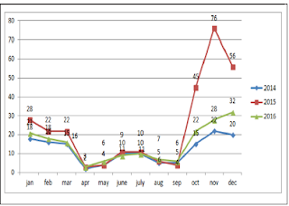
Sums of 608 bluetongue epidemics have been reported in delta districts during the period of study. Then we have analysed the change trend of bluetongue epidemic and the annual variation of meteorological data (Figure1). The results indicated the change trend and the annual variation of the disease epidemics, monthly maximum temperatures, monthly minimum temperature, wind speed and rainfall in delta districts of Tamilnadu from 2014 to 2016. Analyses of time series revealed that the epidemic of bluetongue of a determined month was changed by case numbers registered during the previous years. The epidemic trend was changing with the months were also recorded (Figure 2).

Figure 1: Standard epidemic of Sheep bluetongue in Tamilnadu.

Lupinepublishers-openaccess-Dairy-Veterinaryscience

Figure 2: Changing epidemic of Sheep blue tongue in Tamilnadu.

Lupinepublishers-openaccess-Dairy-Veterinaryscience

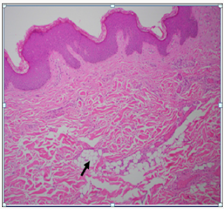
From the year analysis, a seasonal distribution and change in the disease cases from 2014 to 2016 were noticed, which correlates with the climatic factors of our investigation results from the results of data analysis, we found that temperature is the main risk factors which can influence the epidemic of bluetongue (Figure 3). In delta region maximum temperature at the tune of 330C will have higher epidemics during 2015 (288) than in 2013 (142) and in 2016 (178). Rainfall was also showed a influence on the occurrence of disease, a rise in mean monthly rainfall is related to increase in the monthly number of disease cases (Figure 4). High relative humidity positively related to the epidemic of bluetongue (Figure 5). Clinical signs like cutaneous ulcers (Figure 5) and bilateral purulent nasal discharge also evident. Under multiple linear regression model analysis maximum temperature, relative humidity and rainfall were statistically significant (P< 0.05) with the epidemiology of bluetongue (Table 1).

Figure 3: Maximum temperature (0C) data in the study area during 2014 to 2016.

Lupinepublishers-openaccess-Dairy-Veterinaryscience

Figure 4: Trend line of Rain fall (mm) in the study area.

Lupinepublishers-openaccess-Dairy-Veterinaryscience

Figure 5: Relative humidity (%) trend in the study population.

Lupinepublishers-openaccess-Dairy-Veterinaryscience

Table 1:Multiple linear regression model on climatic factor with bluetongue epidemic (P < 0.05).

Lupinepublishers-openaccess-Dairy-Veterinaryscience

Discussion

In this study, we aimed to prove the relationship between climatic factors and the epidemiology of bluetongue in delta districts of Tamilnadu. We found that a positive relationship between rainfall, monthly maximum temperature and relative humidity in the epidemic of Blue tongue in Sheep (Figure 6).

Figure 6: Relative humidity (%) trend in the study population.

Lupinepublishers-openaccess-Dairy-Veterinaryscience

The positive effect of temperature and relative humidity on blue tongue infections is comparable to previous research [11-13]. It has been proven that a hot and humid environment can interfere with the antioxidant system of the animal and increases the synthesis of lipid peroxides, which may cause peroxide damage [14-16] and increases the serum levels of epinephrine and norepinephrine will negatively affect humoral and cell-mediated immune responses and make animals more susceptible to infectious diseases like bluetongue.

Maximum temperature in our study helps in the epidemic of bluetongue because temperature also significantly influences the activity rates of adult insects. The scientific reason behind is adult culicoides are more active than on cooler days and their reproduction will be inhibited by low temperature [17]. However, BTV has a strong environmental resistance and will not lose its infectivity in seven days under 56 oC [18]. Thus, the effect of temperature may be limited in the normal / lower temperature conditions (Figure 7). High Rainfall showed increased cases of bluetongue because of contamination of soil water and that can influence the life cycle of insects. Moreover at the right level, rainfall can create humid microhabitats which promote the activities of [19] and can regulate the growth and activity of Culicoides spp. populations [20,21]. Even though this research found that wind velocity did not favors the spread of Bluetongue virus but, long distance movement of Culicoides is mediated by the prevailing winds [21].

Figure 7: Relative humidity (%) trend in the study population.

Lupinepublishers-openaccess-Dairy-Veterinaryscience

As a final point, mechanisms of transmission of bluetongue are more complex and climate factors only represent some components of the epidemic of blue tongue. Many other risk factors like age, immunization calendar and management practices may also have strong impact on the incidence of the disease. In this paper, we only studied the effect of climatic factors on the epidemiology of bluetongue. This research work can able to help to design in the control of blue tongue in sheep by adopting timely vaccination methods, insects management program and healthy ecosystem to make the flock more positive energy and protein balance which will augment the immunity of the herd sustainably.

Acknowledgement

The authors sincerely thanks the sheep farmers, Director of Animal health studies, TANUVAS, Chennai and The Dean, Veterinary college and research institute, Orathanadu, Tamilnadu for their support.

Read More About Lupine Publishers Journal of Dairy & Veterinary Sciences Please Click on Below Link:
https://lupine-publishers-dairy-veterinary.blogspot.com/

Friday 29 October 2021

Lupine Publishers | Laparoscopic Myomectomy: A Safe and Effective Procedure in Multiple Submucosal Fibroids

 Lupine Publishers | Journal of Gynaecology


Abstract

We report a case of 31 year old nulligravida with multiple sub mucosal and intramural fibroid uteri which was managed by laparoscopic myomectomy. She was asymptomatic and was diagnosed with multiple fibroid uterus and primary infertility. She and her partner were evaluated completely for other causes of infertility and all investigations were normal. She was further evaluated with mapping of myomas on Magnetic resonance imaging and underwent laparoscopic removal of all 10 fibroids in the same sitting. In this case report, we highlight the advantage and the technical challenges in laparoscopic removal of multiple sub mucosal fibroids in the same sitting that overcomes the complications encountered during hysteroscopic removal of multiple fibroids.

Keywords: Laparoscopic myomectomy; Infertility; Sub mucous fibroids; Multiple fibroids

Etiology & Pathogenesis

Fibroids are the most common benign gynaecological tumour that affects reproductive aged women with a prevalence of 20- 50% among them [1]. In general, uterine myomas are found in 5% to 10% of women with infertility. In 1.0% to 2.4% of women with infertility, myomas are the only abnormal findings [2,3]. Submucous myomas account for 5.5 % to 10 % of all uterine myomas [4]. Submucous myoma can be symptomatic irrespective of their size. They cause menorrhagia that can lead to anemia, infertility [5,6]. The route of surgery employed depends on the size, number and location of fibroid. Hysteroscopic resection of the myoma is the gold standard in selected patients with sub mucous fibroids (type 0,1,2). However, in women with associated subserosal and intramural fibroids, multiple sub mucosal fibroids with deep intramural extension(type1-5,2-5), sub mucosal fibroids more than 5 cm in size, laparoscopic route is the appropriate choice [7,8].

Hysteroscopic myomectomy has its associated complications of uterine perforation, fluid overload, uterine bleeding if performed by an inexperienced surgeon. Prolonged hysteroscopic surgery can lead to fluid overload and consequent electrolyte imbalance. In general, sub mucous myomas up to 4 to 5 cm can be resected hysteroscopically by an experienced surgeon but women with larger myomas and multiple myomas (>2 in number) benefit from laparoscopic surgery [8].

Case Report

We report a 31year old nulligravida who presented to.com with a diagnosis of multiple fibroid uterus when she was being evaluated for primary infertility. She has been married for 2 years. Her menstrual history is regular with no complaints of menorrhagia or dysmenorrhoea. She is a known hypothyroid on supplementation with thyroxin 25 mcg. No history of other associated medical co morbidities. There was no history of previous surgery. On clinical examination, the uterus was of 22 weeks size, irregularly enlarged, fibroid was palpable in right lateral, posterior fornices with restricted mobility. She was further evaluated with a Magnetic resonance imaging (MRI) mapping of the myomas which was suggestive of multiple fibroids, 10 in total. Five sub mucosal fibroids, two fibroids were intramural in location and three of them were subserosal. Both the ovaries were normal on MRI.

a. Operative Technique

The patient was operated under general anaesthesia in the modified lithotomic position. In view of multiple fibroids, five sub mucosal, two intramural and three subserosal in location the patient was planned for laparoscopic myomectomy. Hysteroscopic resection is the gold standard in management of sub mucous fibroids. However, laparoscopy was the best choice in our patient due to the presence of multiple (five) sub mucosal fibroids with large subserosal and intramural fibroids. The need for multiple sittings, prolonged operative time, and fluid overload that is more likely after hysteroscopic myomectomy can be completely avoided when managed laparoscopically. The presence of associated pathology like endometriosis can be dealt with concurrently. On abdominal examination, the uterus was noted to be of 22 to 24 weeks size. Pneumoperitoneum was created with veress needle in epigastria followed by closed technique insertion of primary optical 10mm supraumbilical trocar. Following this, three 5 mm and one 7 mm accessory ports were inserted under vision. The first accessory port was placed in the left lateral upper quadrant and the second accessory port was placed in the right lateral upper quadrant. The third and the fourth accessory ports were placed in the right and left lower quadrants respectively (Figure 1).

Figure 1: Port placement for laparoscopic myomectomy.

Figure 2: Uterus with multiple fibroids.

On inspection, uterus was studded with multiple fibroids and enlarged to 24 weeks size. 3 fibroids were visualised, the first one was a 6x4 cm subserosal fibroid of type 6 arising from the fundus, the second one was 3x 3 cm type 5 subserosal fibroid arising from the anterior and left lateral wall and the third one seen was a 2 x 2 cm subserosal type 5 fibroid arising from the anterior wall of the uterus lower body (Figure 2). Posteriorly, only the upper half of body of the uterus was visualised. Lower half of posterior uterine surface was densely adherent to the rectum and large bowel with both drawn up to 1/3rd body of the uterus. Rectum was separated from the posterior surface of uterus and while releasing the adhesions, chocolate material was drained. After releasing the bowel adhesions, entire posterior surface of the uterus was visualised. About 4x 4 cm intramural type 4 fibroid was seen in the posterior wall. Bilateral adnexa were normal. Bilateral uterine arteries were temporarily occluded with a hemostatic clip to minimise intraoperative blood loss that was released after the suturing of myoma bed (Figure 3). The posterior intramural fibroid was enucleated after giving a 5 cm vertical incision. Another 4x3 cm intramural fibroid type 4 was enucleated through the same incision. Through the same incision, the endometrial cavity was opened to 3 cm length (Figure 4). Five intracavitary sub mucous myomas were visualised, 2 sub mucous fibroids type 1, each measuring 2 x 2 cm, seen arising from fundus and posterior wall respectively. 3 sub mucous myomas type 2, each measuring 3 x 3 cm, 3x 2 cm, 2x2 cm were seen arising from the right lateral wall of endometrium.

Figure 3: Temporary occlusion of Uterine artery.

Figure 4: Opening the endometrial cavity.

All the above described fibroids were enucleated. Using laparoscopic endoultrasound, the entire uterus including the cavity was scanned intraoperatively to look for the presence of fibroids (Figure 5). There were no fibroids in endometrial cavity and uterine musculature as well. Myoma bed was sutured. The posterior vertical incision was closed in 4 layers. The endometrial cavity was sutured with No.3-0 PDS, the intramural layer was closed in 2 layers with V loc sutures and finally the uterine serosa was closed with No.2- 0 Vicryl. The 4cm transverse incision given to remove the fundal subserosal fibroid was closed in 3 layers with No. 2-0 Vicryl. The anterior 2 cm transverse incision for removal of the anterior lower segment fibroid was sutured with No.2-0 Vicryl in three layers. Uterine artery occlusion was released and hemostasis was ensured. The right lower port was converted to 12 mm and morcellator was introduced. Specimen was retrieved by morcellation in endobag.

Figure 5: sing an endoultrasound to identify the location of submucous fibroids.

Interceed adhesion barrier was kept over the suture line in the posterior wall, fundus and the lateral wall of the uterus. Abdominal drain of size No.20 was kept which was removed on the second postoperative day. Port closure was done for 12 mm and 10 mm ports with No.1-0 PDS. The estimated blood loss was 150 ml. The duration of the procedure was 3 hours from the time of veress insertion to suturing of skin. Skin was sutured with No.3-0 vicryl interrupted simple sutures. Post-operative recovery of the patient was normal. Urinary catheter was removed after 12 hours. She was started on liquid diet 6 hours after surgery. Intraperitoneal drain was removed on the second post-operative day as there was minimal drain output. Patient was given thromboprophylaxis for deep vein thrombosis with 0.6 ml low molecular weight heparin, 1 dose preoperatively and for 2 days post operatively. Her histopathology report was benign leiomyoma. The patient was doing well in her post-operative review. Postoperatively we have given the patient continuous oral contraceptive pills for 3 months to allow for endometrial healing. She is currently on follow up.

Discussion

Myomas have been known to impair fertility by several mechanisms including the creation of an abnormal site for placental implantation and growth resulting in infertility, an increased risk of spontaneous abortions, preterm labour and delivery [9]. The conception rate is approximately 53%–70% after myomectomy for sub mucous myomas, and 58%–65% after myomectomy with intramural or subserosal leiomyomas [10] Fibroids can be managed by laparotomy, laparoscopy and by hysteroscopic approach. The choice of surgery depends on the patient selection, skill and expertise of the surgeon. The laparoscopic route is employed for subserosal and intramural fibroids whereas hysteroscopy is recommended in the management of carefully selected patients with sub mucous fibroids [11]. There is a lot of debate on the management of sub mucous fibroids. Hysteroscopic resection of the sub mucous myoma has the advantage of avoiding an abdominal incision with faster recovery in well selected patients [11].

Sub mucous myomas (types 0, 1, and 2) up to 4 to 5 cm diameter can be removed under hysteroscopic guidance by experienced surgeons, whereas larger and multiple myomas are best removed abdominally. Type 2 myomas are more likely to require a multistage procedure than types 0 and 1 [8]. The main concern for the hysteroscopic surgeon being the risk of perforation especially when the myoma has a deep intramural component. Other possible complications include fluid overload, uterine bleeding and incomplete resection of the myoma. Prolonged duration of surgery during hysteroscopic resection of large or numerous submucous myomas can cause fluid overload and electrolyte imbalance. Traditionally hysteroscopic myomectomy was recommended in patients with the fibroid size smaller than 5 cm, < 50% of the endometrial surface is involved by myoma size or number, uterine size more than 16 weeks gestation or 10, 12 or 15 cm [11].

Amongst the numerous challenges that we had overcome in managing this case, the vital one was deciding on route of surgery. In our patient, we have managed the patient laparoscopically as the most likely reason for infertility in our patient was the presence of multiple sub mucous myomas(five). There was a high clinical suspicion of coexistent endometriosis. The presence of multiple fibroids involving more than half of the endometrium can cause endometrial damage due to use of energy sources (monopolar and bipolar) during hysteroscopic resection. Choosing hysteroscopic myoma resection in this patient will significantly prolong the duration of surgery and might result in inadvertent consequences. Laparoscopic myomectomy is preferable to the abdominal approach in many ways, in terms of rapid recovery, shorter hospitalization, reduced blood loss, fewer adhesion formation, simultaneous treatment of associated pathology and increased pregnancy rate. Nevertheless, laparoscopic myomectomy is a technically challenging procedure that requires surgical skill and expertise [8].

By choosing laparoscopic approach in this patient, we could enucleate all the myomas including the submucosal fibroids in the same sitting with minimal harm to the endometrium. Hysteroscopic myoma resection in our patient would require multiple sittings, significantly prolong the operative time and can cause increased endometrial damage as larger surface area of the endometrium would be lost during the removal of fibroid. We had performed temporary clamping of the bilateral uterine arteries with hemostatic clip that aided in significantly reducing the blood flow to the myometrium and hence minimized intraoperative blood loss during enucleation and facilitated quicker myoma bed suturing [12]. Permanent occlusion of the uterine vessels in women with leiomyoma associated infertility or the desire for conception must be judiciously employed. These women may have a greater risk of complications such as infection, chronic vaginal discharge, and spontaneous passage of an infarcted leiomyoma [8].

The biggest challenge we faced was planning the uterine incision. Based on the location of fibroids mapped in MRI and discussing with our team regarding the best approach to enucleate maximum fibroids with minimal incisions required, we had planned a posterior vertical incision. As majority of the fibroids on MRI were noted to be posterior to the endometrial cavity, they were easily approached by a posterior incision on the uterus. After enucleation of the intramural fibroids, the sub mucous fibroids were enucleated by opening the endometrial cavity. We had removed all the sub mucous fibroids through the same incision with the use of harmonic scalpel and location of the fibroids was accurately identified by laparoscopic ultrasound. This is a massive advantage in laparoscopic myomectomy as hysteroscopic myomectomy utilises monopolar and bipolar energy during myoma resection. The thermal damage to endometrium has been very minimal due to the use of harmonic in comparison to the use of monopolar or bipolar resectoscope [13]. Removal of five submucous myomas that are occupying more than 50 % endometrium by hysteroscopy may result in more endometrial damage.

The suturing of myoma bed is an integral component of laparoscopic myomectomy. The predominant concerns in the laparoscopic removal of a sub mucous fibroid are the risk of opening the endometrial cavity, healing of the suture line, formation of adhesions over the suture line especially in a posterior uterine incision, outcome of a subsequent pregnancy in a scarred uterus, risk of rupture in pregnancy following myomectomy [14]. Based on the clinical trials and case series, the risk of uterine rupture during pregnancy is no higher than 1% when the myometrial incision is appropriately repaired [15]. The suturing of myoma bed is difficult and technically demanding to achieve proper approximation and hemostasis. Our institute being a tertiary centre for advanced laparoscopic surgeries, the above technical challenges were efficiently managed.

Conclusion

Laparoscopic myomectomy is an effective procedure in patients with multiple sub mucosal fibroids associated with subserosal and intramural fibroids that are desirous of future childbearing as there will be lesser complications, minimal endometrial thermal injury and better chances of future conception. With the adequate equipment and technical skill, sub mucous myomas can be managed safely and effectively by laparoscopic surgery.

Acknowledgement

We thank the management and team of GEM Hospital and Research Institute for all the support and encouragement. This research did not receive any specific grant from funding agencies in the public, commercial, or not-for-profit sectors.

Read More About Lupine Publishers Journal of Gynaecology Please Click on Below Link:
https://lupinepublishers-gynecology.blogspot.com/

Thursday 28 October 2021

Lupine Publishers | The Problems of the Dream in Mexico

 Lupine Publishers | Journal of Psychology


Opinion

As we discussed in our previous talk, we will talk in general about sleep disorders in our country. Fifty years ago, the first national studies began to learn about the dream and its suffering, all this thanks to clinical, neurophysiological and neuropsychological studies to understand the stages of sleep, which helped in turn to understand some statistics in general as is the fact that one third of Mexicans are snorers, which is probably increasing due to the concomitant problem of weight, and it is also known that these snoring patients have moderate to severe apneas, just under 10%, or the fact that that when sleeping badly increases the prevalence of depression and anxiety in the population and at the same time its treatment becomes increasingly difficult.

a) Will you know?

b) Friend reader about the scope of this that I explain?

c) Do you know someone who does not sleep properly or who constantly complains about not sleeping well?

d) Do you have a child or nephew who has a deficit of attention due to poor sleep habits?

e) An acquaintance with mental health problems such as depression, anxiety, impulsiveness, neurosis or frantic behavior, even leading to unfinished suicidal acts?

Well, many answers are found in sleep problems. From the aforementioned sleep investigations, it has been possible to carry out a classification of sleep disorders, in order to better understand which problem corresponds to each one and adopt a clinical attitude in which a “custom-made suit” is placed the needs of each patient. This is best explained because people in common mention the word insomnia to any situation that put him in a “sleep unsatisfied” or that leads to excessive daytime sleepiness, a basic symptom that we must understand and that is understood as the presence of sleep abnormal during the daytime stage. But no, this is not the case, each condition has a name, a surname and is guided by a specific treatment and different measurements. Because friends, not everyone is treated with Diazepam or Rivotril, or antidepressants, or with valerian tea, or melatonin, nor with Dalai, no sir, each person deserves a specific treatment: there are people like snorers who deserve strict control of weight, revision by a doctor specialized in ears, nose and throat, or even some device of nocturnal oxygenation; there are the patients who are watchmen, night workers, doctors who deserve another type of control of schedules and luminous stimuli; they are also found, and really the most common, those who have bad sleeping habits, and only the fact of adhering to sleep hygiene measures is enough and enough for their improvement, in order to talk. Here, therefore, I am commenting on the international classification of sleep disorders, updated in the year of 2005 and that we will be mentioned with a brief breakdown of each one.

Read More About Lupine Publishers Journal of Psychology Please Click on Below Link:
https://lupine-publishers-psychology-journal.blogspot.com/

Wednesday 27 October 2021

Lupine Publishers| An Image Secret Sharing Method Based on Shamir Secret Sharing

 Lupine Publishers| Journal of Computer Science and Applications


Abstract

This paper presents an image secret sharing method based on Shamir secret sharing method. We use the matrix projection to construct secret sharing scheme. A secret image to be divided as n image shares such that:

i) Any k image shares (k ≤n) can be used to reconstruct the secret image in lossless manner and

ii) Any (k-1) or fewer image shares cannot get sufficient information too reveal the secret image.

It is an effective, reliable and secure method to prevent the secret image from being lost, stolen and corrupted. In comparison with other image secret sharing method this approach’s advantages are its strong protection of the secret image and its ability for real time processing.

Keywords: Image secret sharing; Finite field; Matrix projection

Introduction

The effective and secure protection for important message is a primary concern in commercial and military applications. Numerous techniques, such as image hiding and watermarking, were developed to increase the security of the secret. The secret image sharing approaches are useful for protecting sensitive information [1]. The main idea of secret sharing is to transform an image into n shadow images that are transmitted and stored separately. The original image can be reconstructed only if the shadow images that participated in the revealing process from a qualified set [2]. The (k; n)-threshold image sharing schemes were developed to avoid the single point failure. Hence the encoded content is corrupted during transmission. In these schemes, the original image can be revealed if k or more of these n shadow images are obtained. Moreover, the users who with complete knowledge of k 1 shares cannot obtain the original image. Blakley [3] & Shamir [4] independently proposed original concepts of secret sharing in 1979. In these (k; n)-threshold schemes encode the input data D into n shares, which are then distributed among k recipients. D can be reconstructed by anyone who obtains a predefined number k, where 1 k n, of the images.

Noar & Shamir [5,6] extended the secret sharing concept into image research and referred it as visual cryptography. Visual cryptography requires stacking any k image shares (or shadow images) to show the original image without any cryptographic computation. The disadvantages are

i) Image shares have larger image size compared to the size of the original secret image and

ii) The contrast ratio in the reconstructed image is quite poor [7].

A better image secret sharing approach was presented by Thien & Lin [1]. They used Shamir’s secret sharing scheme to share a secret image with some cryptographic computation. The method significantly reduces the size of the secret image and the secret image can be reconstructed with good quality. Ramp secret sharing schemes are another types of secret sharing schemes [8- 11]. In ramp schemes, a secret can be shared among a group of participants in such way that only sets of at least k participants can reconstruct the secret and k1 participants cannot [12]. The rest of this paper is organized as follows. Section II reviews the Shamir’s scheme. The proposed secret image sharing method and experimental results are given in Section III. It is also explained the advantages of proposed scheme in this section. The last section collects concluding remarks.

Review of shamir’s secret sharing scheme

Shamir [4] developed the idea of a (k, n)-threshold based secret sharing technique (k ≤n). The technique allows a polynomial function of order (k -1) constructed as,

, where the value of s0 is the secret and p is a prime number. The secret shares are the pairs of values (xi, yi) where, and 0⩽𝓍11⩽𝓍2⩽.........𝓍n⩽p-1 The polynomial function f(x) is destroyed after each shareholder possesses a pair of values (xi, yi) so that no single shareholder knows the secret value s0 [7]. Actually, no groups of (k 1) or fewer secret shares can discover the secret s0. That is when k or more secret shares are available, then we may set at least k linear equations yi = f(xi) for the unknown si’s. The unique solution to these equations shows that the secret value s0 can be easily obtained by using Lagrange interpolation [4].

Proposed Method

In this section, we examine the application of some secret sharing schemes. We have worked a new approach to construct secret sharing schemes based on field extensions in [13]. In this paper, we generalise the results of [13].

The application of some secret sharing schemes

Digital image consists of by transporting images in the nature through the agency of sensors to the computer. Digital images are sampled signals at regular intervals. These sampling points are called the pixel. The image is a two dimensional matrix which consists of pixels. It should be determined that how many bits of each pixel value will be stored when this matrix is constructed. This value is called the bit depth. For an image with a bit depth of 8, the maximum value that a pixel can have is 255. In general, it is used 3 bands to obtain a color picture. These bands have same size and each matrix represents a different color component. Each color component corresponds to red, green and blue.

Proposed scheme

Consider the matrix I is an image with height of h and wideness of w. The height corresponds to row number of matrix and the wideness corresponds to column number. Let the secret space be Mq for a pixel, where

. This set consists of the elements of the matrix I. Let the secret be the image I and the threshold structure be (k; n). In this case, it can be constructed a secret sharing scheme as follows.

Lupinepublishers-openaccess-computer-sciences-journal

The matrix P(x) is generated which consisting of height of h and wideness of by using I.

Lupinepublishers-openaccess-computer-sciences-journal

The aij entry corresponds to i th row and j th column of matrix I. It is clear that the degree of polynomial pij is (k -1). The columns of the matrix I are divided into pieces that has length of (k -1). j th piece in the i th row is represented by the vector Hij = {h1; h1…. hk-1}. It is used the vector Hij to construct the element pij=1(1≤𝒾≤𝒽',1≤𝒿≤𝓌') of the matrix p(x). The first entry of Hij is located i th row and [(j - 1) (k - 1) + 1] th column of the matrix I. The leading coefficient of polynomial pij(x) is randomly chosen from Mq – {0}. The coefficient of term which is the degree of t = (0 ≤ t ≤ k − 2) of polynomial pij(x) is chosen as (k - t -1) th element of Hij. This corresponds to [(j - 1) (k - 1) + (k - t)] th column in the ith row. The matrix P(x) is written as the elements of matrix T(x) by using Algorithm 1 [13].

Lupinepublishers-openaccess-computer-sciences-journal

It is determined an ID number for each participant. The secret piece is obtained by equality (5) for each participant and These ID numbers are transformed to the.

by Algorithm 1 [13]. Then the matrix is transformed to the matrix T(x) as follows.

Lupinepublishers-openaccess-computer-sciences-journal

This polynomial matrix is written as the matrix Yi by using Algorithm 2 [5].

Lupinepublishers-openaccess-computer-sciences-journal

Secret retrieval procedure

To reach the secret, at least k pieces of secret must be known. On the other hand, the number of elements of must be at least k. In the ordered pair (𝓊ti; Y𝓊ti) the ordered of participant in the W is denoted by i, the order of the set of participant of ti th participant in the W is denoted by 𝓊ti. Y𝓊ti is the secret piece which is given to participant with ID of 𝓊ti. These ordered pairs are transformed to the ordered pairs (𝒱ti;R𝓊ti) i ti t u v R by using algorithm 1 in [13]. It is used to Lagrange Interpolation for the ordered pairs ( ; ) i ti t u v R . Hence it is obtained the matrix T(x) again. Then it is found the matrix P(x). The image is constructed with the coefficients of this polynomial.

Example. Let the secret space be M256 and the irreducible polynomial be f(x) = x8 + x4 + x3 + x2 +1∈(GF(2))[x] to construct GF (256). It can be constructed a (3; 5)-threshold schemes by using the following matrix I.

Lupinepublishers-openaccess-computer-sciences-journal

The matrix P(x) can be constructed as follows. The leading coefficient is randomly selected and the other coefficients are chosen from matrix I.

Lupinepublishers-openaccess-computer-sciences-journal

The coefficient of polynomial in the matrix P(x) is moved to GF(256). Therefore, it is obtained the elements of matrix T(x) = [tij(x)]; (t(x) 2 (GF(q))[x]).

Lupinepublishers-openaccess-computer-sciences-journal

Let the IDs of participants be 𝒰1 = 1, 𝒰2 = 2, 𝒰3 = 3, 𝒰4 = 4 and 𝒰5 = 5. These elements correspond to 2 𝒱1 =1,𝒱2 =θ ,𝒱3 =θ +1,𝒱42 and 𝒱5 =θ +1∈GF(256)

The pieces of participants are as follows.

Lupinepublishers-openaccess-computer-sciences-journal

These elements correspond to the following matrices in M256.

Lupinepublishers-openaccess-computer-sciences-journal

At least 3 participants can recover the image by combining their shares by using Lagrange Interpolation in [13]. It is seen that the original secret image in Figure (1a) and the secret pieces are seen (1b-1d). Reconstructed image is seen in Figure (1f).

Advantages

It is known that a file in the computer environment can be expressed with a bit string. A bit string consists of 8 bits is called a byte. A byte gets value in the range (0-255) and is an element of M256. A file D consisting of m bytes can be expressed as a vector such that D = (a1 a2……… am) (ai∈Mq) . Consider any file (text, image, video, etc.) by using the proposed scheme, the file is also secret. The operations an secret sharing schemes can be applied to this file. The participants know that the secret is the image. The secret sharing scheme is defined over GF(256). So it is a lossless scheme. As in the Shamir’s scheme if the operations were done in GF(251), then the large values of 250 would be lost. That is the file will be corrupted. So, the entire file could be lost. At result the image could not reconstruct again.

Conclusion

We proposed an image secret sharing method based on Shamir secret sharing. We have two techniques. i) Secret sharing scheme using matrix projection and ii) Shamir’s secret sharing scheme. A secret image can be successfully reconstructed from any k image shares but cannot be revealed from any (k-1) or fewer image shares. The size of image shares is smaller than the size of the secret image. Our scheme is defined over GF(256). So it is a lossless scheme. This is another advantage of our scheme. So the proposed scheme stands well, in terms of security.

Read More About Lupine Publisher Journal of Computer Science and Applications Please Click on Below Link:
https://computer-sciences-lupine-publishers.blogspot.com/

Saturday 23 October 2021

Lupine Publishers | Perspectives over the Brazilian Iron-Niobium Production

 Lupine Publishers | Journal of Organic and Inorganic Sciences


Editorial

Niobium is a chemical element, symbol Nb, atomic number 41 (41 protons and 41 electrons) and atomic mass 92.9 u. It is a transition element belonging to the group 5 or 5B of the periodic classification of the elements. The name derives from the Greek goddess Niobe, daughter of Dione and Tantalus - the latter, in turn gave name to another element of the family 5B, tantalum. It is used mainly in alloys of steel for the production of conductive tubes of fluids. Under normal conditions, it is solid. It was discovered in 1801 by the Englishman Charles Hatchett [1].

It is used to bond in the manufacture of specialty steels and is one of the most resistant metals to both corrosion and external temperatures. It is also a superconducting metal and its melting point (melting) is at 2,468 °C, while its evaporation point is at 4744 °C. If added (in grams) proportionally to the ton of steel, it can give greater tenacity and lightness to the material [2]. Nowadays, this mineral element is used in the manufacture of air turbines, automobiles, gas pipelines, tomographs used for magnetic resonance imaging, as well as optical lenses, high intensity lamps, electronic goods and even piercings in the aerospace, nuclear.

98% of niobium reserves are in Brazil; in contrast, the Federal Government does not have a specific policy for the commercialization of niobium in Brazil. The country currently accounts for more than 90% of the volume of metal traded on the planet, followed by Canada and Australia. In the country, reserves are in the order of 842,460,000 tons and the largest deposits are in the states of Minas Gerais (75% of the total), Amazonas (21%) and Goiás (3%).

According to the National Mining Plan 2030 report [3], Brazil currently exploits 55 mineral substances, accounting for more than 4% of global production, and is the world leader in niobium production alone. In the case of iron and manganese, for example, where the country also occupies a prominent position, the share of global production does not exceed 20%.

The total production in Brazil is fully utilized by Companhia Brasileira de Metalurgia e Minera^ao - CBMM (MG), Minera^ao Catalao de Goias (GO) (Anglo American) and, to a lesser extent, Minera^ao Taboca (AM), which operate in a using the concentrate for the production of iron-niobium alloy, other alloys and niobium oxide.

There is no commercialization of the crude or concentrated ore (pyrochlor) in the internal or external market. The niobium reserves in Brazil are located in the states of Minas Gerais, Amazonas and Goias. In 2008, the price of the iron-niobium alloy reached an average price of.com $33 thousand/t, reflecting the appreciation of international commodities. In 2006, the average price was close to.com $14,000/t.

Still according to the Ministry of Mines and Energy [3], in 2015 the production of niobium in the country was 83 kt with direct participation of 880 jobs, already in 2022 is estimated that production reaches 113kt generating 1,199 job vacancies, and by 2030 production reaches 161kt and 1,708 direct jobs. Already the forecast of the investments in mineral transformation (US$/t 6,390) during the period 2010-2015 was 141 million dollars, 2016-2022 is 191 million dollars, and for 2023-2030 is 637 million dollars.

The projections for the demand for niobium, in the form of concentrate, expressed in Nb2O5 contained, are in the order of 83kt for 2015, 113kt in 2022, and 161kt in 2030. The concentrate feeds the miners' own integrated metallurgical plants, with no export of niobium concentrate.

Already the forecast of investments in mineral research in 2010-2015 was 20, 3 million.com$, and in 2023-2030 will reach 32, 3 million.com$. Some Brazilian universities, such as University of Sao Paulo (USP), State University of São Paulo (UNESP) and Federal University of Rio de Janeiro (UFRJ), investigate the characteristics of niobium in order to develop technologies in the superconducting sector, in the generation and transmission of electric energy, in the transportation system and in mining, being widely used in construction of mechanical, aeronautical, biomedical and nuclear equipment [4].

Brazil exports the final product, iron-niobium alloy, super- resistant in the manufacture of rockets, space satellites, turbines for jet engines, missiles and steel power plants. So it is necessary that the country invest in technology seeking its self development through the production of niobium. The main application of niobium is in the steel industry boosts exports of niobium. And the evolution of the price of niobium is also associated with the demand of the steel sector and the application of steel. For this reason, the decline in the market also accompanies the export trend.

According to the Ministry of Development, Industry, Foreign Trade and Services [5], the volume of iron-niobium alloy exported increased 110% in 10 years, from 33,688 tons in 2003 to 70,948 in 2012. The world demand for niobium has grown in recent years at a rate of 10% per year, mainly driven by Chinese purchases. The average price of the ton of ore shipped by Brazil in December was.com$ 50.40 - a 56% increase compared to December 2015 (US$ 32.30) and 28.6% before November 2016 (39.2% dollars). The volume exported in December 2017 of 35.1 million tons was also higher than in November 2017 (31.4 million tons), but fell below the 39.5 million tons shipped in December 2015 - a record time.

Vale S.A is the largest iron ore producer in the world and accounts for most shipments of the Brazilian commodity, while Companhia Siderurgica Nacional (CSN) is the largest steel industry in Brazil and Latin America, and one of the largest in the world, responsible by the production of crude and rolled steel. Still according to the Ministry of Development, Industry, Foreign Trade and Services [5], Brazilian iron ore exports and its concentrates in 2016 reached a record volume of 373.962 million tons, an increase of 1.7 percent over 2015.

With niobium Brazil cannot repeat the same mistake of selling iron ore to other countries in order to process it and return it in the form of ingot or other finished products, thereby losing jobs, incomes, research & development and technology transfer. Reinforcing that Brazil must invest in high technology seeking a partnership between universities, research centers and industries, selling finished products with high added value, always seeking to develop the national technology in sustainable ways.

Read More About Lupine Publishers Journal of Organic and Inorganic Sciences Please Click on Below Link:
https://lupinepublishers-chemicalsciences.blogspot.com/

Friday 22 October 2021

Lupine Publishers | Natural Measures of Normalization 0f Vital Systems of the Person for maintaining of the Healthy Longevity

 Lupine Publishers | Journal of Research & Reviews


Abstract

The World Health Organization reads that the organism status only for 5% depends on services of health care, for 10% of the environment, for 10% of heredity, for 5% of a supply and for 70% of a way of life. The healthy status of people reaches and supports by a healthy lifestyle. The healthy status is reached by setup of vital systems and integral normal functioning of an organism. The scientist and doctor Avicenna was convinced: The person, moderately both timely engaged in physical exercises and realizing natural measures of normalization of vital systems, does not need treatment. The person reaches and supports a healthy status in the environmentally friendly natural and spiritual social environment by the healthy lifestyle including preventive health the supporting spiritual and solid cleanings up and also physical exercises of normalization, first, physiological rhythms gymnastics, secondly, of a power system charging, thirdly, of a tone of muscles of an organism physical culture. For this purpose it is necessary to create health the preserving health care system on the basis of a healthy lifestyle. Natural measures of normalization of vital systems of the person are briefly covered in article.

Keywords: Healthy status; Vital systems; Physiological normalization; Healthy lifestyle; Health care; Self-restoration

Introduction

Control of Biotic Systems

In modern conditions illness is the reason of 75% of absence from work. The aspiration to conservation of health, to be exact to concealment of pathology, was already shown in today's market relations. It is caused by the fact that the population doesn't want not to come to work since it can get laid off and be the unemployed, once again. Therefore, patients have more chances to become the unemployed, than at healthy. Important it because frequent illness of working won’t allow framing material benefits, i.e. profiting to the enterprise; in case of illness or a trauma the employer is obliged to pay to the patient temporary disability benefit. Therefore it is much more favorable to employer to employ healthy faces, than patients with the low level of health. In this regard at the population in the conditions of market economy arises motivations to conservation of health. The population understands that health is an invaluable social quality which needs to be kept and increased greatly [1-4].The person understands that exciting to be more expedient, than the patient.

The modern medical medicine leans, generally, on the pharmaceutical industry. Chemical drugs are carcinogens which impact on an organism increases probability of emergence of foreign matters in an organism, at worst cancer educations. After treatment the patient keeps addictions and former habitual not a healthy lifestyle and hopes only for drugs for improvement of health which cease to help it over time. Prophylaxis of medical medicine is directed to fight against widespread diseases of impact of medicinal preparations on the particular systems of an organism. The medical medicine prolongs life to patients, but doesn't make them healthy. The healthy condition of an organism is supported by all biotic systems: mental, power, physiological, anatomic and spiritual. Their daily control is for this purpose necessary [5-10].

Control of vital systems consists in normalization of operation of mechanisms of realization of their functions. Her person carries out charging, gymnastics, physical culture in the environmentally friendly environment and other measures. Has to be result of control balanced mentality, normal rhythms of walking, respiration and pulse, balance of exaltation and inhibition of a nervous system, hormonal equilibrium, balance of inflow and outflow of a blood of a brain and acid-base balance of all biological mediums, normal integrated functioning of an organism. Control of difficult essence of the person is also carried out at the power and spiritual level. Control is carried out by natural measures and turns into a healthy habit within a month emotional fixing.

Infrastructure of Maintaining of Healthy Longevity

Relevant is a process of formation of the international scientific communities aimed at development of applied scientific research using digital platforms and network forms of cooperation on detection of natural measures of normalization of vital systems of the person and formation of infrastructure of health care on the basis of a healthy lifestyle and health the supporting economy (Figure 1). The main development of the modern health care there shall be a formation at the population of a healthy lifestyle [11-15]. For this purpose it is necessary to create health the preserving medicine. The role of the doctor health of the preserving medicine consists in implementation of natural measures of normalization of vital systems of a difficult entity and a healthy lifestyle of patients.

Figure 1

Lupinepublishers-openaccess-Research-Reviews

Increase in motivation at the population to natural measures of normalization of an organism and to a healthy lifestyle shall be the purpose of the modern health care. Tasks of the modern health care becomes shall: formation health of creative outlook of the population, lecturing and distribution of literature on naturalscience aspects of health and a healthy lifestyle, an involvement of citizens in improving spiritual and sports collective actions, acquisition by citizens of all categories of the useful habits. To provide mass accessibility of the population to objects of physical culture: to stadiums, gymnasiums and platforms, pools. To provide a healthy supply and access to clear water and also diagnostics of a physiological status of citizens; To organize for the population of an action for formation of family and public culture of a healthy lifestyle; To provide physical, energetic and spiritual cleaning to citizens. To rely on ecology of the environment.

Read More About Lupine Publishers Journal of Research & Reviews Please Click on Below Link:
https://lupine-publishers-research.blogspot.com/

Thursday 21 October 2021

Lupine Publishers | Surgical Management of Multiple Nevus Lipomatosis Superficialis: A Case Report

 Lupine Publishers | Journal of Surgery & Case Studies


Abstract

Nevus lipomatosis superficialis is a rare skin lesion characterized by the ectopic dermal deposition of adipose tissue. While benign, it typically presents in the second to third decade of life as regional discomfort. We present the case of multiple lesions requiring serial surgical management.

Presentation of Case: The patient was a 22-year-old male who presented with multiple, soft, warty lesions histologically consistent with nevus lipomatosis superficialis. He underwent excision of the largest mass and is undergoing multiple excisions for adjacent masses.

Discussion: Nevus lipomatosis superficialis is a rare connective tissue hamartoma of unclear etiology and multiple clinical subtypes. While management is typically surgical, efficacy of treatment options for multiple nearby lesions is not well characterized.

Conclusion: We review the literature for nevus lipomatosis superficialis and describe a case with multiple lesions of varying chronicity. Treatment must be chosen to optimize wound healing and cosmetic outcomes.

Keywords: Nevus lipomatosis superficialis; Multiple; Surgery; Skin; Excision

Introduction

Nevus lipomatosis superficialis (NLS) is a rare, benign hamartoma of the skin that was first described by Hoffmann and Zurhelle [1]. It is characterized by mature, ectopic adipose deposition in the dermis [2]. The classical form of NLS as described in 1921, is found on the buttocks, trunk, and thighs and described as multiple collections of soft, cerebriform, non-tender papules or nodules that are yellowish in color and may be pedunculated [3]. Solitary lesions have also been reported on the arms or scalp and are typically dome-shaped or sessile papules [4]. We report the surgical management in a young, otherwise healthy male with multiple pedunculated and sessile NLS lesions. Currently, there is no standardized surgical treatment for multiple nearby lesions.

Case Report

A 22-year-old white male presented to outpatient General Surgery with a 9-year history of a painless mass in the perianal region and left buttock. The mass gradually increased in size and was not associated with any skin changes over the surrounding areas. The mass had never been associated with bleeding, discharge, or itching. The patient reported a second mid-sized mass that had progressively increased in size over the past 4 years. Several smaller satellite masses had emerged in the past year. While the patient did have cosmetic concerns, the largest of the lesions was causing discomfort when sitting and requested the mass be surgically removed. Patients past medical history were unremarkable. Laboratory testing was wholly unremarkable and standard sexually transmitted infection screens were negative. He was not previously vaccinated against human papilloma virus. On physical examination, there was a 5.9 x 4 x 3.5 cm, fleshy, tanpink polypoid lesion arising at the 4 o’clock position approximately 2cm from the anal verge and extending 5cm along the radial axis of the left buttock. The largest lesion had a relatively narrow, pedunculated base and multiple papulo-nodules with a warty surface. Another, mid-sized sessile lesion similar in appearance was present 4cm lateral to the largest mass. Several smaller lesions were also present further lateral. All lesions were soft to palpation and nontender. There was no bleeding or discharge with manipulation. Differential diagnosis included anogenital warts and neurofibromatosis. The patient was offered surgical excision of the largest of the masses. Linear incision was made at the base of the mass, 2cm away from the anal margin radially out. Postoperative recovery was uncomplicated. Patient’s two-week post-operative check showed an appropriately healing wound (Figure 1). Pathology revealed nevus lipomatosis superficialis. There was no atypia or malignancy identified (Figure 2). Patient was subsequently offered serial-excisions for the remaining masses.

Figure 1: (A) Post-excision of largest (5.9 x 4 x 3.5cm) polypoid lesion (B) Sessile, cerebriform masses of varying chronicity including a 4-year-old medium-sized mass. And (C) Multiple 1-year-old smaller, satellite masses

Lupinepublishers-openaccess-Surgery-Casestudies

Figure 2: Spotty Involvement of Dermal Adipose Deposition (Arrow) Creating an Irregular Zone of Fat Splaying Apart Dermal Collagen.

Lupinepublishers-openaccess-Surgery-Casestudies

Discussion

NLS is a rare connective tissue hamartoma with altered epithelial elements with no sex predilection or familial trend [5]. While its incidence has not been reported, NLS descriptions are mostly limited to case reports. NLS may be congenitally present or develop later in life. There are two primary forms of NLS: the classical/multiple form and the solitary form. The lesions of the classical form are either congenital or fully form by the second or third decade of life [6], thought it has been reported to arise in the fifth decade as well [7]. Classical NLS is described as skin-colored papules that coalesce into plaques with cluster-like or linear distribution. Lesions are asymptomatic, unilateral, slow-growing over many years and may have smooth or cerebriform surface that is nontender [8]. Classical lesions most frequently distribute over the lower abdomen, the pelvic girdle, thigh, and gluteal surface. The second NLS subtype is the solitary form (pedunculated lipofibroma [9]) that may appear at any body surface and tends to affect older patients in the third to sixth decade of life [10,11]. The largest study of solitary form NLS is a retrospective case study of 13 cases in Tunisia [12]. A third, much less reported form of NLS is described as “Michelin tire baby syndrome,” an autosomal dominant deletion on chromosome 11 characterized by symmetric circumferential skin folds. These folds harbor underlying NLS and may affect the neck, legs, and arms; they are self-limited and resolve during childhood [13,14]. This is the first reported case of NLS with both sessile and pedunculated features with multiple lesions requiring serial surgery for optimal would healing and cosmetic outcome. Histologically, NLS lesions are classically characterized by ectopic mature adipocytes in the reticular dermis surrounded by dermal collagen fibers. Fat cells may extend to the papillary layer as larger fat lobules blur the boundary between dermis and hypodermis. Adipocytes may be either entirely mature or incomplete. The density of collagen, fibroblasts, and vasculature in the dermis is increased [15]. The epidermis can show acanthosis, elongation, rete ridge obliteration, hyperkeratosis, and hyper basilar pigmentation [16]. On electron microscopy, young adipocytes are of perivascular pericyte origin [17].

The etiology of NLS remains unclear. Originally, Hoffman and Zurhelle theorized that connective tissue degeneration incited dermal fat deposition [1]. In 1955, Holtz postulated that pericapillary lipoblast differentiated into mononuclear cells that grew into preadipose tissues. Others argue that focal heterotropic growths of aberrant adipose tissue occur during embryonic development [18]. Thus far, there have been no studies describing any cytogenic alterations in those with classical or solitary NLS, though Cardot-Leccia et al. [19] report a case of NLS with a 2p24 deletion. As in our case, NLS is typically asymptomatic. Ulceration has been reported secondary to external trauma or ischemia,11as well as café-au-lait macules, hypopigmented spots, leukodermic macules, and comedo-like changes may be present [15]. As such, the differential diagnosis for NLS includes condyloma acuminata, neurofibroma, lymphangioma, skin tag, Fordyce spots, granuloma annulare, and nevus sebaceous. Treatment for NLS is typically cosmetic, as malignant transformation is extremely rare [20]. Surgical excision, as in our patient, is the mainstay of treatment, as recurrence rate are low. Cryotherapy, ultrapulse CO2 laser ablation [21], topical fludroxycortide corticosteroids [7], and intralesional injections of phosphatidylcholine and sodium deoxycholate. Kim HS [22] have been reported as non-surgical options with positive clinical response. For optimal cosmetic results, patients with multiple, separated masses should be serially managed with adequate time for healing and scare formation after each excision.

Conclusion

NLS is a rare, benign skin tumor characterized by aberrant adipocyte deposition. While NLS is typically asymptomatic, lesions can cause discomfort and require surgical management. We report the case of a 22 year-old with multiple large lesions requiring serial surgery for optimal wound and cosmetic outcomes.

Read More About Lupine Publishers Journal of Surgery Please Click on Below Link:
https://surgery-casestudies-lupine-publishers.blogspot.com/

Tuesday 19 October 2021

Lupine Publishers| Reduced Graphene Oxide Via Green Route Exposure of Gamma (γ) Ray for Surface Morphological Investigation

 Lupine Publishers| Journal of Nanomedicine


Abstract

γ-ray radiation was used to reduce graphene oxide thin films to partially reduced graphene oxide films at ambient conditions. Micro-Raman spectroscopy showed that irradiation of GO with γ-rays did not change the structure of graphene. The γ-rays are only active on the oxygen functional groups bonds because they are short and highly energized wavelengths. γ-rays penetrate the C-C lattice isotropically hence sustaining the graphene layer structure. This results in transparent and intact r-GO thin films with no structural defects. The defects on the structure would result to the opaqueness of the C-C lattice hence compromising the transparency of the material. The Scanning electron microscopy, Transmission Scanning electron, Fourier transform infrared and Raman spectrometry are presented.

Abbrevations: ITO: Indium Tin oxide, TCOs: Transparent Conducting Oxide, GFs: Graphene flakes, FE-SEM: Field Emission Scanning Electron Microscope, TEM: Transmission Electron Microscope, GO: Graphene oxide, FTIR: Fourier Transform Infrared

Introduction

Indium Tin oxide (ITO) has been for so many years used as a transparent and conductive material on our mobile phones and television screens. However, since ITO is expensive and environmentally harsh it is therefore, critically important to come up with an alternative material that will replace it. This material should be environmentally friendly, cost effective and much more efficient and accessible. Graphene which is a competitive candidate is atomically thick, two-dimensional platelets featuring carbon atoms in a honeycomb arrangement. These sp2-hybridized sheets of carbon possess a host of desirable properties, including high mechanical stiffness (1 TPa) [1-2], good thermal conductivity (5000Wm_1 K_1) [3-6], and excellent charge carrier mobility (250 000cm2 V_1 s_1) [3]. As such, graphene has been studied for use in a variety of applications that exploit these properties [7-10] ranging from gas sensors [6] to lithium-ion battery electrodes [11- 14] as well as transparent conducting oxide (TCOs) type electrodes. Current reduction methods for producing colloidal graphene for graphene based electrodes from graphene oxide typically afford Nano sheets with high C/O ratios (∼10) [15] that exhibit good electrical conductivity and optical transparency.

Graphene flakes (GFs) and carbon nanotubes are very promising for molecular sensors, single-electron transistors, super capacitors, non-volatile memory devices, integrated circuits, atomic scale switches and other carbon based electronic and magneto-electronic devices [16-18]. Graphene in particular exhibits outstanding properties in various novel applications [19-23]. Specifically, sensing is arousing continuous interests due to the low background noise and the excellent surface activity [24,25], targeting to employ them in practical application. Various graphene Nano sensors have been fabricated in previous works [26-28]. The industrialization and commercialization of graphene Nano sensors require the preparation of graphene in quantity. Several methods have been used to synthesize graphene, among which, there is the reduction of graphene oxide which is a promising path toward high-yield production of graphene. It is however, known that the negative influence rendered by this method, that is the defective graphene structure, hinders the application in the field requiring excellent conductivity such as transparent conductive film [2].

Chemical reagents are fundamental to the reduction of GO. For instances, hydrazine hydrate (H2O4) which is used to reduce GO to r-GO is poisonous and toxic to human life. Taking sensing into consideration, the introduction of chemicals is a serious disadvantage because the intrinsic response of graphene to trace analyte may be masked by the signals caused by the impurities [29]. Therefore, a reduction method without impurities involved should be developed. Herein, we report on facile and environmentally friendly reduction method to synthesis a well-dispersed graphene from graphene oxide as a conductive and transparent dye solar cell electrode. This contribution reports on the synthesis of reduced graphene coatings for TCOs type applications in Gratzel dye solar cells and the description of some fundamental reactions taking place during the reduction phase.

Characterization

The surface measurements were recorded with a JEOL 7500F field emission Scanning electron microscope (FE-SEM). Transmission electron microscope (TEM) images were obtained on Fei Tecnai G2-20 operated at 200kV using an energy filter of 20eV. The Chemical and crystalline nature on the wings was analysed with the Perkin Elmer spectrum 100 (FT-IR) in the transmission mode operating with the wave number range of 400-4000 cm-1 at a scan speed of 0.20cm/s and a resolution of 4cm-1. Raman spectra were carried out using Horiba Jobin Yvon HR 800 model DU 420AOE with power of 20m Watts at an excitation wavelength of 514.5 nm with an argon ion laser from 100 to 4000 cm-1. Gamma ray with Co60 source type GIK-9-4, S/N 08398 and 56 TBq was used.

Experimental Methods & Results

Raw Materials

Natural graphite flakes (Bay carbon) were bought from Germany. 98 % Sulphuric acid (H2SO4), Potassium permanganate (KMnO4), Sodium nitrate (NaNO3), 30 % Hydrogen peroxide (H2O2) were locally purchased from Sigma Aldrich. All chemicals were of analytical grade reagents and were used without any further purification

One-Step Synthesis Of Graphene Oxide (Go)

5g of commercial graphite flakes (Bay Carbon) was added to 3.8g of sodium nitrate while 169 ml of sulphuric acid (H2SO4) was slowly added in agitated ice bath. 22.5g Potassium Permanganate (KMnO4) was also added while agitating for an hour maintaining the temperature below 20 oC. The solution was cooled for 2hrs, then removed from ice bath and allowed to stand for five days in a gentle stir at temperature below 20 oC. A highly viscous liquid was produced. As prepared solution was added with 498 ml distilled water and 1.4ml sulphuric acid was slowly added in this viscous liquid while agitating for 1 hour. Stirring continued for two hours and a liquid turned brown. 15ml of 30% Hydrogen Peroxide (H2O2) was slowly added and stirring continued for another two hours while effervescence was observed. This resulted in brownish solution of Graphene oxide (GO).

GO Reduction

The GO thin films on SiO2/Si glass substrates were directly irradiated using a Co60 γ-ray source at dose rate of 6 Gy min-1 at ambient conditions.

Results and Discussion

The short wavelength and high energy γ-ray interaction with deionized water (d-H2O) produces excited electrons, ions and molecules which reduce GO. The number of layers of exfoliated graphene oxide and the concentration of graphene sheets is not dependent on the γ-rays induced but rather on the quality of graphene oxide synthesized and the fabrication of the thin film. The GO is not totally reduced as this is a very difficult task hence reduced graphene oxide (r-GO).

Scanning Electron Microscope

The morphological properties of the fabricated GO thin films were investigated by Scanning electron microscopy. Figure 1 shows SEM images of a typical large area GO and r-GO film transferred onto SiO2/Si substrates. The SEM images show that the vacuum filtration method produces GO and r-GO thin films which are of uniform thickness and large surface area. When there is chemical conversion of GO to r-GO, holes and defects are easily produced on the carbon grid. This could be attributed to the fact that these holes are a result of removal of oxygen functional group species during the reduction process [30]. But for the γ-ray reduction, that does not seem to be the case because the γ-rays are short wavelengths that are only active and penetrate on the carbon bonds without causing any structural damage. This is demonstrated by ultrathin and homogenous graphene film. However, r-GO image shows rippled graphene layer instead of completely flat layer. This could be attributed to the removal of water from the graphitic gallery.

Figure 1: SEM images (with 80 000xmag) of a large surface area GO (a) and r-GO (b) thin films on a SiO2/Si substrate fabricated using vacuum filtration method.

Lupinepublishers-openaccessjournals-Nanotechnology-nanomedicine

Transmission Electron Microscope

The morphology and structure of the graphene oxide and reduced graphene oxide were also studied by transmission electron microscope (TEM) analysis. The transmission electron microscope spectra of graphene oxide and reduced graphene oxide are presented in Figure 2a- 2c shows large GO sheets which were observed on the top of the copper grid. The most transparent and featureless regions are likely to be layer of GO. Different from bulk GO sheets, the reduced graphene oxide, r-GO sheets, are no longer totally flat and smooth but always exhibit some corrugation, where they resemble crumpled silk veil waves. Especially, any particular area of the particle shows considerable folding Figure 2b.

Figure 2: Transmission electron micrograph GO (a) and r-GO (b) thin films on a SiO2/Si substrate fabricated using vacuum filtration method (c) SEM image of graphene scrolled layers.

Lupinepublishers-openaccessjournals-Nanotechnology-nanomedicine

Fourier Transform Infrared (FTIR)

As reported in Figure 3, the FTIR spectra of the pristine GO shows the presence of oxygen species represented by the hydroxyl groups (3050-3800cm-1), ketones groups (1600-1650cm-1), carboxyl groups (1650–1750 cm-1) and contribution from C-O and C=O groups (1100-1280cm-1). The presence of the sp2-hybridized C=C (1500-1600cm-1) in Pristine GO shows that some parts of the GO material is not completely oxidized. Reduction of GO to r-GO is shown by a decrease or complete disappearance of the following oxygen species (peaks) represented by hydroxyl groups (3050- 3800cm-1), ketones groups (1600-1650cm-1, 1750-1850cm-1). Ketones groups (1600-1650cm-1), carboxyl groups (1650-1750 cm- 1) in the 3.0kGy irradiated GO films.

Figure 3: Typical ATR-FTIR spectra of of GO and r-GO irradiated at different γ-ray doses.

Lupinepublishers-openaccessjournals-Nanotechnology-nanomedicine

The γ-ray treatment was carried at ambient conditions. Continued irradiation of GO films resulted in the reappearance of the following oxygen species represented by the presence of hydroxyl groups (3050-3800 cm-1), ketones groups (1600-1650cm-1), carboxyl groups (1650-1750c) and contribution from C-O and C=O (1100-1280cm-1). This crystal clearly indicates that -radiation can be used to partially reduce GO thin films at ambient conditions but cannot be used to completely reduce GO films to r-GO because -radiation can also break C-C bonds. The breaking of C-C and C=C bonds at ambient conditions results in the formation of C-OH, C-O-C and, C=O bonds. This then explains why graphene undergoes what is termed self-healing process (reorganizing itself to the sp3 hybridized electronic configuration).

Micro-Raman Spectroscopy Investigation

Raman spectroscopy is a non-destructive technique used to obtain structural information about carbon-based materials [31]. Raman spectra have two characteristic peaks of graphitic carbon material that is the first order D and G peaks and their overtones. This first order peaks arise from the vibration of sp2-carbon corresponding to 1355cm-1 and 1585cm-1 respectively. The D peak which represents the breathing mode of aromatic ring arising from the defect in the sample [32] has intensity always used to measure the magnitude of the disorder [32]. The G peak corresponds to the optical E2g photons at the Brillouin zone center resulting from both stretching of sp2 and carbon atoms in both rings and chains while the D peak is due to the breathing modes of the six-atom rings and requires a defect for its activation [33,34]. Figure 4 reports the room temperature Raman spectra at various gamma irradiation doses. The changes in the G band peak positions and the ID/IG ratio of the GO films upon irradiation in Table 1. A slight downshift of the G Peak positions from 1591cm-1 in GO to 1592cm-1 in GO irradiated with 300Gy/min gamma radiation showing that indeed GO was reduced to partially reduced graphene oxide.

Figure 4: Raman spectra of GO and r-GO irradiated at different γ-ray doses.

Lupinepublishers-openaccessjournals-Nanotechnology-nanomedicine

Table 1: The Raman D, G, ratio intensities of samples at different γ-ray doses.

Lupinepublishers-openaccess-Nanomedicine

Conclusion

We have therefore, shown for the first time that γ-radiation can be used to reduce graphene oxide films using distilled water as aqueous media. We propose that the γ-rays break C-O bonds of the carbonyl, carboxylic and the hydroxyl group of the graphene oxide matrix leaving the graphene film intact. The SEM images demonstrate ultrathin and homogenous GO and r-GO films. TEM shows the thermodynamic stability of the 2D membrane as a result from microscopic crumbling via bending or buckling. This phenomenon also supports that the coarse aggregates have been exfoliated completely. FTIR analysis show reduction at 3kGy and reappearance of oxygen functional groups at 6kGy when carbon lattice undergoes self-healing process. The Raman shows the reduction of GO with a more ordered carbon structure at 3 and 4.5kGy irradiated samples.

Read More About Lupine Publishers Journal of Nanomedicine Please Click on Below Link: https://lupine-publishers-nano-science.blogspot.com/