Saturday 30 September 2023

Lupine Publishers | Motivation: We Need Psychobiosocial Model

 Lupine Publishers | Journal of Research & Reviews Health care


Opinion

Motivation has been studied in many ways over many years, historically, the concept of motivation derived from many different lines of inquiry. Morgane (1979) for example attacked the concept as mystical and without representation of nervous system. Now we can see that the concept represents the convergence of different lines of inquiry in the history of philosophy and sciences I will highlight on some viewpoints and global facts of new trends and theories of the concept. Why do we do what we do? Why do we feel what we feel? How can we change what we do and feel? What causes behavior? What starts, maintains, and stops behavior? Why does behavior vary in its intensity? Motivation is the process of initiating, sustaining, and directing psychological and physiological activities, including internal forces such as impulses, drives and desires involved in the process. Motives may operate on a conscious or unconscious level and are frequently divided into physiological (primary or organic, such as hunger and elimination), and psychological (secondary, or personal /social such as af􀏐iliation, competition and interests) [1]. Motivation and emotion derive from movere (Latin for “to move”. Motivation refers to the processes that give behavior energy and direction Motivated behavior leads to rewards or reinforcement, which create in the promotion of new learning, and the maintenance of performance and achievement. On the other hand, the motivation can be measured preferences, interests, choices, aversions, and willingness to overcome barriers to achieve the goal, or to work and perform. At this point the approach/avoidance ply a signi􀏐icant role in understanding the dynamic basis of behavior, especially, the concept of con􀏐licts.

Motivation con􀏐licts is happened when two or more motives with each other resulting in frustration as in animal desiring food but waiting to avoid a 􀏐light with predator. Some motivational con􀏐licts involve acquired motives. According to this view, I see that, social and environmental context ply an important role in the direction of motivated behavior. The motivation is inferred from behavior to account for the shifts in arousal and direction of behavior throughout the day and season. The motivated behavior may be aroused or derived by a change in the internal environment, by naturally arousing stimuli such as incentive, signs stimuli, and previously neutral stimuli that come to arouse because of learning and conditioning. The hypothalamus and other structures lining the ventricles received information from the internal environment through seven known cirecumventricular organs that lie outside the blood-brain barrier. The hypothalamus exerts over the internal environment through its in􀏐luence on pituitary and through its connections with the brainstem such as dorsal vague complex. These connections allow it to participate in neural control over autonomic functions and metabolism. Learning and experience play signi􀏐icant roles in motivation and rewards (reinforcement). It is only with humans that we have a way of knowing about hedonic experience for we can measure pleasantness and unpleasantness with suitable rating scales or magnitude estimation techniques.

There is connection between motivation and creativity. The enjoyment is justi􀏐ication enough for intrinsic motivation, and the intrinsic motivation boosts creativity. On the other hand, personal relevance of a task causes intrinsic motivation [2]. Higher incentives do not always lead to better performance. Monetary incentives worsen performance in tasks that require creative problem solving. For complex tasks, people are driven by autonomy, mastery and purpose. Cultivate emotional ownership. As we suggested at the outset of this article, it is indeed an exciting time for the study of motivation–cognition –emotion interactions. Although studies of motivation have been an active focus within psychology and neuroscience for decades, there has clearly also been a recent rejuvenation of interest [3]. It is very important to investigate the relationship between motivation, emotion and cognition in personality. The 􀏐ield is now poised to make rapid progress on these and related questions, but that such progress will critically depend on the adoption of an integrative, collaborative approach. Psychological study of motivation searches for theories that describe the functions of motivation in natural systems such as humans and animals. New trend in investigating emotion is that the psychological theories of motivation are implementing in arti􀏐icial systems [4].

Conclusion

we need an integrated/global model for investigating the motivation because it is complex phenomena. This model should be containing the psychobiosocial trend for approaching the goal of science: understanding, predicting and controlling the motivated behavior in personality [5]. Rudolf concluded, “Motivated behavior is complex. Obtaining goals — ‘appetitive’ behavior — involves the integration of cognitive knowledge about your goals with habits and the motivational impact of environmental stimuli (CSs). Once you’ve obtained your goal, you need to integrate complex ‘consummatory’ response patterns to use it. Structures within the brain’s limbic system play an important role in appetitive and consummatory behaviors; we can distinguish those structures contributing to each”.

Read More About Lupine Publishers Journal of Research and Reviews Healthcare Please Click on Below Link:
https://lupine-publishers-research.blogspot.com/

Friday 29 September 2023

Lupine Publishers | Covered Perforativnye Ulcers Gastroduodenal Zone

 Lupine Publishers | Journal of Surgery & Case Studies


Abstract

Of the 215 patients with gastroduodenal ulcer probodnymi zone 17 (7.9%) She wore covered nature. The author notes some difficulties in diagnosing this type of pathology. If you open probodenijah symptomatology are manifestnyj nature and errors in diagnosis is usually not observed, then covered the clinical picture had ulcers was wiped out in nature, requiring additional research methods, including x-ray, to recognize this pathology. An inexperienced clinician may resort to using gastrografii contrast barium dredge. As a result of the suspension enters the free abdominal cavity, which further accompanied by the development of severe adhesive disease. This cause of this serious pathology he watched from 2 patients operated in various hospitals of Kuban. The author describes a diagnostic algorithm of this pathology in 17 patients admitted in the hospital, or with acute cholecystitis, or other pathology, and only a few hours when they are accidentally fibro gastroduodenoscopy the true cause has been identified the emergence of pain in epigastralna area is covered with a perforated gastroduodenal ulcer zone.

Keywords:Gastroduodenal Ulcer Zone Covered Perforation; Diagnostic

The aim of the study was the definition of diagnostic methods to identify covered perforating ulcers of the stomach and duodenum (PPJaZhD).

Introduction

Identification of covered probodnyh ulcers gastroduodenal zone represents some diagnostic difficulties. If you open perforations of ulcers leading clinical symptom is triad-Mond or (ulcerative anamnesias, “pain in the abdomen, doskoobraznoe tension of the abdominal wall) and x-ray-presence of free gas in the abdominal cavity, when covered probodnoj ulcer all they are missing [1- 3]. More-over, and other clinical symptoms are not defined, i.e. dullness, no liver kept blunting in sloping are-as of the abdomen and other [4,5,6]. Patients usually complain of mild pain in the area epigastralna, accompanied by diarrheal events malaise [7,8]. As their doctors have had the impression that the patient has occurred, or the aggravation of gastric ulcers or chronic cholecystitis [9,10]. Such patients are hospitalized in a therapeutic institution, and begin to be a typical survey, sometimes with the use of barium dredge that is blunder-its particles embedded in the peritoneum, and remain there, despite intensive abdominal lavage varying fluid. This leads to the development of a total of commissural process. To avoid this error, in the middle of the 20 centuries, Weber resorted to inflation of the stomach using gastric probe. This was accompanied by a delaminating of agglomerated tissues in area of ulcers and gastric cavity air rushed to free abdominal cavity (usually under the left or right of the dome of the diaphragm). This manipulation it produced during rentgeno gastroscopy, which al-lowed him to clearly observe the screen air outlet outside the cavity of the body [1,5]. Something similar can be observed in fibro gastroduodenoscopy-as soon as the endoscopies begins to inflate the stomach for inspection of its walls, the patient occur severe abdominal pain that is associated with the forced penetration of air from the stomach cavity in free abdominal cavity. In such a situation urgently carry out a repeated review x-rays of the abdomen, and if, in the first survey of the air in the abdominal cavity is not detected, then now he’s clearly defined [1,2,10]. So, the diagnosis is established. With this same purpose, some radiologists have resorted to the use of water-soluble contrast media.

Material and Methods

For 3 years in 2-ohm ECHO KGCSMP were treating patients with 215 probodnymi ulcers stomach and duodenal ulcers, of whom 17 (7.9%) There were PPJaZhD. All of these patients, who were all men aged 31 to 67 years, was on the front wall, or duodenal ulcers (12), or pyloric stomach Division (5). At 9 (4.18%) these patients the disease began with the emergence of moderate pain in epigastralna area, which was accompanied by vomiting. District therapists they suspected food poisoning and within days conducted appropriate outpatient treatment effect. By the end of the specified term of treatment in patients pain intensified, and at the same time appeared the strain in the right podreberie. With suspected acute cholecystitis, they were sent to hospital treatment. The remaining 8 (3.72%) disease patients developed similarly but when seeking medical help, through 6-12 hours from onset of symptoms, they immediately were suspected of acute cholecystitis. With this diagnosis were hospitalized in the surgery department. The first survey of all 17 patients was missing symptoms of irritation of the peritoneum. ULTRASOUND of abdomen radiography review pathology. For x-ray study of stomach using barium dredge, not steel, as in earlier Office underwent surgical treatment of patient with 2 adhesive ileus, which developed due to the falling mist abdomen (this error allowed doctors in other hospitals). In this situation, esophagogastroduodenoscopy was shown, but when I try to run it in all patients immediately arose strong abdominal pain. The procedure was discontinued. Within one hour from all 17 patients developed a picture of acute peritonitis. Repeat-ed survey radiography of abdominal cavity revealed the presence of free gas in it. Patient’s emergency laparotomy was performed.

The Result of The

During surgery in the abdominal cavity found muddy effusion. Around probodnoj the holes had inflammatory infiltrate, and it was partially obscured the top adjacent organs and tissues. All 17 patients carried out organ-preserving operations the results of the study. Of the 17 patients with PPJaZhD (5.9%) and 1 died the patient (from cardiovascular insufficiency). The overall mortality in probodnyh ulcers was 4.65% (of 215 patients died 10).

Discussion

When examining the data received on time execution of operations on the stomach and duodenum from these 17 patients, it can be concluded that cover probodnoj ulcers occurred due to the adhesive around the process expressed ulcers, with the rapid development of inflammatory infiltration. This can happen only when expressed protective immunity when around the ulcer occurs hearth with positive electric potential, to which attracted surrounding organs, having all the negative potential, and fabric to stick together. So in vivo survival occurs. When stoking the stomach this protective barrier is destroyed. That’s why even from ancient times people with abdominal pain otljozhivalis and ate almost nothing for a few days. Range that they at that time carried naked stomach ulcers. Apparently, positive role plays and reflex vomiting, during which adopted on the eve of food almost in full erupts outward. Body cavity decompression promotes podsasyvaniju surrounding organs to probodnymi.

Discussion

As you know, the most informative diagnostic process ulcers gastroduodenal zones possess esophagus gastroduodenoscopy and x-ray studies. However, we observed patients with PPJaZhD these methods initially did not give the expected result. And this was due to the fact that when abdominal radiography review absent such Cardinal diagnostic test, as the detection of free gas in it. Increased pain in the abdomen that occurred during the execution of fibro gastroduodenoscopy can be attributed to the violent disintegration of infiltration air, which at this time vduvalsja in the stomach cavity. As a result of its contents from falling into the free abdomen, that led to the development of a peritonitis. Repeated survey radiography of abdominal cavity allowed recognizing the true cause of the critical deterioration of patients, i.e. the development of widespread peritonitis, which is when the hollow organ perforation took only locally limited form. Thus, the leading value in the diagnosis of PPJaZhD has repeated panoramic radiography of abdominal cavity, which is performed after the failed fibro gastroduodenoscopy. The use of barium dredge to diagnose ulcers of stomach stones leads to the development of heavy adhesive disease of abdomen (due to the introduction of particles of barium in thickness of the peritoneum).

Read More About Lupine Publishers Journal of Surgery Please Click on Below Link:
https://surgery-casestudies-lupine-publishers.blogspot.com/

Thursday 28 September 2023

Lupine Publishers | The TUR Syndrome Re-Incarnating as ARDS after Saline use as Irrigating Fluid in Endoscopic Surgery

 Lupine Publishers | Journal of Urology & Nephrology


Abstract

Objective: To demonstrate the TUR syndrome characterized with hyponatraemia (HN) will no longer be seen after using saline as irrigating fluid in urology, but it has re-incarnated as the acute respiratory distress syndrome (ARDS) presenting with the same clinical picture of the multiple organ dysfunction syndrome (MODS).
Material and Methods: A focused objective and relevant narrative review of other eminent authors’ work and mine are used here.
Results: The TUR syndrome characterized with HN will no longer occur in urology after the use of saline as irrigating fluid in endoscopic surgery. It has reincarnated as ARDS presenting with the same MODS clinical picture. It is induced by VO caused by iv fluid infusions. This induces cardiovascular shock (VOS) that cause ARDS. The latter is already common in clinical practice due to the excessive us of iv fluids in the management of shock, acutely ill patients, and prolonged major surgery as iatrogenic complication of fluid therapy. The wrong Starling’s law dictates the current faulty rules on fluid management of shock that mislead physicians into giving too much fluid. The correct replacement is the hydrodynamics of the porous orifice (G) tube which should be the new scientific basis for fluid therapy in shock management. The currently available hypertonic sodium therapy of 5%NaCl and/ or 8.4%NaCo3 is lifesaving therapy for HN, the TUR syndrome and ARDS.
Conclusion: The TUR syndrome may seem to have been eradicated in urology with the use of saline as irrigating fluid in endoscopic surgery. However, it has reincarnated as ARDS with the same clinical picture of MODS. It is an iatrogenic complication of fluid therapy dictated by the wrong Starling’s law for which the hydrodynamic of the G tube is the correct replacement that should be the new scientific basis for a new policy on fluid management of shock.

Keywords: The TUR syndrome; Endoscopic Surgery; ARDS; Shock; Fluid Therapy; Starling’s law, Capillary-ISF transfer

Introduction

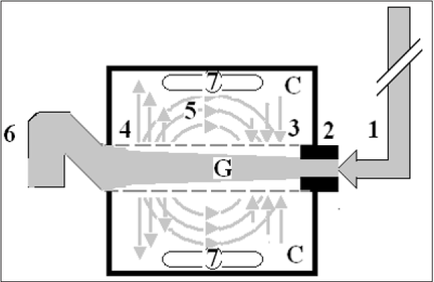
My beginning with the transurethral resection of the prostate (TUR) syndrome started in 1981 after I attended post-mortem (PM) examinations on 3 patients who died after the TURP surgery. I was only an SHO in urology working for the late Mr. KC Perry and JP Ward at DGH in Eastbourne. At the PM examination it was clear and obvious to me that these patients died of internal drowning as result of massive volumetric overload (VO) of fluids used for resuscitation of a cardiovascular shock they suffered, and the fluid was retained in their bodies. When I asked the pathologist why doesn’t he mention that retained VO in his report? He replied: “because it offends treating physicians”? The word offends hit me right hard on my head like a hammer. My next question to myself was if it offends them why do physicians do it? This had led me to immediately replace the term fluid overload with the new and original Volumetric Overload (VO) after adding the cardiovascular hypotension Shock to it to become (VOS) that was introduced to avoid the word offends but it has proved to be a new scientific medical discovery. Another few questions such as: “What is misleading physicians into giving too much fluid during the resuscitation of shock? What shock is it? I communicated with Richard Harrison III (who may be late now) who is the originator of the hyponatraemic shock of the TUR syndrome and the use of 5%NaCl therapy in clinical practice for years during his retirement [1]. I reported later the true pioneer originators of this shock and the hypertonic sodium therapy (HST) were Danowski et al who induced it experimentally in dogs by massive 5%Glucose infusion [2]. Harrison advised me to “put the poison in the honey” that I could not accept. After the PM examination I suspected and incriminated Starling’s law being the scientific basis of fluid therapy in shock that dictates the wrong rules on fluid therapy for shock management documented in articles and books [3-7], for which the hydrodynamics of the porous orifice (G) tube is the correct replacement (Figures 1a&b) [8,9]. I felt so strongly about it that I wrote a letter to the late great professor of physiology Eric Neil and author of Sampson Wright Textbook of Physiology later in 1983 [10,11]. He nicely replied in handwritten letter as he was in retirement asking: Why and how may Starling’s law cause death of patients? The answer is there now after 40 years of hard scientific research and investigations [12].

Figure 1a: shows a diagrammatic representation of the hydrodynamic of G tube based on G tubes and chamber C. This 37-years old diagrammatic representation of the hydrodynamic of G tube in chamber C is based on several photographs. The G tube is the plastic tube with narrow inlet and pores in its wall built on a scale to capillary ultra-structure of pre-capillary sphincter and wide inter cellular cleft pores, and the chamber C around it is another bigger plastic tube to form the G-C apparatus. The chamber C represents the ISF space. The diagram represents a capillary-ISF unit that should replace Starling’s law in every future physiology, medical and surgical textbooks, and added to chapters on hydrodynamics in physics textbooks. The numbers should read as follows:
The inflow pressure pushes fluid through the orifice.
Creating fluid jet in the lumen of the G tube**.
The fluid jet creates negative side pressure gradient causing suction maximal over the proximal part of the G tube near the inlet that sucks fluid into lumen.
The side pressure gradient turns positive pushing fluid out of lumen over the distal part maximally near the outlet.
Thus, the fluid around G tube inside C moves in magnetic field-like circulation (5) taking an opposite direction to lumen flow of G tube.
The inflow pressure 1 and orifice 2 induce the negative side pressure creating the dynamic G-C circulation phenomenon that is rapid, autonomous, and efficient in moving fluid and particles out from the G tube lumen at 4, irrigating C at 5, then sucking it back again at 3,
Maintaining net negative energy pressure inside chamber C.
**Note the shape of the fluid jet inside the G tube (Cone shaped), having a diameter of the inlet on right hand side and the diameter of the exit at left hand side (G tube diameter). I lost the photo on which the fluid jet was drawn, using tea leaves of fine and coarse sizes that run in the centre of G tube leaving the outer zone near the wall of G tube clear. This may explain the finding in real capillary of the protein-free (and erythrocyte-free) sub-endothelial zone in the Glycocalyx paradigm. It was also noted that fine tea leaves exit the distal pores in small amount maintaining a higher concentration in the circulatory system than that in the C chamber- akin to plasma proteins.

lupinepublishers-openaccess-journal-urology-nephrology

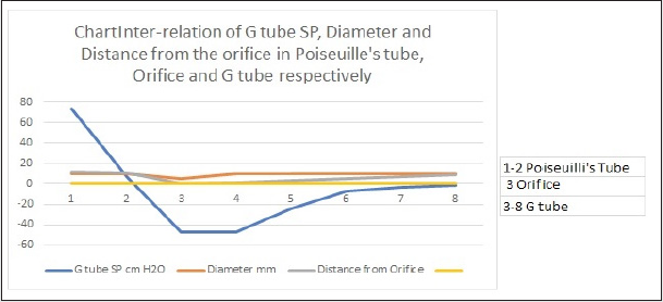
Figure 1b: shows the relationship between SP to the Diameter and length of the G tube which demonstrate a negative SP starting at the orifice (Point 2) (akin to precapillary sphincter) and extends as high negative pressure gradient over the proximal part of the G tube (Point 2-6) to cross 0 line at point 8 and then turn positive of 7 cm water at Point 9. This SP gradient from orifice at Point 2 to G tube lumen (Points 2-6) is negative to become positive DP at point 9 of 7 cm H20 water along the G tube. The wide section diameter of G tube is 7 mm all along the entire tube. The orifice is 5 mm while the distance from orifice to exit represent the tube’ length in which the Fluid jet diameter change with increasing gradient (Figure 1a). Neither Poiseuille’s law nor Bernoulli’s equation can predict SP neither at orifice of Venturi’s effect nor at the G tube proximal part know as Bernoulli’s effect. Thus, the RBCs speed or CBS depend on the dynamic fluid jet diameter not the G tube diameter. Hence the equation in Figure 2g (Figure 2) and graph are wrong giving low RBCs speed or CBS over the capillary length but is correct only at point of the G tube where the jet diameter equals the tube diameter.

lupinepublishers-openaccess-journal-urology-nephrology

What is the TUR Syndrome? And what is causing the “Understanding Gap”? Our prospective cohort study on the TUR syndrome was conducted in 1987-8, a letter to the editor of BJU was reported in 1988 [13], MD Thesis was accepted November 1988 [14], and the article reported in 1990 [15]. The TURP syndrome is a condition induced by gaining large volume of sodium-free fluid overloading the cardiovascular system and spelling over into the interstitial fluid (ISF) space of vital organs and subcutaneous. The fluid of 1.5%Glycine used as irrigating fluid gets absorbed, or rather infused through peri-prostatic veins, during the TURP surgery as well as all endoscopic surgeries performed under sodium-free fluid irrigation of any type such as Mannitol, Sorbitol, Glucose and Cytal. Also, intravenous (iv) infusion of 5% Glucose considerably and significantly contributes to it- as well as saline. What is more, excessive infusion of saline or any sodium-based fluid such as Saline, Hartman, Ringer, plasma, and plasma substitutes, and blood worsens it transferring the shock being treated from VOS 1 into VOS 2 [16] and causing ARDS 1 and 2 [17,18] with apparent correction of HN, and has high morbidity and mortality later.
The TUR syndrome has a characteristic severe drop of serum sodium level causing acute dilutional hyponatraemia (HN) induced by VO 1 (Figures 2 & 3) with severe clinical symptoms affecting all vital organs causing the multiple organ dysfunction syndromes (MODS) (Table 1) or ARDS [17,18] with recognizable clinical picture but one system may predominate such as acute kidney injury (AKI). The HN of <120 mmol/l has 2 paradoxes and 2 nadirs that have eluded authorities and physicians on HN, and that has made the TUR syndrome most elusive and invisible making it though obvious it has remained invisible even to authorities on HN. Professors and consultant urologists who are such swift good resection experts have testified that the TUR syndrome does not exist as no fluid absorption occurs, with a negative prospective study of 100 patients [19]. Off course no such hyponatraemia occurs when the irrigating fluid is saline whatever the volume absorbed and infused. Another important reason that prevents massive 1.5% glycine absorption and the TUR syndrome is for the Urologist not to breach the prostate capsule and not to open the venous sinuses where the irrigating fluid is directly injected intravenously (iv) into the periprostatic veins. There was also another good swift urologist who reported >1000 consecutive TURP surgeries without seeing the TUR syndrome. The risk of VO during endoscopic surgery will continue to occur as long as there are registrars in training and even with the experienced consultants who occasionally and inadvertently breach the prostatic capsule and open the venous sinuses. However, the TUR syndrome due to 1.5% Glycine VO with its characteristic HN has an undoubted reality [13-15] and [20-22]. Our study reported 10% incidence of the TUR syndrome with one near death case that was saved [14] and a similar study done a year earlier in the same department reported 7% incidence of morbidity with 1% mortality [22]. Before the TUR syndrome disappears into oblivion and is totally replaced by ARDS a most comprehensive literature review on the subject was reported in 2018 after the wide use of saline as irrigating fluid in the TURP surgery [23]. Here a distinction between a physiological VO of <2 L infused in less than one hour that is extensively studied by Hahn in volunteers and patients is known as Volume Kinetic (VK) (20) and the pathological VO of 3.5-5 L gained in < 1 h that causes the TUR syndrome [15] is highlighted. This has been a cause of serious misunderstanding gap in the pathogenesis of the TUR syndrome. The physiological response of VK is remarkably different from the pathological response of VO which is paradoxical: VK elevates blood pressure and induces diuresis while VO causes hypotension with bradycardia and causes acute renal failure.

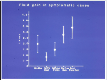
Figure 2: It shows the means and standard deviations of volumetric overload in 10 symptomatic patients presenting with shock and hyponatraemia among 100 consecutive patients during a prospective study on transurethral resection of the prostate. The fluids were of Glycine absorbed (Gly abs), intravenously infused 5% Dextrose (IVI Dext) Total IVI fluids, Total Sodium-free fluid gained (Na Free Gain) and total fluid gain in litres.

lupinepublishers-openaccess-journal-urology-nephrology

Table 1: Shows the manifestations of VOS 1 of the TURP syndrome for comparison with ARDS manifestations induced by VOS2. The manifestations are the same but one vital organ-system may predominate.

lupinepublishers-openaccess-journal-urology-nephrology

Table Abbreviation
SBB1: Sudden Bilateral Blindness
COC2: Clouding of Consciousness
MBCI3: Paralysis mimicking bizarre cerebral infarctions, but is recoverable on instant use of HST of 5%NaCl and/or NaCO3, and so is coma and AKI
FAM4: Frothing Around the Mouth
APO5: Acute Pulmonary Oedema.
RA6: Respiratory Arrest.
CPA7: Cardiopulmonary Arrest; ARDS$: Occurs on ICU later.
Annuria8: That is unresponsive to diuretics but responds to HST of 5%Ncl and/or 8.4%NaCO3; AKI8: Acute Kidney Injury. Also occurs the excessive bleeding at
AKI9: Acute Kidney Injury
DGR10: Delayed Gut Recovery; CV Shock*:
Excessive bleeding may occur at the surgical site and leucocytosis occurred in the absence of sepsis and septic shock.

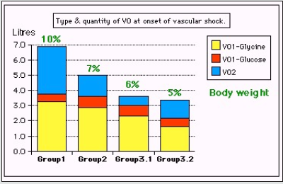
Figure 3: Shows volumetric overload (VO) quantity (in liters and as percent of body weight) and types of fluids. Group 1 was the 3 patients who died in the case series as they were misdiagnosed as one of the previously known shocks and treated with further volume expansion. Group 2 were 10 patients from the series who were correctly diagnosed as volumetric overload shock and treated with hypertonic sodium therapy (HST). Group 3 were 10 patients who were seen in the prospective study and subdivided into 2 groups; Group 3.1 of 5 patients treated with HST and Group 3.2 of 5 patients who were treated with guarded volume.

lupinepublishers-openaccess-journal-urology-nephrology

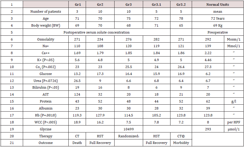
The TURP syndrome starts by presenting with cardiovascular hypotension shock to anaesthetists and surgeons in theatre [24,25] and at times by cardiac or cardiopulmonary arrest [26] and sudden death. By next morning the surviving patients present with coma, convulsion and bizarre paralysis to physicians, neurologists, and ICU specialists [15]. It has the characteristic serum hypo-osmolality. BUT other solute contents dilutions seem to be apparently spontaneously improving due to water shift into cells [Table 2, Figures 1 and 2]. The HN of <120 mmol/l causes cardiovascular hypotension shock. Volumetric overload (VO) is the most highly significant factor causing its patho-aetiology with a (p=0.0007). Osmolality was also significantly low (p=0.02) while all other serum solute changes including the most remarkable drop in serum sodium and huge elevation in serum glycine did not reach statistical significance in the multiple regression analysis, yet it did alone when pre- and post-operative levels are compared!? [Table 2 and 3]. This cardiovascular shock of VOS is easily confused with and mistaken for haemorrhagic or septicaemia shock and is wrongly treated with further massive volume expansion that usually kills the patient as happened in the 3 patients mentioned above!?

Table 2: Shows the mean summary of data, therapy and outcome comparing the 3 groups of 23 case series patients who’s (whose) VO is shown in Figure 3. Groip-1 was the 3 patients who died and had post-mortem examination, Group-2 were a series of severe TURP syndrome cases successful ly treated with hypertonic sodium therapy (HST), and Group-3 were 10 patients encountered in the prospective study who were randomized between HST (3.1) and conservative treatment (CT) (3.2). The significant changes of serum solute contents are shown in bald font with the corresponding p- value. Most of the patients showed manifestation of ARDS of which the cerebral manifestation predominated, being on initial presentation (Regional Anaesthesia) and representation of VOS 1 (General Anaesthesia). However, most patients were given large volume of saline that elevated serum sodium to near normal while clinical picture became worse. They suffered VOS2 that caused ARDS. The VO of patients to whom these data belong is shown.

lupinepublishers-openaccess-journal-urology-nephrology

Table 3: Shows the multiple regression analysis of total per-operative fluid gain, drop in measured serum osmolality (OsmM), sodium, albumin, Hb and increase in serum glycine occurring immediately post-operatively in relation to signs of the TURP syndrome. Volumetric gain and hypo-osmolality are the only significant factors.

lupinepublishers-openaccess-journal-urology-nephrology

The toxic theory of the TUR syndrome and septic theory of ARDS.

Sepsis and septic shock in the pathogenesis of ARDS is as innocent as the wolf in Josef story [18], so is glycine in the aetiology of the TUR syndrome [15], particularly as correctly mentioned that the TUR syndrome occurs with Mannitol, Sorbitol, and Glucose. Professor Alan Arieff has clearly reported the morbidity and mortality of hyponatraemia (HN) of the TUR syndrome induced by 1.5%Glycine as well as the excessive 5%Glucose infused intravenously during prolonged surgery in healthy women [27]. That does not mean that I deny the toxicity of glycine and the seriousness of sepsis. I am just saying they are misleading like a mirage to someone thirsty and lost in the desert. While thinking about it please, try to attend the PM examination of some patients who died from the TUR syndrome and ARDS. Every anaesthetist should examine own practice when he embarks on Bolus Fluid Therapy (BFT) during anaesthetic induction and watch out how much fluid is given during prolonged major surgery. Review the scientific basis of fluid therapy in the management of septic and all other types of shock on which bases the current practice is implemented.

Fluid therapy Regimen and Iatrogenic complications

The TUR syndrome occurs because of combination of fluid absorption and direct iv infusion of the irrigating fluid when the prostatic capsule is breached, and venous sinuses are open. In clinical practice all ARDS cases occur as result of iv infusion of fluids. In our study 7 cases of capsule breaching occurred among the 10 TUR syndrome cases as observed by the surgeon. The iv infusion occurs with both the liberal regimen of Early Goal-Directed Therapy (EGDT) and Bolus fluid therapy (BFT) of the conservative regimen. Hahn is a professor and consultant of anaesthesia and intensive care. He is also a leader and world authority on fluid therapy and the editor of a book on the same subject. I would and have recommended him as the head of a committee to write the new guidelines on fluid therapy in shock management. He has my new book that will help him for >8 months now, please read it if you’ve not done so already. Like all anaesthetists, Intensive care therapists, surgeons, and physicians of the whole world who remain to practice the liberal fluid therapy regimen also well known as EGDT in the management of shock, don’t you? Go to any ICU near you and observe the swollen-up ARDS patients mostly with trunk oedema comparing their body weight on hospital admission with their current weight while suffering from ARDS. Try to attend the PM examination of the TURP patients and ARDS patients. Allow me to reproduce this section from my article later that is most recommended reading to all physicians interested in the subject of fluid therapy, the TUR syndrome, HN, VOS and ARDS [18].

The role of Starling’s law

Starling’s law [28,29] dictates the current faulty rules on fluid therapy in the management of shock. It thus misleads physicians into giving too much fluid during shock resuscitation [30]. More than 21 reasons were reported to show that Starling’s law is wrong [31], none of it can be denied or refuted. The correct replacement is the hydrodynamic of the porous orifice (G) tube [8,9] (Figure 1 a & b) that was built on capillary ultrastructure anatomy of having precapillary sphincter [32] and a porous wall [33] that allow the passage of plasma proteins-hence nullify the oncotic pressure. It follows that the extended Starling Principle is wrong and a misnomer [34,35] and all the equations are also wrong.

Two types of VO inducing VOS and causing ARDS of type 1 and 2

There are two types of VO: Type 1 induced by sodium-free fluid and Type 2 induced by sodium-based fluid. These in turn induce VOS 1 and VOS 2 which cause ARDS 1 and ARDS 2, respectively. The clinical picture is the same for both types (Table 1). Type 1 is characterized with HN of the TUR syndrome with which the cerebral neurological manifestations of coma, convulsions, and bizarre paralysis predominate while type 2 may have moderate hypoproteinemia if induced by crystalloids and none when plasma, plasma substitutes and blood are used. Type 2 may complicate Type 1 or may occur do novo. Manifestations of the multiple organ dysfunction syndrome (MODS) are the same and appear in every case, but one system may predominate. When Hahn sent me his article on Revised Starling Principle calling for revalidation [34] I immediately responded with an article: Revised Starling’s Principle (RSP): a misnomer as Starling’s law is proved wrong. I considered research on validating RSP is a total waste of money, time, and efforts.

Proof by eminent authors on the VO role in the aetiology of the TUR syndrome and ARDS

Professor Robert Hahn from Sweden has done lots of research infusing various types of fluid used in clinical practice to normal adult volunteers and patients, as well as animal research and clinical studies and reported >340 articles on the TURP syndrome alone (PubMed 2017) and 532 articles in total (PubMed search 2021): Here is what Robert Hahn said: in the abstract of an article reported in 2017 [36]:

Abstract [36]:

“Adverse effects of crystalloid fluids are related to their preferential distribution to the interstitium of the subcutis, the gut, and the lungs. The gastrointestinal recovery time is prolonged by 2 days when more than 2 liters is administered. Infusion of 6-7 liters during open abdominal surgery results in poor wound healing, pulmonary oedema, and pneumonia. There is also a risk of fatal postoperative pulmonary oedema that might develop several days after the surgery. Even larger amounts cause organ dysfunction by breaking up the interstitial matrix and allowing the formation of lacunae of fluid in the skin and central organs, such as the heart.” Thank you, Professor Hahn for a most impressive work indeed. New guidelines based on currently available evidence on fluid therapy for resuscitation of sepsis, septic shock, trauma patients, critically ill patients, ARDS and patients undergoing prolonged major surgery are badly needed. Professor Hahn is the expert witness on fluid therapy.
Why does not Hahn believe his own results? Why doesn’t he make the most obvious conclusion based on what he said in the abstract above? What and how much more evidence and years that he needs to believe that the pathological VO of massive fluid infusions induces cardiovascular shock that is VOS of both types and causes ARDS? If my articles referenced here and the books [3- 7] particularly the one Hahn has now for 8 months and being held in the press awaiting his introduction, then allow me most sincerely and humbly to give you a helping hand to lift you up to where I stand and clearly see the picture on the real issues discussed here. Hahn does not need to do any more research studies. Just report a re-analysis of data from previously reported articles he has done and reported before, based on his previous published articles on the TUR syndrome and saline-based fluid infusions. Please, reexamine and re-analyse your own research work in a manner and method identical to your article reported here [20]. Please, Hahn don’t bother with equations that are hard to understand and are meaningless and perhaps misleading or even wrong. Do not use fancy sophisticated graphs that does not impress me. I would love, most sincerely and humbly, to give you a hand to get you out of the huge maze you have been lost inside it for >3 decades. All you need to do my friend now is to liberate yourself from the illusive and misleading concepts of the toxic/septic hypotheses of glycine and sepsis!? One must unlearn old bad habits to be able to receive and acquire the new correct ones.

Evidence for the VO Theory causing VOS and ARDS

“The prevalence of “liberal fluid infusion” in resuscitation of all types of shocks not only septic shock in clinical practice all over the world is attributed to an impactful article by Rivers et al, reported at The N Engl J Med 2001 [37]. Dr Rivers’ investigation reported EGDT in the treatment of severe sepsis and septic shock. In this singlecenter study published more than 20 years ago involving patients presenting to the emergency department with severe sepsis and septic shock, the conclusion was: “mortality was markedly lower among those who were treated according to a 6-hour protocol of EGDT, in which intravenous fluids, vasopressors, inotropes, and blood transfusions were adjusted to reach central hemodynamic targets, than among those receiving usual care” Usual care means conservative fluid regime. There is something grossly wrong with this conclusion, but I cannot tell what is it? Not yet. Let us see what other author investigators have said first. The EGDT of liberal fluid infusion has been termed “aggressive” by some authors. However, it has been adopted all over the world not only for the therapy of septic shock but also whenever fluid therapy is required for the management of all types of shocks.
“In another article by Dr Rivers 11 years later in 2012 [38] he compared the liberal to the conservative approach concluding in his last statement: “In contrast to what is true in politics, in fluid management of acute lung injury, it is OK to be both liberal and conservative.” So, Dr Rivers says it is OK to have it both ways: “one for the ebb and one for the flow”! Sorry, sir, I disagree. It is not OK. It is not politics either. No, you cannot have it both ways. The right way is only one. The issue here is how much fluid should be infused during the ebb phase of shock and does it have a maximum limit? Replace the loss but do not overdo it. Since the cardiovascular system (CVS)’ maximum capacity of an adult is 7 L and the normal blood volume is 5 L, the maximum infused volume of fluid should be limited by the maximum capacitance of the CVS. What do you expect when you try to fit 10-15 L of fluid into a 7 L capacity container? Simple physics and common sense indicate that it must spell over if it is open system or burst if closed! The cardiovascular system is no exception. Dr Rivers should re-examine his own data and tell us where and why he went so grossly wrong.” The EGDT has spread like fire in a haystack, and it remains operative in current clinical practice all over the world that is why ARDS is so common yet remains under recognized and underestimated affecting and killing hundreds of thousands of patients per year.” Other authors have confirmed the significant role of VO of crystalloids in causing the morbidity and mortality of ARDS both in adult and children of trauma patients [39,40]. All authors have stopped short of recognizing VOS as Cause of ARDS or MODS morbidity and mortality. Quoting also from this article [18] I mention here the remarkable multicenter study by Rowan et al. [41] Like Hahn they reported results that demonstrate the massive VO retained in the body of surviving ARDS patients. After sending 3 emails to Rowan commending the authors on their results and asking about the dead patients retained fluid VO, none of the 40+ authors replied. “The PRISM Investigators reported its Trial by Rowan et al at NEJM 2017 [41] concluded: “In this meta-analysis of individual patient data, EGDT did not result in better outcomes than usual care and was associated with higher hospitalization costs across a broad range of patient and hospital characteristics.” Thank you, Dr Rowan and colleagues for the excellent research and report. This is good evidence-based medicine, but more is needed, from you, and you have the data to provide it. Based on this conclusion that agrees with other multicenter trials I wonder is time to say goodbye Dr Rivers? The aggressive and deleterious liberal approach of EGDT is no longer wanted. It should be abandoned immediately. Even when the nasty liberal approach goes away, hopefully soon, it remains bad enough with the conservative regime as it is now that must be sorted out! I wonder what Dr Rivers has to say about this, particularly as authors of 3 other huge prospective multicenter trials of The ProCESS/ARISE/ProMISe reported similar conclusion by Huang et al. [42]. So, Rowan gave the results of: The cumulative VO was -136 ml in the conservative-strategy group, as compared with 6992 ml in the liberal-strategy group (P<0.001). For patients who were in shock at baseline, the cumulative seven-day VO was 2904 ml in the conservative-strategy group and 10,138 ml in the liberal strategy group (P<0.001). For patients who were not in shock at baseline, the cumulative VO was −1576 ml in the conservative-strategy group and 5287 ml in the liberal-strategy group (P<0.001)”. “First, the negative sign (-) indicating negative fluid balance has appeared in the data above and is very important. It characterizes the nonsymptomatic patients among the conservative-strategy group.
These patients should be used as the controls for the statistical analysis of the data. I have been waiting for 40 years to see these VO results. I am still waiting to see VO data with statistical significance in mortality patients. I plead with and urge the respected authors of major randomized Trials of FACCT, PRISM, ProCESS, ARISE, and ProMISe to come forward with these data, please,

Clinical picture of (VOS, The TUR syndrome, ARDS and MODS)

The clinical picture of ARDS is that of the multiple organ dysfunction syndrome (MODS) (Table 1) reported previously by Khadarow and Marshal in 2002 [43]. Another remarkable article was reported by Schrier in 2010 [44]. Demonstrating the link of the TUR syndrome with ARDS by having identical clinical picture with minor variations was reported by Ghanem as complications of VO covering the cardiovascular/hematological that appear first under general Anaesthesia with bradycardia [45], the cerebral/ neurological with coma appear first under spinal/epidural Anaesthesia and convulsions and bizarre paralysis predominate in the TUR syndrome, not in ARDS [46], the respiratory of ARDS and hepatic/gastrointestinal manifestations [47] and AKI predominate later were documented recently in individual specific reports. Excessive bleeding and leukocytosis in the absence of sepsis also occur.

Therapy of VOS, the TUR syndrome and ARDS [17] Prevention

Based on the above discussion, ARDS is an iatrogenic complication of fluid therapy in hospital, never in community, that is overlooked and underestimated. Being iatrogenic; means it is preventable. In order to prevent VOS and ARDS a limit to the maximum amount of fluid used during shock resuscitation or major surgery must be agreed upon. Professor Hahn [36] found that infusing 2 L of saline to human volunteers produces symptoms. Infusing >3 L is pathological. More than 5 L is associated with deleterious morbidity [38,39]. So, the maximum volume of fluids that can be infused safely to an adult patient is 3 L which is the daily fluid requirement, and no more fluid of any kind is given for 24 hours except replacing the actual loss that does not include urine loss. The patient should be put on a weighing scale every day from hospital admission till discharge or death. Any retained volume of fluid above his body weight on admission is pathological. On using CVP for monitoring fluid therapy, please refrain from persisting to elevate CVP to levels above 12 and up to 18-22 cm saline [48]. This is a major cause for inducing VO and VOS and ARDS during shock resuscitation, particularly septic shock [37]. Look up any physiology textbook to find out that the normal CVP is 0 and it swings between -7 and +7 cm saline which is the level that should be aimed at in monitoring fluid replacement in shock of sepsis, trauma, and bleeding, acutely ill and during major surgery. Elevating CVP is not synonymous with elevating arterial pressure. If hypotension develops later during ICU stay, inotropic drugs, hydrocortisone 200 mg and HST should be used. The latter restores the pre-capillary sphincter tone (peripheral resistance) so that the capillary works as normal G tube again [9], but no isotonic crystalloids or colloids infusions of above the daily fluid requirement should be given. If persistence with the current liberal regimen of Early Goal-Directed Therapy (EGDT) and conservative Bolus Fluid Therapy regime continues, then more reports on ARDS will continue. Future authors will be hopefully taking into consideration the mentioned above data concerning VO/Time, or the retained fluid VO at the time of inducing ARDS or death on reporting new trials or case reports.

Treatment of ARDS [6]

Hypertonic sodium therapy (HST) of 5%NaCl and/or 8.4%NaCo3 has truly proved lifesaving therapy for the TUR syndrome and acute dilution HN [17,18] as well as Secondary VOS 2 that complicates fluid therapy of VOS 1 causing ARDS. It works by inducing massive diuresis; being a potent suppressor of antidiuretic hormone. My experience in using it for treating established ARDS with sepsis and primary VOS 2 that causes ARDS is limited. However, evidence on HST suggests it will prove successful if given early, promptly, and adequately to ARDS patients while refraining from any further isotonic crystalloid or colloid fluid infusions using saline, Hydroxyethyle starch and/or plasma therapy- just give the normal daily fluid requirement and no more. After giving HST over one hour using the CVP catheter already inserted, the patient recovers from AKI and produces through a urinary catheter massive amount of urine of 4-5 L as you watch. This urine output should not be replaced. Just observe the patient recovering from his AKI, coma and ARDS and asks for a drink. This is done in addition to the cardiovascular, respiratory, and renal support on ICU. Patients with AKI on dialysis, the treating nephrologist should aim at and set the machine for inducing negative fluid balance. The HST of 5%NaCl and/or 8.4%NaCo3 is given in 200 ml doses over 10 minutes and repeated. I did not have to use more than 1000 ml during the successful treatment of 16 patients. Any other hypertonic sodium concentration is not recommended- I know Hahn tried 1.8%NaCl and it does not work. A dose of intravenous diuretic may be given but it does not work in a double or triple the normal dose. A dose of 200 mg of hydrocortisone is most useful. Antibiotic prophylactic therapy is given in appropriate and adequate doses to prevent sepsis and septic shock. No further fluid infusions of any kind of crystalloids, colloids and blood is given. The urinary loss should not be replaced as this represent a surplus in the body and must be discarded otherwise defeats the objective of treatment.

Addendum: Relevant articles on the history of the TUR syndrome and ARDS

This addendum is dedicated to important landmark articles on the history of the TUR syndrome and ARDS that could not be fitted directly on the above focused narrative review on how the TUR syndrome has been reincarnated into ARDS. It is optional reading for the interested reader, but it completes this review. The first part is dedicated to eminent authors on the TUR syndrome and ARDS whether directly or indirectly. The second part is a section on selfreferences by the author that report important issues that highlight aspects of the presentation.

A. Other Eminent Authors

Creevy was the first author to report the TUR syndrome as acute water Intoxication [49]. Ashbaugh et al were the first to report ARDS in the Lancet in 1967 [50]. Lessels et al. reported in a letter to the editor as the only article on death during prostatectomy [51]. Hendry was first to report that the osmotic pressure of various body fluid is the same as plasma [52]. Guyton and Coleman reported the negative pressure of the subcutaneous space of -7 cm water, a fact that cannot be explained by Starling’s law [53]. Calnan et al reported the negative pressure in lymphatic vessels [54]. Renkin was the first to call for reconsideration of Starling’s law [55]. The Coshran injuries Group, Finfer, Vincent and futier et al demonstrated that oncotic pressure does not work and the argument on albumin versus saline is obsolete [56-59].

B. Self-references

Articles 60 and 61 have educational and entertainment value. Articles 62 and 63 shows the relevance of my work on ARDS to Covid-19 pandemic ARDS. Article 64- 66 corrects other received misconceptions on capillary physiology to augment the discovery of the G tube hydrodynamics and its impact on the capillary- ISF transfer. Articles 67 and 68 report the two clinical studies on which the above article is based. Article 68 corrects some errors and misconceptions on fluid therapy. Article 70 is on preventing renal failure in the critically ill patients. Article 71 reports my Experience with cystoprostadenectomy with “prostatic capsule sparing” for orthotopic bladder replacement. Article 72 is on Features and Complications of Nephroptosis Causing the Loin Pain and Haematuria Syndrome. Article 73 reports “New Discoveries in Medicine and Physiology Originated in Urology”. Article 74 is on an Update on Ghanem’s new scientific discoveries in physics, Physiology, and Medicine, Article 75 is on Goodbye Starling’s law, hello G tube.

Conclusion

The TUR syndrome as defined and characterized with acute dilutional hyponatraemia will no longer be seen in urology after the use of saline as irrigating solution in endoscopic surgery. However, the ARDS will replace it with identical clinical picture of MODS that continue to occur with high morbidity and mortality that is underrecognized and underestimated. The ARDS is common in clinical practice and is induced by excessive sodium-based fluid infusion and is likely to occur in urology due to the added risk of irrigating fluid absorption and infusion through periprostatic veins. Neither the toxic theory nor the septic theory plays the significant assumed rule in the pathogenesis of the TUR syndrome and ARDS. Both are iatrogenic complications of fluid therapy, induced by VO of > 3 L in <1 h time and is severe at 7-10 L of retained fluid VO in surviving ARDS patients wile mortality occur with 12 L, and both have preventative and curative therapy of HST of 5%NaCl and/or 8.4%NaCo3.

Conflict of Interest

Conflict of Interest: None

Funds received

Funds received: None

Read More About Lupine Publishers Journal of Urology Please Click on Below Link:
https://lupine-publishers-urology-nephrology.blogspot.com/

Wednesday 27 September 2023

Lupine Publishers | Children’s Mouthwash; Commercial Product or Oral Health Guarantor

 Lupine Publishers | Journal of Dentistry and Oral Health Care


Opinion

Nowadays, when we enter a pharmacy, we come across a variety of healthcare products that come to the market every day. Types of masks, gels, detergents and etc., which may look identical in name and application but claim to be totally different and each has its own user extent. Some of these products are commercial in nature, and some are not so important in maintaining health, and in fact, one’s health is not dependent on them. Perhaps that’s why each of us never consumed some of these health and beauty products, and we do not even know them at all. If the product does not directly interact with our health, we do not need to carefully look at the need to how to consume them and do not curious about them, but when it comes to products that claim to protect our child’s health, the matter becomes more quite critical and worrying. Mouthwash is one of these products which are found at all pharmacies. So many oral health officials have been able to highlight the need to use mouthwashes among people. With the training and information provided in this area, people should use mouthwashes in addition to toothpaste and dental floss. But what is the child’s mouthwash, and do the children really need them to use? Do these products guarantee the health of their teeth or is it more a commercial product to fill the pockets of some of the companies?

Mouthwashes have different types. Some have pharmaceutical uses and should be used in certain cases only with physicians’ prescriptions. Mouthwashes that are on the market for the public use contains fluoride which prevents tooth decay. Many pharmaceutical companies have produced and marketed mouthwashes for children to facilitate the use of mouthwashes for children. These mouthwashes have been manufactured considering the least risk of swallowing fluoride. The proper taste of these mouthwashes is a feature that encourages children to use them. Mouthwash is one of the complementary methods of oral home care. These mouthwashes provide oral and dental care along with tooth brushing and dental floss, but they should never be replaced by each other. In other words, application of none of them alone has the significant effect. Before planning to buy the product, if parents tend to use pediatric mouthwashes, they should have a consultation with the pediatric dentist so that the dentist can select the better of the most appropriate one and the least harmful mouthwash. However, they should be used according to the instructions; these instructions vary from product to product and depending on the content and concentration of fluoride, the application may be different.

In other words, it should be noted that some types of mouthwashes are highly recommended by most dentists in the routine oral care program. This is due to the ease and speed of its use, and its effectiveness. In general, along with toothbrushes and dental flosses, many types of mouthwashes are also produced, each of which has its own interests in the beauty and health of teeth. These mouthwashes can be prescribed by the dentist or can be purchased OTC from the pharmacy. Nonetheless, alcohol-free mouthwash is a product that depends on the individual’s needs. The choice of the mouthwash that meets personal needs is very important. Some mouthwashes on the market contain alcohol, in particular, Ethanol, which can cause burning sensation, unpleasant taste, and dryness in the mouth. It is not recommended for children at all because the burning and spicy tastes force the child to stop using the kid’s mouthwash for future use. Consequently, if the parents intend to choose an appropriate mouthwash, first they should not choose any kinds of mouthwashes, and secondly, it is advisable to have a consultation with the pediatric dentist so that they don’t become bewilderment when choosing the proper mouthwash. To finish this point of view, for some time, fluoridecontaining mouthwash has been commonly used in children. But, in my opinion, some kids do not need to use them at all. That is, if fluoride is adequately contained in fluoride-containing toothpaste, in drinking water, or even the consumption of foods such as seafood or tea which contains this material, then, there is usually no need for fluoride mouthwashes for children.

Read More About Lupine Publishers Journal of Dentistry and Oral Health Care Please Click on the Below Link: https://lupine-dentistry-oral-health-care.blogspot.com/


Tuesday 26 September 2023

Lupine publishers | Continuous or Intermittent? Which Regimen of Enteral Nutrition is Better for Acute Stroke Patients? a Systematic Review and Meta-Analysis

 Lupine Publishers | Journal of Neurology and Brain Disorders

Abstract

Background and purpose: Enteral nutrition via nasogastric tube in acute stroke patients with dysphagia is an important determinant of patient outcomes. It is unclear whether intermittent or continuous feeding is more efficacious. The aim of this review is to examine the current evidence comparing the effectiveness of intermittent versus continuous feeding in stroke patients in terms of nutritional status, gastrointestinal intolerance and other complications.

Methods: A systematic review of randomized controlled studies comparing intermittent with continuous nasogastric feeding in acute stroke patients was conducted in accordance with PRISMA (Preferred Reporting Items for Systematic Reviews and Metaanalyses) guidance using predefined search terms. The search was conducted in MEDLINE and EMBASE up to 1st March 2019. Two independent reviewers assessed study quality using the Joanna Briggs Institute Critical Appraisal Tool. Meta-analyses were conducted, where appropriate, using a random-effects model to pool risk ratio with corresponding 95% CI.

Results: Three studies including a total of 184 patients were identified. All three were medium to low quality. The definition of intermittent enteral nutrition within each study varied considerably in terms of volume, rate and mode of delivery. Achievement of nutritional targets was the same for both feeding patterns in the one study it was reported. Only aspiration pneumonia and diarrhea were measured by all three studies. There was no significant difference in the incidence of aspiration pneumonia (RR 0.91, 95% CI 0.53-1.57, p=0.74, I2=50%) and diarrhea (RR 1.74, 95% CI 0.70-4.30, p=0.23, I2=42%) between the two patterns of feeding. Other outcomes including, vomiting, gastric retention, mortality, pre-albumin and nasogastric tube complications showed no significant differences.

Conclusion: There is very little and low-quality evidence to inform patterns of enteral feeding after stroke. The available evidence shows no significant difference in nutritional achievement and complications between intermittent and continuous nasogastric tube feeding in acute stroke patients.

Keywords: Stroke; Enteral; Nutrition; Nasogastric; Dysphagia

Background

Dysphagia occurs in up to 50% of patients following a stroke [1- 4] and increases the risk of pneumonia almost ten-fold [5]. Strokerelated pneumonia is associated with longer length of hospital stay, worse levels of disability and increased mortality [6-9]. In most dysphagic patients, adaptation of the consistency of diet and fluids is sufficient to ensure that the swallow is safe. However, in a small proportion insertion of a Nasogastric Tube (NGT) is required to ensure safe and adequate nutrition. Despite this, more than twothirds of NGT-fed stroke patients still develop pneumonia [10] Gastric dysmotility is a well-documented phenomenon that occurs in critically ill patients, including acute stroke patients, whereby incomplete gastric emptying results in stasis, heightening the risk of reflux and aspiration of gastric contents [10-13]. NGT bolus feeding was first described by Morrison et al. [14] in 1895 for children with Diphtheria, who received 6-ounce bolus feeds 3 times a day via NGT. However, it wasn’t until 1910s when Morgan et al. [15] and Jones et al. [16] began administering their enteral feeds “drop by drop” rather than as a bolus. Contemporaneously, the regimen most frequently used in most patients requiring enteral feeding is continuous (i.e. low volume pumped feed lasting 16-24 hours without interruption). However, recent attention has been afforded to examining whether a discontinuous feeding strategy - often described as either intermittent or bolus (i.e. high volume of feed administered over a short period multiple times a day) - could reduce patients’ risk of pneumonia and achieve better nutrition and digestive tolerance.

Intermittent feeding reflects normal human feeding patterns more closely than continuous feeding. A period of fasting interrupted by the ingestion of a discrete meal causes gastric distension and subsequent stimulation of gut motility, secretion of digestive enzymes and metabolic responses to nutrient loading [17- 18]. This physiological gastrointestinal response to intermittent feeding has been demonstrated in healthy adults, neonates and intensive care populations [17-20]. While there are good theoretical reasons to assume that intermittent feeding is more physiological, most stroke patients in the UK receive nasogastric feeding continuously, as there are concerns that intermittent feeding may be less well tolerated. Guidance and practice relating to enteral feeding after stroke differs between countries; with the American Heart Association [21] and the Royal College of Physicians [22] not addressing the issue, Australian Guidelines allowing for both options [23] and intermittent feeding described as “traditional” in China [24]. The aim of this systematic review is to determine whether there are differences in the achievement of adequate nutrition, gastrointestinal tolerance, and metabolic stability between intermittent and continuous nasogastric feeding.

Methods

This systematic review and meta-analysis were prepared according to PRISMA (Preferred Reporting Items for Systematic Reviews and Meta-Analyses) guidelines [25].

Criteria for Considering Studies for this Review

The inclusion criteria for this review were:
a. Population: Acute stroke patients aged 18 or more with a nasogastric tube receiving enteral nutrition
b. Intervention: Intermittent enteral nutrition: by bolus, gravity systems or infusion pump several times a day with a rest between feeds
c. Control: Continuous enteral nutrition: with gravity systems or infusion pumps, without interruption for a minimum period of 12 hours/day
d. Outcomes: Nutritional status, aspiration pneumonia, diarrhea, vomiting, gastric distension, gastric retention, hyperglycemia, pre-albumin, mortality, length of stay, and NGT complications
e. Study Design: Randomized controlled trials or pseudo-randomised controlled trials (a study without true randomisation) that compared continuous and intermittent enteral feeding methods.

Search Strategy

A literature search was performed using MEDLINE (1966 – 1st March 2019) and EMBASE (1974– 1st March 2019). Studies were searched for using the terms enteral, nutrition, nasogastric, gastrointestinal, feeding as Medical Subject Heading (MeSH) and free text terms. These were combined with the set operator “AND” with following terms: intermittent, continuous as both MeSH and free text terms. Publications were restricted to those studying adult populations, defined as greater than 18 years old, with a documented diagnosis of stroke according to accepted international criteria [26]. This search strategy is described in Appendix 1. The reference lists of all eligible studies that were identified were also comprehensively searched for studies not identified using the initial search strategy. This search was performed independently by two reviewers.

Selection of studies

Two reviewers (GDP and ET) assessed the studies independently for inclusion using the title and abstract. In cases where relevance could not be determined solely from the abstract, the full text was consulted. Any disagreements were resolved by consensus with a third reviewer (CR).

Data extraction and management

Data extraction was done manually by two reviewers (GDP and ET). Differences were discussed and adjudicated in faceto- face meetings. Foreign language papers were translated, and descriptions of each study were derived. This included authors, year of publication, type of participant, location, study design, sample size, age and gender of participants, exclusion criteria, when feeding was started, monitoring period, nasogastric tube size, type of feed and definitions of each intervention. In addition, data was extracted for definition and results of each outcome from all studies.

Assessment of risk of bias in included studies

Methodological quality of the studies was assessed using the Joanna Briggs Institute Meta-Analysis of Statistics Assessment and Review Instrument (MAStARI) Critical Appraisal tool for experimental studies [27].

Data synthesis

The studies presented in this review all fitted the conceptual definitions of intermittent and continuous enteral nutrition, as outlined in the inclusion criteria. However, there were differences in the volume, rate and temperature of nutrient delivered. In addition, two of the studies did not use true randomisation. Taking into consideration these limitations, a meta-analysis has been carried out with the outcome’s diarrhea and aspiration pneumonia, as these were the outcomes assessed by all studies. Narrative synthesis was used where outcomes did not allow meta-analysis. The meta-analysis was performed using Review Manager (RevMan) Version 5.3. Copenhagen: The Nordic Cochrane Centre, The Cochrane Collaboration, 2014. Data was extracted from all three studies for the outcome’s diarrhea and aspiration pneumonia. We calculated risk ratios (RR) and 95% CIs using the Mantel–Haenszel model. Statistical heterogeneity among trials was assessed by the I2 test, with I2 >50 representing possible substantial heterogeneity. The meta-analysis was performed with a random‐effects model irrespective of the level of heterogeneity as the included trials varied considerably in a number of methodological features.

Results

Study selection

Figure 1: PRISMA flowchart for study selection process.


The PRISMA flow-chart for study selection is shown in Figure 1. Following the removal of duplicates the number of potentially relevant studies identified from this search was 1,377. Four studies met the criteria of relevance and no studies were added following a secondary manual search. On review of the full-texts, one study [28] was excluded due to a cross-over study design with no washout period and the outcomes reported were not clinically relevant to this review. Three studies [25] [29,30] remained including a total of 184 patients.

Study characteristics

Table 1 shows the characteristics of the included studies and patients. Two studies were conducted in China [24,25,29] and one in Turkey [30]. Population sizes (52-69) and age (mean 61-69 years) were similar in all three. A summary of the studies is given in Appendix 2.

Table 1: Characteristics of included studies. SD: Standard Deviation; M: Male; F: Female; NG: Nasogastric; BMI: Body Mass Index.


Risk of bias and quality of the evidence

Appendix 3 shows details of the quality assessment with moderate risk of bias (9/13 quality criteria fulfilled by Wang, and 6/13 by Chen and Gungor respectively). Only one study (Wang) was truly randomized (random numbers table), while Chen used alternate assignment for allocation of treatment groups, and Gungor randomized patients into two groups taking into account the age and gender, with no more detail has been given regarding how they randomised. Wang randomised patients using a random number table. Blinding of participants and assessors was not feasible due to the nature of the intervention and the outcomes measured. Only Chen commented on removal of patients from the study for clinical reasons. Four patients were excluded within three days of enrolment because of left ventricular failure, cerebral herniation, gastrointestinal haemorrhage, and respiratory failure respectively. It was not reported whether these patients were included in an intention to treat analysis. The other two studies did not refer to removal of patients after allocation to treatment groups. Chen was the most comprehensive in demonstrating similar baseline characteristics using age, gender, Glasgow Coma Scale, [31] the Acute Physiology, Age, Chronic Health Evaluation- II scale, [32] the National Institutes for Health Stroke Scale, [22] and the Barthel index [33]. Gungor used age, gender and a stroke subscale, and Wang only used age, gender and the Glasgow Coma Scale. The only outcome measure that is likely to be unreliable is the assessment of gastric distension in Wang’s study. This was ascertained by palpation combined with measurement of abdominal circumference, a method which is not validated and has no defined criteria.

Delivery of the feeds

Feed was given via wide bore NGTs (16 and 14 F for Wang and Gungor respectively). Details of administration given in Appendix 4. Continuous enteral nutrition was delivered via an infusion pump in all three studies, initially at less than 50 ml/h increasing to 75-100 ml/h as tolerated. Gungor started at a slower rate (10 ml/h) than the other two studies and increased feeding rates more gradually. Two studies (Wang and Gungor) continued feeding overnight without a period of rest, while Chen discontinued the feed for a period of 7 hours overnight. Intermittent regimens were considerably different between the studies. Wang delivered each feed (200-300 ml) over 10-15 minutes at a rate of 800-1800 ml/h, while Gungor infused a smaller volume of feed (120 ml) over a longer period of time (30-60 min) at a much slower rate of 300-600 ml/h. Wang administered the feed manually with a 50 ml syringe, which may have resulted in an even quicker administration time by the nurses than appreciated by the assessors of the study.

Outcomes

The effects of intermittent and continuous feeding on clinically relevant outcomes are given in Table 2. Definitions for key outcomes are detailed in Appendix 5.

Table 1: Comparative effects of intermittent and continuous enteral nutrition on clinically relevant outcomes. g/L: grams per litre.


Achievement of nutritional targets

This was only reported in one study [29]. There was no significant difference in achievement of the nutritional target and in levels of pre-albumin between intermittent and continuous feeding.

Complications of nasogastric feeding

One of the three studies (Chen) showed a significantly higher incidence (58.3%) of pneumonia with continuous feeding than with intermittent feeding (33.3%), with no difference in the other two studies. Diarrhea was significantly more frequent with intermittent feeding (64.0% vs. 14.3%) in Wang, but not in the other two studies. Wang also reported significantly more hyperglycaemia with intermittent feeding. No significant differences were found for vomiting (Gungor), gastric retention (Gungor, Wang), and NGT complications (Gungor).

Other outcomes

One study (Gungor) reported mortality and length of stay. No significant differences were identified between feeding patterns.

Meta-analysis

Figure 1: Meta-analysis of intermittent compared with continuous enteral nutrition on the incidence of aspiration pneumonia and diarrhea in acute stroke patients.


CI: Confidence interval; Chi2: Chi-squared test; Tau: Tau test

Only aspiration pneumonia and diarrhea were assessed by all three studies and could be included in the meta-analysis. There was no significant difference between intermittent and continuous feeding in either incidence of aspiration pneumonia (RR 0.91, 95% CI 0.53-1.57, p=0.74, I2=50%) or diarrhea (RR 1.74, 95% CI 0.70- 4.30, p=0.23, I2=42%). A funnel plot is not presented here as there were only 3 trials. This is analysis is displayed in Figure 2.

Discussion

The systematic review identified three studies comparing intermittent and continuous nasogastric feeding including 184 acute stroke patients. There was no significant difference between feeding regimes for most outcomes in individual studies with the exception of pneumonia, which was higher with continuous feeding in one study [29] and diarrhea, gastric distension and hyperglycemia, which were seen more frequently in another study [24]. The only outcomes which were assessed by all three studies and could be included in the meta-analysis were aspiration pneumonia and diarrhea, neither of which were significantly different in the two feeding regimens. Intermittent feeding would be expected to improve achievement of nutritional goals, as it is closer to normal feeding patterns allowing for more physiological gastrointestinal and metabolic responses. There is insufficient evidence to determine the effect of feeding pattern on the achievement of nutritional goals in this patient group. In the one study [29], where nutritional goals were addressed, no significant difference was found. Studies in intensive care patients found that calorific objectives were more likely to be achieved with intermittent than with continuous enteral nutrition [34,35] and this was confirmed through systematic review [21]. Furthermore, studies examining these two methods of administering enteral nutrition in older adults on general wards also found no discernible difference in the calories achieved [36,37] This was in keeping with the results observed from this review.

Aspiration pneumonia is a major complication of dysphagic stroke and may be affected by the pattern of feeding. Our metaanalysis did not find a significant difference in pneumonia between intermittent and continuous feeding. In all three studies the minimum incidence of aspiration pneumonia in acute stroke patients fed by NGT was regardless of intervention. Chen was an outlier with almost twice the incidence of aspiration pneumonia in the continuous group, and this difference might have been due to chance. However, this was the only study to specify that they recruited patients within 7 days of admission, and this could have ensured that patients hadn’t had a significantly long starvation period in which gastric dysmotility would have developed. Interestingly, it was the only study which discontinued feeding during the night, a practice usually considered to reduce the risk of pneumonia. Studies of intermittent versus continuous feeding in other settings give mixed results with a reduction of pneumonia with intermittent feeding in intensive care,38 but no difference in older people nursed on general wards [37]. Gastrointestinal tolerance is a major determinant of choice of feeding pattern. There was no significant difference in the incidence of diarrhea in our meta-analysis. Looking at individual studies, Wang consistently reported more gastrointestinal and metabolic adverse effects in the intermittent feeding group than with continuous feeding with a significantly higher incidence of diarrhoea, gastric distension, and hyperglycaemia. While this might have been a chance effect, it could have been due to differences in the delivery of the feeds. They gave intermittent feeding manually via a 50 ml syringe rather than by pump and at a much higher rate (200-300 ml over 10-15 minutes). Wang was the only study to warm their feed to body temperature (37 degrees in the intermittent group and 40 degrees in the continuous group to allow for slower infusion rates).

This would be expected to improve tolerance [39-41] especially with the larger volumes in the intermittent feeding group [42]. Both gastric distention and retention are known to be affected by gastric motility/emptying, which has been shown to be improved by the use of intermittent enteral nutrition in healthy adults[17,18]. However, in intensive care patients, where gastric dysmotility is common, studies have consistently demonstrated no difference between intermittent and continuous nutrition [43-48]. Several previous studies, largely conducted in intensive care, demonstrated that gastrointestinal tolerance was similar with intermittent and continuous enteral nutrition. However, [36] 1992 found a very high frequency of diarrhea in older adults on intermittent compared with continuous feeding (96% v 66%, p <0.008).36 Not to the same extent, this finding was also reported by Hiebert et al 1981 in adult patients with burns.44 However, in a systematic review in intensive care patients by Martinez 2014 [20] there was no significant difference between intermittent and continuous enteral nutrition with regards to gastrointestinal tolerance. This is corroborated by our results, which has also shown no significant difference in incidence of diarrhoea when comparing intermittent with continuous enteral nutrition.

The measurement of gastric residual volume (GRV) is not standard practice for acute stroke patients admitted in the UK, although it is carried out in patients on intensive care units. Two of the studies used GRV to assess gastric retention as an outcome measure; this will have required large bore NGT (French 14- 16 as described in Wang and Gungor). These size NGT are not normally required for standard feeding regimens and would have facilitated the faster rates of feeding seen in these studies. Glycaemic responses to feeding were only assessed in one study (Wang), where hyperglycaemia was found to significantly more common with intermittent feeding. In this study, blood glucose was measured every 4 hours and a blood glucose of more than 8.0 mmol/L was documented as an episode of hyperglycaemia. It has previously been shown that increasing gastric emptying heightens postprandial glycaemic excursions, [49] which is likely to be the case in intermittent feeding. In an of itself, hyperglycaemia potentiates the slowing of gastric emptying [50,51] which is an important factor considering its sequential impact on gastric retention. However, this may not mean that the overall glycaemic control is worse than with continuous feeding, which would be better assessed through 24-hour blood glucose monitoring. The limitations of the review are the small number of studies, the limited number of participants, and the moderate quality of the evidence. There is a risk of bias which was evident when significant findings in individual studies were no longer evident in systematic review. While the interventions and populations where comparable, there were variations in the definition of intermittent and continuous feeding and delivery of feeds which may have accounted for some of the differences observed between individual studies.

Conclusion

In conclusion, there are only few studies comparing intermittent with continuous feeding in stroke patients, and these are of low quality with small sample sizes. The definitions of intermittent enteral nutrition varied, and the findings were inconsistent. Based on this review, no definitive conclusion can be made as to which method of delivery of nutrition by nasogastric tube is safer and more effective in acute stroke patients. Further research is warranted to address this.


Monday 25 September 2023

Lupine Publishers | The Cone of Events in Anthropokinetics

 Lupine Publishers | LOJ Medical Sciences


Abstract

The author presents the physical structure of the light cone, which divides the space-time into two parts. The information and events in the first one is connected with each other and may form the cause-effect chains. The information and events in the other one (dubbed “elsewhere”) cannot influence the run of events under consideration. The same general philosophy might be applied to the motor operation patterns in humans (and other living beings) while taking into consideration temporal constraints of various rungs of the modalities’ ladder. The latter is a mental structure originated in N.A. Bernstein’s “brain skyscraper”. Author shows at the practical manifestations of the application of the mental model termed events’ cone.

Keywords: Anthropokinetics; Modalities’ ladder; Light cone; Events’ cone

Introduction

Let us start from three simply banal statements:

A. Firstly: The only manifestation of any mental activity, and the only way to affect the environment, is the movement. Consequently, there are no other behaviors than the motor ones.

B. Secondly: The main task of the Science (with great “S”) probably most consciously has been expressed by Auguste Comte in the words “To know in order to predict; to predict in order to can” [1]. The first element of this statement may be substituted with the word “understand”. If one wants to predict, it is not enough to simply know; it is necessary to understand the essence of phenomena and processes under consideration and their mutual relations. The accurate prediction causes-nearly directly-the potentiality of realization of actions reliably resulting with desired effects. In general, “to understand” (or, may be, more precisely- “to grasp”) might be regarded as a product of philosophy, “to predict”-a product of science, and “can”-a product of technology.

C. Thirdly: Let us remember that mathematics is the science on relations, which facilitate understanding. In the non-living world, where the things passively obey the laws external against them, establishing of the net of such laws enables predicting the behavior of such things also in the future. On the other hand, in the living world the laws are not external against the entities (no longer “things”!). In biology, just these entities contribute to creation of such relations. More, in psychological processes, where various relations are being actively and sometimes “online”, i.e., consciously shaped by living entities. Not rarely such relations act only in very short periods. Therefore, it is not possible to establish a universal net of relations, reliably governing the behavior of living entities. Therefore mathematics-being the science on relations, which may be described with a “stiff” formalism-is not eligible for description of biological phenomena, where the evolution destroys any “stiffness” of a formalism (or any “formalism-like” structure). The same concerns, more, the psychological processes, which are not prone to any “formalism-like” constraints, external against entities being described and taking no into consideration their internal determinants [2,3].

The term “anthropokinetics” from the title of this paper should be described more precisely. The position of this discipline in the general system of sciences on human motor behavior has been shown in the Table 1. It is worth noting that specific disciplines, which in such a system have been termed “sub-disciplines”, in other systems may play the function of supra-disciplines. Such a flexibility makes one of the aspects of beauty and usefulness of a system. However, in the system of sciences on motor behavior such an order seems to be the most effective.

Table 1: The system of sciences on motor behavior of living beings, especially humans (Petryński, 2019, in print).

Lupinepublishers-openaccess-journals-medical-science-journal

The physical light Cone

In physics there is known the notion of “light cone” (Figure 1). It is “a surface in space-time that marks out the possible directions for light rays passing through a given event” [4]. Let us look closer at this cone. The basic rule of its construction is the fact that “nothing can move faster than light” [4]. Therefore, if anything lies in the distance greater than that, which during observation might be travelled by light, is located “elsewhere”, i.e., in the space, from which no information may be received by observer. Accordingly, such an information cannot influence the run of events in the spacetime region encompassed by the light cone. And vice-versa. If a given event starts a cause-effect chain, it may act only inside the light cone (Figure 1). Therefore, such a representation of reality divides the whole space-time of events into two parts. Inside the cone, there are some mutually related cause-effect chains, which shape the run of events, but the information from outside the cone (“elsewhere”) cannot influence such a run. On the other hand, the actions inside the cone have no effect on what is going on outside it. The light cone is no doubt a mathematical structure. Therefore, according to earlier statements, it should not be useful in the description of psychical processes underlying human motor behavior.

Figure 1: The light cone. The events from “elsewhere” cannot influence the events inside the cone. It would be possible only when the information from this region would be able to travel faster than light. After time “t” from the moment of event, the light will reach the distance “r” from the place of event.
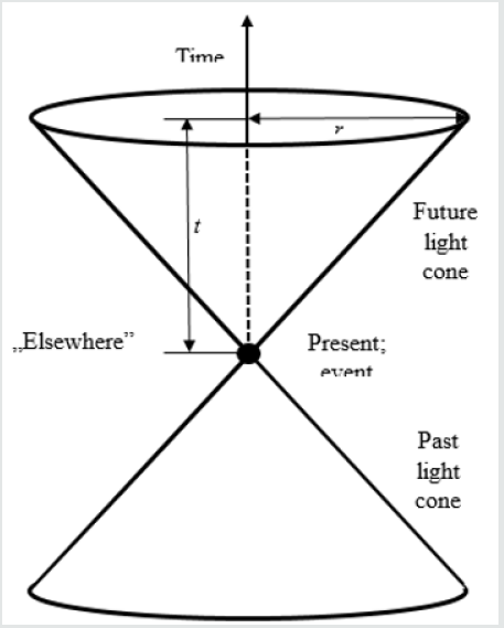
Lupinepublishers-openaccess-journals-medical-science-journal

Anthropokinetics and physics

However, the anthropokinetics is still young discipline, which searches for its scientific identity. Therefore, it is forced to adopt what might be termed the “Foraminifera-strategy”. The Foraminifera are one-cellular organisms, which build around their bodies the shells of sand. However, they select only such grains that under microscope their tests look as if they were polished [5]. Anthropokinetics should take any suitable “grains of knowledge”, no matter, where they come from, either. Accordingly, let us listen to novelist Jo Nesbø, who wrote: “You can discover new things by changing your perspective and your location (in science their equivalent is the methodology-WP). You can compensate for any blind spots” [6]. Accordingly, let us try to look at anthropokinetics from slightly different perspective. Already in 19th century philosopher, Auguste Comte has divided the whole science into two parts: “physique organique” and “physique inorganique” [7]. The former might be-roughly-identify with the biology, whereas the latter-with physics. The common element is the “physics”. Hence, one may perceive it no as a sum, but as a system. In such a situation justified seems to be the presumption that-may be-some relations are active in both these regions, but in “physique inorganique” they are better visible, whereas in “physique organique” are hidden deeper. In such a context highly illustratively sounds the statement by Niels Bohr that “It is wrong to think that the task of physics is to find out how Nature is. Physics concerns what we say about Nature”.

While coming out from such a “starting point”, one might put a question, whether the physical-mathematical structure of the light cone (or, more precisely, the philosophy underlying its mental construction) might be useful also in description of any psychological processes (in general) and anthropokinetical ones (in particular)? In other words-whether in anthropokinetics we have to do with relations independent of an individual, which executes a motor operation, which are able somehow “from outside” impose specific constraints on potentialities of performing by this individual specific motor actions? And whether to description of such a relation one might-even marginally-use a physical model?

Brain skyscraper and modalities ladder

The questions put above may be answered positively. Such external (against, e.g., an individual human) system of constraints is the “brain skyscraper”, shaped by evolution. It has been invented by Nikolai A. Bernstein [8-10], and its “intellectual daughter” is the modalities’ ladder [2,3]. The latter is fully coherent with the “skyscraper”, but devoid of evolutionary and neurophysiological components; it is mainly information-processing structure. Both are hierarchic, systemic structures. The former has five levels, the latter–five rungs. One if the main rules by Bernstein states that each motor operation has its main level of control (“master”), where the attention of the executor is being focused, and the lower ones (“slaves”) play the function of “background” (not “subconsciousness”, whatever this term might mean) and their action does not need attention concentration. Let us emphasize: the main criterion is not a division into “consciousness” and “subconsciousness”, but into elements, which need attention focusing and such ones, which do not need such a concentration. Before comparing the “brain skyscraper” and the modalities’ ladder, let us remember that one and only manifestation of each mental, psychical process in living beings-including humans-is the movement. This is why philosopher Andrzej Wohl wrote: “All that we dispose of, all what constitutes the resource of our culture, all the pieces of art, science and technology-all that results from motor activities” [11]. In short, there are no other conducts than the motor ones. The basis of such a behavior is the consciousness. Before further considerations, let us formulate the two definitions:

A. Motor operation: Motor action of a living being aimed at solving of a given task in environment; it may be evoked either by extrinsic stimulus (trigger; in such a case it is the motor response), or by intrinsic motivation without any contact with environment [12].

B. Consciousness: A dynamically changing component of a quasi-static whole; the multimodal knowledge of an individual, activated at given moment by perception directed by attention, aimed at dealing with a task at hand [3].

Let us add that the consciousness is a multifaceted phenomenon. Knowledge might be described with various codes-e.g., haptic, visual or verbal-but the general term “consciousness” encompasses all these modalities of information processing. It seems worth remembering that the term “modality” includes a specific code of information storing and processing, a logic specific to them, certain scale of phenomena and processes, the definite time period and depth of information processing. The characteristics of the “brain skyscraper” and modalities’ ladder, as well as the phenomena related to them, have been presented in Table 2. In short, the brain skyscraper has been built on structural, whereas the modalities’ ladder-on functional basis.

Table 2: Bernstein’s “brain skyscraper” and the modalities’ ladder.
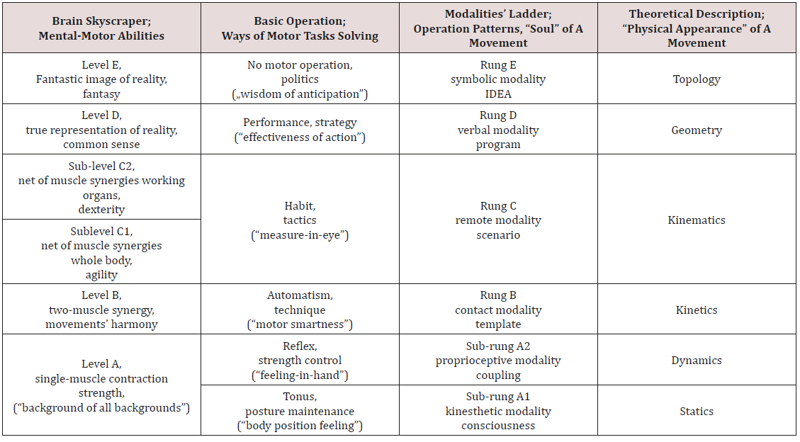
Lupinepublishers-openaccess-journals-medical-science-journal

As one can see, the divisions in both these structures are not identical. The equivalent of the single A-level in “brain skyscraper” are two sub-rungs (A1 and A2) in the modalities’ ladder. A single C-rung in the modalities’ ladder has two sub-levels (C1 and C2) in the brain skyscraper. Some comment needs the function of the tonus (sub-rung A1) in the structure of any motor operation in a human. The skeleton of Homo sapiens amounts to about 200 bones. Each of them may move against other ones; such movements may be described with the term “degrees of freedom”. In sum, human skeleton disposes of very many degrees of freedom. However, if a muscle should to move a given bone lever, then one its end has to be fixed relatively stiffly. In other words, all bones in a kinematic chain ending with this “stiffly fixed” end of the muscle should be properly immobilized; Bernstein dubbed this process “reduction of freedom degrees”. It makes the main task for the muscle tonus. Thanks to it, non-controllable system has been transformed into a controllable one. Hence, the muscle tonus makes a basis for the all other motor operations. Therefore, Bernstein termed it “background of all backgrounds”. In the modalities’ ladder, the notion of “degree of freedom” has been generalized and encompasses the abstract “information chunks”, related to movements or set of movements, specific to higher rungs of the modalities’ ladder.

The anthropokinetic events cone

The modality of each rung of the modalities’ ladder includes a specific type of coding, logics of information processing and temporal limits of the phenomena under consideration. Therefore, facing angry grizzly bear somewhere in Alaska, I would prefer company of experienced trapper with Winchester rather, and not ingenious Albert Einstein. Just the temporal limits, peculiar to rungs, may make a structure similar to the physical light cone. Let us term it “events’ cone” (for the sake of simplicity, because it should be named “the cone of abstract representation of real events”). A given modality may effectively “deal” with events, which belong to a specific period. Hence, the events lying beyond these limits should be categorized as being “elsewhere” (Figure 1). Consequently, they cannot influence the information processing inside the events’ cone. It is possible, then, to use the general rule of construction of the physical light cone, i.e., the division of the space of events into two parts. One of them includes such events, which may make parts of cause-effect chain shaping the future, and the other, which are to be found “elsewhere” and cannot influence the run of events (Figure 2).

The structure of the light cone differs essentially from that of events’ cone. In the former the time axis is positioned vertically (Figure 1), whereas in the latter-horizontally (Figure 2). However, the general philosophy-division of events and information into potentially active and unable to any activity-remains the same. It is worth noting that the time axis in Figure 2 should be perceived as a logarithmic scale, and not a linear one. Nevertheless, clearly visible are time periods specific to rungs, and the fact that the higher the rung, the longer the time period for analysis of events and information processing (thinking).

As a result, one might consider the space inside the events’ cone (bold dashed line) makes the room for analyses and information processing, whereas the space outside the cone represents the “elsewhere”. In short, the temporal constraints-specific to rungs of the modalities’ ladder-disable the events from “elsewhere” and make them ineffective in shaping of a given motor operation. The higher rung, the longer “working” period. The price, which inevitably must be paid for its extension, is higher and higher level of abstraction, i.e., getting further and further from reality. Therefore, the processes and phenomena at distant to the “tangible” reality highest rungs of the modalities’ ladder cannot be tested experimentally. Therefore, at those rungs the only tools for scientific description are hypotheses and theories. The techniques of intellectual work, which may be applied in this region of abstraction, are, e.g., the logic of loops by Michał Heller [13] or “inference to the best explanation” (IBE) by Gilbert Harman (Harman, 1965). Otherwise, both of them are nearly identical. Such a “moonshine” way of science creation evokes almost contempt of “genuine scientists”, i.e., the worshippers of arithmetical average and standard deviation. Nevertheless, the science is being composed of theories, and not “new, original experimental data”. Their amorphous ashes may merely fertilize the intellectual ground, on which the theories should grow. This has succinctly expressed by biologist (Nobel Prize winner) Peter Medawar with the words “theories destroy facts” [14]. Unfortunately, as its physicist Edward Teller aptly stated, “A fact is a simple statement that everyone believes. It is innocent, unless found guilty. A hypothesis is a novel suggestion that no one wants to believe. It is guilty, until found effective.”

However, let us look once more at the Figure 2. Let the symbols A, B, C, D and E symbolize rungs of the modalities’ ladder, tightly related to Bernstein’s brain skyscraper levels. The grasping of time is possible only at C-level-at that level appear remote sensory organs, which enable observation of motion in the environment; it is the only phenomenon, which makes possible to shape the notion of time-but it does not mean that it cannot be used to description of phenomena also from the lower rungs. The bold dashed line symbolizes the anthropokinetic events’ cone, including rungs of the modalities’ ladder. Not without reason the borders between rungs are marked with the dotted line. In fact, they symbolize not sharp limits, but fluid zones rather. The information may cross them, indeed, but in the zone between the rungs, its modality is being transformed. This is a non-linear process, i.e., elements from one rung are differently amplified in the other one. This phenomenon is probably responsive, to the main extent, for unpredictable, qualitatively new system effect produced by such a system. Let us notice that such a structure is generally coherent with division of memory into short-term sensory store (STSS), short-term memory (STM) and long-term memory (LTM) by Richard Atkinson and Richard Shiffrin. Roughly, STSS might be associated with the lowest rungs of the modalities’ ladder, STM-with the middle ones, and LTMwith the highest rungs. The vertical relations make the system. The horizontal extension, limited by dashed line, represents the period specific to the information processing modality at a given rung.

Figure 2: The events’ cone in anthropokinetics. White field inside – consciousness; grey field outside – unconsciousness (“elsewhere”); bold dashed line – half-consciousness.
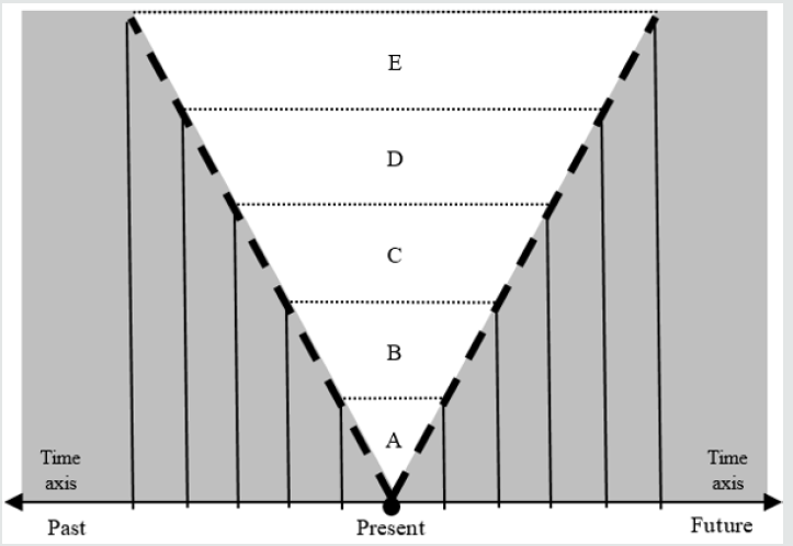
Lupinepublishers-openaccess-journals-medical-science-journal

Consciousness, half-consciousness and unconsciousness

At that moment of our analyses appears the space for mental construction of what might be associated with the phenomenon commonly termed “sub-consciousness”. This term seems to be incorrect, because it not describes the essence of the phenomenon under consideration. It may be regarded as a specific “black box”, where one may put all, what scientists are not able to properly describe scientifically. In such a situation, the item put into black box termed “sub-consciousness” remains not understandable, indeed, but marvelously gains the attribute of “scientificity”. However, one might imagine that the borders of the events; cone are not sharp as the cut of Japanese sword, but they make rather some fluid zones. While approaching the inside of the cone, the image of a given phenomenon or process becomes more and more pronounced, and inside the cone are completely clear. In such a model, each of the rungs-which dispose of its “own” modality of information processing and temporal limits of abstract representations of phenomena and processes-has also its own “zone of twilight of perception”. Such a model would enable description of the phenomenon of gradual forgetting of a particular event. It would transfer from the inside of the events’ cone-in this region, its abstract representation is immediately accessible-to the “twilight zone”. Its retrieving from this zone is possible indeed, but it is more difficult and time consuming. Finally, when it goes out from the zone of “twilight”, it becomes completely forgotten. Such a “twilight zone” from the side of future one might dub “precognition”, and that from the side of past- “shadows of oblivion”. The representations of events in this zone exist, indeed, but they are not precise and indistinct. Therefore, the interior of the events’ cone may be identified with the consciousness, the border zone-with half-consciousness, and the region of “elsewhere”-the unconsciousness.

To avoid creation of a “moonshine” term (like “subconsciousness”), let us try to invent a rationale for what has been roughly dubbed “precognition”. Motor operations are always faced towards future-closer or farther, according to the rung of the modalities’ ladder. The main “processor”, which produces the abstract, mental pattern of a motor operation is the intellect. It may be perceived, roughly as a system consisting of three mechanisms of information processing: intelligence, intuition, and instinct. Intelligence makes the “armed forces” of the intellect. It is responsible for final shape of the motor operation pattern. However, to produce such a pattern, it needs full information necessary to given task solution and knowledge of all the rules of such information processing. We are very rarely in such a luxurious situation; we have not such a complete knowledge. Hence, if an information lacks, it must be guessed to get intelligence going. This makes the task for intuition. Finally, the instinct directs the searching for lacking information towards these regions of memory, where its finding seems to be most probable. The half-consciousness zone faced towards future cannot include the full information about a given task; otherwise, it would be the full consciousness. Hence, the intelligence itself cannot be effective in this region. As a result, here opens the wide field of action for intuition. The term “precognition” might be described, then, as a way of processing of not complete information, where the main tool is the intuition (“I don’t know, but I suppose”), and only marginal role plays the intelligence.

The Events’ cone in practice

The system presented in the Figure 2 may make sense only when the time period, assigned to a given rung, is sufficiently long to enable practical realization of the task related to this rung. Therefore, the lower rung, the simpler operation and the swifter its execution. And vice-versa: the higher rung, the more time-consuming preparation and execution of a specific motor operation. Let us imagine such a situation. During a solemn, international scientific conference, I am presenting my work. I am moving freely in the room and using a pilot for changing slides remotely. Suddenly, I take a pin, hidden in my sleeve, and acutely sting the buttock of a dignified, gray-haired scientist. What will happen? No doubt, the scientist will jump. It is natural reaction in such a situation: to take a distance from the source of pain. Does s/he realize immediately, what happened? For sure-not! Such an event would be so astonishing, so improbable, without any equivalents in the past, to which it might be related. The scientist would have to build the abstract model of the event, what inevitably must be time-consuming. Hence, at the contact B-rung the stimulus is received, response-prepared and executed, before at the verbal D-rung the stimulus is barely identified. However, if it happens, I would be far away.

The content of events’ cone depends not only on information processing modality, but also on the level of pre-preparation of a needed operation pattern. In this respect instructively sound the words by Ben Johnson-mysterious racing driver “Stig” from the BBC program “Top Gear”. In the interview, he stated: What defines a good driver? What attribute is necessary, and what merely useful? The anticipation. Racing driver is a person, who does not look for solutions of the problems that occur in a race. S/he knows those solutions, and when the situation comes, when the reaction becomes necessary, s/he simply performs the operations leading to its successful solving [15]. While seen from the perspective of the modalities’ ladder, in this case we have to do with the D-rung depth of information processing being “pushed down” to the C-rung temporal constraints. In daily language, such a process may be identified with what is commonly termed “experience”. It has been described by Nikolai A. Bernstein [10,16].

On the other hand, if an individual has to his/her disposition C-rung time, one cannot expect the information processing with depth specific to D-rung. In such a situation suitable information processing should be located in the region of “elsewhere”. For example, the analysis of car accident or ships’ collision at sea, where the teams of expert witnesses have plenty of time for D-rung calculations and analyses, cannot be compared to the situation of a driver or captain, who was able to make only C-rung assessments, basing on previous experience, and had to his/her disposal merely seconds or minutes. Such a situation has been brilliantly presented in the movie by Clint Eastwood “Sully”, about landing on 15th January 2009 on the Hudson River of the Airbus 320, in charge of the captain Chesley “Sully” Sullenberger.

Conclusion

Well, is it possible-based on the presented analyses-to formulate the conclusion that the laws of physics in their “pure” form may be applied also in anthropokinetics? For sure-no! One might merely assume that some mental structures, invented by physicists, may be used-after specific modifications-also in anthropokinetics. However, such an analogy reminds the similarity of the shark and the dolphin rather, and not a common law being in force in both these disciplines. It is not possible, then, to perceive it as a rule. In science there are no any well-worn, simple templates-like, e.g., calculations (not mathematics!) or any other commonly accepted methodologies”-which would release scientists from thinking. In this respect highly instructively sound the words by Niels Bohr: “You are not thinking; you are just being logical.”

Philosopher Paul Feyerabend has invented an image of knowledge built by Truth and Freedom. The former has its feet firmly on the ground; the latter flies freely in the sky. Where they meet, the Science (with great “S”) is being born. However, the Truth is harnessed with the stiff constraints; hence, it cannot for long remain in union with the unhampered Freedom. Hence, sooner or later, they must part their ways. Until next meeting. Analogously, also the similarity of the light cone and the events’ cone should be treated as a result of a momentary meeting of the Feyerabend’s Truth and Freedom, and not as a basis for formulation of more general theories.

Read More About Lupine Publishers Journal of Medical Sciences Please Click on the below link :

https://lupine-publishers-medical-sciences.blogspot.com/