Showing posts with label Journal of Surgery and Case Studies. Show all posts

Friday, 29 September 2023

Lupine Publishers | Covered Perforativnye Ulcers Gastroduodenal Zone

 Lupine Publishers | Journal of Surgery & Case Studies


Abstract

Of the 215 patients with gastroduodenal ulcer probodnymi zone 17 (7.9%) She wore covered nature. The author notes some difficulties in diagnosing this type of pathology. If you open probodenijah symptomatology are manifestnyj nature and errors in diagnosis is usually not observed, then covered the clinical picture had ulcers was wiped out in nature, requiring additional research methods, including x-ray, to recognize this pathology. An inexperienced clinician may resort to using gastrografii contrast barium dredge. As a result of the suspension enters the free abdominal cavity, which further accompanied by the development of severe adhesive disease. This cause of this serious pathology he watched from 2 patients operated in various hospitals of Kuban. The author describes a diagnostic algorithm of this pathology in 17 patients admitted in the hospital, or with acute cholecystitis, or other pathology, and only a few hours when they are accidentally fibro gastroduodenoscopy the true cause has been identified the emergence of pain in epigastralna area is covered with a perforated gastroduodenal ulcer zone.

Keywords:Gastroduodenal Ulcer Zone Covered Perforation; Diagnostic

The aim of the study was the definition of diagnostic methods to identify covered perforating ulcers of the stomach and duodenum (PPJaZhD).

Introduction

Identification of covered probodnyh ulcers gastroduodenal zone represents some diagnostic difficulties. If you open perforations of ulcers leading clinical symptom is triad-Mond or (ulcerative anamnesias, “pain in the abdomen, doskoobraznoe tension of the abdominal wall) and x-ray-presence of free gas in the abdominal cavity, when covered probodnoj ulcer all they are missing [1- 3]. More-over, and other clinical symptoms are not defined, i.e. dullness, no liver kept blunting in sloping are-as of the abdomen and other [4,5,6]. Patients usually complain of mild pain in the area epigastralna, accompanied by diarrheal events malaise [7,8]. As their doctors have had the impression that the patient has occurred, or the aggravation of gastric ulcers or chronic cholecystitis [9,10]. Such patients are hospitalized in a therapeutic institution, and begin to be a typical survey, sometimes with the use of barium dredge that is blunder-its particles embedded in the peritoneum, and remain there, despite intensive abdominal lavage varying fluid. This leads to the development of a total of commissural process. To avoid this error, in the middle of the 20 centuries, Weber resorted to inflation of the stomach using gastric probe. This was accompanied by a delaminating of agglomerated tissues in area of ulcers and gastric cavity air rushed to free abdominal cavity (usually under the left or right of the dome of the diaphragm). This manipulation it produced during rentgeno gastroscopy, which al-lowed him to clearly observe the screen air outlet outside the cavity of the body [1,5]. Something similar can be observed in fibro gastroduodenoscopy-as soon as the endoscopies begins to inflate the stomach for inspection of its walls, the patient occur severe abdominal pain that is associated with the forced penetration of air from the stomach cavity in free abdominal cavity. In such a situation urgently carry out a repeated review x-rays of the abdomen, and if, in the first survey of the air in the abdominal cavity is not detected, then now he’s clearly defined [1,2,10]. So, the diagnosis is established. With this same purpose, some radiologists have resorted to the use of water-soluble contrast media.

Material and Methods

For 3 years in 2-ohm ECHO KGCSMP were treating patients with 215 probodnymi ulcers stomach and duodenal ulcers, of whom 17 (7.9%) There were PPJaZhD. All of these patients, who were all men aged 31 to 67 years, was on the front wall, or duodenal ulcers (12), or pyloric stomach Division (5). At 9 (4.18%) these patients the disease began with the emergence of moderate pain in epigastralna area, which was accompanied by vomiting. District therapists they suspected food poisoning and within days conducted appropriate outpatient treatment effect. By the end of the specified term of treatment in patients pain intensified, and at the same time appeared the strain in the right podreberie. With suspected acute cholecystitis, they were sent to hospital treatment. The remaining 8 (3.72%) disease patients developed similarly but when seeking medical help, through 6-12 hours from onset of symptoms, they immediately were suspected of acute cholecystitis. With this diagnosis were hospitalized in the surgery department. The first survey of all 17 patients was missing symptoms of irritation of the peritoneum. ULTRASOUND of abdomen radiography review pathology. For x-ray study of stomach using barium dredge, not steel, as in earlier Office underwent surgical treatment of patient with 2 adhesive ileus, which developed due to the falling mist abdomen (this error allowed doctors in other hospitals). In this situation, esophagogastroduodenoscopy was shown, but when I try to run it in all patients immediately arose strong abdominal pain. The procedure was discontinued. Within one hour from all 17 patients developed a picture of acute peritonitis. Repeat-ed survey radiography of abdominal cavity revealed the presence of free gas in it. Patient’s emergency laparotomy was performed.

The Result of The

During surgery in the abdominal cavity found muddy effusion. Around probodnoj the holes had inflammatory infiltrate, and it was partially obscured the top adjacent organs and tissues. All 17 patients carried out organ-preserving operations the results of the study. Of the 17 patients with PPJaZhD (5.9%) and 1 died the patient (from cardiovascular insufficiency). The overall mortality in probodnyh ulcers was 4.65% (of 215 patients died 10).

Discussion

When examining the data received on time execution of operations on the stomach and duodenum from these 17 patients, it can be concluded that cover probodnoj ulcers occurred due to the adhesive around the process expressed ulcers, with the rapid development of inflammatory infiltration. This can happen only when expressed protective immunity when around the ulcer occurs hearth with positive electric potential, to which attracted surrounding organs, having all the negative potential, and fabric to stick together. So in vivo survival occurs. When stoking the stomach this protective barrier is destroyed. That’s why even from ancient times people with abdominal pain otljozhivalis and ate almost nothing for a few days. Range that they at that time carried naked stomach ulcers. Apparently, positive role plays and reflex vomiting, during which adopted on the eve of food almost in full erupts outward. Body cavity decompression promotes podsasyvaniju surrounding organs to probodnymi.

Discussion

As you know, the most informative diagnostic process ulcers gastroduodenal zones possess esophagus gastroduodenoscopy and x-ray studies. However, we observed patients with PPJaZhD these methods initially did not give the expected result. And this was due to the fact that when abdominal radiography review absent such Cardinal diagnostic test, as the detection of free gas in it. Increased pain in the abdomen that occurred during the execution of fibro gastroduodenoscopy can be attributed to the violent disintegration of infiltration air, which at this time vduvalsja in the stomach cavity. As a result of its contents from falling into the free abdomen, that led to the development of a peritonitis. Repeated survey radiography of abdominal cavity allowed recognizing the true cause of the critical deterioration of patients, i.e. the development of widespread peritonitis, which is when the hollow organ perforation took only locally limited form. Thus, the leading value in the diagnosis of PPJaZhD has repeated panoramic radiography of abdominal cavity, which is performed after the failed fibro gastroduodenoscopy. The use of barium dredge to diagnose ulcers of stomach stones leads to the development of heavy adhesive disease of abdomen (due to the introduction of particles of barium in thickness of the peritoneum).

Read More About Lupine Publishers Journal of Surgery Please Click on Below Link:
https://surgery-casestudies-lupine-publishers.blogspot.com/

Friday, 20 January 2023

Lupine Publishers | Endometriosis Involving Colon and Rectum: A Literature Review and Laparoscopic Management

 Lupine Publishers | Journal of Surgery & Case Studies


Abstract

Introduction: Endometriosis is characterized by the presence of functional endometrial tissue consisting of glands and/ or stroma located outside the uterus [1], although implanted ectopically, this tissue presents histopathological and physiological responses that are similar to the responses of the endometrium [2].

Clinical Features: Endometriosis usually becomes apparent in the reproductive years when the lesions are stimulated by ovarian hormones. Forty percent of the patient’s present symptoms in a cyclic manner, which are usually related with menses Pelvic pain, infertility and dyspareunia are the characteristic symptoms of the disease, but the clinical presentation is often non-specific [1].

Diagnosis and Investigations: A precise diagnosis about the presence, location and extent of rectosigmoid endometriosis is required during the preoperative workup because this information is necessary in the discussion with both the colorectal surgeon and the patient. Furthermore, almost all patients with intestinal endometriosis have lesions in multiple pelvic locations and it is difficult to know what symptoms are caused by the intestinal disease versus the pelvic disease.

Treatment: Treatment must be individualized, taking the clinical problem in its entirety into account, including the impact of the disease and the effect of its treatment on quality of life. Pain symptoms may persist despite seemingly adequate medical and/ or surgical treatment of the disease. In such circumstances, a multi-disciplinary approach involving a pain clinic and counselling should be considered early in the treatment plan.

Endometriosis is characterized by the presence of functional endometrial tissue consisting of glands and/ or stroma located outside the uterus [1], although implanted ectopically, this tissue presents histopathological and physiological responses that are similar to the responses of the endometrium [2].

Prevalence and Epidemiology

The reported prevalence of endometriosis is 1%-20% in asymptomatic women, 10%-25% in infertile patients and 60%- 70% in women with chronic pelvic pain [1]. Endometriosis is a common benign disease among women of reproductive age and affects the intestinal tract in 15%-37% of all patients with pelvic endometriosis [3]. Multiple births and extended intervals of lactation decrease the risk of being diagnosed with endometriosis, whereas nulliparity, early menarche, frequent menses, and prolonged menses increase the risk [4]. Endometriosis also appears to be associated with a taller, thinner body habitus and lower body mass index [5]. The prevalence appears to be lower in blacks and Asians than in Caucasians [6]. Growth and maintenance of endometriotic implants are dependent upon the presence of ovarian steroids. As a result, endometriosis occurs during the active reproductive period: women aged 25 to 35 years [6]. Other factors that appear to play important roles in determining if a woman will develop the clinical condition include [7]:

a) Reproductive lifestyle, especially a delay in childbearing

b) Poorly understood immunological factors

c) Some environmental factors, probably including exposure to a range of environmental toxins

d) Reproductive tract occlusion, such as an imperforate hymen.

Pathogenesis

Endometriosis is a common disease of unknown etiology. Many theories have been proposed to explain this condition: retrograde menstruation theory, metaplastic, transformation, the migration of cells through the lymphatic system or via hematogenous spread, Iatrogenic during CS. However, other factors, immunological, genetic and familial, could be involved in the pathogenesis of this disease [1].

Sampson’s Theory of Retrograde Menstruation

The implantation theory proposes that endometrial tissue passes through the fallopian tubes during menstruation, then attaches and proliferates at ectopic sites in the peritoneal cavity. Recent studies using laparoscopy have demonstrated that retrograde menstruation is a nearly universal phenomenon in women with patent fallopian tubes. Classic studies performed in the 1950s demonstrated viability of sloughed endometrial cells and the capacity to implant at ectopic sites. Patients with mullerian anomalies and obstructed menstrual flow through the vagina may have an increased risk of endometriosis. The anatomic distribution of endometriosis also provides evidence for Sampson’s theory [8].

Coelomic Metaplasia Theory

The theory of coelomic metaplasia proposes that endometriosis may develop from metaplasia of cells lining the pelvic peritoneum. Iwanoff and Meyer are recognized as originators of this theory. A prerequisite of the coelomic metaplasia theory is that mesothelial cells lining the ovary and pelvic peritoneum contain cells capable of differentiating into endometrium. An attractive component of the coelomic metaplasia theory is that it can account for the occurrence of endometriosis anywhere mesothelium is found. This includes reports of endometriosis occurring in the pleural cavity. Pleural endometriosis could result from local metaplasia of pleural mesothelium. On the other hand, it could also result from transdiaphragmatic passage of peritoneal fragments of endometrium as well as vascular metastasis of endometrium. Coelomic metaplasia is thought to account for the rare occurrences of endometriosis reported in males. In these reports of endometriosis, the men were all undergoing estrogen therapy. Although coelomic metaplasia was a possibility, estrogen stimulation of mullerian rests could not be excluded. Likewise, the occurrence of endometrial carcinoma in males is thought to possibly arise from mullerian remnants. Still, further support for the coelomic metaplasia theory may be found in the study of benign and malignant epithelial ovarian tumors. Both are considered to be derivatives of germinal epithelium. The presence of ovarian surface endometriosis could be accounted for by this type of transformation [8].

Induction Theory

The induction theory is an extension of the coelomic metaplasia theory. This theory proposes that menstrual endometrium produces substances that induce peritoneal tissues to form endometriotic lesions [8].

Embryonic Rests Theory

Von Recklinghausen and Russell are credited with the theory that endometriosis results from embryonic cell rests. These embryonic rests, when stimulated, could differentiate into functioning endometrium. As described above, rare cases of endometriosis have been reported in men. Transformation of embryonic rests is a plausible explanation for this phenomenon [8].

Lymphatic and Vascular Metastasis Theories

The lymphatic metastasis theory of endometriosis is often referred to as Hal ban’s theory. He reported that endometriosis could arise in the retroperitoneum and in sites not directly opposed to peritoneum. Sampson had also suggested that endometriosis could result from lymphatic and hematogenous dissemination of endometrial cells. An extensive communication of lymphatics has been demonstrated between the uterus, ovaries, tubes, pelvic and vaginal lymph nodes, kidney, and umbilicus. Metastasis of endometrial cells via the lymphatic system to these areas is therefore anatomically possible. These findings are consistent with a literature review showing a 6.7% incidence of lymph node endometriosis in 178 autopsy cases. Lymphatic and vascular metastasis of endometrium has been offered as an explanation for rare cases of endometriosis occurring in locations remote from the peritoneal cavity. In addition to pleural tissue, endometriosis has been reported in pulmonary parenchyma. Vascular or lymphatic metastasis may also explain cases of endometriosis that have been reported in bone, biceps muscle, peripheral nerves, and the brain [8].

Composite Theory

Javert proposed a composite theory of the histogenesis of endometriosis which combines the implantation, vascular/ lymphatic metastasis, as well as a theory of direct extension of endometrial tissue through the myometrium. Along similar lines, Nisolle and Donnez have recently argued that the histogenesis of endometriosis depends on the location and ‘type’ of the endometriotic implant. For example, peritoneal endometriosis can be explained by the implantation theory. Ovarian endometriomas could be the result of coelomic metaplasia of invaginated ovarian epithelial inclusions. Rectovaginal endometriosis, which often resembles adenomyosis, could result from metaplasia of Mullerian remnants located in the rectovaginal septum. These composite theories are attractive in that they recognize a multifaceted mechanism of histogenesis. It seems logical that a disease with such variable manifestations may originate via several mechanisms [8].

Altered Immunity

Alterations in immunologic response to retrograde menstruation have been implicated in the genesis and maintenance of the endometriotic lesion. This defective immunosurveillance may lead to decreased clearance of menstrual debris from the peritoneal cavity and may allow for attachment of ectopic endometrium to peritoneal surfaces. An abnormal immune response could also promote the persistence and growth of ectopic endometrial tissue [8].

The “Neurologic Hypothesis”

It is a new concept in the pathogenesis of endometriosis: There is a close histological relationship between endometriotic lesions of the large bowel and the nerves of the large bowel wall. Endometriotic lesions seem to infiltrate the large bowel wall preferentially along the nerves, even at distance from the palpated lesion, while the mucosa is rarely and only focally involved [9].

Pathology and Sites of Involvement

Sites

Endometriosis can be divided into intra- and extraperitoneal sites. In decreasing order of frequency, the intra-peritoneal locations are ovaries (30%), uterosacral and large ligaments (18%-24%), fallopian tubes (20%), pelvic peritoneum, pouch of Douglas, and gastrointestinal (GI) tract. Extra-peritoneal locations include cervical portio (0.5%), vagina and rectovaginal septum, round ligament and inguinal hernia sac (0.3%-0.6%), navel (1%), abdominal scars after gynaecological surgery (1.5%) and caesarian section (0.5%). Endometriosis rarely affects extraabdominal organs such as the lungs, urinary system, skin and the central nervous system [1]. Endometriosis affects the intestinal tract in 15% to 37% of patients with pelvic endometriosis [10], involvement have been reported from the small bowel to the anal canal, but more frequently the disease involves the rectum and the sigmoid colon (74%), followed by the rectovaginal septumn (12%), cecum (2%), and appendix (3%) . When the ileum is involved, the most common tract is the distal part. A full-thickness involvement of the colonic wall is infrequent since the mucosa is usually spared. One of the classic locations is the anterior rectal wall in the region of the pouch of Douglas. This can be single nodule or can simulate a cancer. Because of the invasive appearance, the disease can be mistaken for cancer [11].

Gross and Microscopic Pathology of Bowel Endometriosis

The appearance and size of the implants are quite variable. Areas of endometriosis appear as raised flame-like patches, whitish opacifications, yellow-brown discoloration, translucent blebs, or reddish or reddish-blue irregularly shaped islands. The peritoneal surface may be scarred or puckered.

The Microscopic Appearance

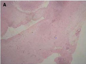
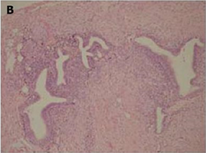
Of endometriotic tissue is similar to that of endometrium in the uterine cavity; the two major components of both are endometrial glands and stroma. Unlike endometrium, however, endometriotic implants often contain fibrous tissue, blood, and cysts (Figure 1(a) & 1(b)).

Figure 1(a): Low-power image of the colonic wall, with a few endometrial glands and stroma embedded in the muscular layer.
Lupinepublishers-openaccess-surgery-case-studies-journal

Figure 1(b): High-power view of the colonic wall, with endometrial glands and stroma embedded in the smooth muscle of the colon [12].
Lupinepublishers-openaccess-surgery-case-studies-journal

Link to Cancer

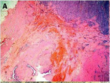
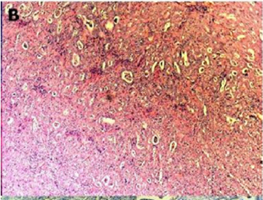
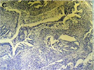
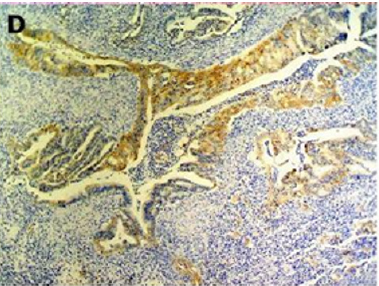
Endometriosis is considered a benign disorder; however, it shares some of the characteristics of malignancy, such as abnormal morphology, deregulated cell growth, cellular invasion, and neoangiogenesis. The glandular epithelium occasionally displays DNA aneuploidy. In vitro evidence suggests that endometriosis may have a monoclonal origin. In addition to being monoclonal, endometriotic deposits showed loss of heterozygosity in 28% of lesions. In 2002, Nezhat et al. with immunohistochemistry, found that alterations in bcl-2 and p53 may be associated with the malignant transformation of endometriotic cysts [12]. The development of a malignancy is a relatively common complication of endometriosis. In fact, several publications have reported malignant neoplasms arising from endometriosis. Most of these publications are case reports or refer to a small series of patients presenting either ovarian carcinomas with associated endometriosis or invasive endometrioid adenocarcinomas involving adjacent pelvic structures. Malignant transformation of extraovarian endometriosis, including the intestinal tract, however, has not been reported as frequently. The largest reported series of neoplastic changes in gastrointestinal endometriosis includes 17 cases [10] (Figure 2(a-d)). Some studies suggest that the development of malignancies may occur in up to 5.5 % of female patients with endometriosis. Only 21.3% of the cases arise from extragonadal pelvic sites, and endometriosis-associated intestinal tumors are even rarer. Malignant transformation of primary gastrointestinal endometriosis without pelvic involvement is uncommon, and its real incidence is unknown. It can mimic a primary gastrointestinal neoplasm. Most of these neoplasms are carcinomas, but sarcomas and müllerian adenosarcomas have also been described. Petersen et al, in a large review of the previously published endometrioid adenocarcinomas arising in colorectal endometriosis, report less than 50 cases of neoplastic transformation, 22 of which were adenocarcinomas. The others included sarcomas and mixed müllerian tumors. The progression to invasive cancer has been related with hyperestrogenism, either of endogenous or of exogenous origin. A possible genetic background favoring the onset of cancer has been reported in some patients without hyperestrogenism and with a family history of cancer. The anatomic distribution and frequency of these cancers parallel the occurrence of which benign endometriosis is found at various sites. In order to classify a malignancy as arising from endometriosis, strict histopathologic criteria need to be fulfilled. Sampson first proposed these criteria in the year 1925. He suggested that the following should be fulfilled:

Figure 2(a): Rectal endometriod adenocarcinoma with adjacent focus of endometriosis (hematoxylin-eosin, 20x).
Lupinepublishers-openaccess-surgery-case-studies-journal

Figure 2(b): Rectal endometriod adenocarcinoma endometriosis (hematoxylin-eosin, 100x).
Lupinepublishers-openaccess-surgery-case-studies-journal

Figure 2(c): Cytokeratin 20 immunostaining negative (100x).
Lupinepublishers-openaccess-surgery-case-studies-journal

Figure 2(d): Cytokeratin 7 immunostaining positive (100x). [10]
Lupinepublishers-openaccess-surgery-case-studies-journal

a) the presence of both malignant and benign endometrial tissue in the same organ.

b) the demonstration of cancer arising in the tissue and not invading it from elsewhere.

c) the finding of tissue resembling endometrial stroma surrounding characteristic glands.

Years later, Scott suggested an additional qualification to complete Sampson’s criteria: the demonstration of microscopic benign endometriosis contiguous with the malignant tissue [10]. Endometriosis and its possible malignant changes should be taken into account in the differential diagnosis of intestinal masses in females. Also, clinical suspicion for malignancy should be aroused in patients with abdominal pain or rectal bleeding and a previous history of quiescent endometriosis. Recognition of these lesions is important because of the different management required by primary gastrointestinal neoplasms and by those arising from endometriosis. These differences may have significant clinical implications [10].

Clinical Features

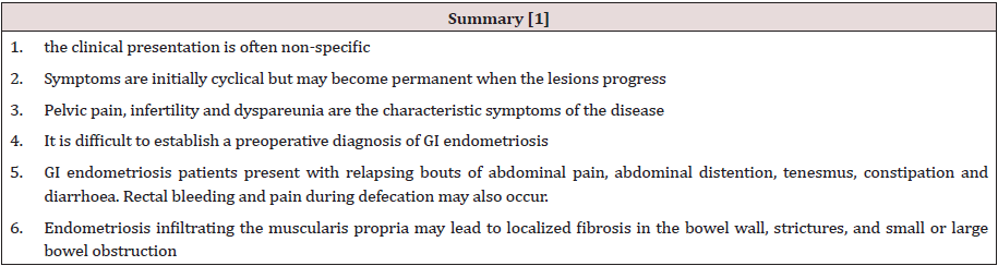
Endometriosis usually becomes apparent in the reproductive years when the lesions are stimulated by ovarian hormones. Forty percent of the patient’s present symptoms in a cyclic manner, which are usually related with menses Pelvic pain, infertility and dyspareunia are the characteristic symptoms of the disease, but the clinical presentation is often non-specific [1]. Symptoms are initially cyclical but may become permanent when the lesions progress. It is difficult to establish a preoperative diagnosis of GI endometriosis, because GI tract symptoms can mimic a wide spectrum of diseases, including irritable bowel syndrome, infectious diseases, ischemic enteritis/colitis, inflammatory bowel disease and neoplasm. GI endometriosis patients present with relapsing bouts of abdominal pain, abdominal distention, tenesmus [1], constipation and diarrhoea. Rectal bleeding and pain during defecation may also occur. Endometriosis infiltrating the muscularis propria may lead to localized fibrosis in the bowel wall, strictures, and small or large bowel obstruction. The true incidence of endometriosis causing bowel obstruction is unknown, although complete obstruction of the bowel lumen occurs in less than 1% of cases. Endometriosis of the distal ileum is an infrequent cause of intestinal obstruction, ranging from 7% to 23% of all cases with intestinal involvement. The incidence of intestinal resection for bowel obstruction is 0.7% among patients undergone surgical treatment for abdominopelvic endometriosis [1]. Rectal bleeding may be caused by mucosal injury during the passage of stools through a stenosed colon with the intramural endometriotic tissue increased at the time of menses if it occurs. Colonic mucosa heals rapidly, and no signs are detectable at endoscopy [1] (Table 1).

Table 1:
lupinepublishers-openaccess-surgery-case-studies-journal

Differential diagnosis [1]

a) irritable bowel syndrome,

b) infectious diseases,

c) ischemic enteritis/colitis,

d) inflammatory bowel disease

e) neoplasm

f) Other causes of intestinal obstruction (Acute/chronic, small/large bowel)

Diagnosis and Investigations

A precise diagnosis about the presence, location and extent of rectosigmoid endometriosis is required during the preoperative workup because this information is necessary in the discussion with both the colorectal surgeon and the patient. Furthermore, almost all patients with intestinal endometriosis have lesions in multiple pelvic locations and it is difficult to know what symptoms are caused by the intestinal disease versus the pelvic disease. In particular, in the case of sigmoid endometriosis, the lesion cannot be suspected at clinical examination, which is why sigmoid endometriosis is often diagnosed only during surgery. Although several radiological techniques have been proposed for the diagnosis of bowel endometriosis, data are inconclusive, and no gold standard is currently available [13].

Colonoscopy

Although endoscopic diagnosis of colonic endometriosis has been reported, the mucosa is usually normal or shows minimal mucosal abnormalities, friability, extrinsic process or fibroses stenoses [1]. Endoscopic biopsies usually yield insufficient tissue for a definitive pathologic diagnosis as endometriosis involves the deep layers of the bowel wall [14]. Endometriosis can induce mucosal changes without any specific pattern, which mimic findings of other diseases such as inflammatory bowel disease, ischemic colitis or neoplasm [1]. Colonoscopy is helpful to rule out colorectal malignancy [11].

Double Contrast Barium Enema

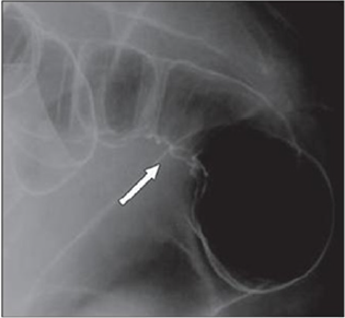
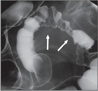
Radiologically, lesions of endometriosis are either of constricting and polypoid type or both. On barium studies, radiographic findings caused by implants in the ileum are similar to those in the colon. Rectosigmoid or cecal endometriosis on double contrast barium enema studies is seen as an extrinsic mass with speculation and tethering of folds [1]. Shortening or flattening of the bowel wall, crenulation of the mucosa, or a combination of these factors [15], Double-contrast barium enema may be effective in determining the precise location of the endometriotic nodules, but it cannot clearly demonstrate the depth of parietal involvement. Furthermore, the experience of the radiologist in the diagnosis of bowel endometriosis remains a critical limit of this technique [13] (Figures 3 & 4(a & b)).

Figure 3: Thirty-four years old woman with suspected intestinal implants of endometriosis. A and B, Lateral A and oblique B spot images show three endometriotic lesions exhibiting extrinsic mass effect with crenulation of contour and speculation that are direct signs of infiltration of bowel wall (arrows). Small polypoid lesion (arrowhead) is benign tubular adenoma confirmed at surgery [15].

Lupinepublishers-openaccess-surgery-case-studies-journal

Figure 4(a): Twenty-eight years old woman with suspected intestinal implants of endometriosis and finding of rectal localization of intestinal endometriosis. DCBE image shows extrinsic mass effect and speculation (arrow) of rectal wall that appears infiltrated [15].
Lupinepublishers-openaccess-surgery-case-studies-journal

Figure 4(b): Twenty-three years old woman with suspected intestinal implants of endometriosis. DCBE examination showing pathologic pelvic process involving bowel serosa at rectosigmoid junction. Finding of extrinsic mass effect and speculation (arrows) owing to poor wall distention after air insufflation suggesting wall infiltration [15].
Lupinepublishers-openaccess-surgery-case-studies-journal

Transvaginal Us

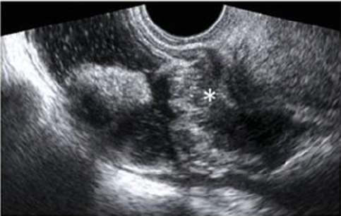
Transvaginal ultrasonography can be useful not only in the first-line exploration of the pelvic cavity, but also in diagnosing rectosigmoid endometriosis. However, relevant limitations of transvaginal ultrasonography consist in the impossibility of determining the exact distance of rectal lesions from the anal margin and of evaluating precisely the depth of rectal wall involvement. In addition, locations above the rectosigmoid junction might be beyond the field of view of a transvaginal approach and limited by the presence of air for a transabdominal approach [15]. Transvaginal us combines with rectal water contrast is more accurate than TVS in diagnosing rectal infiltration reaching at least the muscularis propria in women with rectovaginal endometriosis. However, this exam cannot determine whether the infiltration reaches the rectal submucosa. RWC-TVS may be more painful than TVS, therefore it could be used when TVS cannot exclude the presence of rectal infiltration in women with rectovaginal endometriosis [15] (Figure 5).

Figure 5: A large rectovaginal nodule infiltrating the bowel muscularis (indicated by the asterisk) demonstrated by Rectal Water Contrast- Transvaginal Sonography (RWC-TVS) [16].
Lupinepublishers-openaccess-surgery-case-studies-journal

CT & MSCT

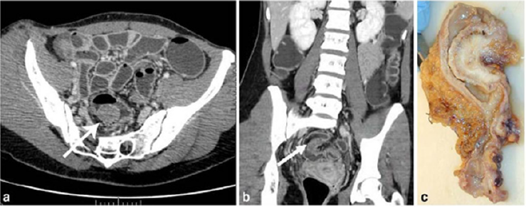
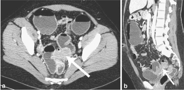
CT is not the primary imaging modality for evaluation of bowel endometriosis, although it can occasionally demonstrate a stenosing rectosigmoid mass. Multislice CT (MSCT) has a great potential for detecting alterations in the intestinal wall, especially if it is combined with enteroclysis (MSCTe). Biscaldi et al carried out a study on 98 women with symptoms suggestive of colorectal endometriosis and MSCTe identified 94.8% of bowel endometriotic nodules [1]. Biscaldi et al reported the usefulness of multislice CT combined with distention of the colon by rectal enteroclysis for bowel endometriosis. The sensitivity was 98.7% and specificity was 100% in identifying women with intestinal endometriosis. This method is thought to be very helpful for diagnosing intestinal endometriosis, but requires bowel preparation, such as the need for a low-residue diet for 3 d, drinking of 4-6 doses of a granular powder dissolved in 500 mL of water per dose and intravenous administration of iodinated contrast medium. This technique is thus inappropriate for patients with obstructive symptoms or allergy to iodinated contrast medium [3] (Figures 6 & 7).

Figure 6: Endometriotic nodule infiltration the muscular layer, A: Axial MSCTe image of the abdomen, the arrow indicates the endometriotic nodule. The lesion is enhanced, and it infiltrates the bowel wall involving the muscular layer. B: Coronal reconstruction demonstrating the extension of the sigmoid endometriotic nodule (indicated by the arrow) C: Formaldehydefixed resected bowel segment, the endometriotic nodule of the sigmoid colon was previously demonstrated by MSCT [14].
Lupinepublishers-openaccess-surgery-case-studies-journal

Figure 7: Endometriotic nodule infiltration the muscular layer, A: Axial MSCTe image of the abdomen, the arrow indicates the endometriotic nodule. The lesion is enhanced, and it infiltrates the bowel wall involving the muscular layer. B: Coronal reconstruction demonstrating the extension of the sigmoid endometriotic nodule (indicated by the arrow) C: Formaldehydefixed resected bowel segment, the endometriotic nodule of the sigmoid colon was previously demonstrated by MSCT [14].
Lupinepublishers-openaccess-surgery-case-studies-journal

Magnetic Resonance Imaging (MRI)

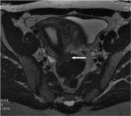
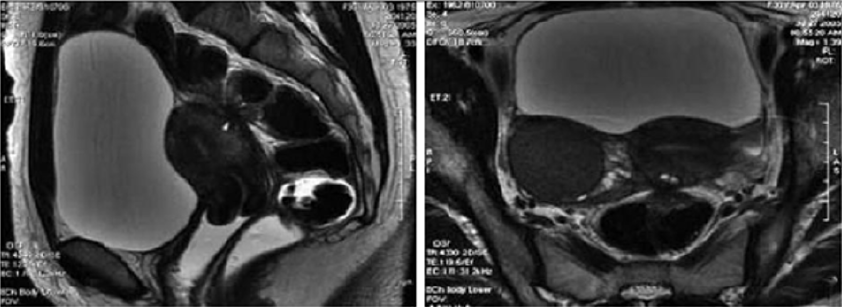
Magnetic resonance imaging (MRI) has a high sensitivity (77%- 93%) in the diagnosis of bowel endometriosis. The depth of rectal wall infiltration by endometriosis is poorly defined by MRI. A combination of MRI and rectal endoscopic ultrasonography (EUS) has recently been proposed. When retroperitoneal infiltration is present, it is mandatory to know if the bowel wall is involved in order to identify patients requiring bowel resection. Both rectal EUS sensitivity and negative predictive value range from 92% to 100%. The specificity and positive predictive value are rather poor, which are 66% and 64%, 83% and 94%, respectively, as reported in two different studies [1]. Imaging examination is thus essential for the preoperative diagnosis of intestinal endometriosis, but some reports have described preoperative confusion between this disease and cancer according to colonoscopy and CT with barium enema, particularly in patients with lesions involving the mucosal surface. In such patients, MRI is helpful for differential diagnosis. In a typical endometrial lesion, MRI showed signal hyperintensity on T1-weighted imaging and signal hypointensity on T2-weighted imaging. However, smooth muscle components are reportedly recognized frequently in endometrial lesions. In such lesions, as seen in the present case, MRI indicates signal hypointensity on both T1- and T2-weighted imaging, and differential diagnosis from other diseases such as cancer and gastrointestinal stromal tumor is thus difficult. In fact, Chapron et al reported that MRI specificity for deeply infiltrating endometriosis was 97.9%, but sensitivity was only 76.5% [3] (Figures 8 & 9).

Figure 8: T2- weighted axial view: fecal matter attached to the rectal wall, simulating thickening of the rectal wall [17].
Lupinepublishers-openaccess-surgery-case-studies-journal

Figure 9: T2- weighted sagittal (a) and axial (b) views.

Nodule of the rectosigmoid junction adhering to the posterior surface of the uterus [17].
Lupinepublishers-openaccess-surgery-case-studies-journal

Transrectal EUS

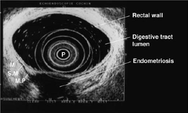
The involvement of the colon is difficult to detect because the implants rarely invade through the intestinal mucosa. For this reason, the rectal ultrasound is of primary importance to assess the rectal involvement [11]. Also, the depth of rectal wall infiltration by endometriosis is poorly defined by MRI. A combination of MRI and rectal endoscopic ultrasonography (EUS) has recently been proposed. When retroperitoneal infiltration is present, it is mandatory to know if the bowel wall is involved in order to identify patients requiring bowel resection [1]. Endoscopic ultrasonography is also a useful and noninvasive examination for the diagnosis of intestinal endometriosis. Sensitivity and specificity are reportedly about 97% for the diagnosis of rectal involvement in patients with known pelvic endometriosis. In addition, EUS-FNAB provides accurate tissue and may be the only procedure for correct preoperative diagnosis of intestinal endometriosis, but the overall specificity, sensitivity and accuracy of EUS-FNA for neoplasms of the gastrointestinal tract are reportedly 88%, 89% and 89%, respectively [3]. Among these examinations, it is considered that MRI and EUS (and/or EUS-FNAB) are the most useful examinations for intestinal endometriosis. However, it is important to perform valuable examinations for diagnosis of intestinal endometriosis, including radiological, histological and etiological examinations, as the condition basically involves a benign lesion requiring minimally invasive treatment [3] (Figure 10).

Figure 10: Rectal endoscopic ultrasonography showing a uterosacral endometriosis nodule (2 cm x 3 cm) with bowel infiltration.

P = probe, M = mucosa, SM = submucosa, MP = muscularis propria [18].
Lupinepublishers-openaccess-surgery-case-studies-journal

7- Serum Markers

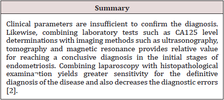
There is a great interest in the use of serum markers to diagnose endometriosis, but they are not sufficiently accurate for use in clinical practice. Cancer antigen CA-125 has been used to monitor the progress of endometriosis [16]. CA19-9 has a lower sensitivity than CA-125, and cytokine interleukin-6 may be more sensitive and specific than CA-125 [1]. Mol et al reported a systematic review of the diagnosis of endometriosis and concluded that serum CA125 level may be elevated in endometriosis, but this measurement had no value as a diagnostic tool compared to laparoscopy [3].

Laparoscopy

Laparoscopy is a primary diagnostic and therapeutic tool providing the opportunity to explore the abdominal cavity and obtain biopsies. The magnified vision enables the surgeon to operate with the best possible exposure. Although it was once believed that intestinal endometriosis was best managed by hormonal regimens or surgical castration, the advent of laparoscopic surgery has dramatically changed this approach [11] (Table 2).

Table 2:
lupinepublishers-openaccess-surgery-case-studies-journal

Treatment

Despite being a gynecologic pathology, deep infiltrating endometriosis is not of exclusive gynecologic concern. A multidisciplinary approach involving urologists and colorectal surgeons therefore is recommended strongly for complete evaluation and correct management. A minimally invasive approach offers convenient advantages concerning the surgical management of multifocal deep infiltrating endometriosis. Traditionally, radical surgery [17] was considered the best measure to prevent disease relapse. However, because of the prevalence of endometriosis among women of reproductive age and the advances in surgical techniques, minimally invasive conservative surgery now is encouraged more [18]. Treatment must be individualized, taking the clinical problem in its entirety into account, including the impact of the disease and the effect of its treatment on quality of life. Pain symptoms may persist despite seemingly adequate medical and/or surgical treatment of the disease. In such circumstances, a multidisciplinary approach involving a pain clinic and counselling should be considered early in the treatment plan. It is also important to involve the woman in all decisions; to be flexible in diagnostic and therapeutic thinking; to maintain a good relationship with the woman, and to seek advice where appropriate from more experienced colleagues or refer the woman to a centre with the necessary expertise to offer all available treatments in a multidisciplinary context, including advanced laparoscopic surgery and laparotomy [19]. The objective of the treatment in pelvic endometriosis is to cease the endometrial stimulus in order to ameliorate the symptoms. Thus, danozol, gonadotropin- releasing hormones, oral contraceptives, and prostaglandin inhibitors can be used. The conclusive treatment of endometriosis is total abdominal hysterectomy, bilateral salpingo-oophorectomy and removal of all endometrial foci. Because malignant transformation cannot be excluded preoperatively and medical treatment may cause fibrosis, the definitive treatment is surgical. Also, in the case of intestinal obstruction and severe rectal and abdominal pain, surgery is indicated. The main objective of surgery is the resection of the affected bowel segment, enabling the histopathological examination of the resection material. Limited surgery, such as excision or cauterization of superficial lesions, following confirmation through frozen section analysis could be performed. In conclusion, intestinal endometriosis is a disease that may imitate various gastrointestinal system diseases. The definite diagnosis could only be done by histopathologic confirmation, since there are no pathognomonic radiological or colonoscopic findings. In female patients who have unexplained digestive complaints, endometriosis should also be considered in the differential diagnosis [20].

The Treatment of Uncomplicated Intestinal

It depends on the patient’s age and intention to conceive. Bowel resection is indicated if there are symptoms of obstruction or bleeding, and if malignancy cannot be excluded. In patients of childbearing age, resection of the involved colon followed by hormonal treatment may be sufficient; otherwise, hysterectomy and bilateral oophorectomy is the treatment of choice [21]. Medical suppressive therapy may be beneficial in some patients with symptomatic rectovaginal endometriosis, but often it is either ineffective or only temporarily effective, whereas surgical therapy is effective in relieving pain conditions. Other studies have shown that operative therapy of rectovaginal endometriosis does not modify reproductive prognosis but significantly reduces pain and improves quality of life. The best long-term results are obtained after complete excision of the endometriotic tissue [22]. The surgeon’s judgment on bowel involvement with the consequence of bowel resection is of the utmost importance [22]. Redwing has suggested a severity scoring system for intestinal endometriosis based on the form of surgical management required: grade I, superficial seromuscular; grade II, partial thickness to mucosa; grade III, full thickness; grade IV, segmental. The surgical approaches to intestinal disease include simple excision (with cautery or laser), mucosal skinning, full thickness disc excision with primary closure, and formal bowel resection [23]. Full thickness disc resection of bowel endometriotic lesion is often incomplete, at least one-third of patients with bowel endometriosis treated by full thickness disc resection have persistent disease. The surgeons must always weigh the risk of potential complications of surgery against the benefit of the complete removal of bowel endometriotic lesions. To date, no clear guideline exists for the pre-operative assessment of patients with suspected endometriosis; therefore, bowel resections should only be performed after a careful pre-operative evaluation of patients’ symptoms and a radiological examination of the bowel [23]. Bowel resection can be performed according to previously published criteria (Remorgida et al.): single lesion >3 cm in diameter, single lesion infiltrating >50% of the bowel wall, three or more lesions infiltrating the muscular layer [15].

Operative Technique

The collaboration of a laparoscopically skilled gynaecologist and colorectal surgeon has been recognised as ideal in the surgical management of colorectal endometriosis. Patients undergo bowel preparation 24 h prior to surgery with a Fleet® ACCU-PREP® Bowel Cleansing System (C.B. Fleet Co., Braeside, Vic., Australia) [24]. Prophylactic anticoagulant therapy was given the evening before the operation, and prophylactic antibiotic therapy was given at the beginning of the operation [25]. Surgery is performed with the patient in the lithotomy position. Five ports are used with placement as shown in Figure 11. Five-millimetre 0° and 5-mm 30° endoscopes are used and most dissection is undertaken using the harmonic scalpel (Ethicon Endo-Surgery, Inc., Cincinnati, OH, USA). Pneumoperitoneum is maintained at 12-14 mmHg with a flow rate of 40 L/min. For an anterior segmental bowel resection, the descending colon is mobilised up to the level of the splenic flexure. The ureter is identified crossing the pelvic brim on the left, and mobilisation continued inferiorly to open the para-rectal space medial to the uterosacral ligament. The right mesocolon is then opened and dissection extended down to the right pararectal space medial to the uterosacral ligament. The right ureter is identified during this dissection. The sigmoid colon is then elevated with bowel grasping forceps, and the space posterior to the sigmoid mesocolon is opened. During this dissection the inferior mesenteric vessels are identified and divided using an endoscopic linear cutter 45 mm (ATW45 Ethicon Endo-Surgery, Inc.) once the requirement for a bowel resection is established. Further elevation of the sigmoid colon allows the posterior dissection to continue inferiorly to the presacral plane, allowing mobilization of the rectum. Having mobilized the rectosigmoid laterally and posteriorly, the rectum is then dissected free from the posterior cervix. This is the most difficult part of the dissection and an attempt is made to free disease from the posterior cervix and posterior vaginal wall as completely as possible. If there is coexisting invasive uterosacral disease, this is excised end bloc with the affected rectal segment. The inferior dissection is complete when the normal tissue in the rectovaginal septum is encountered. Once the level of rectal transection is identified, the mesorectum is divided at that point leaving the rectal tube. An endoscopic articulating linear cutter 45 mm (ATG45 Ethicon Endo-Surgery, Inc.) is introduced and applied transversely across unaffected distal rectum. The stapler is fired to separate the affected rectal segment from the distal rectal stump. Two firings may be required. The lower right 12 mm port site is then converted to a minilaparotomy incision, approximately 3-4 cm in length. The affected rectal segment is then delivered through this wound, clamped and divided above the level of disease. The anvil from an endoscopic curved intraluminal stapler 29 mm (ECS29 Ethicon Endo-Surgery, Inc.) is secured into the proximal colon with a purse-string suture. The proximal segment is then returned to the abdominal cavity and the mini laparotomy wound closed. Pneumoperitoneum is reestablished. The ECS29 is passed transanally, and the distal rectal stump is elevated. The circular stapling device is opened, passing a metal spike through the distal rectal stump adjacent to the staple line. The anvil within the proximal segment is then docked to the spike and the circular stapling device closed. The circular stapling device is then fired to complete the anastomosis. After removing the transanal stapler, an integrity check is performed by distending the rectum with Betadine after occluding the sigmoid colon at the pelvic brim. Further check of integrity is undertaken by instilling air into the rectum after flooding the pelvis with saline. A 17-gauge drain is left in the operative site, after which all ports are removed [24]. Terminal-to-terminal anastomoses were classified according to distance from the anus as high/medium (>8 cm), low (5-8 cm) or ultralow (<5 cm). The choice to perform primary ileostomy or colostomy was based on intraoperative findings [26]. For a disc excision, a lesser degree of descending colon mobilization is required, less posterior rectosigmoid dissection may be required and there is no requirement to divide the inferior mesenteric vessels. The principles of pelvic dissection are otherwise as described. Once the rectal disease has been identified and isolated, a figure of eight suture is placed through the lesion. An ECS33 stapling device is passed transanally with the anvil intact. The device is then opened and angled towards the rectosigmoid lesion. The suture is grasped with laparoscopic forceps, and the disease drawn down into the open stapler. The stapling device is then closed, rotated slightly to ensure that the posterior rectal wall has not become entrapped, and then fired. An anterior arc of rectal wall containing the lesion is therefore removed and the rectal wall stapled in a single action. Rectosigmoid integrity checks are then undertaken as described above [24]. The extent of the lesions as well as the severity of the symptoms justifies the extensive nature of the surgery undertaken. The findings of additional areas of the bowel that are macroscopically normal but microscopically involved, as well as involvement of the lymph nodes, suggest to us that simple local excision of a disc may on occasions be inadequate to remove the whole of the involved area of the bowel [12]. There are no data to justify hormonal treatment prior to surgery to improve the success of surgery [19]. However, according to ESHRE guidelines Postoperative treatment for endometriosis in general might include danazol or a GnRH agonist for 6 months after surgery as it reduces endometriosis associated pain and delays recurrence at 12 and 24 months compared with placebo and expectant management. However, postoperative treatment with a COC is not effective [19].

Figure 11: Placement of five port sites used in Surgery.

Lupinepublishers-openaccess-surgery-case-studies-journal

Postoperative Complications

The risk of complications depends on the clinical conditions, such as the level of bowel stenosis, opening of the vaginal wall, the extent of endometriosis infiltration, and the surgeon’s experience. Moreover, the possibility of performing this kind of surgery (complete eradication with colorectal surgery) in a referral center reduces the risk of complications and improves clinical outcomes. Indeed, women who undergo intestinal surgery are at higher risk of complications mainly in the short-term, but close surveillance reduces the risk of need for reintervention and allows a good recovery within a few weeks [27]. Complications include:

a) Internal hemorrhage

b) Bowel fistula

c) Vaginal fistula

d) Retention of urine

e) Constipation

f) Abdominal wall hematoma

g) Ureteral injury and stenosis

h) Bladder perforation

i) Uterine perforation

j) Cystitis

k) Adynamic ileus

l) Mechanical bowel obstruction

m) Peritonitis

n) Peritoneal effusion

Outcome after Surgery

The indications of colorectal resection for endometriosis are controversial, and the likely risk/benefit ratio must be discussed with each patient. No menstrual pelvic pain, pain on bowel movement, cramping, and cyclic rectal bleeding improved or disappeared in all the women concerned, in keeping with previous studies of colorectal resection for endometriosis. dysmenorrhea, dyspareunia, pain on defecation, and no menstrual pelvic pain improved significantly, on the basis of visual analog scores, whereas no impact was noted on pain on bowel movement, lower back pain, or asthenia. Recent results confirm those of Redwing and Wright, showing that women with dysmenorrhea, dyspareunia, pain on defecation, or no menstrual pelvic pain associated with complete endometriotic obliteration of the sac of Douglas are the best candidates for extensive resection [28]. Bowel resection is not completely free of recurrence of endometriosis, but the incidence of recurrence is significantly lower [29]. In conclusion, laparoscopic rectosigmoid resection and end-to-end anastomosis seem safe and effective in women with deep infiltrating colorectal endometriosis, where the bowel lumen is largely restricted, and bowel function is greatly impaired. Results of long-term follow up demonstrate significant reductions in painful and dysfunctional symptoms associated with deep bowel involvement [30]. Laparoscopic segmental colorectal resection for endometriosis is associated with a significant improvement in quality of life and gynecological and digestive symptoms [25].

Read More About Lupine Publishers Journal of Surgery & Case Studies Please Click on Below Link:
https://surgery-casestudies-lupine-publishers.blogspot.com/

Friday, 18 November 2022

Lupine Publishers | Bempedoic Acid a Small Molecule Drug Process and Synthesis, Innovation And /Or Advantages, Development Status And /Or Regulatory Status

 Lupine Publishers | Journal of Surgery & Case Studies


Abstract

Bempedoic acid (ETC-1002), a small molecule drug, promotes low density lipoprotein (LDL) receptor mediated clearance of LDL-cholesterol (LDL-C) by inhibition of adenosine triphosphate citrate lyase (ACL), a mechanism complementary to those of existing lipid-modifying therapies. Bempedoic acid is a pro-drug activated specifically within the liver where it inhibits ACL, a regulatory checkpoint within the cholesterol biosynthesis pathway. By inhibiting ACL, bempedoic acid reduces cholesterol synthesis in liver cells and triggers compensatory LDL receptor upregulation. Inhibiting ACL with bempedoic acid complements other mechanisms targeted by current therapies, resulting in additional lowering of LDL-C, without leading to increases in adverse events (AEs).b In the phase III clinical trials (NCT02666664, NCT02991118) of patients with high cardiovascular risk and elevated LDL-C not adequately controlled by their current therapy, patients are given a daily dose of 180 mg bempedoic acid as an oral tablet, whilst remaining on ongoing lipid-modifying therapy. The present paper describes the process and synthesis, innovation and /or advantages, development status and /or regulatory status of Bempedoic acid.

Keywords:Cardiovascular Disease; Hyperlipidemia; ETC-1002; Low Density Lipoprotein Cholesterol; Synthesis; Statin Associated Muscle Symptoms; Statin Intolerance; Regulatory Status

Introduction

The current standard of care for patients with hypercholesterolaemia is primarily statins which can reduce LDL-C. However, some patients, particularly those with heterozygous familial hypercholesterolaemia, coronary heart disease (CHD), CHD-risk equivalents, and other clinical manifestations of atherosclerotic cardiovascular disease [1] (ASCVD), require additional LDL cholesterol lowering on top of what can be achieved with maximum tolerated statin therapy. Additionally, there are patients who are unable to tolerate statins due to adverse advents such as muscle pain, or increased blood glucose. There is an unmet medical need for patients unable to achieve sufficient reduction in LDL cholesterol with existing treatment options and thus remain at increased risk of cardiovascular disease and the consequences thereof. Bempedoic acid requires activation by a specific enzyme acyl-CoA synthatase (ACSVL1), which is largely restricted to the liver. Therefore, it is believed that unlike statins, myotoxicity is unlikely to occur with bempedoic acid because it does not inhibit cholesterol biosynthesis in skeletal muscle due to the absence of ACSVL1 in these cells. The effect of bempedoic acid is additivenot redundant-to that of statins, because the target of bempedoic acid, ACL, is a distinct regulatory checkpoint on the cholesterol biosynthesis pathway than HMG-CoA reductase, the primary target of statins. Inability to tolerate statins because of muscle symptoms contributes to uncontrolled cholesterol levels and insufficient cardiovascular risk reduction. Bempedoic acid, a prodrug that is activated by a hepatic enzyme does not present in skeletal muscle, inhibits ATP‐citrate lyase [2], an enzyme upstream of β‐ hydroxy β‐methylglutaryl‐coenzyme A reductase in the cholesterol biosynthesis pathway. Statins are widely prescribed for lowering LDL-cholesterol (LDL-C) and reducing the risk of cardiovascular disease. However, many patients are statin intolerant and unable to achieve sufficient LDL-C lowering due to muscle-related side effects. Bempedoic acid (ETC-1002)’s mechanism of action is similar to that of statins, but because it does not inhibit the cholesterol biosynthesis pathway in skeletal muscle, myotoxicity is unlikely to occur. ETC-1002 was first discovered at the original Esperion Therapeutics, which was acquired by Pfizer in 2004 and subsequently spun-out as Esperion Therapeutics in 2008 along with ETC-1002 and other assets. Esperion continues development of ETC-1002 [3], which is currently in phase III trials as an LDL-Clowering agent in patients with hypercholesterolemia. This review discusses the drug candidate’s mechanism of action, effects, safety and clinical data. This paper explaining how the process of synthesis was carried and conclusion of Bempedoic acid offers a safe and effective oral therapeutic option for lipid lowering in patients who cannot tolerate statins.

a) The phase 3 CLEAR (Cholesterol Lowering via Bempedoic acid, an ACL‐Inhibiting Regimen) Serenity clinical trial demonstrates the lipid‐lowering efficacy of bempedoic acid, a firstin‐ class, prodrug, small‐molecule inhibitor of ATP‐citrate lyase, among patients with established statin intolerance and elevated low‐density lipoprotein cholesterol who were receiving stable background therapy.

b) Muscle‐related symptoms contributed to the history of statin intolerance for almost all patients.

c) Although bempedoic acid acts on the same cholesterol biosynthesis pathway as statins, the muscle‐related adverse event rate in CLEAR Serenity with bempedoic acid, which is not activated in skeletal muscle, did not differ from placebo, even among patients who had experienced muscle‐related symptoms while on statin therapy [4].

Development Status And/or Regulatory Designations

Bempedoic acid does not currently have Marketing Authorisation in the EU/UK for any indication. Bempedoic acid or bempedoic acid with ezetimibe in a fixed-dose combination are in phase III clinical trials for the treatment of primary hypercholesterolaemia [5] (heterozygous familial and nonfamilial) or mixed dyslipidaemia in patients who are statin-intolerant, or for whom a statin is contraindicated. Bempedoic acid monotherapy and bempedoic acid with ezetimibe in a fixed-dose combination is also in phase III clinical trials for the treatment of primary hypercholesterolaemia (heterozygous familial and non-familial) or mixed dyslipidaemia in patients unable to reach LDL-Cholesterol goals with the maximum tolerated dose as an adjunct to diet in combination with a statin or statin with other lipid lowering therapies. Esperion Therapeutics was founded in April 2008 by former executives of, and investors in, the original Esperion Therapeutics which was founded in July 1998 and was bought by Pfizer for $1.3 billion in 2004 and then spun out in 2008. ETC-1002 was first discovered at the original Esperion [5], and Esperion subsequently acquired the rights to it from Pfizer in 2008. Esperion own the exclusive worldwide rights to ETC-1002.

Preparation

Bempedoic acid [5,6] was prepared by condensation of 1,5-dibromopentane (I) with ethyl isobutyrate (II) by means of LDA in THF in the presence of DMPU at −78 °C to give ethyl 7-bromo-2,2-dimethylheptanoate (III), which is dimerized with tosylmethyl isocyanide (IV) in the presence of NaH and Bu4NI in DMSO to yield diethyl 8-isocyano-2, 2,14,14-tetramethyl-8-(tosyl) pentadecanedioate (V). Reaction of intermediate (V) with aqueous HCl in CH2Cl2 affords 2,2,14,14-tetramethyl-8-oxopentadecanedioic acid diethyl ester (VI), which is hydrolyzed with aqueous KOH in refluxing EtOH/H2O to provide the dicarboxylic acid ESP-15228 (VII) (1-4). Ketone (VII) is finally reduced by means of NaBH4 in MeOH (1). Scheme 1[6].

7-Bromo-2,2-Dimethylheptanoic Acid Ethyl Ester

7-Bromo-2,2-dimethylheptanoic acid ethyl ester [5]

Under argon atmosphere and cooling with an ice-bath, a solution of lithium diisopropylamide in THF (1.7 L, 2.0 M, 3.4 mol) was slowly dropped into a solution of 1,5- dibromopentane (950 g, 4.0 mol) and ethyl isobutyrate (396 g, 3.4 mol) in THF (5 L) while keeping the temperature below +5 DC. The reaction mixture was stiπed at room temperature for 20 h and quenched by slow addition of saturated ammonium chloride solution (3L). The resulting solution was divided into three 4-L portions. Each portion was diluted with saturated ammonium chloride solution (5L) and extracted with ethyl acetate (2 ‘2L). Each 4-L portion of ethyl acetate was washed with saturated sodium chloride solution (2L), 1 N hydrochloric acid (2L), saturated sodium chloride solution (2L), saturated sodium bicarbonate solution (2L), and saturated sodium chloride solution (2L). The three separate ethyl acetate layers were combined into a single 12-L portion, dried over magnesium sulfate, and concentrated in vacuo to give the crude material (1.7L) which was purified by vacuum distillation. Two fractions were obtained: the first boiling at 88 – 104 °C / 0.6 ton (184.2 g), the second at 105 – 120 °C / 1.4 ton (409.6 g) for atotal yield of 60 %. 1H NMR (300 MHz, CDC13/ TMS): δ (ppm): 4.11 (q, 2 H, J = 7.2 Hz), 3.39 (t, 2 H, J = 6.8 Hz), 1.85 (m, 2 H), 1.56 – 1.35 (m, 4 H), 1.24 (t, 3 H, J = 7.2 Hz), 1.31 – 1.19 (m, 2 H), 1.16 (s, 6 H). 13C NMR (75 MHz, CDCI3/TMS): δ (ppm): 177.9, 60.2, 42.1, 40.5, 33.8, 32.6, 28.6, 25.2, 24.2, 14.3. HRMS (El, pos): Calcd. for CπH22Brθ2 (MH+): 265.0803, found: 265.0810.6.18.

2,2,14,14-tetramethyl-8-oxo-pentadecanedioic acid diethyl ester

Under Air atmosphere, to a solution of 7-bromo-2,2- dimethylheptanoic acid ethyl ester5 (26.50 g, 100 mmol), tetra-nbutylammonium iodide (3.69 g, 10 mmol) and p- toluenesulfonyl methyl isocyanide (9.80 g, 50 mmol) in anhydrous DMSO (300 mL) was added sodium hydride (4.80 g, 20.5 mmol, 60 % dispersion in mineral oil) at 5 – 10oC The reaction mixture was stiπed at room temperature for 20 h and quenched with ice-water (300 mL). The product was extracted with dichloromethane (3D 100 mL). The combined organic layers were washed with water (200 mL), half-saturated NaCl solution 100 mL), and saturated NaCl solution (200 mL), dried over MgS04, and concentrated in vacuo to get the crude 8-isocyano-2,2,14,14-teframethyl-8-(toluene-4- sulfonyl)-pentadecanedioic acid diethyl ester (36.8 g) as an orange oil, which was used in the next step without purification. To a solution of this crude product (36.8 g) in dichloromethane (450 mL) was added concentrated hydrochloric acid (110 mL) and the mixture was stiπed at room temperature for 1h. The solution was diluted with water (400 mL) and the aqueous layer was extracted with dichloromethane (200 mL). The combined organic layers were washed with saturated NaHC0 solution (2 x 150 mL) and saturated NaCl solution (150 mL). The organic solution was dried over Na2S04 and concentrated in vacuo. The residue was subjected to column chromatography (silica gel, hexanes: ethyl acetate = 11:1) to give 2,2,14,14-tetramethyl-8-oxo-pentadecanedioic acid diethyl ester (12.20 g, 66 % over two steps) as a colorless oil. lH NMR (300 MHz, CDC13/TMS): δ (ppm): 4.11 (q, 4 H, J – 6.9 Hz), 2.37 (t, 4 H, J – 7.5 Hz), 1.58 – 1.47 (m, 8 H), 1.35 – 1.10 (m, 8 H), 1.24 (t, 6 H, J = 7.2 Hz), 1.15 (s, 12 H). 13C NMR (75 MHz, CDC13/TMS): δ (ppm): 211.6, 178.3, 60.5, 43.1, 42.5, 40.9, 30.1, 25.5, 25.1, 24.1, 14.7. HRMS (LSIMS, nba): Calcd. for C23IL3O5 (MH+): 399.3110, found: 399.3129.

8-Hydroxy-2.2.14,14-Tetramethylpentadecanedioic Acid

Under nitrogen atmosphere, sodium borohydride (0.06 g, 1.6 mmol) was added to a stiπed solution of 8-oxo-2,2,14,14- tetramethylpentadecanedioic acid (1.18 g, 3.4 mmol) in methanol (50 mL) at 0 °C. The reaction progress was momtored by thin layer chromatography (silica; hexanes: ethyl acetate = 50: 50). Additional sodium borohydride was added after 1h (0.48 g, 13 mmol). After 8 h, the reaction mixture was hydrolyzed with water (50 mL) and acidified with concentrated hydrochloric acid (3 mL) to pH 1. The solution was diluted with water (50 mL) and exfracted with dichloromethane (4 x 25 mL). The combined organic layers were washed with saturated sodium chloride solution (2 x 30 mL), dried over magnesium sulfate, concentrated in vacuo, and dried in high vacuo to give 8-hydroxy-2,2,14,14-tetramethylpentadecanedioic acid (0.7 g, 60 %) as a very viscous oil. 1H NMR (300 MHz, CDC13/TMS): δ (ppm): 7.42 (br. s, 3 H), 3.59 (br. s, 1 H), 1.65 – 1.00 (m, 20 H), 1.18 (s, 12 H). 13C NMR (75 MHz, CDC13/TMS): δ (ppm): 184.5, 71.8, 42.1, 40.5, 37.0, 29.8, 25.2, 25.1, 24.9, 24.8. HRMS (FAB): Calcd. for Cι9H3705 (MH+): 345.2635, found: 345.2646. HPLC: 83.8 % purity. Keto-substituted hydrocarbons with 11−19 methylene and bis-terminal hydroxyl and carboxyl groups have been synthesized and evaluated in both in vivo and in vitro assays for their potential to favorably alter lipid disorders including metabolic syndrome. Compounds were assessed for their effects on the de novo incorporation of radiolabeled acetate into lipids in primary cultures of rat hepatocytes as well as for their effects on lipid and glycemic variables in obese female Zucker fatty rats [Crl:(ZUC)-faBR] following 1 and 2 weeks of oral administration. The most active compounds were found to be symmetrical with four to five methylene groups separating the central ketone functionality and the gem dimethyl or methyl/aryl substituents. Furthermore, biological activity was found to be greatest in both in vivo and in vitro assays for the tetramethyl-substituted keto diacids and diols (e.g., 10c, 10g,14c), and the least active were shown to be the bis(arylmethyl) derivatives (e.g., 10e, 10f,14f). Compound 14c dose-dependently elevated HDL-cholesterol, reduced triglycerides, and reduced NEFA, with a minimum effective dose of 30 mg/kg/day. Compound 10g dose-dependently modified non-HDL-cholesterol, triglycerides, and no esterified fatty acids, with a minimum effective dose of 10 mg/kg/day. At this dose, compound 10g elevated HDL-cholesterol levels 2−3 times higher than pretreatment levels, and a dose-dependent reduction of fasting insulin and glucose levels was observed.

Only Keto Compd Described

2,2,14,14-Tetramethyl-8-oxopentadecanedioic Acid [6] (10g).

According to the procedure given for 10f, 9g (8.54 g, 21.4 mmol) was saponified with KOH (85%, 4.53 g, 68.6 mmol) in EtOH (13 mL) and water (5 mL) at reflux for 4 h. The solid product obtained after usual workup was recrystallized from Et2O/hexanes (50 mL/50 mL), affording 10g (4.16 g, 57%) as colorlessneedles. Mp: 82−83 °C. 1H NMR (CDCl3): δ 11.53 (br, 2H), 2.39 (t, 4H, J = 7.3), 1.60−1.50 (m, 8 H), 1.30−1.20 (m, 8 H), 1.18 (s, 12 H). 13C NMR (CDCl3): δ 211.7, 185.0, 42.8, 42.3, 40.4, 29.7, 25.1, 24.8, 23.8.

HRMS (LSIMS, gly): calcd for C19H35O5 (MH+) 343.2484, found 343.2444. HPLC: Alltima C-8 column, 250 × 4.6 mm, 5 μm; 60% acetonitrile/40% 0.05 M KH2PO4, flow rate 1.0 mL/min; RI, tR 6.50 min, 92.6% pure. Bempedoic acid is an oral medicinal product that is in clinical development for the treatment of people with primary hypercholesterolaemia or mixed dyslipidaemia [7-9] with high cardiovascular risk. Abnormal levels of lipids in the blood characterises dyslipidaemia. High levels of cholesterol in the blood (hypercholesterolemia) may be caused by genetic defects as seen in familial hypercholesterolaemia or may occur when genes and other factors such as lifestyle habits interact, as seen in non-familial hypercholesterolaemia. Most people with hypercholesterolaemia have mildly or moderately increased low-density lipoprotein cholesterol (LDL-C) levels (often considered the “bad” cholesterol that may cause blockages of blood vessels). Elevated levels of LDL-C increase the risk of cardiovascular disease, which is responsible for many deaths and disabilities. Bempedoic acid lowers LDL-C via a different mechanism of action and offers the potential advantage of reduced muscular adverse effects when compared to statins which are the current standard of care. Bempedoic acid is being developed for patients at high cardiovascular risk who are unable to reach LDL-C goals with the maximum tolerated dose of statins. The effect of bempedoic acid is additive-not redundant-to that of statins, and if licensed, may offer additional and effective treatment option to use in combination with dietary changes and other lipidmodifying therapies to treat primary hypercholesterolaemia or mixed dyslipidaemia. Bempedoic acid is claimed in U.S. Patent No. 7,335,799 that is scheduled to expire in December 2025, which includes 711 days of patent term adjustment, and might be eligible for a patent term extension period of up to five years. U.S. Patent Nos. 9,000,041, 8,497,301 and 9,624,152 claim methods of using bempedoic acid. PRODUCT: Bempedoic acid.

Bempedoic Acid (Esperion Therapeutics, Inc.)

With a targeted mechanism of action, bempedoic acid is a firstin- class, orally available, once-daily ACL inhibitor that reduces cholesterol biosynthesis and lowers elevated levels of LDL-C by up-regulating the LDL receptor, [10] but with reduced potential for muscle-related side effects. Completed Phase 1 and 2 studies in more than 800 patients treated with bempedoic acid have produced clinically relevant LDL-C lowering results of up to 30 percent as monotherapy, approximately 50 percent in combination with ezetimibe, and an incremental 20+ percent when added to stable statin therapy.

Mechanism of Action

In November 2016, we announced the publication of “Liverspecific ATP-citrate lyase inhibition by bempedoic acid decreases LDL-C and attenuates atherosclerosis,” by Stephen L. Pinkosky, our Associate Director of Translational Research and Biology, et al., in Nature Communications. The paper systematically outlines the experiments and analyses undertaken by us and our collaborators to fully understand the mechanism of action for how bempedoic acid reduces LDL-C, including its specificity for the liver. Bempedoic acid is a prodrug that once activated, inhibits ACL, an enzyme upstream of HMG-CoA reductase [11,12], (the molecular target of statins) in the cholesterol synthesis pathway. Like statins, bempedoic acid decreases cholesterol synthesis in the liver, which results in decreased intracellular cholesterol, up-regulation of LDL receptor activity and increased LDL-C clearance from the blood. Although bempedoic acid and statins both inhibit cholesterol synthesis in the liver, an important differentiating feature is that, unlike statins, bempedoic acid is inactive in skeletal muscle. Specifically, bempedoic acid is a prodrug which requires activation by a specific enzyme, very long-chain acyl-CoA synthetase, or ACSVL1, to convert bempedoic acid to its CoA activated form. This enzyme is present in the liver but not in skeletal muscle. Therefore, bempedoic acid does not inhibit the cholesterol biosynthesis pathway in skeletal muscle, thus providing a mechanistic basis for reduced potential for muscle-related adverse effects. Bempedoic acid has been shown to provide incremental lowering of LDL-C when used in combination with both ezetimibe and statins at all doses [13].

Fixed Dose Combination Bempedoic Acid and Ezetimibe (BA+EZ)

In the second quarter of 2016, the Food and Drug Administration, or FDA, accepted our submission of an Investigational New Drug, or IND, application for the fixed dose combination of bempedoic acid 180 mg and ezetimibe 10 mg, or BA+EZ, which is in development for the same indications as bempedoic acid monotherapy [14,15] (LDL-C lowering and CV risk reduction). We recently completed a bioavailability study and a formulation of BA+EZ has been selected for manufacturing, development and, if approved, commercialization. We expect to announce clinical development and regulatory plans for BA+EZ in the first half of 2017.

Cardiovascular Disease and Elevated LDL-C

Cardiovascular disease, which results in heart attacks, strokes and other cardiovascular events, represents the number one cause of death and disability in western societies. The American Heart Association, or AHA, estimates that approximately 800,000 deaths in the United States were caused by cardiovascular disease in 2013. Elevated LDL-C is well-accepted as a significant risk factor for cardiovascular disease and the CDC estimates that 78 million U.S. adults have elevated levels of LDL-C. A consequence of elevated LDL-C is atherosclerosis, which is a disease that is characterized by the deposition of excess cholesterol and other lipids in the walls of arteries as plaque. The development of atherosclerotic plaques often leads to cardiovascular disease. The risk relationship between elevated LDL-C and cardiovascular disease was first defined by the Framingham Heart Study, [15] which commenced in 1948 to define the factors that contributed to the development of cardiovascular disease. The study enrolled participants [16].

(Esperion Therapeutics, Inc)

a) Licenses

In April 2008, we entered into an agreement with Pfizer pursuant to which we acquired a worldwide, exclusive, fully paidup license from Pfizer to certain patent rights owned or controlled by Pfizer relating to bempedoic acid, and we granted Pfizer a worldwide, exclusive, fully paid-up license to certain patent rights owned or controlled by us relating to development programs other than bempedoic acid. The license to us covers the development, manufacture and commercialization of bempedoic acid. We may grant sublicenses under the license. Under the license agreement, Pfizer is restricted from making, using, developing or testing any of the compounds claimed under the same patents that claim or cover the composition of matter of bempedoic acid. [17], Neither party is entitled to any royalties, milestones or any similar development or commercialization payments under the license agreement, and the licenses granted are irrevocable and may not be terminated for any cause, including intentional breaches or breaches caused by gross negligence [18].

Intellectual Property of Esperion Therapeutics, Inc.

As of December 31, 2016, our patent estate, including patents we own or license from third parties, on a worldwide basis, included approximately 25 issued United States patents and four pending United States patent applications and 23 issued patents and 15 pending patent applications in other foreign jurisdictions. Of our worldwide patents and pending applications, only a subset relates to our small molecule program which includes our lead product candidate, bempedoic acid. Bempedoic acid is claimed in U.S. Patent No. 7,335,799 that is scheduled to expire in December 2025, which includes 711 days of patent term adjustment, and may be eligible for a patent term extension period of up to five years. U.S. Patent Nos. 9,000,041 and 8,497,301 claim methods of treatment using bempedoic acid. We also have a pending U.S. patent application directed to bempedoic acid. There are currently three issued patents and four pending application in countries outside the United States that relate to bempedoic acid [19].

Overall Safety Observations (Esperion Therapeutics, Inc statement)

To date, in completed studies, over 800 patients have been treated with bempedoic acid for periods of up to 12 weeks at maximum repeated doses of 240 mg per day. Bempedoic acid has been safe and well-tolerated with no dose-limiting side effects identified to date in our ongoing or completed clinical studies. No clinical safety trends have emerged to date.

Conclusion

The synthesis of Bempedoic acid was prepared by condensation of 1,5-dibromopentane with ethyl isobutyrate by means of LDA in THF in the presence of DMPU at −78 °C to give ethyl 7-bromo-2,2-dimethylheptanoate , which is dimerized with tosylmethyl isocyanide in the presence of NaH and Bu4NI in DMSO to yield diethyl 8-isocyano-2, 2,14,14-tetramethyl-8-(tosyl) pentadecanedioate . Reaction of intermediate with aqueous HCl in CH2Cl2 affords 2,2,14,14-tetramethyl-8-oxopentadecanedioic acid diethyl ester, which is hydrolysed with aqueous KOH in refluxing EtOH/H2O to provide the dicarboxylic acid ESP-15228 (1-4). Ketone is finally reduced by means of NaBH4 in MeOH (1). Esperion announced new details about its phase 3 program for bempedoic acid, its unique oral, once-daily cholesterol-lowering compound. The company said the phase 3 program would include patients with hypercholesterolemia on any statin at any dose, including those with LDL levels not adequately controlled on current statin therapy in intolerant” patients unable to take even low doses of statins. Bempedoic acid does not currently have Marketing Authorisation in the EU/UK for any indication. Bempedoic acid or bempedoic acid with ezetimibe in a fixed-dose combination are in phase III clinical trials for the treatment of primary hypercholesterolaemia (heterozygous familial and nonfamilial) or mixed dyslipidaemia in patients who are statin-intolerant, or for whom a statin is contraindicated. Bempedoic acid monotherapy and bempedoic acid with ezetimibe in a fixed-dose combination is also in phase III clinical trials for the treatment of primary hypercholesterolaemia (heterozygous familial and non-familial) or mixed dyslipidaemia in patients unable to reach LDL- Cholesterol goals with the maximum tolerated dose as an adjunct to diet in combination with a statin or statin with other lipid lowering therapies.

Read More About Lupine Publishers Journal of Surgery Please Click on Below Link:
https://surgery-casestudies-lupine-publishers.blogspot.com/

Saturday, 3 September 2022

Lupine Publishers | The Optimal Pain Management Methods Post Thoracic Surgery: A Literature Review

 Lupine Publishers | Journal of Surgery & Case Studies


Abstract

Post-operative pain control is one of the key factors that can aid in fast and safe recovery after any surgical interventions. Thoracic surgery can cause significant postoperative pain which can lead to delayed recovery, delayed hospital discharge and possibly increased risk of chest complications in the form of atelectasis and even lower respiratory infections. Therefore, appropriate pain management following thoracic surgery is mandatory to prevent development of such morbidities including chronic pain.

Keywords:Thoracic Surgery, Analgesia, VATS, Robotics, Thoracotomy

Introduction

Thoracic surgical procedures can result in severe pain which can present as a challenge to be appropriately managed postoperatively. In particular, thoracotomies are well known for their severity of pain due to the incision, manipulation of muscles and ligaments, retraction of the ribs with compression, stretching of the intercostal nerves, possible rib fractures, pleural irritation, and postoperative tube thoracotomy [1]. Recognition of this has contributed to the development of minimally invasive techniques such as video assisted thoracoscopic surgeries (VATS) and lately robotic surgery [1]. These techniques not only aim to produce better aesthetic results, but also reduce post-operative pain and enhance recovery without compromising the quality of treatment offered. Poor pain management can lead to several and serious complications such as lung atelectasis, hypostatic pneumonia due to avoidance of deep breathing in these patients as a result of pain and superimposed infection [1]. Pain management as a result, does not only lead to greater patient satisfaction, but it also reduces morbidity and mortality in patients undergoing thoracic surgery [2]. Historically, post-operative pain management for thoracic surgery involved the use of narcotics alongside parenteral or oral anti-inflammatory agents [2]. Post chest tube removal patients typically are transitioned to oral analgesia. Multiple additional pain control adjuncts were also implemented with differing levels of success [1]. Over time, intra-operative techniques have been developed which aims to target pain reduction postoperatively [2]. As our understanding of both pain management and the factors that play a role in the development of pain has increased, we have been able to target these and improve postoperative pulmonary morbidity and pain scores [1,2]. We aim to review different means of pain control in this paper in order to assess their effectiveness in achieving optimum results.

Thoracotomy

The mechanism of pain in thoracotomy involves the innervation of the intercostal, sympathetic, vagus and phrenic nerves [3]. Additionally, shoulder pain may result from stretching of the joints during the operation.

After a thoracotomy, pain can persist for two months or more, and in certain incidences it recurs after a period of cessation. The incidence of chronic pain post thoracotomy is reported to be 22-67% in the population [4]. Good surgical technique and effective acute post-operative pain treatment are evident means of preventing post-thoracotomy pain and consequent pulmonary complications [4]. Due to the multifactorial character of the pain, a multimodal approach to target pain is advised. Typically, both regional and systemic anaesthesia are administered. A combination of opioids such as fentanyl or morphine are typically used [5]. A variety of techniques for the administration of local anaesthetics are available at present, and the effectiveness of each is assessed in this paper.

a) Thoracic Epidural Analgesia (TEA)

TEA was the most widely used method of means of analgesia. It was the gold standard means of pain relief [6,7]. It is typically inserted prior to general anaesthesia, at the level of T5-T6, midway along the dermatomal distribution of the thoracotomy incision. A study by Tiippana et al. [8] measured the visual analogue scale (VAS) in order to assess the presence of pain during rest and at the time at which they coughed in 114 patients of whom 89 had TEA and 22 who had other methods of pain control. TEA was effective in alleviating pain at rest and during coughing. In TEA patients, the incidence of chronic pain of at least moderate severity was 11% and 12% at 3 and 6 months, respectively. The study found that at one week after discharge, 92% of all patients needed daily pain medication. The study advised for extended postoperative analgesia for up to the week post-discharge to be administered in order to manage this. The study however concluded overall, that TEA was effective in controlling evoked post-operative pain. However, the study did encounter problems of technical form in 24% of the epidural catheters. The incidence of chronic pain, however, was lower compared with previous studies where TEA was not used. Several other studies support that TEA is superior to less invasive methods. According to Shelley B. et al. [9] TEA was preferred by 62% of the respondents over paravertebral block (PVB) with 30% and other analgesic techniques with 8%. Limitations of this technique included hypotension and urinary retention. Certain patients with active infection and on anticoagulation are excluded from epidural placement.

b) Paravertebral Block (PVB)

PVB is considered an effective method for pain management and its use has been increased in the recent years. This technique involves injecting local anaesthetic into the paravertebral space and it is able to block unilateral multi-segmental spinal and sympathetic nerves. Previous studies have shown that it is effective in achieving analgesia and is associated with a lower incidence of side effects such as nausea, vomiting, hypotension and urinary retention [10,11]. As the lungs are collapsed, it is associated with a lower risk of pneumothorax.

In a study by Davies R.G. et al. [10] there was no significant difference in pain scores, morphine consumption and supplementary use of analgesia between TEA and PVB. The rate of failed technique was lower in PVB (OR =0.28, p=0.007). Respiratory function was improved at both 24 and 48 hours with PVB but only significantly improved at 24 hours.

c) Intercostal Nerve Block (ICNB)

ICNBs are generally administered as single injections at least two dermatomes above and below the thoracotomy incision [12]. It is performed percutaneously or under direct vision, using single injections or through placement of an intercostal catheter. It can also be formed using cryotherapy. It is associated with reduced post-operative pain scores; however, it is less effective than TEA in controlling chronic pain [12]. This was illustrated by a study by Sanjay et al. [12] which found that patients that underwent ICNB had higher pain scores 4 hours post-operatively, than those who received epidural anaesthesia using 0.25% bupivacaine (p<0.05). The study concluded that in the early post-operative period there was significant impact in pain relief for both techniques, but thereafter, epidural anaesthesia was proven to significantly reduce post thoracotomy pain over ICNB. Due to the multifactorial nature of post-thoracotomy pain, various approaches are required in order to target pain. ICNBs are useful in the blockade of intercostal nerves, whilst PVB and TEA appear to block the intercostal and sympathetic nerves. Due to the inability of regional anaesthesia to block the vagus and phrenic nerves which are implicated in the pathophysiology of pain, NSAIDs and opioids are required as adjuncts. TEA is proven to be the most effective means of treating pain alongside PVB; however, it is associated with more side effects than PVB. At present, there are a limited number of studies directly comparing pain control and post-operative outcomes between PVB and TEA. There is no conclusive evidence that either method is superior to the other regarding pain control.

Video-Assisted Thoracoscopic Surgery (VATS)

Existing evidence supports the noninferiority of thoracic PVB when compared to TEA for postoperative analgesia [13]. PVB is versatile and may be applied both unilaterally or bilaterally. It can be used to avoid contralateral sympathectomy, consequently minimising hypotension. This is an apparent advantage it has over thoracic epidural. Furthermore, it offers a more favourable side effect profile when compared to epidural anaesthesia. At present, the factors taken into consideration when selecting a regional technique include tolerance of side effects associated with TEA, consensus on best practice/technique, and operator experience [13]. A randomised controlled trial by Kosiński et al. [14] compared the analgesic efficacy of continuous thoracic epidural block and percutaneous continuous PVB in 51 patients undergoing VATS lobectomy. The primary outcome measures were postoperative static (at rest) and dynamic (coughing) visual analogue pain scores (VAS), patient-controlled morphine use and side-effect profile. The study found that pain control (VAS) was superior in the PVB group at 24 hours, both at rest (1.7 vs3.3, p=0.01) and on coughing (5.8 vs 6.6, p=0.023), and control of pain at rest was also superior in the PVB group at 36 hours (3.0 vs 3.7 (p=0.025) and at 48 hours (1.2 vs 2.0, p=0.026). There were no significant differences in the postoperative morphine requirements. In regard to side-effect profile, the study showed that the incidence of postoperative urinary retention (defined as no spontaneous micturition for 8 hours or ultrasound-assessed volume of the urinary bladder >500ml) was greater in the epidural group (64.0% vs 34.6%, p=0.0036), as was the incidence of hypotension (32.0% vs 7.7%, p=0.0031). There was no significant difference in the incidence of atelectasis (4.0% vs 7.7%, p=0.0542). However, the incidence of pneumonia was significantly more frequent in the PVB group (3.8% vs 0%, p=0/0331). Kosiński et al. concluded that PVB is as effective as thoracic epidural block in regard to pain management as it offers a superior safety profile with minimal postoperative complications. A further randomised controlled trial by Okajima et al. [15] compared the requirements for postoperative supplemental analgesia in 90 patients who received wither a PVB or thoracic epidural infusion for VATS lobectomy, segmentectomy or wedge resection. The main outcome measures were pain scores at rest (verbal rating scale 0= none and 10=maximum pain), blood pressure, side effects and overall satisfaction scores relating to pain control (1=dissatisfied and 5=satisfied). The study found a similar frequency of supplemental analgesia (50mg diclofenac sodium suppository or 15mg pentazocine intramuscularly) for moderate pain in both groups, with 56% of those in the PVB group requiring ≥2 doses, compared to 48% in the epidural group (p=0.26). Hypotension, defined as a systolic blood pressure <90mmHg, occurred more frequently in the epidural group (21.2% vs 2.8%, p=0.02). There was no difference in the incidence of pruritus (3.0% vs 0%, p=0.29) and post-operative nausea and vomiting (30.3% vs 25.0%, p=0.62) between both groups. The study found no statistical difference between patient-reported satisfaction in pain control between epidural and PVB using the verbal rating scale (5.0 vs 4.5, p=0.36). The study concluded that PVB offered additional to equivalent analgesia to epidural, a lower incidence of haemodynamic instability postoperatively. A further study by Khoshbin et al. [16] performed an analysis on 81 patients undergoing VATS for pleural aspiration +/- pleurodesis, lung biopsies or bullectomy. The main outcome was postoperative pain levels, documented every 6 hours and scored against the Visual analogue Scale (0= no pain, 10= worst possible pain). In both PVB and epidural groups, bupivacaine 0.125% was the local anaesthetic of choice, with clonidine added to the epidural infusion at 300μg in 500ml. The study showed that there was no significant difference in mean pain scores between PVB or EP (2.1 vs 2.9, p=0.899), therefore concluding that PVB is as effective as epidural in controlling pain post-VATS.

Robotic Lung Surgery

Minimally invasive techniques are considered advantageous over open surgical approaches due to their shorter recovery times, reduced perceived levels of pain post-operatively and shorter postoperative length of stay in hospital [17-19]. Robotic surgery has become a popular method in recent years. Debate remains regarding whether robotic surgery is superior to VATS in regard with pain reduction. A case control study by Louie et al. [19] compared 45 robotic assisted lobectomies (RAL) to 34 VATS lobectomies. The study showed that both groups had a similar mean ICU stay (0.9 vs 0.6 days) and a mean total length of stay (4.0 vs 4.5 days). The study showed that patients that underwent robotic lobectomies had a shorter duration of analgesic use post-operatively (p=0.039) and a shorter time resuming to normal everyday activities (p=0.001). A limitation in this study was an inaccurate record of the amount of pain relief used by the patients, ultimately working as a confounding factor when interpreting the results. In a separate study by Jang et al. [18] 40 patients undergoing RAL were compared retrospectively to 80 VATS patients (40 initial patients and 40 most recent patients), all with resectable non-small cell lung cancer. The study showed that the post-operative median length of stay was significantly shorter in RAL patients compared to the initial VATS patients. The rate of post-operative complications was significantly lower in the RAL group (10%) compared to the initial VATS group (32.5%) and similar to the recent VATS group (17.5%). Post-operative recovery was easier for patients in both the RAL and VATS group due to earlier mobilisation, allowing them to return to their everyday activities quicker. In a retrospective review by Kwon et al. [17] 74 patients undergoing robotic surgery, 227 patients undergoing VATS and 201 patients undergoing anatomical pulmonary resection were assessed and compared with regard to acute (visual pain score) and chronic pain (Pain DETECT questionnaire). The study showed that there was no significant difference in acute or chronic pain between patients undergoing robotic assisted surgery and VATS. Despite no significant difference in pain scores, 69.2% of patients who underwent robotic-assisted surgery felt the approach affected their pain versus 44.2% of the patients who underwent VATS (p=0.0330). These results all support the superiority of robotic surgery over VATS and open approaches with regard to pain, length of hospital stay and recovery times. Both robotic surgery and VATS have their benefits i.e. two-versus three-dimensional view, instrument manoeuvrability, and reduced post-operative pain.

Conclusion

Since post-thoracotomy pain is multifactorial, a multimodal approach is required. In particular, ICNB blocks the intercostal nerves, and PVB and TEA appear to block the intercostal and sympathetic nerves. NSAIDs and opioids are required as valgus and phrenic nerve cannot be blocked by regional anaesthesia. TEA is evident to be the most effective in treating pain alongside with PVB. It is however associated with more side effects than PVB.

Read More About Lupine Publishers Journal of Surgery Please Click on below Link:
https://surgery-casestudies-lupine-publishers.blogspot.com/