Showing posts with label Journal of Anesthesia; Journal of Pain Medicine; GJAPM. Show all posts

Saturday, 6 August 2022

Lupine Publishers | Giant Pedunculated Lipoma Over Left Upper Limb: An Unusual Case Report

 Lupine Publishers | Journal of Anesthesia & Pain Medicine


Introduction

Lipomas are the most common mesenchymal tumours. They arise from primordial adipocytes and gradually increase in size with age. They usually develop as well-circumscribed, encapsulated masses that are freely mobile beneath the skin. Lipomas can occur in many locations all over the body, but most commonly in the subcutaneous tissue of the head, neck, shoulders and back. Lesions that are larger than 5 cm are termed as giant lipomas [1]. Giant lipomas of the upper extremity are rare and require an appropriate workup. This should be followed by adequate open surgical excision and repeat examination over time to monitor for recurrence.

Case Report

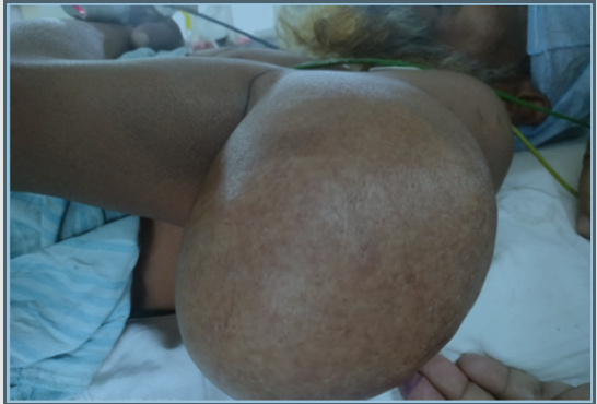
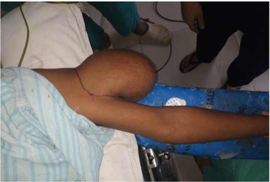
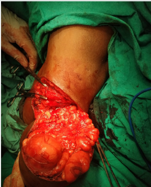
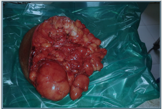
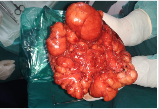
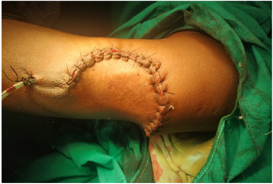
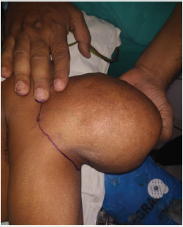
A 45-year-old gentleman presented with chief complaints of a large swelling over the left upper limb for 20 years. The swelling was initially small to begin with but increased gradually over the past 20 years to reach the present size. There were no associated complaints of weakness, numbness, difficulty in upper limb movements, loss of sensations or overlying skin colour changes. There was no history of any other swellings over the body. Patient was not having any associated comorbidities. On examination, a large pedunculated swelling of size 25x 15cm over left arm extending from proximal one third to distal third of arm. No tenderness or local rise in temperature was present. Swelling was mobile with no fixity to skin, muscle or bone. Ultrasonography of local swelling was suggestive of lipoma. Fnac was done which was suggestive of lipoma. Patient underwent an elective open excisional surgery for the swelling under general anaesthesia. Patient was extubated and tolerated the procedure well. Histopathology reports were confirmatory for lipoma. Patient was discharged uneventfully (Figures 1-7).

Figure 1: Lipoma over left upper limb.
Lupinepublishers-openaccess-anesthesia-pain-medicine-journal

Figure 2: Lateral view

Lupinepublishers-openaccess-anesthesia-pain-medicine-journal

Figure 3: Size and extent of lipoma.

Lupinepublishers-openaccess-anesthesia-pain-medicine-journal

Figure 4: Intraoperative finding.

Lupinepublishers-openaccess-anesthesia-pain-medicine-journal

Figure 5: Excised specimen.

Lupinepublishers-openaccess-anesthesia-pain-medicine-journal

Figure 6: Gross specimen.

Lupinepublishers-openaccess-anesthesia-pain-medicine-journal

Figure 7: Postoperative status.

Lupinepublishers-openaccess-anesthesia-pain-medicine-journal

Discussion

The standard management of giant lipomas is open excision [2]. A full histopathology report of the specimen is required to determine the possible need for any further treatment, as the main concern while evaluating a giant lipoma in the upper extremity is to rule out malignancy. Excisional surgery although being the standard treatment of giant lipomas also involves large incisions that may be associated with complication like infection, seroma or haematoma, as well as an unacceptable scar. Liposuction was employed for giant lipomas with added advantage of avoiding scar formation, but it may be complicated by skin puckering and dimpling as well as chances of recurrence [3]. It is suggested that if a fatty subcutaneous tumour is more than 10 cm in diameter and has shown a rapid growth in recent months, then the possibility of liposarcoma must be considered [4,5]. Clinically, the age of the patient and onset of symptoms must be thoroughly evaluated, and if possible, a magnetic resonance imaging or a tissue sample must be obtained in order to prove that the tumour is not malignant. Although if tumour is proved negative by these measures, it does not exclude the probability [6-8].

Conclusion

Giant lipomas are of size more than 5cm and warrant a workup for malignancy. They rarely occur in upper limbs, unlike in our case, making it a rare finding. All lipomas in the upper extremities measuring larger than 5 cm in a single dimension should be surgically removed due to malignant potential.

Read More about Lupine Publishers Journal of Anesthesia Please Click on below Link:
https://lupinepublishers-gynecology.blogspot.com/

Thursday, 3 June 2021

Lupine Publishers | Ganglion Impar Pulsed Radiofrequency for Intractable Coccydynia

Lupine Publishers| Journal of Anesthesia & Pain Medicine


Abstract

Patient with coccydynia are often not easy to manage, the aim of this single case study was to describe the outcome of ganglion impar pulsed radiofrequency (PRF) of patient with intractable coccydynia. A 31 years old presented with severe coccydynia, symptom provocation occurred during long sitting, pain reached high level (NRS: 8/10). MRI showed degenerative changes in the sacrococcygeal junction. A first session of impar ganglion PRF for 8 min was applied and the patient was improved 80% for few months (NRS: 2-3/10), although the results of this case report cannot be generalized, it describes the successful outcome of patient with severe coccydynia.

Keywords: Coccydynia; Impar ganglion; Pulsed radiofrequency

Introduction

Ganglion impar is the lowest paravertebral sympathetic chain, which is placed at the anterior aspect of sacrococcygeal disc used to be blocked in many chronic pain conditions originating from pelvic structures such as the coccyx. Coccydynia is one of chronic pain syndrome which is usually intractable to known therapeutic modalities such as medications, steroid injection and physical therapy manipulation, in addition, there are invasive approaches such as neurolysis with alcohol, phenol or ablative radiofrequency, these approaches are neurodestructive techniques. PRF is a neuromodulative application that has been used for years for chronic pain syndromes. We present a case of coccydynia successfully treated with PRF for neuromodulation of the ganglion impar.

Patient Description

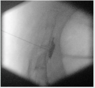
A 31-year-old man healthy, working as software developer, had pain over the coccyx started in 2015 without any history of trauma, increasing with time. The pain triggered with the prolonged sitting which made his work very difficult and start thinking to change his job, seen by many doctors, MRI of spine coccyx requested concluded degenerative changes in the sacrococcygeal junction Figure 1, he had been diagnosed as coccydynia and treated with conservative management including medications, coccyx steroid infiltrations and physical therapy manipulations , after the failure of conservative methods patient was referred to our pain clinic by his orthopedist, at this stage he became depressed because of functional limitation and job problems he described the pain as 8/10 point on verbal numerical rating scale (NRS). The clinical examination found the pain originated from coccyx. The ganglion impar block test was explained to the patient, done with lidocaine 1 % (5ml) which came positive, so patient planned for PRF of ganglion impar. He was admitted to the operating theater in prone position on the table, with a pillow under the anterior superior iliac spine to flatten the normal lumbar lordosis. First, intravenous access was inserted, and monitors were attached. The midline of the sacrococcygeal area was cleaned with antiseptic and sterile drapes were placed. Lidocaine 1% (5mL) was used for skin infiltration over the coccyx. A 22-gauge RF needle 10cm (Cosman electrode CU TM) with an active tip of 5mm was inserted through the sacrococcygeal disc to the anterior surface of the coccygeal disc, with the guidance of anteroposterior and lateral fluoroscopy view of the sacrum and coccyx. After negative aspiration, the fluoroscopic image was observed as 2mL of nonionic radiocontrast was injected to confirm the good placement of the needle Figure 2. Before applying PRF, sensory and motor tests were checked, which showed a sensory response at 0.35V (50Hz), a motor response below 2Hz. The treatment was maintained for 4min twice when the temperature at the needle tip did not exceed 42C. The total treatment time was 8min. He described the pain as zero on the NRS post procedure. After the procedure, the patient kept under close monitoring to check for any complications. Five months later, patient improved almost 80% and he was asked to describe the pain once more, and it was within two to three points NRS [1-4].

Figure 1: MRI of spine coccyx: degenerative changes of sacrococcygeal junction.

Lupinepublishers-openaccess-journals-anesthesia-pain-medicine-journal

Figure 2: Impar ganglion PRF: trans-sacrococcygeal needle placement with contrast injection.

Lupinepublishers-openaccess-journals-anesthesia-pain-medicine-journal

Comment and Discussion

Coccydynia is one of chronic pain syndrome difficult to manage, adversely affects quality of life, and the outcome of conservative methods is very poor. There are trials showed that PRF application is not only effective but also safe for nerve tissue because of its application of 42C. There are also other choices in the relief of coccydynia, such as ablative RF application and coccygectomy. These methods are more invasive and have higher risks of adverse effects compared with PRF application. Pulsed radiofrequency as new alternative practice in chronic pain is not widely used in the management of coccydynia and We believe that the transsacrococcygeal disc approach using PRF for ganglion impar block is not only simple but also effective for the treatment of coccydynia [5-8].

Read More About Lupine Publishers Journal of Anesthesia & Pain Medicine Please Click on Below Link:
https://lupine-publishers-anesthesia-pain.blogspot.com/