Showing posts with label Journal of Pain Medicine; GJAPM. Show all posts

Thursday, 15 December 2022

Lupine Publishers | Ventilation and Oxygenation Management in the Traumatic Brain Injury Setting

 Lupine Publishers | Journal of Anesthesia & Pain Medicine


Introduction

Early management of ventilation and oxygenation in the patients with traumatic brain injury, can be of importance to reduce secondary injury risks and improve outcomes. Increasing in the intracranial pressure of the patients with traumatic brain injury is an important risk which these patients are faced with. Hypocapnia due to hyperventilation and vasoconstriction in the brain’s blood vessels as the result, causes reduction in blood flow. Therefore, the risk of secondary injury due to hypoperfusion and ischemia would increase. It is recommended to keep PaCO2 levels in the range between 35 and 40 in these settings. Minimizing the bag-ventilating of the patients with traumatic brain injury who have been intubated, is an important note which should be kept in mind to reduce the risk of hyperventilation in such patients. These patients should be placed on the mechanical ventilator at the earliest possibility. 7 to 8 liters/min can be a reasonable starting minute ventilation in these settings, because of the possibility of presenting hypermetabolic state in the patients with traumatic brain injury [1-5].
Permissive hypercapnia is inappropriate in these settings since these patients are at risk for Acute Respiratory Distress Syndrome. In such settings Capnography and ABG should be considered to correlate End-tidal CO2 with the Partial pressure of carbon dioxide. Normoxia is important in such settings to prevent secondary injury and improve the outcomes. The ABG should be checked in a time period about 15 to 20 minutes after intubation. Based on oxygen-haemoglobin dissociation curve in different cases, FiO2, PaO2 and O2 saturation ranges should be defined. Hemodynamic of the patients with traumatic brain injury should be managed appropriately. Hypotension and hypertension in these patients should be corrected to avoid further complications. It is important for the neurointensive care professionals to have knowledge about appropriate management of ventilation and oxygenation in the patients with traumatic brain injury.

Read More About Lupine Publishers Journal of Anesthesia & Pain Medicine Please Click on Below Link:
https://lupine-publishers-anesthesia-pain.blogspot.com/

Friday, 27 May 2022

Lupine Publishers | Do You Really Want to Improve the Results of Treatment for Acute Pneumonia?

 Lupine Publishers | Journal of Anesthesia & Pain Medicine


Editorial

The question raised in the title of this letter is a natural consequence of the findings and conclusions that have been growing steadily in recent years regarding the results of treatment for acute pneumonia (АР). If you look at the publications of recent years in this field of medicine, it turns out that one of the main obstacles to progress in improving the results of treatment of this disease is the lack of appropriate methods for determining the pathogen. Thus, the lack of timely diagnostic information about the etiology of the disease excludes the possibility of targeted antibiotic therapy. In recent years, such regrets have become more and more relevant, playing the role of the main explanation for treatment failures .Continuing to narrow the unidirectional view of the problem and to pay attention only to the microbial factor as the main cause of the disease, such views are in fact another illusion, which, even in the case of its hypothetical implementation, will not make significant changes in the overall trend. This statement is easy to verify if you rely on well-known facts, and not use as arguments assumptions and guesses. First of all, it is necessary to remember the features of the initial period of clinical use of antibiotics. On the one hand, the first results showed their phenomenal effectiveness, and the greatest preference for many years was given to penicillin. On the other hand, bacteriological methods of verification of the pathogen at that time were significantly inferior in their speed and quality indicators to the methods of modern Microbiology. However, treatment of patients with AР for a long time was successfully carried out only by one penicillin and the question of the role of determining the causative agent of inflammation in those years did not have such a critical importance. I think it should be obvious to readers with medical education that such success in the use of antimicrobials in a dynamic and living biological environment could not continue indefinitely. The further course of events developed according to the so-called law of scissors, but the dynamics of these changes occurred very slowly, which made it difficult to monitor and timely assessment. On the one hand, each new generation of doctors was brought up in the spirit of priority and irreplaceability of antibiotics, gradually forming the image of a kind of panacea. One of the reflections of the new mentality was the wide spread of the principle “antibiotics alone” for the treatment of many diseases (including AP). Declaring this principle of treatment, no one was confused by the fact that the antibiotic in many patients is the only means of treatment and that the same drug is used in radically different processes. And these are indirect signs of the attitude to antibiotics as a panacea. As you know, the basic principle of antibiotics is only intended to suppress the pathogen, is not it? Other medicinal properties of these drugs do not have. All other changes that have already occurred in the focus of inflammation, and in the body in General, cannot be eliminated with the help of antibiotic therapy, no matter how powerful and long it was not. The latter task falls on individual protection and adaptation mechanisms and depends on the direction of additional assistance methods. The gradual narrowing of views on the nature of AР to the direct dependence of all sides of the disease on the type of the pathogen was accompanied by the formation of relevant normative documents recommending and prescribing the implementation of specific actions and prescriptions. Today, a specialist working on the basis of a license cannot go beyond these requirements, even if he had doubts about their effectiveness. In parallel with the deformation of the image of AР as an inflammatory process of nonspecific etiology in the lungs, there is another important process, the results of which are increasingly recorded in recent years. The symbionts of our body are also biological objects. Exposed to mortal danger, they have learned not only to survive, but also to create strains resistant to such aggression. To date, the group of resistant strains to antibiotics is already quite impressive, and its representatives are increasingly identified among the symbionts of healthy people [1]. In itself, the presence of such microflora in the body does not necessarily mean the fact of the disease, but in the case of the beginning of the last, conventional antibiotic therapy can meet with serious difficulties. In fact, we are talking about microbes that have always been found in the composition of human symbionts and that have only gained additional stability, not aggressiveness. Hardly anyone doubts the fact, that the emergence of such strains is the result of long-term and widespread use of antibiotics. In addition, the frequency of detection of such strains in a healthy population will increase as a result of continued antimicrobial therapy. At the same time, it is even theoretically impossible to stop this already started process, since such an initiative can lead to disastrous results. Therefore, it is necessary to realistically assess the current situation in bacteriology, which is not the worst option, and consider the gradual increase in the number of carriers of such strains among the healthy population as a future inevitable reality. It is known that the uncertainty of what is happening has always been one of the main prerequisites for the emergence of fears. In this regard, it is especially important to note that the author’s point of view is presented to the reader not for emotional impact, but only to develop their own rational assessments of already known facts. If for many years, the AР microbe is considered to be the main cause of the disease and the entire policy in the treatment of these patients was aimed only at suppressing this factor, the decrease in the effectiveness of treatment and the increase in the resistance of the microflora led scientists into complete confusion. In this case, modern ideas about the nature of the disease can indicate only one way out of this impasse - to speed up the process of determining the pathogen. However, the accumulated evidence shows the futility of trying to realize these hopes. Although the participation of various microorganisms in the inflammatory process of lung tissue differs significantly in its frequency, and the leaders of this list change periodically, it should be recalled that to date more than 100 microbes have already been established that can act as pathogens of AР [2]. In addition to these data, the literature on this problem always notes the fact that AР pathogens are not only bacteria, but also viruses, fungi, parasites. In this regard, it is especially advisable to pay attention to acute inflammation in the lungs, which is a consequence of a viral infection. According to the statistics of this disease, at least a third of patients in the world have the viral nature of AР [3,4]. These indicators cannot be ignored, as the continuation of the therapeutic principle of “antibiotics alone” in the AP reveals the following fact. All patients with viral etiology of the disease do not have targeted treatment, and antibiotic therapy is only an attempt to prevent the possible addition of a bacterial component. Therefore, successful treatment in such observations should be considered more as a merit of the patient’s body than as a triumph of medical efforts. At this stage, the etiology of AР is widely interpreted on the basis of the results of bacteriological studies of the microflora of the nasopharynx and oropharynx [5-7]. However, such studies require clarification of the breadth of their application, and the evaluation of their results raises logical objections. For example, it is known that bacteriological examination is usually carried out mainly in hospitalized patients. The etiology of AР in most patients undergoing outpatient treatment remains unknown. But, even among hospitalized patients, the duty of such studies refers to cases of severe forms of the disease, and positive results (including cultures of blood) are found in less than half of the cases [4,8]. However, even the interpretation of the positive results of the examination does not explain such a paradox as the presence of less virulent microflora in a number of AP observations compared with bacteriological examination of the upper respiratory tract in healthy people, when the simple presence of aggressive strains without any signs of the disease is revealed. Serological diagnosis is also not the result of studying the material directly from the area of inflammation. Allowing to identify trace reactions, this type of examination refers to indirect signs of contact of the body with a certain pathogen but is not an absolute proof of its presence at the moment directly in the inflamed organ. If we continue to discuss the role of the material in the reliability of the results, the most accurate method of determining the agent of AP, of course, is to study the content of the affected area. The possibility of practical implementation of such a study appears only in a limited group of patients with purulent pleural complications that develop at a certain stage of the process and previous treatment. However, even the results of this relatively small group of patients show that in 30 and more % of cases microflora was not detected [4,9] and there is no reasoned explanation for this fact. As a result, several hundred million AР cases are diagnosed each year in the world, but their etiology remains an open question, and the choice of antimicrobials has been and is being conducted empirically [4,8-10].

Despite the intuitive choice of antimicrobials, their use remains a very important support for the body. For most patients with AР, the suppression of an unexpected mutiny of the accompanying microflora is enough for the body to cope with inflammation in the future. However, in the most severe situations with the rapid and aggressive development of the inflammatory process, another aspect of the problem is revealed, which currently continues to be explained by the extremely high virulence of the pathogen. For example, among patients hospitalized with severe AP, mortality reaches 13-28%. Among the total number of hospitalizations with AP, septic shock is diagnosed in 30% of cases, and mortality in this group increases to 50% [4,11-13]. The statistics of these results seem, at first glance, quite understandable and logical: the more severe and aggressive the disease develops, the more difficult it is to eliminate the catastrophic violations that have arisen. However, the logic of such arguments collapses completely, if we take into account some very important, in my opinion, facts. First, if we consider shock as a septic complication of aggressive forms of AР, it is necessary to have conclusive evidence of this option, the cause of which is the penetration of pathogens into the bloodstream. However, the detection of bacteria in the blood is recorded only in 10-12% of all hospitalizations with AР without connection with the shock pattern [8,9]. That is, the latest figures reflect the frequency of diagnosis of bacteremia, which is not always accompanied by a clinic of shock. Secondly, interpretations of shock in AP as its septic variant consider this complication only from the point of view of the microflora and leave without attention such an important feature of inflammation as an individual reaction of the body to the process. It is the juxtaposition of these two causes (microorganism against microorganism) that creates the uniqueness of the manifestations of the inflammatory process even in the presence of the same pathogen. However, despite this long-known fact, the severity of AР in recent years is increasingly attributed solely to the properties of microflora. Finally, the main feature of the nature of the shock in the clinic, the AP determines, from my point of view, the undeniable fact that this intense process is the only inflammatory disease of nonspecific etiology of all known, which occurs in the vascular system of the pulmonary circulation. Rapid change of stages of the process makes it difficult to adapt the body to new conditions. In the most aggressive cases, the body gives us a clear signal of its catastrophic situation. Demonstration of shock in AP is a reflection of the body’s efforts to facilitate the work of pulmonary vessels, which could not provide the necessary blood flow and synchronous operation with a large circle of blood circulation [14]. The causes and pathogenesis of this form of shock differ significantly from other variants and require a different treatment solution. So, this form of shock has been highlighted by us in a separate section as pulmonal shock. Understanding the pathophysiology of current disorders involves eliminating the reflex effect of the source of inflammation in the lungs on pulmonary blood flow [15-18]. Instead, shock in AР is compared with its manifestations of another origin, and such patients begin to receive standard therapy in the form of intravenous injections with the addition of hormones and vasopressors [4,8,9,11,13,19-22]. Intravenous fluids increase blood flow to the pulmonary vessels and further disrupts blood circulation in the small circle, stimulating further hemodynamic changes. In parallel, the processes of exudation in the area of inflammation are stimulated. In response to such aggression, the body of many patients stops responding contrary to expectations and then begins the use of vasopressors and hormones. The result of such therapeutic efforts in pulmonal shock in patients with AР is the loss of many patients, and the frequency of pleural complications among hospitalized patients reaches 60% or more [4]. The above brief considerations, together with some known facts and figures, allow us to present only the contours of the modern problem of AР treatment and draw the attention of readers to another direction of its solution. It is important to note another fact that a non-standard approach to solving this problem has already been tested in the clinic and its results allowed us to note that early pathogenetically justified assistance with AР guarantees the prevention of a complicated course of the disease [23,24]. Therefore, the question that was raised in the title of this letter has a real basis. In order to answer this question in the affirmative, it is necessary first of all to radically reconsider the understanding of the nature of the disease and the mechanisms of its development. Without this step it is impossible to move on. Do you really want to improve the results of treatment for acute pneumonia? So, go ahead and improve!

Read More About Lupine Publishers Journal of Anesthesia & Pain Medicine Please Click on Below Link:
https://lupine-publishers-anesthesia-pain.blogspot.com/

Friday, 6 August 2021

Lupine Publishers | Is There A Neck-Shoulder Syndrome?

 Lupine Publishers | Journal of Anesthesia & Pain Medicine


Abstract

Concomitant presentation of neck and shoulder pain is a common clinical scenario which can present a significant diagnostic and therapeutic dilemma. Neck and shoulder pain presentations can be separated into four different categories: Primary neck pathology with referred pain to the shoulder, primary shoulder pathology with referred pain to the neck, primary neck and primary shoulder pathology, and primary neck pathology resulting in secondary shoulder pathology. Primary neck pathology resulting in secondary shoulder pathology is mechanically plausible but not proven. Authors are proposing this scenario to be described as “neck-shoulder syndrome.” For instance, C5 and/or C6 cervical radiculopathy can result in rotator cuff, deltoid, biceps and scapular muscle weakness as these nerve roots innervate the shoulder girdle musculature which in turn could produce shoulder/scapular muscle imbalance resulting in shoulder impingement signs. A patient may present with features of both cervical radiculopathy and shoulder impingement syndrome in this scenario. At this time there are no agreed clinical criteria for a diagnosis of “neck-shoulder syndrome.” As with any other syndrome, management differences can only be well studied once the entity has been properly defined. In this article, authors set out to summarize how to best approach patients presenting with both neck and shoulder pain while describing features of proposed “neck-shoulder syndrome.” It is paramount to take a comprehensive and holistic approach towards patients presenting with concomitant neck and shoulder pain as the symptoms may not always represent isolated entities.

Keywords: Neck and shoulder pain; Neck-shoulder syndrome; Pain treatment; Differential diagnosis of neck and shoulder pain

Introduction

Co-existent neck and shoulder pain has been described in limited fashion in the literature as a unique diagnosis, but the concomitant presentation of neck and shoulder pain is a common scenario in primary care and orthopedic offices [1,2]. Gorski et al described “shoulder impingement syndrome” where patients presented with neck pain secondary to rotator cuff tendinopathy [1]. Compere et al described a “neck, shoulder, and arm syndrome” which primarily referred to neuropathic pain in the neck, shoulder and arm resulting from a brachial plexus lesion [2]. When patients present with both neck and shoulder pain, it can present a significant diagnostic dilemma[3]. “Hip spine syndrome” has recently been described, and “neck-shoulder syndrome” likely represents an analogous entity involving the cervical spine and upper limb [4]. It is estimated that among primary care office visits, neck pain accounts for approximately 20-30% and shoulder pain for 10-20% of musculoskeletal complaints. From this population, combined neck and shoulder problems account for approximately 6-10% [3,5,6,7].

Discussion

Concomitant neck and shoulder pain presentations can be separated into four different categories: Primary neck pathology with referred pain to the shoulder, primary shoulder pathology with referred pain to the neck, primary neck and primary shoulder pathology, and primary neck pathology resulting in secondary shoulder pathology

Primary neck pathology with referred pain to the shoulder

An isolated C5 and/or C6 radiculopathy without shoulder pathology could certainly present with neck and shoulder pain due to C5 and C6 dermatomal symptoms corresponding to the shoulder region. C5 or C6 myotomal pain can cause pain in the deltoid, scapula and biceps, and can mimic shoulder pathology [8,9]. This scenario is typically straightforward as the physical examination will be absent of shoulder impingement signs. Classically, cervical radiculopathy examination can demonstrate positive cervical root impingement signs (Spurling’s maneuver), myotomal weakness, dermatomal sensory abnormalities and blunted reflexes in a specific root distribution. Several neuropathies involving brachial plexus and its proximal branches will also refer pain to neck and shoulder simultaneously.

Primary shoulder pathology with referred pain to the neck

Primary shoulder pathology should not directly lead to neck pathology, and such cases are not well described in the literature. Nevertheless, patients with shoulder pathology may develop pain and tightness in the trapezius muscle on the ipsilateral side and referred pain in the cervical area. Restricted motion at the glenohumeral joint may also lead to overuse and pain in the scapulothoracic musculature. A general concern in musculoskeletal medicine is that symptomatic pathology in a joint may refer pain to a joint below and/or above.

Primary neck and primary shoulder pathology

Degenerative arthritis can affect multiple joints. Thus, many patients may have both glenohumeral arthritis and cervical spondylosis. The radiographic incidence of glenohumeral arthritis is reported as 32.8% in people over 60 years of age [10]. Radiographic evidence of cervical spondylosis is present in 50% of people over 50 years of age and 75% of individuals over 65 years of age [11]. As both conditions are common, both can present as “pain generators.”

Primary neck pathology resulting in secondary shoulder pathology

Primary neck pathology resulting in secondary shoulder pathology is mechanically plausible although not proven. For instance, C5 and/or C6 cervical radiculopathy can result in rotator cuff, deltoid, biceps and scapular muscle weakness as these nerve roots innervate the shoulder girdle musculature. This could produce muscle imbalance and poor shoulder/scapular mechanics. A patient may present with features of both cervical radiculopathy and shoulder impingement syndrome in this scenario. In clinical practice, it is not uncommon to see a patient with chronic neck pain presenting with insidious onset of shoulder pain later in the course. Authors are proposing this unique presentation be referred to as “neck-shoulder syndrome.” Although most clinicians would treat this as separate neck and shoulder pain, they may be related diagnoses.

Literature Search

We conducted a comprehensive search in the PubMed database in order to identify relevant studies on “neck-shoulder syndrome.” Based on the review of the available literature, there are no agreed upon clinical criteria for a diagnosis of “neck-shoulder syndrome” despite its common clinical presentation nor is there a well described “neck-shoulder syndrome.” As with any other syndrome, management differences cannot actually be studied until the entity has been appropriately defined. This article will concentrate on how to best approach patients presenting with both neck and shoulder pain while describing features of proposed “neckshoulder syndrome.”

Presentation

In patients presenting with neck and shoulder pain, a thorough history is paramount in identifying the etiology of the patient’s pain.

Location: Patients with primary neck pathology can experience pain extending beyond the neck based on the etiology. Disorders that affect the lower cervical nerve roots will often result in pain distal to the shoulder which can be characterized by radiation into the arm in a clear dermatomal or myotomal distribution [12]. In addition, Dwyer et al described reproducible pain patterns that can refer into the shoulder, trapezius and occiput from cervical zygapophyseal joint pathology [13-15]. Pain from a primary shoulder problem can also refer pain to the neck, periscapular region and distally into the arm although not typically extending below the elbow [16]. Associated paresthesias are not classically associated with a primary shoulder problem.

Onset: Onset of symptoms is also a key component of the history. Degenerative cervical pathology can have insidious onset although acute disc herniations can have a sudden onset that may be precipitated by trauma. Whiplash injuries are known to precipitate neck pain which can be of myofascial and/or cervical facet in origin. Shoulder disorders can also be of insidious (overuse injuries) or acute onset (trauma). Sudden onset of shoulder pain with restricted motion can be associated with acute calcific tendinitis or adhesive capsulitis. Neuralgic amyotrophy (Parsonage Turner syndrome/ brachial neuritis) has a unique presentation where patients usually experience severe, acute pain following exercise, recent illness, immunization, surgery or trauma [17]. As the initial severe pain starts to resolve, neurological deficits will become apparent, which is in contrast to most presentations of cervical radiculopathy where pain will continue with associated neurological symptoms. Onset of symptoms plays a key role in proper identification of proposed “neck-shoulder syndrome.” Development of shoulder pain (especially in the absence of injury) after onset of neck/radicular pain can be considered primary neck pathology with secondary shoulder pathology and can be referred to as “neck-shoulder syndrome.” Shoulder pain in this scenario is likely secondary to rotator cuff and periscapular muscle weakness/imbalance caused by C5 and/or C6 cervical radiculopathy. This clinical scenario is not well studied in the literature, hence prevalence and incidence is not known. Among patients with cervical radiculopathy, studies report a frequency of C5 nerve root involvement at 5-10%, C6 at 20-25%, and C7 at 45-60% [12,18].

Exacerbating factors: Pain with overhead arm movements generally suggests primary shoulder pathology. However same pattern can also be present in peripheral neuropathies like thoracic outlet syndrome and spinal accessory, suprascapular, or axillary neuropathy. Shoulder pain with side lying on the affected upper limb tends to be associated with shoulder impingement and acromioclavicular joint arthropathy.

Features of systemic diseases: In patients who present with neck and shoulder pain in the absence of trauma, the history will need to include an assessment for widespread involvement that may suggest systemic disease processes like fibromyalgia, polymyalgia rheumatica, myofascial pain syndrome and myopathy.

Red flags: The history should also include an evaluation for findings to suggest a disease process that requires more urgent evaluation. Red-flag symptoms to assess for include gait imbalance, hand clumsiness, bowel/bladder dysfunction (cervical myelopathy), pain after high impact trauma (fractures), unintentional weight loss (Pancoast tumor), chest pain (cardiac ischemia), blurry vision, nausea/vomiting and vertigo (vertebral artery dissection/ insufficiency).

Physical Examination

In addition to a detailed history, a thorough physical examination is key for proper diagnosis and identification of the pain generator(s). A thorough neurological exam plays an essential role in distinguishing neck from shoulder pathology. Sensory, motor and reflex changes in a specific nerve root distribution are characteristic of cervical radiculopathy. C5 and C6 cervical radiculopathies may result in periscapular and shoulder/rotator cuff muscle weakness while C7 radiculopathy is unlikely to cause shoulder weakness. Rotator cuff pathology may result in shoulder weakness with preserved elbow flexion while a C5 radiculopathy can result in weakness of both. Testing deltoid strength with the arms at the sides instead of in shoulder abduction can aid in differentiating pain inhibition versus true weakness.

Provocative Testing

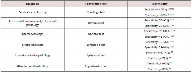
Provocative testing can assist in the diagnosis of cervical and shoulder disorders [9, 19]. Among them, provocative tests for shoulder impingement may help distinguish primary versus secondary shoulder pathology in proposed “neck-shoulder syndrome”(Table 1) [18-30].

In patients with neck and shoulder pain, one test by itself may not have enough sensitivity and specificity to make a diagnosis and most physical exam maneuvers are not pathognomonic. A combination of multiple exam components and a thorough history are necessary to accurately identify the etiology of symptoms.

Table 1: Provocative tests for common cervical and shoulder problems and reported validity.
Lupinepublishers-openaccess-journals-anesthesia-pain-medicine-journal

Diagnostic Testing

When presented with both neck and shoulder pain, history and physical exam should dictate appropriate use of diagnostic tests. Although imaging studies such as radiography, CT and MRI can reveal many pathologies, further testing should be done to identify the true pain generator. When suspecting pain mediated by a cervical zygapophyseal joint, cervical diagnostic medial branch blocks can be considered. A shoulder injection of lidocaine with or without corticosteroid can be done for diagnostic and perhaps therapeutic reasons. In cases of secondary shoulder pathology, this may give the patient partial benefit, but a primary cervical pathology should still be investigated [9]. Electrodiagnostic testing with electromyography (EMG) and nerve conduction studies (NCS) can be employed when suspecting myopathy, brachial plexopathy, peripheral neuropathy or radiculopathy. When evaluating neck and shoulder pain, scientific evidence suggests using a combination of history, physical examination, imaging modalities, diagnostic injections and electrodiagnostic study to make the appropriate diagnosis.

Treatment

Success of any proposed treatment algorithms will depend on an accurate diagnosis. There is scant evidence on how to approach the concomitant presentation of neck and shoulder pain. Treatment should be geared toward the primary site of pathology whether it be the cervical spine or the shoulder [3]. Lack of current literature evidence underscores the importance of describing a “neckshoulder syndrome,” as it can lead to studies looking at management differences. It can be hypothesized that in patients with cervical radiculopathy with secondary rotator cuff impingement, treatment of the primary lesion will likely yield eventual improvement at the secondary site although no studies have been done on this topic.

Conclusion

The concomitant presentation of shoulder and neck pain is a common scenario in primary care and orthopedic offices and can present a diagnostic and therapeutic dilemma. A careful history and thorough physical examination along with ancillary studies can often yield the correct diagnosis and successful treatment. Primary neck pathology resulting in secondary shoulder pathology is mechanically plausible but not proven. Authors are proposing this scenario to be described as “neck-shoulder syndrome.” Appropriately describing a “neck-shoulder syndrome” can lead to further studies looking at management differences. A prospective study looking at incidence of shoulder impingement signs in chronic C5 and/or C6 radiculopathy patients could be helpful in delineating diagnostic criteria for “neck-shoulder syndrome.” Above all, it is paramount to take a comprehensive and holistic approach towards patients presenting with concomitant neck and shoulder pain as the symptoms may not always represent isolated entities.

Acknowledgements

The authors would like to thank Dr. David Janerich for his help with the development of the article.

Read More About Lupine Publishers Journal of Anesthesia Please Click on Below Link:
https://lupine-publishers-anesthesia-pain.blogspot.com/