Saturday, 5 August 2023

Lupine Publishers | Swathed Ureter, an Enigma in Diagnosis- A Pictorial Essay

 Lupine Publishers | Journal of Urology & Nephrology


Abstract

This pictorial essay is an educational article aiming to provide both textual and visual portrayals consisting of a collection of images and texts on an important issue by reviewing and extrinsic encasing pathology of the ureter to provide a guide to those who are involved in diagnostic intervention. While considering diseases of the urinary system, physicians mainly focus on the kidneys and the bladder. Only scant attention is paid to the ureters. Most of the UTO due to calculi are readily identifiable whereas many cases of ureteric exterior encasement are frequently missed from early detection even by experienced clinicians and radiologists. Failure in recognition of the encasement of ureters and its causes may lead to mistaken diagnosis with resultant inappropriate management. However, problems with the ureters can adversely affect the functioning of the kidneys and could even be lethal. In this article we focus only ‘encasement of ureters’ with a few common examples and salient signs that help in the diagnosis.

Keywords: Encasement of Ureters; Endometriosis; MRI; RPF; Obstructive Uropathy

Background

Urinary tract Obstruction (UTO) an alarming but common clinical condition affecting more women than men at any age, though common in 20 to 60 years of age having an overall incidence of hydronephrosis in 3.1% of autopsy. Over time, UTO results in irreversible loss of numerous nephrons leading to obstructive nephropathy and end-stage renal failure. If the obstruction of the ureter is partial and brief and if intervention is done at correct time, after relief of obstruction. complete recovery of renal function is possible. One has to be aware that UTO lasting more than 24 hours may cause irreversible loss of renal function. Radiology investigations may show UTO without ureteric dilation and dilation of ureter without UTO creating potential pitfall in radiologic diagnosis of UTO. Most of the UTO due to calculi are readily identifiable whereas many cases of ureteric exterior encasement are frequently missed from early detection even by experienced clinicians and radiologists. Failure in recognition of the encasement of ureters and causes may lead to mistaken diagnosis with resultant inappropriate management [1]. The most common benign cause of encasement of ureters is retroperitoneal fibrosis and the most frequent malignant causes are extension from an adjacent primary tumour such as sarcoma, lymphoma, (E.g.: sarcomas and lymphomas of uterus, ovaries, urinary bladder and prostate). Among benign conditions of swathed ureters, Extrinsic benign tumours, Retroperitoneal lymphadenopathy, Retroperitoneal abscess, Retroperitoneal fibrosis, Inflammatory abdominal aortic aneurysm or iliac artery aneurysm, and Endometriosis are significant. Chronic fibrosing conditions of the abdomen may involve multiple systems by their proliferative deep fibromatoses which form pseudotumor which cannot be differentiated from neoplastic conditions at imaging. Peri-ureteral inflammation (E.g.: peritonitis, salpingitis, and diverticulitis), multifocal idiopathic fibrosclerosis and schistosomiasis are some other known causes. Encasement may also be associated with blocked ureter [2-4]. Besides the inherent features of the disease causing the encasement of the ureters, in general, clinical features include recurrent fever, pain abdomen, oliguria, frequency, dysuria, haematuria nocturia, and hypertension. Patients may also present with fatigue, anorexia, weight loss, fever, hydroceles, scrotal pain, lower extremity oedema, and pulmonary embolism. Since ureters may be affected, various degrees of ureteral obstruction, hydronephrosis, and renal failure are also considered early and common clinical manifestations. In this article, we will review the four important diseases that cause ureteric encasement with some key imaging features for the diagnosis [5,6].

Imaging

On imaging ureteral pathology, plain abdominal radiography does not play a major role in imaging. Intravenous excretory urography (EUG or IVU) was once the study of choice for evaluating the renal collecting system and ureters; although now replaced by Computed Tomography urography (CTU), IVU is still performed in some centres. IVU has limited utility in patients with impaired renal function. In patients with renal insufficiency the risk for contrast-induced nephropathy from iodinated contrast media is high [7-10]. Systemic IV contrast should be avoided, and direct injection of a contrast medium can be performed with antegrade or retrograde pyelography. This allows evaluation of the collecting system and ureters and the opportunity for interventions such as stent placement. In cases where a detectable mass is not present, computed tomography and ultrasound, while helpful, are probably less sensitive and less specific than the retrograde ureterogram. Ultrasonography is not at all used to evaluate mid ureteral stricture or encasement; but is useful to diagnose if ureters are well distended in urinary obstruction due to a retroperitoneal mass and retroperitoneal fibrosis. However, overlying bowel often obscures visualization of the mid-ureter.

CT scan is the first choice to demonstrate ureters CT urography (CTU) is the major imaging modality for evaluating the ureters and secondary findings that help to narrow the differential diagnosis of the cause of the ureteral stricture and allows visualization of adjacent structures to differentiate an extrinsic pathology from an intrinsic process. For CTU, a three-scan CT protocol is used. After voiding completely all patients are asked to drink 800-1000 mL of water immediately prior to the examination [11]. The urinary system is imaged in both unenhanced and contrast enhanced CT (CECT) scan, by using multidetector helical CT scanner. On CECT, 100 mL of Non-ionic Low-osmolar contrast media (LOCM) is injected(2.5ml/sec); the kidneys are scanned 25-80 seconds after intravenous administration of the contrast for cortico-medullary phase, after 100 seconds for nephrographic phase, 8-10 minutes after contrasting medium injection for pyelographic phase, by using a maximum collimation of 1.0 mm. All three scans are performed with a tube voltage of 120 kVp and a tube current of 200 mAs.

Magnetic resonance urography (MRU) is very useful in patients with renal failure and or with Iodine contrast allergy. Absence of ionizing radiation, evaluation of paediatric patients and pregnant women, the high T2 signal intensity of urine in the dilated collecting system, are the advantages. Disadvantages of MRU are higher cost, motion artifacts from respiration and ureteral peristalsis, small field of view because of coil size nephrogenic systemic fibrosis secondary to gadolinium and bowel artifacts, However, when compared with CT, MRI offers better contrast resolution, but CT has higher spatial resolution [12-14]. Among Nuclear Medicine imaging in the setting of ureteral stricture and obstruction, diuretic renograms can be used to differentiate collecting system dilation from urinary obstruction. Radionuclide studies are not used for the detection of encasement of the ureters although positron emission tomography (PET) scan may show increased uptake. The role of PET in evaluating urothelial lesions is limited. Occasionally, ureteral obstruction may be identified in patients undergoing PET for other processes [15,16]. In general, on imaging, fibrosis displays hypoechogenic with acoustic shadowing at ultrasonography (US), hypovascularity at Doppler imaging, isoattenuating to muscle at computed tomography (CT), and isointensity at T1W1 and markedly low signal intensity at T2-weighted imaging. Due to reduced cellularity, the fibrous tissue typically does not show significant restriction at diffusion-weighted imaging. Contrastenhanced CT and MR will not be informative although delayed phase enhancement may be present. Other diseases mimicking Chronic fibrosing conditions such as mesenteric panniculitis, retractile mesenteritis, mesenteric carcinoid, Crohn disease with fibrofatty proliferation, and desmoid (mesenteric fibromatosis), carcinoid and carcinomatosis, mesenteric lipogenic liposarcoma will be the primary differential diagnoses [17].

IgG4-related sclerosing disorders

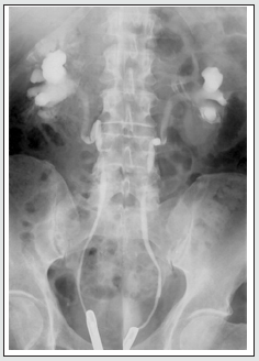
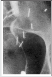
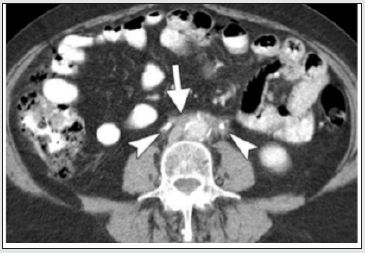
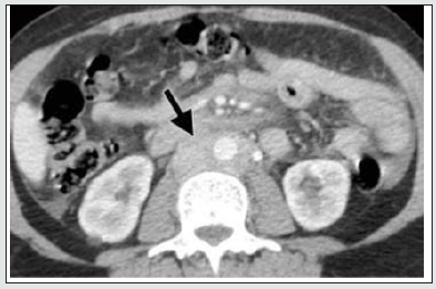
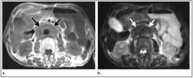
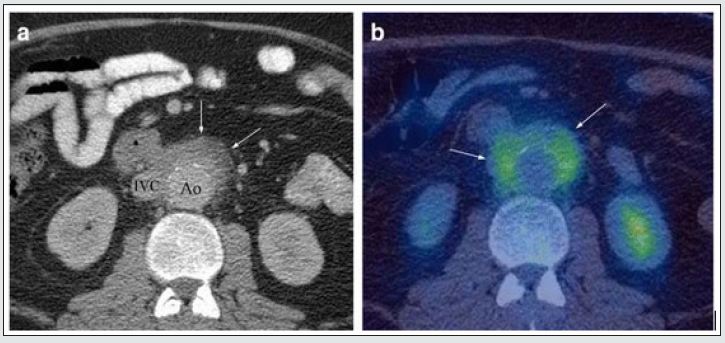
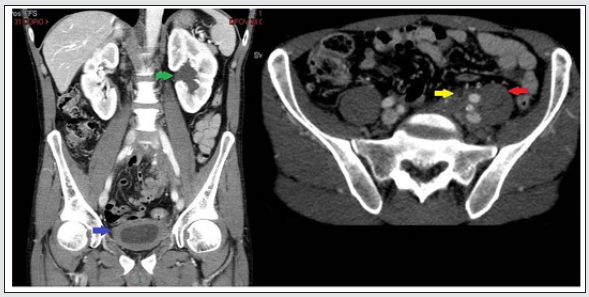
Retroperitoneal Fibrosis (RPF) is now considered an important abdominal component of the group of IgG4-related sclerosing disorders. It is an aggressive, rare fibro- proliferative process causing deposition of unencapsulated fibrous tissue masses in the retroperitoneal space usually centered at the lumbosacral junction. it is typically seen in men (2-3 times commoner than in women) between the 5th and 7th decades of life. Up to 15% of RPF patients have additional fibrotic processes in the mediastinum, thyroid (Riedel thyroiditis), biliary tree. sclerosing mesentery, and orbital tissue(pseudotumor). It leads to progressive encasement of the retroperitoneal structures, especially the ureters. and may cause hydronephrosis secondary to the extrinsic compression of the ureters [18]. RPF plaque may involve both ureters and may draw them medially towards the spine. This appearance on retrograde pyelogram resembles a narrow-waisted maiden (Figure 1a). Encasement of the ureter cause abrupt tapered narrowing of the ureteral lumen The dilated proximal ureter (the bullet) and the nondilated, encased distal ureter may appear as ‘The bullet and bodkin’ and this appearance can be caused by malignancy such as lymphoma, or rarely by RPF (Figure 1b). However, on the above imaging pictures, the ureteric encasement is not visually demonstrable. On CT, the area of fibrosis and encasement appears as a soft-tissue mass with variable contrast enhancement. The active and inactive stages of the disease can be differentiated on CT and MRI. In the retroperitoneum, RPF tissue surrounds the aorta (arrow) sparing posterior aspect (Figure 2a) indicating a benign stage. In contrast to the figure above, in malignant RPF, the RPF has mass effect with adjacent structures (Figure 2b below). Another noteworthy finding is the intensity difference of RPF mass on MRI imaging (Figure 3). A high intensity signal of the mass on T2WI is suggestive of early active RPF whereas a low intensity signal on T2WI is highly suggestive of benign RPF in a late inactive stage. On FDG PET/CT imaging, the fibrous tissue in the affected areas exhibit avid FDG uptake (Figure 4b). When the infiltrating tissue encompass both ureters and compromise the lumen, bilateral hydronephrosis will ensue regardless of the location of the mass in the abdomen or pelvis (Figure 5a-b). Abdominal pain and obstructive uropathy should, therefore, raise suspicion for retroperitoneal fibrosis and imaging should be obtained. On the other hand, new onset of hypertension, flank pain radiating to groin in a known case of RPF or malignancy should arise the suspicion of ureteral encasement and appropriate imaging should confirm [19]. Failure in recognition of the encasement of ureters and causes may lead to mistaken diagnosis with resultant inappropriate management.

Figure 1: Retrograde ureteropyelogram 1a. Maiden waist deformity.
lupinepublishers-openaccess-journal-urology-nephrology

Figure 1b: Bullet and Bodkin Sign (Courtesy: Classic Signs in Uroradiology; Raymond B. Dyer, Michael, Ronald)
lupinepublishers-openaccess-journal-urology-nephrology

Figure 2a: Plaque-like Soft tissue surrounding the aorta (arrow) sparing posterior aspect without elevating the aorta from the spine; encases both ureters (arrow heads) findings indicative of benign RPF. (courtesy: Ali Jiwani https://www.slideshare.net/AliJiwani/retroperitoneal-fibrosis- radiology)
lupinepublishers-openaccess-journal-urology-nephrology

Figure 2b: Axial CECT demonstrates well defined mass surrounding Aorta and IVC pushing the aorta anterior to the spine, a finding suggestive of malignant RPF. (courtesy: Ali Jiwani https://www.slideshare.net/AliJiwani/retroperitoneal-fibrosis-radiology)
lupinepublishers-openaccess-journal-urology-nephrology

Figure 3a: Histologically confirmed idiopathic RPF mass surrounding the abdominal aorta and IVC, showing medium intensity on T1WI (3a); and low signal intensity on T2WI (3b) a finding suggestive of late stage. (courtesy: Ali Jiwani https://www. slideshare.net/AliJiwani/retroperitoneal-fibrosis- radiology)
lupinepublishers-openaccess-journal-urology-nephrology

Figure 4: Example of IgG4 retroperitoneal fibrosis. 4a Axial CECT showing enhancing soft tissue deposit partly encasing the IVC and atherosclerotic aorta (arrows). b Axial FDG PET/CT showing increased FDG uptake in the abnormal retroperitoneal soft tissue (arrows). (Courtesy: Christopher Siew Wai Tanghttps://insightsimaging.springeropen.com/articles/10.1007/ s13244- 018-0618-1).
lupinepublishers-openaccess-journal-urology-nephrology

Figure 5a: Axial and 5b: coronal computerized tomography images demonstrating left-sided pelvic mass causing hydroureteronephrosis (green arrow) with diffuse bladder wall thickening (blue arrow). Asymmetric pelvic mass extending to posterior presacral region and psoas (red arrow), with ureter encased by pelvic mass (yellow arrow). Courtesy: R Bechtold, M I Shaff; PMID: 6879267; DOI: 10.1097/00007611-198308000-00023.
lupinepublishers-openaccess-journal-urology-nephrology

Perianeurysmal Fibrosis

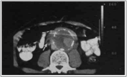
Reports of 56 previous cases showed striking male predominance. An abdominal mass may be palpable in 45% of cases. Fibrosis is often seen in association with atherosclerosis of the aorta (Figure 6), possibly due to fibrosis as an immune response to the leakage of ‘ceroid’ from the atheromatous plaque into the perivascular tissues. When perianeurysmal fibrosis occurs in association with an abdominal aortic aneurysm it may encase and produce ureteric obstruction and renal function impairment. The computerized tomography scan is able to provide accurate details of aortic aneurysm, intraluminal thrombosis, calcification, periaortic inflammation, entrapped ureters and hydronephrosis (Figure 6).

Figure 6: At L3/4 DISC SPACE level – Aorta enlarged with calcifications in the wall. The left ureter (black arrow) is encased by the periaortic aneurysmal fibrosis and the right ureter (white arrow) lies adjacent to fibrosis (partly encased). Both ureters are small, the possibility of Ureteric obstruction secondary to perianeurysmal fibrosis should be in the pre-operative evaluation of abdominal aortic aneurysms. (Courtesy: https://www.researchgate.net/ R N Gibson Alison Halliday A Mansfield).
lupinepublishers-openaccess-journal-urology-nephrology

Neoplasm

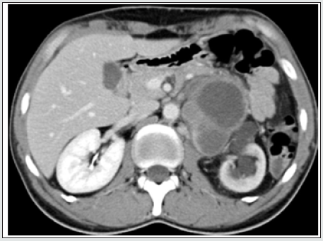
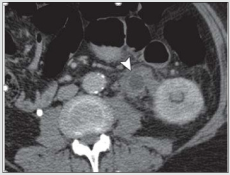
Locally invasive neoplasm in the retroperitoneum and pelvis can involve ureters by three ways: 1. by malignant infiltration of their walls causing narrowing of their lumen (stricture) and affecting the peristalsis; 2. by external compression by the mass itself that may encroach, encompass and compress and displace the kidneys and ureters (Figure 7a) or by the adjacent aorto-caval and common iliac lymphadenopathy around abdominal ureter and internal and external iliac nodes around the pelvic ureter (carcinoma of prostate, uterus, cervix and or colon cause lymph node enlargement or organ enlargement); 3. by encircling and encasing the ureter as a whole. True hematogenous or lymphatic metastases to the ureter may occur from primary somewhere (Figure 7b). The primary may be in stomach, pancreas, lung, breast, neuroendocrine tumour) But it is exceedingly uncommon to find ureteral obstruction due to metastases to the ureter from distant primary tumors. Until now, only about 400 cases confirmed by post-mortem have been reported [20,21].

Figure 7: Retroperitoneal soft tissue sarcoma Encased and dilated left ureter (https://radiopaedia.org/cases/retroperitoneal).
lupinepublishers-openaccess-journal-urology-nephrology

Figure 7b: Ureteral metastasis of lung cancer Encased and dilated left ureter https://cdn.amegroups.cn/journals/amepc/fil.
lupinepublishers-openaccess-journal-urology-nephrology

Endometriosis

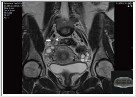
Currently endometriosis is considered between benign and malignant status and is known as a “malignant” benign disease by experts. Its histopathologic, and molecular data suggest that endometriosis has malignant potential and is associated with ovarian cancer [22,24]. Although urinary tract endometriosis occurs in ~1% of women mainly in women of child-bearing age with pelvic endometriosis it may cause hydronephrosis by involving the ureters secondarily by encompassing and or compressing them. Any lapse or delayed diagnosis can lead to renal failure. Ultrasound, CT and MRI (Figure 8) may be revealed and or laparoscopy may reveal the encasement of ureters.

Figure 8: MRI scan of the pelvis showing abnormal tissue (arrowhead) which encases the right distal ureter (arrow). (Courtesy: Apostolos Vrettos, Maria Prasinou, Rob Frymann doi: 10.21037/qims.2016.01.02)
lupinepublishers-openaccess-journal-urology-nephrology

Conclusion

Both ureters are retroperitoneal throughout their course in the abdomen. An encompassing inflammation and or infiltrative malignant process usually cause circumferential wall thickening and an abrupt change in the diameter of the ureter resulting in dilated proximal ureter with a narrow or normal-calibred distal ureter. RPF is usually diagnosed through clinical presentation and imaging studies. The severity of RPF is emphasised by the fact that, in about 56%–100% of patients with idiopathic RPF, the fibroinflammatory tissue entraps the ureters and causes obstructive uropathy and subsequent renal failure. However, in any case biopsy should be considered to exclude malignancy. Any of the above discussed pathologies may involve the renal vessels and contribute to renal insufficiency or cause renovascular hypertension. New onset of hypertension, flank pain radiating to groin in a known case of RPF or malignancy should arise the suspicion of ureteral encasement and appropriate imaging should confirm. Failure in recognition of the encasement of ureters and its causes may lead to flawed diagnosis and irrecoverable damage to the kidneys. Early detection, corticosteroids, Radiation therapy, Retrograde ureteral stent and PCN placement, bilateral ureterolysis and resection of the aneurysm and other surgical interventions are some in the management algorithm to be chosen to broaden the treatment arena and provide encouraging results.

Acknowledgement and Disclaimer

The author would like to thank Taylor’s University, Malaysia for the time granted for reviewing work. I am also grateful for the insightful comments offered by the anonymous peer reviewers of the Journal of Urology & Nephrology studies. This paper would not have been possible without the exceptional support of my amazing partner A. Riaz Ahmed. His enthusiasm, contributions and the responses kept my work on track.

Read More About Lupine Publishers Journal of Urology & Nephrology Please Click on Below Link:
https://lupine-publishers-urology-nephrology.blogspot.com/

Friday, 4 August 2023

Lupine Publishers | Factors of Early Marriage and its Consequences on Reproductive Health from Teenage Mothers’ Perspectives and their Families

 Lupine Publishers | Journal of Nursing & Health Care


Abstract

Background: Earlier marriage (EM) is a threatening concern for young females. The healthy young females reproduce a healthy prospect generation. While women’s reproductive health complications such as bleeding, low birth weight, premature birth, miscarriage, and unwanted pregnancy will reproduce weak, and ill generation. Purpose of the study to determine early marriage factors and its consequences on reproductive health from teenage mothers’ perspectives and their families.

Method: The study used a descriptive-cross-sectional design. The study conducted at the outpatient clinic in Women’s Health Hospital and four maternal and child health centers in Assiut University. Study Sample: A convenient sample of teenage mothers recruited to achieve the aims of this study. A Structured interview questionnaire used for data collection.

Results: The total teenage mothers were (112). The mean age + (SD) at the time of marriage is 16.62±1.74.and current age is 24.95±5.95. About 60% of the currently married women were aged (18-22) years old. Two-thirds of participants revealed that early fertility was one of the essential factors of early marriage. Complications of early marriage were CS, bleeding, perineal tear, preeclampsia and one-fifth of newborn had physiological jaundice and health problems. There was a statistically significant difference between teenagers’ mothers and parents’ perspectives regarding consequences of early marriage.

Conclusion: The study reveals significant differences in most of the factors and also consequences. The study concludes that perspectives of adolescents’ women match with the findings of the previous studies, reflecting their accurate perspectives regarding early marriage and its consequences.

Implications for practice: Initiates an obligation session for each woman in maternal outpatients/clinics, especially in rural/ remote areas, to increase awareness regarding early marriage consequences and integrates the health education regarding early marriage consequences in the school curriculum to increase awareness level among adolescents’ girls.

Keywords: Early Marriage; Factors; Consequences; Reproductive Health; Teenage Mothers and Families

Introduction

Early marriage defined as any marriage carried out below the age of 18 years before the girl is physically, physiologically, and psychologically ready to carry the responsibilities of marriage and childbearing [1]. Child marriage is a human rights violation and has adverse effects on the future of their girl’s children (overwhelmingly girls) who enter into these marriages, creating an intergenerational cycle of disadvantage [2]. Adverse health consequences of child marriage include poor maternal and reproductive health.

Early marriage negatively affects the health and well–being of women and children [3]. It leads to early child-bearing, closed spaced pregnancies, unwanted pregnancy, pregnancy termination, maternal morbidity, and mortality [4]. It also increases the risk of intimate partner violence [5], which further linked to sexually transmitted diseases, including HIV. Early Child Marriage also associated with increased risk of children’s physical growth, lower educational attainment, morbidity, and mortality [6,7].

The World Health Organization (WHO) defines adolescents as those between 10 and 19 years of age. It considered a period of transition from childhood to adulthood. Adolescent girls constitute about 1/5th of the total female population in the world. The period of adolescence for a girl is a period of physical and psychological preparation for safe motherhood. As direct reproducers for future generations, the health of adolescent girls influences not only their health but also the health of future generations [8].

Millions of girls are affected by child marriage (CM) throughout the world. It widely practiced in the countries of South Asia, where millions of girls-preteens and teens become the wives of older men every year. Young girls are married when they are still children. It is a violation of human rights. Their development is limited due to early marriage (EM) and often results in early pregnancy and social isolation (UNCF, 2014).

The causes of early child marriage occurrence depend on the condition, the community’s social life, the culture, and contextspecific norms. First, early marriage is a strategy to survive economically [9]. Poverty is one of the main factors that pivot the foundation for early marriage [10]. Poverty forces parents to marry–off their daughter at a very early age because the demand for dowry is low for younger girls. [11,12].

The second cause is to protect their daughter. Marriage is one way to ensure their daughter protected as a wife from non-marital sexual behavior or non-marital pregnancy. [Wikigender 2016,12]. Besides education and economic status, there are several other factors such as level of development, socio-cultural and religious norms, women’s status, and geographical residence have significant influence in determining girls’ age at marriage [13,4].

Significance of the Study

Globally, 36% of women aged 20-24 were married or in a union, forced or consensual, before they had reached 18 years. An estimated 14 million teenage women between the ages of 15 and 19 give birth each year. They are twice more likely to die during pregnancy or childbirth than women in their 20 years. Girls who marry between the ages of 10 and 14 are five times as likely to die during pregnancy or childbirth as women in their early 20s [UNICEF,2019]. early child-bearing increases risks to women’s health, as maternal disorders1 including complications during pregnancy and childbirth, are the leading cause of death among women aged 20–24 years globally and the second leading cause among adolescent girls aged 15 to 19 years.

In Egypt, early marriage is very high. The women ages 18-22 who married as young is 16.5 percent. In addition to very early marriage, of female before the age of 15-year-old. Early marriage is allied with poverty, lesser education levels, and higher nonemployment condition or low wages. These are however only correspondences, in addition to negative series of consequences and potential causal effects such as reproductive consequences [14]. So, this study aimed to determine factors of early marriage and its consequences on reproductive health from teenage mother’s perspectives and their families.

Research Question

1-What are the factors and consequences of early marriage on reproductive health from teenage mother’s perspectives.

Subjects and Method

Research design: A descriptive- cross-sectional design used in this study.

Study settings: The study conducted at Women’s Health Hospital, Assiut University. It included many units that provided the services the clients needed; these units included labor, postpartum, high-risk maternity unit, and gynecologic units. In addition to four maternal and child health centers in Asyut city, as El-Arbaen, Qulta, El-waleedea, and Gharb district.

Sample: A convenient sample of teenage mothers recruited to achieve the aims of this study. They recruited during their follow up at the previously mentioned clinical settings from March to June 2016. The participants selected according to the following:

Inclusion criteria: mothers who fewer than 18 years old and agreed to participate voluntarily in the study. The study conducted within three months from the period of the beginning of June until to August 2018

Sample size: The sample size calculated based on Raosoft application program (2004) the margin error (α error probability = 0.05) and confidence level (Cl) = 0.90) and the population was 160 per 3 months, so the sample size in adolescents’ mothers’ group = 101 but to avoid withdrawal rate we increased the sample size to be 112.

Tools of the study: Tool (1) A Structured interview questionnaire developed by the researchers based on previous studies [15-34] Prakash et al. 2011; ARWAO, 2006]. It consisted of three parts: The tool composed of 75 items divided into the following parts.

a) First part: demographic data included six items: marital status, age at marriage, and current age and residence, religion, and education levels of the wife. Obstetric data included 12 items such as current mode of delivery, the complication of delivery, new-borns condition of after delivery, skilled attendance at birth, number of children born, died, live births, most recent pregnancy, first pregnancy, unintended pregnancy, complication during pregnancy and antenatal visit during pregnancy. Contraceptive data included (3) items such as using family planning method, types of family planning, duration of uses family planning, and termination of pregnancy. Labor data included 3 items such as complications of labor, new-born status, and new-born weight.

b) Second part: Factors affecting early marriage included 22 items, 12 items regarding social factors, five items regarding economic factors, and five items regarding education factors.

c) Third part: Consequences of early marriage included 28 items, four items regarding economic consequences of early marriage, and seven items psychiatric consequences of early marriage. Nine items were regarding early marriage’s social consequences, and the last eight items were about the reproductive health consequences of early marriage.

Validity and Reliability

The content validity of tools was established by five experts in (obstetrics and gynecology and gynecological nursing and community health nursing) who reviewed the data collection tool for clarity, relevance, comprehensiveness, understanding, and applicability. The modifications made accordingly, and then the tool is re-designed for the final format and tested. The content reliability was estimated by Cronbach’s test. The tool proved to be reliable and acceptable at (0.81).

Ethical consideration

The written approval obtained from the faculty, research committee, and Ethical committee. In addition to the approval from the director of Woman health hospital and all maternal and child health centers in Assiut to collect the necessary data. Then the researcher obtained written informed consent from each woman intend to participate in the study after explanation of the study aim. Participant’s privacy considered during the collection of data. Anonymity, confidentiality assured, and the right to refuse participation emphasized to the participants.

Statistical Analysis

Data was collected and coded through SPSS (Statistical Package for Social Science) program version 20. The descriptive statistics are done in the form of frequency and mean ± SD also used t-Test to compare the groups of the quantities data. In addition to correlation test (Pearson r test) to correlate between teenage mothers and parents’ perceptions. Also, P-value considered statistically significant if it is less than 0.05. The analysis is done in two categories in the first category. The study focused on factors and consequences of early marriage of women, while in the second category, the analysis focused on the association between selected demographic factors and consequences on women’s reproductive health outcomes. There was no missing data in the statistical analysis.

Results

Table 1 shows the three categories of participants’ age at marriage per socio-demographic characteristics. The mean age + (SD) at the time of marriage is 16.62±1.74.and current age is 24.95±5.95. The most sociodemographic characteristics (45.5%)) categorized in age (15-17), (42.9%) aged (<18) and 11.6% aged (<14) years old respectively. About 60% of the currently married women were aged (18-22) years old. The majority (80 %) lived in rural areas aged (15-17), (100%) were Muslims aged (<18) while (11.8 %) were Christians and aged (15-17) years old. Regarding education, the majority (84.6%) of the primary level was (<14) years old, while (37 .5%) of secondary and (16 .7%) of universitylevel were aged (<18) years old.

Table 1: Distribution of socio-demographic data for Study sample (N=112).

lupinepublishers-openaccess-nursing-healthcare

Figure.1 illustrates the obstetric characteristics of the total (112) participants. The mean (SD) of Gravidity, number of dead and living children, reveals 2.95±1.86, 1.62±0.94, and 2.88±1.78, respectively. Two-thirds (66.1%) of participants reveals that early fertility was one of the essential factors of early marriage, while (1.8%) not have the desire in the first pregnancy. Complications during pregnancy, occurred in (44.8 %) and revealed that (21.4%) had an abortion, ectopic pregnancy and unintentional were (5.4%), stillbirth and IUGR) occurred equally (3.6%) illustrates the childbirth complications among the participants. More than One-third (38%) of participants delivered by CS, while the vast majority (94.6%) of the labor process managed by obstetricians. Regarding childbirth complications, it reveals that (14.3 %) had bleeding, (4.6%) had a perineal tear, and (3.6%) had pre-eclampsia. Regarding new-born condition, about one-fifth (21.4%) born had physiological jaundice, (8.9%) admitted to nursery, while (17.9%) had health problems. Table 2 shows the difference between teenage mothers and parents’ perspectives toward the factors of early marriage. Social and tradition factors reveal that both adolescents and parents referees to customs and traditions among families, the desire to drop out of education, desire to multiply offspring, and family keen not to fall in the underworld girl as a factor of early marriage and there is a statically significant difference at (p=0.008, p=0.001, p=0.001p=0.004 ) respectively. The Psychological factors reveal a statistically significant difference in the belief in marriage as destiny and protecting youth from delinquency (p= 0.001& p=0.000). Regarding the economic factors, the participants reveal a statistically significant difference in the feeling of that girl is an economic load, and the family preserves a financial wealth and the poor economic status at (p= 0.001, p= 0.021 & p= 0.035) respectively. Factors associated with education, reveals a statistically significant difference in the inability of the family resources to complete the education stages, family direction to education, preference between males and the inability of mothers either widow or divorced to educate their children at (p=0.002, p=0.001, p=0.005 & p=0.021) respectively.

Figure 1: Correlation between age at marriage and reproductive outcomes. **Correlation is significant at the 0.01 level (2-tailed).

Lupinepublishers-openaccess-Nursing-healthcare

Table 2: Comparison between perspectives participants and their parents toward factors of early marriage.

lupinepublishers-openaccess-nursing-healthcare

*” The significance level p<0.05”.

Table 3: Teenage mothers and parents’ perspectives on early consequences marriage on reproductive health. Total are more than 112 due to more than one perspective for each participant.

lupinepublishers-openaccess-nursing-healthcare

*” The significance level p<0.05”

Table 3 illustrates that the difference between teenage mothers and parents’ perspectives toward the consequences of early marriage on the reproductive health. The consequences on maternal health revealed that (76.9 vs. 35.4) % of adolescent mothers versus parents confirmed a fatigue due to load married life was one of the consequences, there is a statistically significant difference (p= 0.001). However, the maternal mortality consequence showed that (61.5 vs. 16.9) % with a statistically significant difference (p= 0.001) while increase maternal morbidity surprisingly, showed that (44.6 vs. 76.9) % with a statistically significant difference (p= 0.001). Regarding childbirth consequences, the early abortion and post-partum hemorrhage showed similarity (76.9 vs. 44.6) % and (75.4 vs. 56.9) % with a statistically significant difference (p= 0.001 and p= 0.026) respectively. The violence consequence showed that Physical violence was similar in both groups (60.0 vs. 58.5) % with no statistically significant difference, while sexual violence showed (50.8 vs. 13.8) % with a statistically significant difference at (p= 0.001). The consequence on the newborn showed (73.8vs. 12.3)% while low birth babies showed (47.7 vs. 18.5)% and neonatal mortality showed (26.2 vs. 63.1)% with a statistically significant difference at (p= 0.001, p= 0.001 & p= 0.001) respectively. illustrates the correlation between age at marriage and reproductive outcomes. This figure showed longitudinal Axis (r values) and horizontal Axis (Reproductive outcomes) and Red, and the blue line is the P values of age in correlation with reproductive outcomes. There is no correlation between the reproductive outcomes and the age of marriage. The only clear correlation occurs between the age and the increasing maternal mortality rate, which showed a significant relationship with (Pearson r = 0.259) and (p=0.007) between age at marriage and increased maternal mortality rate. Table 4 shows the relationship between age at marriage and the complication during pregnancy and labor. This table shows that the most categorized in age (15-17), (52.9) % (61.5) % aged <14) and (37.5) % aged (<18) years old complain from abortion. While (30.8) % <14 and (35.3) % (15-17) aged years old were IUGD and equally in pre-eclampsia. There was a statistically significant with p≤0.05) association between participant’s age, and sepsis.

Table 4: Relationship between age at marriage and the complication during pregnancy and labor.

lupinepublishers-openaccess-nursing-healthcare

*” The significance level p<0.05”

Discussion

Early or child marriage (CM) considered a violation of young female’ rights because they cannot complete their education, and their health is influenced by it. The timing of first marriage is an important factor in women’s reproductive behavior. Adolescents are mostly affected by CM, mainly adolescent girls. (Zannatul & Zebunnesa, 2019). The study of Knox, 2017, recommended that there is a need to search about responding to the indirect factors encouraging adolescent girls towards early marriage. However, the reasons for early marriage depend on the culture and specific traditions or poor economic status, which is a common cause due to the release of the financial burden of girls’ education (UNCF, 2015). So this study highlighted the perspectives of young women who married early during the adolescent stage and their families toward social, psychological, economic, and educational factors that motivated the early marriage and its consequences on reproductive health. Therefore, this study revealed that women who married early more compromised by severe consequences on reproductive health, including childbirth outcomes.

Relationship Between Socio-Demographic Characteristics and Early Marriage

Since this study about the early marriage of females so, the socio-demographic characteristics of participants age showed that the age at early marriage ranged from less than fourteen up to eighteen years old. The most socio-demographic characteristics categorized in the first category early age, which was less than 18 years-old as the majority lived in rural areas. The majority were in the primary level of education. Similarly, Prakash et al. study, 2011 & Afrouz et al 2018) revealed that the majority of who got married at an early age lived in rural areas. Hence, the age category in our study ended at 18 years-old to focus on the consequence of early marriage on reproductive health; however, Prakash’s study was based on the whole reproductive age from fifteen to forty-nine to compare the early marriage before eighteen and after eighteen-years- old.

Regarding increased early marriage based on residence area, a study by Barry et al. 2016 who reflected similarity as indicated that education and place of residence have a significant impact on early marriage which matching the fact that for each extra year of education, the possibility of early marriage reduced by 4.5 percent (DHS/MICS, 2012). Moreover, Iranian study by studies Mardi et al. 2018, showed that teenage marriage end the education process, as adolescents forced to leave school before or after marriage, while in continuing. In the same line, Safavi et al. 2015 reported that the majority of teenage women only had elementary education. Similarly, Zahangir et al. 2011, confirmed their findings and concluded that early marriage was an obstacle to public education. Our findings revealed a statistically significant difference between adolescents and parents regarding customs and traditions among families, which encouraged the parents to drop out of the adolescents from education, desire to multiply offspring, and avoidance of girls’ underworld problem. However, the psychological factors revealed a statistically significant difference in the belief in marriage is destiny, and protecting youth from delinquency. On the other hand, the economic factors revealed a statistically significant difference regarding the feeling that girl is an economic load. The family preserves a financial wealth and the poor economic statuseven the economic status associated with the inability of the family resources to complete the education stages. Likewise, Barry et al. study 2016 in Nigeria showed that early marriage considered a value, as marriage improves the social status and self-respect of the women in the local societies. In the same way, (IntHEC, 2010). & Abedokun, Adeyemi and dauda (2016) confirmed that marriage also serves a social purpose: preventing potential family humiliation associated with early sexual behavior and premarital pregnancies.

The Consequences of Early Marriage on The Reproductive Health

The main goal of this study, and the findings, to find out the effect of early marriage on the reproductive health. So, it showed that two-thirds of participants reported that early fertility was an important factor in early marriage, while less than twopercentage did not want in the first pregnancy. During pregnancy, complications occurred in fifty percent and revealed abortion, ectopic, stillbirth, unintentional pregnancy, and IUGR. In the same line, Prakash et al. study, 2011 confirmed that the same group age of early marriage intensifying the risk of pregnancy complications such as higher fertility and more unplanned pregnancies, abortions, and stillbirths. The findings our study, also revealed maternal and infant complications as more than One-third of participants delivered by Cesarean section, in addition to postpartum bleeding, perineal tear, and pre-eclampsia. As well as new-born condition, as physiological jaundice, and admission to nursery. In the same line, Irani and Latifnejad study in 2019 reported that early marriage threats sexual and reproductive health in different ways. Such as the maternal and infants who exposed to the adverse outcomes of early marriage. Reproductive health problem experiences by early married respondents were infection, bleeding LBW, premature birth, and unwanted pregnancy. In the present study, most items in consequences agree with Rosmala, et al. 2019 early–age marriage and the impact of health reproduction women. The highest percentage of disorder was hemorrhage, infection, preterm babies, and malnutrition. There was a significant relationship between the age of early marriage ant disorders; this was presumably because pregnancy and childbirth for women below 20 years have not matured physically and psychologically so that the risks of death were much higher than the age of 20 years and above. Not only mothers and children who born also have a risk of death or highrisk disability. In the same lines with Abedokum, et al. 2016 who found that very high reproductive health disorders occurring in married couples at an early age that allows for a long reproductive period unwanted pregnancies and abortion and disagree with the present study in a negative effect on nutrition status. That evidenced (DHS/MICS 2012 and WHO, 2011) reports, which reflected that more than one-third (35 percent) of maternal mortality occurred among adolescents 15 to 19 years of age, also increases the risk for dystocia, fistula, and other damaging outcomes to the reproductive system. The study from Niger similarly confirmed that neonatal mortality is high among adolescent pregnancies. Fifteen percent of children born from married adolescent girls were stillborn or died soon after birth. Also, 14 percent of adolescent mothers gave low birth babies less than 2.5 kilograms (Barroy, et al. 2016).

The Participants’ Perspectives Regarding Early Marriage

Participants’ perspectives regarding the consequences of early marriage on maternal health revealed that a statistically significant difference between adolescent mothers versus parents confirmed fatigue due to load married life was one of the consequences. Also, the maternal mortality consequence showed a statistically significant difference, while increase maternal morbidity surprisingly showed a statistically significant difference, but the parent perspective was higher than adolescents’ perspectives. Besides, the correlation between age at marriage and reproductive outcomes showed a significant relationship between age at marriage and increased maternal mortality rate. The consequences from participants ‘perspectives on the childbirth showed a statistically significant difference regarding early abortion and postpartum hemorrhage. In accord with Mardi et al. 2018 study, which confirmed the consequence of the new-born showed a statistically significant difference regarding low birth babies and neonatal mortality. However, disagrees with Nasrullah et al. (2014). Knowledge and attitude towards child marriage practice among women married as a children-a qualitative study in Lahore, Pakistan, among married age at 11-17 years revealed the majority participants were unaware of the adverse health outcomes of early marriage. This difference interpreted by the age group started with 11 years while our group started with 14 years-old which reflect little awareness in our study participants. Regarding to the marriage life load, our findings showed a statistically significant difference between adolescent mothers and parents perspectives which confirmed that the fatigue due to load married life was one of the consequences. This finding matches the Mardi et al. 2018 study, which showed that most teenage women complained about the heavy load of life and the ambiguous future. Regarding maternal mortality, Ganchimeg et al. 2014 study confirmed that early marriage means a high mortality rate due to the complication of early pregnancy and delivery. Since, the violence perspectives is a necessary consequence, our study focused only on Physical violence, in which, two-thirds of adolescents and parents confirmed it as an extraordinary consequence.

In contrast, sexual violence showed a statistically significant difference in both group perspectives, with fifty percent of adolescents’ women versus only thirteen of parents who confirmed it; this reflected that sexual violence is an impressing issue for parents to disclose while the physical violence is visible and cannot hide. In the same line, a study in Niger reported that sexual violence was more likely to affect younger than older adolescents. However, instead of that, sexual violence was confirmed as rare in Niger as it was socially condemned. Therefore, the families in rural areas handled the retribution by hiding this issue, which is similar to our finding as to the majority the parents denied it (Barroy, et al. 2016). Moreover, the findings of Mardi et al. 2016 showed that early marriage has led to unpleasant or coercive sexual experiences for some of them. Correspondingly, Allen et al. 2017 found that teenage women suffered from the health risks and social costs of early enforced marriage and coercive sexual relationships.

Conclusion

This study spotting lights on the factors motivate early marriage and its consequences on reproductive health from the two essential members of this issue “adolescent women and their parents” to detect how their perspectives are matching or different. The study reveals significant differences in most of the factors and also consequences except in maternal morbidity consequences perspective was higher among parents than adolescents’ women may be because the parents always in the first line when their daughters diseased. The study concludes that perspectives of adolescents’ women matching with the findings of the previous studies, which reflects their accurate perspectives regarding early marriage and its consequences and reflects that the circumstances drive parents and their daughters to accept the undesirable early marriage.

Implications for practice

a) Initiates an obligation session for each woman in maternal outpatients/clinics, especially in rural/remote areas, to increase awareness regarding early marriage consequences.

b) Integrates the health education regarding early marriage consequences in the school curriculum to increase awareness level among adolescents’ girls.

c) Further studies should be on the actual plans of adolescents’ mothers regarding the avoidance of early marriage to their daughters.

d) Strengths: This is the first study done Egypt to discuss the problem of early marriage from participants and their parents who are the main backbone of the problem.

Limitation

The number of participants supposed to be more than current sample but it was limited due to the estimated time for data collection.

Conflicts of interest:

There is no conflicts of interests regarding this study.

Funding:

There is no funding resources issued in this study.

Read More About Lupine Publishers Journal of Nursing Please Click on Below Link:
https://lupinepublishers-nursing-healthcare.blogspot.com/

Thursday, 3 August 2023

Lupine Publishers | Knowledge, Awareness, Attitudes and Practices Regarding Cystic Echinococcosis in Khartoum State, Sudan

 Lupine Publishers | Journal of Clinical & Community Medicine


Background

Cystic echinococcosis (CE) is a neglected disease of public health significance worldwide, especially in low- and middle-income countries caused by the larval form (hydatid cyst) of Echinococcus species tapeworm. Control of CE is difficult and requires a community-based integrated approach. The objectives of this study were to describe, using a questionnaire survey, the characteristics, attitudes, knowledge, awareness and practices of population regarding CE and tounder stand some of the risky practices that could contribute to spread of such disease.

Introduction

Cystic echinococcosis (CE) is a zoonotic parasitic disease caused by larval stage of small taeniid type tapeworm known as dog tapeworm (Echinococcus granulosus) that may cause infection in herbivorous animals and humans. Echinococcosis is one of the 17 neglected tropical diseases (NTDs) stated by the World Health Organization. E. granulosus is responsible for causing CE, which affects more than 1 million people around the world and responsible for over $3 billion in expenses every year [1]. It is characterized by the development of cysts either unilocular or may be multilocular of different extents ranging from the medium sized football to the size of a pea [2]. Genus Echinococcus comprises four species, i.e., Echinococcus multilocularis, Echinococcus granulosus, Echinococcus vogeli, and Echinococcus oligarthrus. There are two more Echinococcus species E. ortleppi and E. equinus on the basis of host-parasite interaction and their probable geographical distribution [3]. Analysis of mitochondrial and nuclear genes of different Echinococcus species has led to taxonomic revisions and the genotypes G1-G3 are now grouped as E. granulosussensustricto, G4 as Echinococcus equinus, G5 as Echinococcus ortleppi, G6-G10 as Echinococcus canadensis, and the “lion strain” as Echinococcus felidis [4]. E. granulosus life cycle is maintained by its definitive cannid host, i.e., dogs that nourish the adult worm in their smaller part of their intestine while wide range of domestic livestock and human acts as an intermediate host. CE is responsible for extensive livestock and human mortality and morbidity [5]. In details life cycle of E. granulosus, adult tapeworms that are usually 3-6 mm long reside in the small intestine of definitive hosts, then hydatid cyst stages occur in herbivorous intermediate hosts, such as sheep, cattle, goats, camels, horses, pigs and humans as well. In a typical dog-sheep cycle, tapeworm eggs are passed in the feces of an infected dog and may subsequently be ingested by grazing sheep; they hatch into embryos in intestine, penetrate intestinal lining, and are then picked up and carried by blood throughout the body to major filtering organs (mainly liver and/or lungs). After localization of developing embryos in a specific organ or site, they transform and develop into larval echinococcal cysts in which numerous tiny tapeworm heads called protoscolices are produced via asexual reproduction. A single cyst can have thousands of protoscolices, and each protoscolex is capable of developing into an adult worm if ingested by the definitive host [6]. There are many social reasons favouring the life cycle of E. granulosus and prevalence of CE in various parts of the world. Many families in rural have small plots of land and live-in close proximity with their flocks and dogs.

The gathering and grazing together of groups of animals belonging to different owners lead to circulation of infections, including CE. Home slaughter and feeding of dogs with raw offals favour the parasite’s life cycle [7]. Various small and poor equipped slaughterhouses built in the area of human settlements, lack of public health education are other factors that favour the life cycle of E. granulosus. Stray dogs and other canids, especially wolves may feed on dead animals and garbage, and hunt intermediate hosts. Dogs and livestock living in close proximity with man leads to circulation of zoonotic infection. Humans are incidental intermediate hosts; they do not play a role in the transmission cycle [8]. Dogs are particularly important in zoonotic transmission due to their close relationships with humans. E. granulosus is distributed worldwide, and it occurs on all continents, including Sudan. Cystic echinococcosis is one of the most important parasitic zoonoses in all regions of Sudan, resulting in high economic losses both in the public health sector and in the livestock industry. The epidemiology of hydatidosis varies from one area to another so control measures appropriate in one area are not necessarily of value in another [9,10]. It is essential to have adequate knowledge of the disease before contemplating control programs. For achieving an operative CE control program, it is vital to assess the level of understanding about the knowledge and awareness of disease and its preventive measure and hazardous acts that spread the infection more rapidly within the community. For these reasons, an investigation is conducted to explore the CE-related knowledge and awareness among human in different localities of Khartoum state, Central Sudan.

Methods

cross-sectional survey was conducted in Khartoum state – Sudan to find out recent knowledge, attitudes and practices on the occurrence of cystic echinococcosis. The quantitative data was collected in the form of questionnaire to investigate the knowledge and awareness of CE among community members and their routine practices that were behind the factors involved in hydatid cyst infection. The descriptive survey was performed between December 2017 and April 2018 by face-to-face communication.

Materials and Methods

Study design

This is a descriptive cross-sectional study was conducted over a period from December 2017 and April 2018todetermine up on the knowledge, awareness, attitudes, and practice regarding cystic echinococcosis among human in Khartoum state, Sudan.

Study area

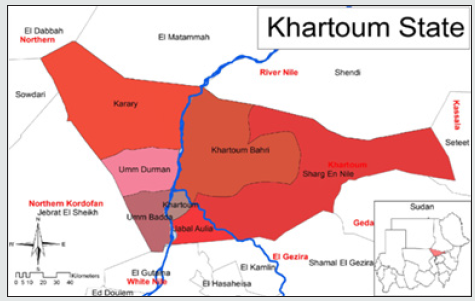
This study conducted in three townships in Khartoum state (Khartoum, Omdurman and Bahry), a part of central Sudan (Figure 1).

Figure 1: A map of the localities included in the study area of Khartoum State, Sudan [10].

lupinepublishers-clinical-community-medicine

Khartoum State lies at the junction of the two rivers, the White and the Blue Niles in the Northeastern part of central Sudan. It lies between latitude 15-16 N and longitude 21-24 East with a length of 250 k and a total area of 20,736 km2 the surface elevation ranges between 380 to 400 m a.s.l. Most of Khartoum State falls within the semi-arid climatic zone while the Northern part of it falls within the arid climatic zone. The state is prevailed with a hot to very hot climate with rainy season during the summer and warm to cold dry winter. Rain falls ranges between 100-200 mm at the Northeastern parts to 200-300 mm at the Southern parts with 10-100 mm at the Northwestern parts. Temperature in summer ranges between 25- 40 CO during the months of April to June and between 20-35 CO during July-October Period. Temperature degrees continue to fall during the winter period between November-March to the level of 15-25 CO. Khartoum State is divided into three clusters (cities), built at the convergence of the Blue and White Niles: Omdurman to the northwest across the White Nile, North Khartoum, and Khartoum itself on the southern bank of the Blue Nile.

Data collection

Clinical data were collected by the facility medical staff via study structured questionnaire.

Questionnaires

Individuals ≥ 18 years of age were included in the study. After receiving individual verbal permission, a questionnaire was administered to obtain basic epidemiological and individual information regarding known CE risk factors. Age, sex, educational level, residence location, occupation, personal hygiene, dog ownership, dog contact, and treatment of domestic dogs were recorded. The questionnaire was administered in face-to-face interviews. Only one investigator administered the surveys, in order to prevent inter-observer differences.

Study variables

The following are the variables utilized by this study:

a) Demographic data

b) Dog ownership

c) Dog contacts

d) Treatment of owned dog

e) Home slaughtering and raw offal disposal

f) Education level

g) Knowledge of disease

Study population

five hundred and twelve individuals were enrolled in this study, 198 males and 314 females, their place of resident is in Khartoum state and their age range from ≥18 years.

Statistical analysis

Descriptive statistics was applied in order to analyze the data using Statistical Package for Social Science (SPSS) version 21.0 software. Data was analyzed descriptively using the frequency table and cross tabulation.

Ethical approval

This study was approved by the Institutional Review Board Committee of Alneelain University, Khartoum, Sudan. Then study participants were first asked whether they accept to take part in the survey.

Results

Socio-demographic characteristics of the study population

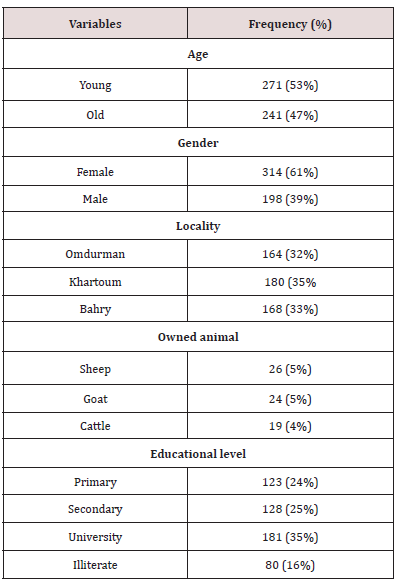
We interviewed 512 individuals, 314 (61%) female and 198 (39%) male, all of them were originated from Khartoum state. Their ages range from 18 -90 years with age mean (41.3 ± 18.08 SD) (Table 1). Regarding the populations’ level of education, 24% of the population had completed primary school, 25% for secondary school, 35% at university level, and 16% of them were unable to read and write (Table 1). Sheep and goat was the main livestock species owned by the population interviewed in this study (Table 1).

Table 1: Socio-demographic characteristics of people (N = 512) participated in cystic echinococcosis (CE) knowledge, awareness and practices survey in Khartoum, Sudan.

lupinepublishers-clinical-community-medicine

Practices towards CE prevention

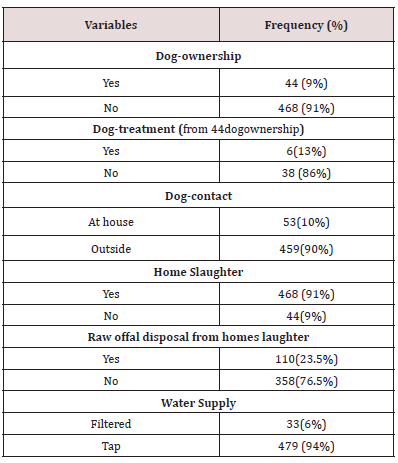
Of all the participants interviewed, 9% (44/512) indicated owning dogs on their houses (Table 2). Our result highlights some negligent dog management practices. For instance, among participants owning dogs, 13% (6/44) dewormed their dogs. Added to that, approximately all 91% (468/512) of the participants reported slaughtering livestock at home for their families’ consumption, of whom, 23.5% (110/512) they provide raw infected organ and uneatable offal like lung, intestine, etc. for their dog and cats. The interviews also revealed that the vast majority of the participants 94% (479/512) they never boiled water prior to drinking it. On the other hand,10% (53/512) of study participants come in close contact to their dogs (Table 2).

Table 2: Descriptive results of study population practices relevant to CE prevention and control.

lupinepublishers-clinical-community-medicine

Knowledge and awareness about CE infection

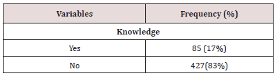
The interviews revealed that 83% (427/512) of the study subjects did not know about the possibility of transmission of certain diseases (zoonoses) between livestock and humans. Small proportions 17% (85/512) of study participants had heard of hydatid disease prior, but 4.7%from them had wrong knowledge about this disease as some of them think that the hydatid cyst is a sebaceous cyst (Table 3).

Table 3: Descriptive results of study populations’ knowledge about and awareness of sources of infection with CE.

lupinepublishers-clinical-community-medicine

Discussion

The purpose of this descriptive study was to determine sociodemographic characteristics, awareness, household practices, and attitudes of population in Khartoum state, Sudan regarding the preventive measures against such important neglected zoonosis (cystic echinococcosis), and risky practices that could contribute to spread and persistence the disease. Parasites leading to chronic diseases can be fatal to humans and animals in warm and temperate climate countries [11]. Sudan is a country where parasitic diseases are widespread because of its geographical characteristics, wide variety of animal populations, and various other environmental and socio-cultural factors. Several potential risky practices have been underlined among the community interviewed in this study, notably practices related to dog management and treatment. Fortyfour (9%) of the surveyed person owned one or more dogs. Of these, six reported to deworm their dogs. This finding reflects a poor awareness among the population regarding the role of dogs in CE transmission. The majority of the interviewees did not de-worm their dogs, and more than half of them indicated feeding uncooked viscera to their dogs when slaughtering at home for household consumption. Such risky practices have been among the most important factors that increase contamination of the environment with faeces containing Echinococcus eggs [12]. Similar findings have been documented in other CE endemic settings. A study in Sardinia (Italy) reported that the majority of the interviewed people used raw offal, after home slaughtering, for feeding their dogs [13]. In addition, a study in Tibet demonstrated that feeding dogs with uncooked viscera is a risk factor for increasing the likelihood of human infection with E. granulosus [14].

In highly endemic areas it is quite possible for individuals to contract CE through indirect transmission through contaminated food or water [15]. Our results show that almost 94% of the respondent’s drink water directly without boiling. Water supply has been also found to be associated with infection with CE, and this may be due to water contamination with dog faeces [15,16]. Studies in Jordan [17] and Kenya [18] established that contaminated drinking water was a risk factor for human CE and detected Echinococcus eggs in water used by both people and livestock. Consequently, treatment of water prior to drinking is an important process to minimize the risk of disease transmission. Adequate hygienic handling practices and heat treatment (cooking food or boiling water) should contribute to minimizing the risk of foodborne echinococcosis. In Sudan, the incidence of Echinococcosis has probably increased in recent years as a result of increased ownership of dogs, and presence of stray dogs in close contact with people in markets and around the slaughterhouses. Also, the slaughter practices in Sudan play a vital role in maintenance of the parasite cycle because of low quality and number of slaughterhouses. In addition, slaughter at house level without any practices of safe slaughter increases chances of maintenance of the parasite. The level of knowledge of Sudan’s citizens must be increased to enable the successful control of parasites. Several studies determining high risk groups for CE and discussing the theory of “lack of knowledge can increase the disease” have been published [19-21]. We conclude that, the participants of this survey were found to be with insufficient knowledge regarding CE. Therefore, it is concluded as the disease is less aware in the community, and it is very important to impart public health education to build up public awareness about the sources of infection and its control and collaboration between veterinary and medical personnel in sharing knowledge on zoonoses and working together to identify and control zoonose.

Results

We interviewed 512 people, 314(61%) female and 198(39%) male, all of them were originated from Khartoum state (three localities: Khartoum, Omdurman and Bahry). Their ages range from 18 -90 years with age mean (41.3 ± 18.08 SD). 9% of people dog ownership in the area. Of whom only (13.6%) dewormed their owned dogs. Moreover, participants’ response also showed that 10% had contact with dogs. Approximately all (91%) of the participants reported slaughtering livestock at home for their families’ consumption, of whom, (23.5%) they provide raw infected organ and uneatable offal like lung, intestine, etc… for their dog and cats.94% reported that they never boiled water prior to drinking it. On the other hand, small proportions (17%) of study participants had heard of hydatid disease prior, but (4.7%) from them had wrong knowledge about this disease as some of them think that the hydatid cyst is a sebaceous cyst, more than half of participants knew about the hydatid disease were at university educational level. The majority of the interviewed persons were not aware about how humans get infected with CE disease.

Conclusion

The participants of this survey were found to be with insufficient knowledge regarding CE. Therefore, it is concluded as the disease is less aware in the community, and it is very important to impart public health education to build up public awareness about the sources of infection and its control and collaboration between veterinary and medical personnel in sharing knowledge on zoonoses and working together to identify and control zoonose. This study highlighted a gap in health education efforts regarding CE in Khartoum state, we advocate the implementation of training programs to improve public awareness on this important disease.

Authors’ contributions

SSA, MEA designed and coordinated the work. SSA produced the questionnaire, which was reviewed by MEA and MPG. MKM , SSA,MEA collected the data. SSA analyzed the data and wrote the manuscript. MEA revised and edited the manuscript. All authors read, commented and approved the final manuscript.

Read More About Lupine Publishers Journal of Clinical & Community Medicine Please Click on Below Link:
https://journalofclinicalcommunitymedicine.blogspot.com/


Wednesday, 2 August 2023

Lupine Publishers | Signs of Tooth Eruption in Infants

 Lupine Publishers | Journal of Dentistry and Oral Health Care


Abstract

Teething is one of the challenges of medical science in Infancy period. Many studies show that teeth eruptions might have a lot of symptoms, including bad mood, skin rashes on the cheeks and around the mouth, increasing saliva secretion, swollen gums, Sucking a finger and etc. These signs and symptoms are mild in some infants, and in others may be severe; it is difficult for parents to tolerate these conditions. The peak of the severity of teething-related symptoms is when the dental buds have grown sufficiently in the gum and try to exit from the swollen gingiva.

Keywords: Teething; Saliva Secretion; Bad Mood; Skin Rash; Swollen Gum; Dental Buds

Introduction

As important as it is, tooth eruption is a matter of difficulty for both parents and children. Note that maintaining the health of teeth and gums in the child is the basis of the health of his teeth and gums for whole life. Teeth eruption is one of the most important stages of the growth, and through this stage, the baby will be able to have chewable foods. Commonly, there is a timetable for each primary tooth to erupt in the mouth. Signs of infant’s teeth eruption include disturbances in sleep and night-time wakening, mouth-watering, redness of cheeks, chewing fingers and objects, swollen gums, child malaise, bad temper, and the appearance of tooth buds [1-3]. In this article, we refer to any of these cases.

Schedule the Growth of Different Teeth in the Baby

Generally, new teeth grow in pairs. First, we see the eruption of the two lower primary central incisors teeth, and about a month later, the two upper primary central incisors teeth would erupt. Of course, in some cases, the four lower primary anterior teeth might erupt at first, and then we see the eruption of the upper primary anterior teeth or vice versa. In general, the eruptions of the primary teeth are scheduled to be as follows: [4]

a) At 6 months of age: lower primary central incisors teeth

b) At 8 months of age: upper primary central incisors teeth

c) At 10 months of age: upper and lower primary lateral teeth

d) At 14 months of age: first primary molars teeth

e) At 18 months of age: primary canines’ teeth

f) At age 2: second primary molars.

Signs and Symptoms

Given that the primary teeth are much smaller when they come out of the gum, and the process of tooth eruption is different in children, the symptoms that can usually be expected are:

a) Distracted Dreams: In some children, teething might be a painful process that can awake the baby at night. Therefore, if the baby suddenly wakes up at night, it may be due to her teeth. At this time, the best thing that mothers can do is taking her to the cradle and calm down.

b) Excessive Mouthwatering: Increasing saliva secretion can be one of the symptoms of the eruption of a new tooth. This is, of course, one of the natural stages of the baby’s teeth growth, so too much water in the mouth is not always a sign of baby’s teething. There is no way to detect the association between excessive saliva secretion and the eruption of new teeth. In a baby who is teething, his chin is often wet. Excessive saliva can cause injury and irritation of the baby’s face, so mothers should dry the baby’s face and mouth with a soft, delicate cloth. To protect the skin of the baby’s face, they can consult with their physician.

c) Swollen Gums: In some cases, before germinating of the tooth, the growth site in the gingiva of the infant becomes red, swollen and bruised. Sometimes, the rise of a tooth below the gum causes a bulge so that if parents can persuade her child to open the mouth enough, they can see the whitish shadow of the tooth under the swollen gingiva.

d) Chewing Things: Children like to take everything in their mouths, but if the baby too much does that, she may be teething. The pressure that unerupted teeth from below have on the gums, make the pain to be relieved by applying pressure from the opposite direction. Hence, most infants who are teething are willing to bite on different objects. Mothers should try to give them a teether rings to make them a little quiet. Of course, chewing on different things can also be the innate response of the baby to the strange sense of the mouth.

e) Sucking a Finger: In addition to biting on things during teething, it may be accompanied by sucking a finger. The baby can suck and bites for a few hours during the day. By doing this, he also tries to eliminate the pruritus of the gums. The joy of this action helps the baby to eliminate the pain and pressure of the teeth. Therefore, a mother may find that her baby calms down by chewing his fingers. She should try to keep her baby’s hands clean so no microorganisms can enter the baby’s mouth.

f) Change in Eating Habits: Wounds and gum’s swelling can make sucking painful for the baby. If the baby is hungry but runs away from feeding by mother’s breast or bottle, he may be teething. In this case, babies who eat solid foods tend to breastfeed or feed on the bottle, because the spoon annoys their inflamed gums. Some other babies also do quite the opposite of doing this, that is, they eat more because the bilateral pressure gives them a good feeling. On the other side, babies who still feed on the breast or bottle may eagerly start feeding at the beginning but quickly refrain from eating, because the sucking action puts very uncomfortable pressure on the gums and ear canals.

g) Baby’s Temper Tantrum: Primary tooth eruption (the outward movement of the tooth in the bone and gum) is usually done in a manner so that this action takes place more often overnight compared to the daytime, consequently, the baby will be more restless at night. The pain of teething can cause her sensitivity and irritability, and make him constantly cry.

h) Acne:In some children, teeth eruption may be accompanied with symptoms such as acne. Of course, this symptom is not definitive like fever, and it may have other causes. Skin hypersensitivities are more likely in children due to delicateness and vulnerability of their skin, and occasionally these acnes are signs of gastrointestinal symptoms.

i) Pulling the Ears: Holding, gripping or even pulling the ears, although sometimes indicative of ear infections, it can also be a sign of tooth eruption; in this situation, the pain that occurs in the jaw could be a transferred pain to the ears.

j) Cough: In some cases, when children are teething, they start coughing at that period of time.

Two Misconceptions about Signs of Teething

Many still believe that a child can have a fever during teething 5]] (even healthcare providers!), but on the contrary, some believe the temperatures above 102 degrees Fahrenheit are not related to teething [6]. If fever exists in this period, it is a mere accident and must be caused by other factors [7] . However, if we compare the temperature of the baby’s body who is teething with a child who does not, his body temperature may be slightly high, but this increase is not important enough to be called fever. Hence, why do many babies with teeth eruption have a fever? What is the explanation for that? Another misconception is that teething causing diarrhea [8,9]. Severe diarrhea or constipation is not associated with teeth eruption, and these two events should normally not be accompanied with each other. Although fever and diarrhea are not so much related to the teething, interestingly these signs are seen in some children with the onset of teeth eruption. The concurrency of diarrhea, fever and tooth eruption has two main causes: first, around the age of 4-6 months, when the teeth are emerging, the child’s immune system gradually become independent of the mother reduces the body’s resistance, and therefore this causes symptoms such as fever and diarrhea. Secondly, at this time, children take everything in their mouths to relieve the itching of the gums and discomfort that they develop during teething, but these objects may be contaminated to microbes [10], hence, there is the expectation of diarrhea in infants. Consequently, the emerging of teeth itself does not cause severe diarrhea. On the other hand, at this time, the safety of the immune system from the mother to the infant will diminish, [11] and the child’s body must build the safety components. Therefore, the reduction of maternal immunity which is transferred from the mother to the child can be a reason for fever or infection, [11] which is only a concurrency with teething; and the tooth eruption alone, do not cause this symptom.

Final Words

To summarize the article, the eruption of the teeth is a natural occurrence that occurs without acute and severe problems. It is a physiological phenomenon that will be associated with the other physiological phenomena such as increased saliva in the child, gums swelling, biting on any objects, disturbance in night sleepless, changes in eating habits, and so on.,. The tooth eruption does not pose a problem for the child. Some people mistakenly think that when the baby wants to have a new tooth, he is prone to having a fever, severe diarrhea or even constipation. The growth of teeth will never be accompanied by high fever. The teeth eruptions are different in newborns, but in general, most of them have first teeth at six months of age. Noteworthy that some of the symptoms of teething are similar to those of some diseases, which should be contacted with a pediatric pediatrician in the event of worsening of the symptoms.


Read More About Lupine Publishers Journal of Dentistry and Oral Health Care Please Click on the Below Link: https://lupine-dentistry-oral-health-care.blogspot.com/


Tuesday, 1 August 2023

Lupine Publishers | Concomitant Cervical Spine Infection with Mycobacterium Tuberculosis and Pyogenic Bacteria Causing Spinal Cord Compression

 Lupine Publishers | Journal of Neurology and Brain Disorders


Case Report

A 57-year-old man presented to the emergency room with neck back pain for about 2 months, unresponsive to nonsteroidal antiinflammatory drugs and progressive course of upper and lower extremity weakness with no sphincter dysfunction. The patient had no predisposing risk factors such as recent spinal surgery, trauma, instrumentation, distal site of infection, immunosuppression, diabetes. He was apyrexial. Physical examination showed marked mid neck tenderness, no palpable masses were felt, no lymph nodes were felt. Neurological examination of his extremities, spasticity was positive, and power was decreased 3/5 in both lower extremities, 2/5 in both upper extremities. Bilateral Babinski signs were present and deep tendon reflexes were increased.

Full blood count and biochemistry showed white blood cell count (WBC) 10,269/L (neutrophils 71.3%; lymphocytes 21.8%; monocytes 2.2%; WBC 4.4 to 11.3/L); C-reactive protein 13.86 mg/dL (0.1 to 6 mg/dL). Magnetic Resonance imaging of the cervical spine showed the collapsed body of C4 with epidural abscess formation, complicating with spinal cord compression. He underwent urgent anterior cervical decompression and evacuation of anterior epidural abscess with fusion. The material underwent histologic examination and aerobic, anaerobic, fungal, mycobacterial cultures. A tuberculous granuloma was detected on histology. Ziehl-Neelsen stain confirmed the diagnosis. Cultures also detected Staphylococcus aureus. Treatment was started with rifampin (600 mg), Isoniazid (300 mg), ethambutol (25 mg/kg), pyrazinamide (25 mg/kg), and levofloxacin 750 mg for two months. This was followed by seven months of isoniazid and rifampin. The patient was referred to rehabilitation. One year later, the patient is able to walk independently, and the back pain is gone.

Figure 1: T2-weighted axial MRI showing an intraductal cystic lesion lateralized to the left and protruding in the adjacent neuro foramina. Squeezing the cervical spinal cord

Spondylodiscitis can be etiologically classified as pyogenic, granulomatous (tuberculosis, brucellosis, or fungal infection), or parasitic. Pyogenic spondylodiscitis commonly affects the lumbar column and more rarely affects the thoracic and the cervical column [1,2] (Figure 1). S. aureus is the predominant pathogen in pyogenic spondylodiscitis, followed in older people by enterobacteria, mainly Escherichia coli, Proteus, Klebsiella, and Enterobacter [2-4]. Mycobacterium tuberculosis is the most common cause of spondylodiscitis worldwide. Tuberculosis affects mostly the thoracic spine and involves two or more vertebral segments. The main contamination routes are hematogenous spread, external inoculation, or involvement from adjacent tissue [5]. Isolation of pyogenic bacteria from an abscess may guide the clinician to disregard the possibility of spine tuberculosis. It is recommended, therefore, to made mycobacterial culture and histopathological examination for all suspicious cases even when there is positive culture of pyogenic bacteria.

Read More About Lupine Publishers Journal of Neurology and Brain Disorders Click on the Below Link: https://brain-disorders-lupine-publishers.blogspot.com/