Friday, 28 February 2020

Acute Liver Failure and Thyrotoxicosis Managed with Liver Transplant and Thyroidectomy_Lupine Publishers


Abstract


Acute liver failure and hyperthyroidism are not typically common, although some cases have been reported. The mechanisms involved and optimal management are not well-defined. This article presents the case of a 32-year-old African American female referred for evaluation of abdominal pain and jaundice, with a past medical history of systemic lupus erythematosus and Grave’s disease. She had thyrotoxicosis after administration of contrast and developed acute liver failure culminating in liver transplant and subsequent total thyroidectomy with a favorable outcome.
Keywords: Grave’s Disease; Autoimmune Hepatitis; Thyrotoxicosis; Acute Liver Failure; Liver Transplant; Thyroidectomy; Contrast Induced
Abbrevations: OLT: Orthotopic liver transplant; SLE: Systemic Lupus Erythematosus; TT: Total thyroidectomy; HD: Hospital day; POD: Post-operative day; ANA: Anti-nuclear antibodies; NaMELD: Sodium model for end-stage liver disease; PTU: Propylthiouracil.

Introduction


The association between liver function abnormalities and thyroid dysfunction is well established dating back to at least 1874 when Habershon presented a case of exophthalmic goiter and jaundice to the London Medical Society [1]. The range of liver dysfunction can go from mild elevation of hepatic enzymes to acute liver failure, which is defined as the presence of hepatic encephalopathy within 8 weeks of initial symptoms in a patient without underlying liver disease. Multiple scales have been devised to predict mortality and determine which patients would benefit from orthotopic liver transplant (OLT), with the presence of hepatic encephalopathy as a key indicator [2]. Here we present a case of a 32-year-old female with systemic lupus erythematosus (SLE) and exacerbation of Grave’s disease after iodinated contrast that presented with acute liver failure and thyrotoxicosis requiring OLT and total thyroidectomy (TT) after medical management of both conditions.

Case


A 32-year-old African American female was referred to the emergency room for evaluation of a 3-week history of worsening abdominal pain and jaundice. She also reported loss of appetite and occasional epigastric discomfort. Her past medical history included SLE and Grave’s disease [3]. Both conditions were well controlled, and her medications included multivitamins, hydroxychloroquine and ferrous sulfate. Three weeks prior to admission she underwent a CT scan with iodinated contrast for abdominal pain at an outside hospital. At that time, she received prednisone (60mg PO qd) and diphenhydramine for a presumed diagnosis of autoimmune hepatitis with positive antinuclear antibodies (ANA) at 1:320. Ferritin, alpha 1 antitrypsin, acetaminophen and viral serologies were all negative prior to admission. Her initial vital signs showed BP 110/70 mmHg, HR 134 bpm, RR 20 bpm, T 37°C and SpO2 100%. Physical examination was significant for generalized jaundice and moderate abdominal pain on palpation.
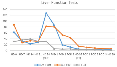
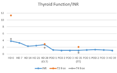
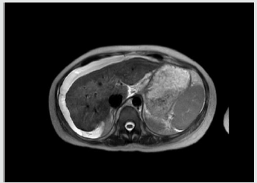
At that time her laboratories were INR 3.8, WBC 36.6 k/uL, K 2.7 mmol/L, lactate 2.4 mmol/L, Alk Phos 187 IU/L, AST 625 IU/L, 872 IU/L, Built 30.2 mg/dL, Bild 21.9 mg/dL, T3 11.3 pg/mL, T4 4.46 ng/dL, TSH 0.02 uIU/mL and ceruloplasmin 26 mg/dL. On HD #11 her 24hr copper excretion was 241.6 ug/d. Her admission Named was 35 and 31 at time of transplant. Renal function remained normal throughout her hospitalization. Trends for her hospital stay are shown in (Tables 1 & 2) After initial stabilization, Doppler ultrasound revealed patent vasculature, and MRI showed nodular appearance of the liver consistent with edema or early cirrhosis (Figure 1). The liver transplant service was consulted and a standard workup for potential liver transplant was started.
Table 1: Liver Function Tests. OLT: Orthotopic Liver Transplant. TT: Total Thyroidectomy.
Lupinepublishers-openaccess-Surgery-Casestudies
Table 2: TSH remained undetectable at <0.02.
Lupinepublishers-openaccess-Surgery-Casestudies
Figure 1: MRI of abdomen showing nodular appearance of liver consistent with edema or early cirrhosis.
Lupinepublishers-openaccess-Surgery-Casestudies
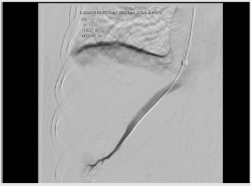
A transvenous liver biopsy on HD #6 was positive for marked active hepatitis with bridging necrosis, parenchymal collapse and minimal focal staining for copper. At the time of biopsy, a hemodynamic study was performed consistent with portal hypertension (Figure 2). Blood cultures were positive for E. coli and urine cultures for E. faecalis on admission, and she received ceftriaxone and meropenem with negative conversion of cultures. Her condition continued to deteriorate with worsening hepatic encephalopathy despite optimal treatment with rifaximin and lactulose. Oral prednisone was continued at 40 mg PO/qd. Her thyrotoxicosis was managed with propranolol and potassium iodide oral solution. An echocardiogram ruled out congestive heart failure. At HD #26 she was admitted to the intensive care unit and due to her condition, it was deemed she was at risk of death within one week and was listed as emergency status 1A on the united network for organ sharing.
Figure 2: Transvenous hemodynamic study consistent with portal hypertension. Free hepatic pressure 19 mmHg, wedge hepatic pressure 26 mmHg, hepatic venous gradient 7 mmHg.
Lupinepublishers-openaccess-Surgery-Casestudies
A suitable donor became available on HD #28 and she underwent OLT with methylprednisolone and mycophenolate induction without complications. She received 250 mcg of potassium iodide the night prior to transplant. Liver explant revealed extensive bridging necrosis, parenchymal collapse, no definite areas of fibrosis and minimal copper staining. She recovered without incidents from transplant and underwent total thyroidectomy on POD #7, she was then started on levothyroxine. She was discharged on HD #39 to an acute rehabilitation facility. At 1-year follow up, she has excellent graft function, remains euthyroid and has not experienced flare ups of SLE.

Discussion


Exposure to iodinated contrast is well known to cause thyroid dysfunction, with an iodine content of 320 to 370 mg/mL it is well above the recommended daily allowance and can precipitate thyrotoxicosis via the Jöb-Basedow phenomenon in patients with Grave’s disease [3]. Hyperthyroidism, particularly in Grave’s disease, is associated with primary biliary cirrhosis and autoimmune hepatitis. In patients without heart failure and hyperthyroidism, the liver biopsy has demonstrated fatty infiltration, cytoplasmic vacuolization, nuclear irregularity and hyperchromatism [4]. Although the exact mechanism for the observed liver-thyroid interactions is unknown, the following have been proposed:
a. systemic effects of thyroid excess,
b. direct toxic effects of thyroid hormone,
c. intrinsic liver and thyroid autoimmune mechanisms,
d. abnormal thyroid metabolism due to liver disease, and
e. subclinical physiologic effects of thyroid hormone [5].
feasible explanation is the presence of a hypermetabolic state with increased hepatic oxygen consumption, but without increases in hepatic blood flow, affecting the centrilobular zones and interfering with bile transport. These findings are consistent with the picture of cholestasis usually present in such cases [6]. In a series of 84 patients with acute liver failure, Anastasiou and colleagues reported a 50% incidence of thyroid hormone abnormalities, with a worse outcome in patients with hyperthyroidism, believed to be secondary to an increase in oxygen consumption and decreased organ perfusion [7]. In the current case, the presence of drug-induced liver injury had to be ruled out, hydroxychloroquine has been associated with acute liver failure requiring liver transplantation, but it appears within two weeks of starting therapy [8]. There is a well-known correlation between anti-thyroid medications and hepatic dysfunction, for example, propylthiouracil (PTU) and methimazole are associated with an incidence of severe liver injury in 0.1% and 0.1-0.2% of patients, respectively [9].
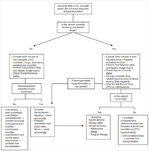
Figure 3: Proposed algorithm for management of patients with symptoms of liver dysfunction and previous diagnosis of hyperthyroidism. [Figure note: Figure obtained from de Campos Mazo, 2013; © 2013 Mazo et al, publisher and licensee Dove Medical Press Ltd. This is an Open Access article which permits unrestricted noncommercial use, provided the original work is properly cited.] [10].
Lupinepublishers-openaccess-Surgery-Casestudies
To our knowledge, our patient was not exposed to these medications. In the absence of established guidelines, a useful algorithm for the management of liver dysfunction with hyperthyroidism has been proposed, (Figure 3) This algorithm is based on a case series of 8 patients in which two cases developed acute liver failure suitable for OLT [10]. Nonetheless, the timing and sequence of OLT and thyroidectomy is not well established. Although it has been proposed not to delay the thyroidectomy as it might represent a potentially life-saving procedure [11]. The incidence of acute liver failure in the setting of hyperthyroidism remains a rare event, with patients undergoing OLT and thyroidectomy even more unusual. In this case report, adequate control of the thyrotoxicosis was achieved prior to transplant with oral potassium iodide and propranolol. In this patient with rapidly progressive liver failure, it appeared clinically prudent to undergo emergent OLT followed by TT.

Conclusion


The presence of ANA in a patient with SLE and Grave’s disease suggest the diagnosis of autoimmune hepatitis, however in this case report, the administration of iodinated contrast and the presence of abnormal copper metabolism made the diagnosis more difficult. The optimal management of these patients is not well established, with only a few cases reported in the literature. Based on our experience, medical stabilization followed by OLT and TT appears to have been an adequate course of treatment. Further studies are needed to help determine guidelines.


Lupine Publishers | The Influence of Yoga on Traumatic Brain Injury Related to Sleep and Mood

Lupine Publishers | Open access Journal of Complimentary & Alternative Medicine


Abstract


Sustaining a Traumatic Brain Injury (TBI) has a significant effect on an individual’s physical and mental abilities. Residual effects of TBI include sleep and mood disorders. Sleep disorders include any disturbance in an individual’s quality of sleep and daytime functioning. Mood disorders include depression, anxiety, and adjustment to injury. Rehabilitation after TBI involves a range of therapeutic services in which a holistic approach to therapy addresses both the mind and the body. Yoga may be used to improve functioning for individuals with TBI. The purpose of this convergent mixed methods study was to examine the influence of yoga on the sleep and mood in individuals with TBI. This research study involved an eight-week yoga intervention at a large rehabilitation hospital in the southern United States. Seven individuals who sustained a TBI were recruited for the intervention. Sleep and mood were assessed pre-, mid-, and post-intervention. Upon completion of the intervention, participants and their caregivers took part in focus groups to share their perceptions of changes in sleep and mood. Data were analyzed and describe the influence of yoga on individuals with TBI. Quantitative data revealed no statistical significance, though percent change calculations of pre- and post-data showed a substantial decrease in anxiety and an improvement in adjustment to injury. Qualitative data were consistent with the calculated percent change in addition to an emerging theme of social support amongst individuals with TBI.

Keywords:
Yoga; Therapy; Traumatic Brain Injury; Sleep; Mood; Depression; Anxiety; Adjustment


Introduction

A Traumatic Brain Injury (TBI) is defined as an acquired injury that is the result of direct damage to the brain [1]. A TBI can occur quickly and unexpectedly, but often has a long-term effect on an individual’s physiological and neurological abilities [2,3]. In the United States, approximately 1.7 million people per year are admitted to the emergency room due to sustaining a TBI [4], many of whom continue to live with residual effects [5]. The residual effects of a TBI include, but are not limited to, trouble sleeping, changes in mood, and difficulty adjusting to life after injury [6,7]. Sleep disorders are defined as any consistent internal disturbance in sleep [8]. Regarding people with TBI, poor sleep quality is common [7] and has the potential to decrease emotional and physical abilities, as well as slow the recovery process [9]. In addition to the negative impacts from sustaining a TBI, individuals are also susceptible to mood disorders as a residual effect of TBI. Common behavioral impairments for people with TBI include mood disorders, which can manifest as depression, anxiety, and adjustment to injury [3,6]. Depression is a common secondary factor for clinical conditions related to TBI [10]. Depression is defined by the Diagnostic and Statistical Manual of Mental Disorders (DSM-5) [8] as depressed mood or loss of pleasure in life activities for more than two weeks, change from an individual’s baseline mood, and compromised functioning. Generalized anxiety is defined in the DSM-5 as extreme or unrealistic worry for the majority of the days within six months [8]. Anxiety after TBI may first be seen as a normal reaction to trauma, but individuals with TBI appear to have an increased risk of developing generalized anxiety in comparison to the general population [11]. Individuals with TBI also experience an adjustment to life after injury [12]. Level of adjustment after sustaining a TBI can be observed through the presence of depression, anxiety, fatigue, and irritability [13].

Due to the physical, cognitive, and emotional impacts of sustaining a TBI, treatment for TBI needs to be approached from a multidisciplinary perspective. As an emerging element of physical rehabilitation, complementary integrative health (CIH) interventions are health practices used in combination with traditional medicine [14]. CIH includes a wide variety of healing interventions that counteract illness or assist in increasing health and wellbeing [15]. CIH interventions, such as yoga, can be used as a holistic and complementary treatment to address the physical and mental needs of individuals with TBI [16-17].

In the West, yoga focuses on three main practices: breathing (pranayama), meditation (dhyana), and physical poses (asanas) [18]. Yoga interventions have been utilized in several rehabilitation settings [19-22], for the purpose of providing a complementary form of therapy. Research on the perceptions of yoga, when integrated into inpatient rehabilitation hospitals, shows patients’ rehabilitation was enhanced by the use of yoga due to the added benefit yoga provided, including self-management skills and assisting longterm recovery [21,23]. Yoga for individuals with TBI is likely a useful intervention due to the adaptability of yoga sequences, the potential physical and cognitive benefits, and the research pointing to the potential sleep and mood benefits [19-24]. While there is limited research on yoga for TBI, one small, exploratory study found that when yoga was administered 16 times over the course of eight weeks, individuals with TBI expressed improvement in physical, emotional, and mental domains [25]. In an analysis of the influence of yoga on sleep for people with TBI through sleep-wake diaries, a substantial improvement in sleep quality was found after eight weeks of yoga treatment [19]. Following an adapted yoga group intervention for individuals with TBI, participants expressed favorable improvements in comfort with approaching balance and relaxation, as well as an increased self-awareness that helped with sleep [26]. There is limited research on yoga for individuals with a TBI and yoga, thus there is need for further studies related to the influence of yoga on sleep and mood in this population. Therefore, the purpose of this study was to observe, analyze, and discuss the influence of yoga on TBI related to their sleep and mood.


Methods

Design

This convergent mixed methods pilot study examined the influence of yoga participation on sleep and mood among individuals with TBI. Quantitative data was collected using a repeated measures design, with pre-, mid-, and post-intervention assessments given. Qualitative data was collected through two post-intervention focus groups, consisting of one focus group with participants and one with the participants’ caregivers. Prior to the start of this study, approval through the Rehabilitation Hospital’s Institutional Review Board (IRB) and the Clemson IRB were obtained.

Recruitment and Participants

Purposeful, criterion-based sampling was employed in this study to decrease the variation of diagnosis amongst subjects [27]. Fifteen individuals who sustained a TBI and were prior patients at a large rehabilitation hospital in the Southeastern United States, that provides a continuum of care for individuals with TBI, were contacted by the project coordinator. The project coordinator, a Recreational Therapist at the rehabilitation hospital, screened all individuals interested in the study using the Six-Item Screener (SIS) to assess cognitive status in order to determine eligibility for a program or intervention [28]. The SIS has been used as a screener into yoga studies for individuals with TBI [20]. After screening the individuals, the project coordinator reviewed the inclusion and exclusion criteria with the individuals with TBI as well as their caregivers, to determine if they met the inclusion and exclusion criteria for the study. Inclusion criteria for persons with TBI required that they:
I. Had diagnosis of moderate-to-severe TBI, verified by the individual’s Glasgow Coma Scale score upon admission to the rehabilitation hospital [29],
II. Were a fluent speaker of English, by self-report,
III. Were 18 years of age or older,
IV. Were able to move into different seated, standing, and supine postures without assistance (based on self- and caregiver-report),
V. Had a caregiver that was willing to assist with participant transportation needs throughout the study, and
VI. Had sufficient cognitive status to participate, as determined by a score of at least 4/6 on the Six-Item Screener.
The presence of any one of the following criteria resulted in exclusion from the study:
A. were unable to attend 12 or more yoga classes during the eight-week intervention,
B. had current drug or alcohol abuse, per self-report, and
C. enrollment in another intervention study that could affect sleep or mood. Inclusion and exclusion criteria were also established for caregivers of participants with TBI to ensure they were able to fulfill the role of caregiver throughout the study, although a caregiver was only required if the individual with TBI needed assistance with daily tasks.
Inclusion criteria for the caregivers required that individuals:
a. were age 18 or older,
b. had no prior history of TBI,
c. were the self-identified caregiver of person with TBI,
d. were a fluent speaker of English, per self-report, as being willing to transport participant to all yoga sessions related to the study (as needed).
Exclusion criteria for caregivers of people with TBI were as follows:
i. were unable to report on participant for whom they provide care, and
ii. had current drug or alcohol abuse based on self-report. All participants provided written informed consent prior to the start of the study. Participants admitted to the study were given a $25 incentive, funded by the rehabilitation hospital research department for clinician research projects, upon completion of the study.


Intervention

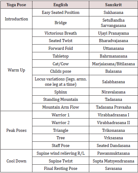
Yoga sessions were conducted in groups in a yoga room within a large rehabilitation hospital in the Southeastern United States. Sessions occurred twice a week for eight weeks, for a total of 16 yoga sessions. A recreational therapist who is a yoga teacher and specializes in yoga for individuals with TBI taught all yoga sessions. The sequences of yoga poses were designed based on the Love Your Brain (LYB) Foundation yoga program, which is designed for individuals with TBI [30]. The project coordinator of this study adapted the LYB yoga sequences to fit this specific study group [31], to focus on influencing sleep and mood. Changes to the LYB protocol included increased time for meditation and a decrease in poses accomplished on hands and knees. See Table 1 for yoga sequence. Each yoga class was one hour long and included a 15-minute centering and focusing of the mind, 30 minutes of gentle physical yoga postures in supine, prone, seated, and standing positions, and 15 minutes of meditation and relaxation. The yoga sessions remained at the same level of difficulty from start to finish, in order to facilitate the transition from the rehabilitation setting to the community setting by encouraging growth towards mastery of the postures as opposed to growth in the number of postures.

Table 1:
Yoga Sequence.
Lupinepublishers-openaccess-complementary-alternative-medicine-journal

Data Collection

Quantitative measures were chosen to focus on sleep and mood for individuals with TBI. Qualitative data were collected through post-intervention focus groups. The primary researcher conducted all data collection.

Quantitative Measures

Sleep quality was measured using the Pittsburgh Sleep Quality Index (PSQI), a self-report questionnaire used to assess the quality of sleep over a one-month period [32]. The 24-items inquire about sleep duration, sleep medication, sleep latency, sleep quality, and how sleep effects an individual’s daytime activity [33]. An individual may be diagnosed with poor sleep if he or she has a global PSQI score of greater than five. The PSQI has been used to screen for insomnia in individuals with TBI in post-acute care [34]. The PSQI has a diagnostic sensitivity of 89.6%, and a specificity of 86.5% when differentiating between individuals who experience ‘poor’ or ‘good’ sleep [32]. Depression was measured using the Patient Health Questionnaire-9 (PHQ-9). The PHQ-9 was developed based on the DSM-V criteria of depression [8] and can be self-administered [35]. The PHQ-9 is a nine-item depression scale that measures level of depression over the past two weeks using four-point likert responses, where 0=not at all, to 3=nearly every day [36]. Once completed, the total score was summed to assess level of overall depressive symptoms. The PHQ9 classifies level of depression based on the sum of responses, with 0-4=minimal depression, 5-9=mild depression, 10-14=moderate depression, 15-19=moderately severe depression, greater than 20=severe depression [37], and a score greater than 12 is the cutoff for being diagnosed with major depressive disorder [38]. The PHQ-9 was also effectively used in a study on combat-related TBI [39].
Anxiety was measured using the Generalized Anxiety Disorder-7 (GAD-7) survey. The GAD-7 is a seven-item anxiety scale that measures level of anxiety of the past two weeks using four-point likert responses, where 0=not at all, to 3=nearly every day [40]. This self-report questionnaire has shown reliability and validity [40,41] and can be used to analyze anxiety in the general population [41]. The GAD-7 classifies level of anxiety based on the sum of responses, with 0-4=minimal anxiety, 5-9=mild anxiety, 10-14=moderate anxiety, 15-21=severe anxiety, and a score greater than 10 is the cutoff for being diagnosed with generalized anxiety disorder [40]. The GAD-7 was validated in primary care facilities [36] but has also been used to measure anxiety in a study on sleep and psychological conditions after sustaining TBI [42] and used to measure anxiety related to mild TBI related to combat [39]. Adjustment was analyzed using Part B of the Mayo-Portland Adaptability Inventory (MPAI-4). The MPAI-4 has four parts, each of which address a different aspect of adjusting to injury. Part B was selected due to the specific focus on adjustment to injury related to an individual’s mood (irritation, aggression, pain, depression, anxiety, fatigue, social interaction, self-awareness, and sensitivity to symptoms). The rating scale ranges from 0-4, from 0=no problem to 4=severe problem that interferes with activities more than 75% of the time [43]. A sum score of 0-7= mild limitations, 8-15=mild to moderate limitations, 16-24=moderate to severe difficulties, and >25=severe limitations with a score of less than seven indicating a good outcome [44]. This scale was designed to assist in the clinical evaluation of participant adjustment during the post-acute (post hospital) period following an acquired brain injury [13]. This scale has been used in multiple rehabilitation settings, including post-acute rehabilitation, comprehensive day treatment, and community-based rehabilitation [45-47].

Qualitative Data Collection. As a convergent mixed methods study, this intervention was best examined through multiple forms of data, addressing research questions in a general and broad quantitative fashion, as well as providing a narrative and explanatory qualitative aspect [48]. The participant focus group focused on the participant’s experience in the yoga intervention, giving an account of their experience, any change they noticed in sleep, depression, anxiety, or adjustment to injury, and any additional comments they had about the influence of yoga over the past eight-weeks. The caregiver focus group facilitator asked similar questions and focused on the caregiver’s observation of participant behavior over the past eight-weeks. These focus groups were held in the private yoga room at the rehabilitation hospital and recorded using two audio recorders.


Data Analysis

Quantitative Analysis

Descriptive statistics were used to describe demographics, which included age, gender, marital status, race, work status, education, time (in years) since injury, and cause of injury. Nonparametric analysis was indicated because of the low sample size; thus, the Friedman Test was used to compare mean ratings of each assessment, using the Statistical Package for the Social Sciences (SPSS) software version 24. Comparisons were made between the group mean Pittsburgh Sleep Quality Index (PSQI) scores, depression scores (PHQ-9), anxiety scores (GAD-7), and adjustment scores (MPAI-4, Part B) from pre, mid, and postintervention assessments. To further examine the quantitative results using the means from each assessment, percent change was calculated using the following formula:
Pre-intervention = [(post-intervention value–pre-intervention value)/pre-intervention value] x 100%.

Qualitative Analysis

The qualitative focus groups were transcribed verbatim to increase descriptive validity [49], and participants and caregivers were assigned a subject number to ensure confidentiality. The project coordinator observed the focus groups to ensure interpretive validity [49], reporting that the project coordinator and primary researcher shared the same perceptions of the focus group discussion. After initial transcription, the primary researcher reviewed the qualitative data for themes, and categorized the responses based on their connection to sleep, depression, anxiety, and adjustment to injury. The project coordinator and an additional researcher reviewed the transcripts from the focus groups before and after analysis to check for consistency and establish interrater reliability [50]. In accordance with Creswell and Creswell’s sequential process of qualitative analysis [50], focus group transcriptions were organized and read thoroughly by the primary researcher. Coding was deductive, to identify patterns within the data relevant to predetermined outcomes (i.e., sleep and mood), and to determine the existence of any emergent codes.

Mixing Quantitative and Qualitative Data

Qualitative and quantitative data were collected and analyzed separately [50]. After individual data analysis, quantitative and qualitative data were compared to discover converging or differing results [48].

Results

Overall, 15 people were contacted and invited to participate in eight weeks of yoga. Ultimately, seven people passed the SIS, met the inclusion criteria, and committed to the study, while eight declined despite having passed the SIS, citing scheduling conflicts, distance from home, lack of interest, and inability to commit to eight sequential weeks. Six people completed the study, five of whom had caregivers, while one person dropped out of the study 1.5 weeks prior to completion due to travel conflicts. Of the six participants who completed the study, four (67%) were female, and the average age was 31, with the ages ranging from 21-43 years old. The majority of participants were White (66%), and most were single (83%). Half of participants had a graduate degree, although 50% were unable to work. The average time since injury was 4.67 years. On average, participants attended 14 of the 16 sessions, with an attendance rate of 89% based on total number of sessions offered. See Table 2 for additional participant demographics. In the following sections, both quantitative data and qualitative data are provided by outcome, as the intent of this convergent mixed methods design was to compare converging or differing results [48]. See Table 3 for the mean pre and posttest, p-value, and percent change.

Table 2:
Participant demographics.

Lupinepublishers-openaccess-complementary-alternative-medicine-journal

Table 3:
Participant demographics.

Lupinepublishers-openaccess-complementary-alternative-medicine-journal

Sleep

The Friedman Test revealed that quality of sleep did not differ significantly when comparing pre-, mid-, and post-intervention PSQI scores (X2=1.46; p=0.48). The percent change from the preand post-intervention scores yielded a result of -5.7% change, indicating a minor decrease in reported issues related to sleep. The qualitative data on sleep was convergent with the quantitative data, supporting that there was no significant change in sleep quality for most participants. Most caregivers and participants commented on an improvement in sleep since the individual sustained the injury, but most did not identify further improvement as a result of the yoga intervention. However, one caregiver believes yoga has enabled her loved one to have deeper rest while sleeping. The caregiver stated that her loved one has “deeper sleep, she sleeps longer in the morning, has trouble to wake up, and she dreams. And she remembers her dreams!” In addition, one participant commented on her ability to sleep, saying sleeping in the past year “I would hear any little noise, it’d just bother me and wake me. So, sleep with earplugs, I slept with earplugs and an eye mask for light. Now I’m much better and I don’t need earplugs or a mask.”

Depression

The quantitative and qualitative data showed converging results regarding depression, as neither form of data collection identified substantial changes following the yoga intervention. The Friedman Test showed insignificant results regarding pre-, mid-, and post- PHQ-9 data (X2=0, p=1.00), while the percent change from the preto post-intervention assessment was -14.9%, indicating a slight decrease in depression. Depression was briefly highlighted in the participant focus group, as one individual stated “I’ve never seen myself as depressed,” and later said “I don’t think I’m depressed but again, the doctors have attributed my past tiredness and sluggishness to depression, and they say that now that I am active, it helps that aspect.”

Anxiety

No significant difference in anxiety was found using the Friedman Test (X2=2.33, p=0.31). However, the percent change from pre- to post-test was -39.9%, representing a substantial decrease in anxiety after the yoga intervention. Complementing the percent change calculation, both caregivers and participants provided meaningful comments related to a decrease in anxiety during focus groups. Caregivers stated that yoga was “calming,” “relaxing,” and “increased the awareness” of their loved ones. Participants shared similar thoughts, using the words “calming” and “relaxing” throughout their discussion of their yoga experience. One caregiver stated: What my daughter seems to get out of it more than anything is the mindfulness and the meditation and just calming her down. Because we go at a high pace, and so this is a good way for her to just relax and help her brain get better. In addition, another caregiver said “she’s maybe more relaxed I would say. Less anxious.” Later on, this same caregiver explained, that yoga “sets her back and somehow it’s relaxing in order to let other things than the panic in her mind.” Participant responses aligned with the caregiver perspectives, as participants commented, “yoga has always relaxed me,” and “it helps me loosen up.” Another participant expressed her appreciation of yoga, saying: It’s perfect how the practice slows down, repeats, and just focuses on just a healthy mind. So, whereas out in the world, we’re supposed to go, go, go. Here we can just slow down, be in our minds, be present, and just be.

Adjustment

Though quantitative data regarding adjustment to injury produced non-significant findings based on the Friedman Test (X2=2.80, p=0.25), the calculated percent change from the preto post-intervention MPAI-4 Part B assessment was -57.6%, indicating a considerable decrease in issues related to adjustment to injury. In addition, the qualitative data showed an improvement in adjustment. Qualitative data showed an increased interest in activity and self-esteem, as well as a decrease in irritability from the perspective of both the caregivers and the participants. When asked about a change in amount of activity for individuals with TBI, one caregiver said, “he’s interested in doing more than just this.” When asked the same question, a participant stated, “I do want to do more activities outside of the house.” Moreover, one participant explained, “I do have more endurance of being able to take on more activities throughout the course of the day.” Caregivers emphasized an increase in self-esteem following the yoga intervention. One caregiver commented on the relationship between improvement in self-esteem, and the eight weeks of yoga, saying:
Self-esteem I think is a big problem. I mean, a huge problem. But um, maybe for the past two months she, I think she’s more aware and more in acceptance. So, it seems like the self-esteem is less of a problem.
While another caregiver explained that her husband is considering taking initiative on a project that she relates to an increase in self-esteem. Concerning irritability, a caregiver stated her son is “definitely getting more pleasant to be with,” and a participant said “yoga, being mindful, the whole practice of presence and really being intentional and present with what you’re doing has positively affected the way I approach anything.” Social Support in the TBI Community. Though not included in the purpose of this study, appreciation of the community that formed as a result of the yoga intervention was evident as a theme throughout the caregiver and participant focus groups. In the profound words of a caregiver, yoga has provided “a place [for the participants] to be injured.” Caregivers expressed “it’s just nice to be with people who are maybe dealing with the same things,” “they need groups to socialize, to exchange because they’re very lonely,” and yoga has “been wonderful for him because the rest of the time he is in the home alone.” In line with caregiver responses, a participant stated that yoga helps in “having community support others who know your situation, experience, having gone through the same things.” One participant expressed an appreciation of the ability to share experiences, saying “it’s better to have friends that you can meet actually, all of you, and to know that they’re doing the same thing that you have to.” The community developed through yoga is unique due to the emphasis on rest and relaxation, which one caregiver highlighted by saying “yoga allows them to have time to think… we’re not the ones that are gonna settle down with them like ‘ah, let’s rest’…we don’t have the time and probably not the patience either.”


Discussion

The primary purpose of this pilot study was to examine the influence of yoga on individuals TBI related sleep quality and mood after eight weeks of bi-weekly yoga. There was not a substantial change in sleep based on the PSQI. The data in this study differ from previous research that found yoga to improve sleep [19,51]. Though sleep disorders are common for individuals with TBI [7], the majority of this study population did not express complaints with sleep prior to or after the yoga intervention, resulting in little to no change in quantitative and qualitative results related to sleep. Considered to be a residual effect of sustaining TBI [52], depression was expected to be present in this study population. The pre-intervention average depression score from the PHQ9 was 4.57, (just beneath the mild depression score of 5-10), showing that participants did not initially experience significant depression symptoms. Depression was not significantly impacted by the yoga intervention, though the percent change showed a slight reduction in depressive symptoms, consistent with previous research claiming yoga yielded decreased reports of depression [53].
The findings of this study support previous work that yoga has the potential to decrease symptoms of anxiety [7,16,54]. Though quantitative measures yielded insignificant results, the percent change showed a substantial decrease in symptoms of anxiety. The qualitative data also demonstrated a reduction in anxiety, which participants identified was due to the emphasis on the calming and relaxing effect of yoga. Furthermore, a study by Verma et al. identified a decrease in anxiety continued beyond the yoga session was supported by caregiver and participant perspective shared during the focus groups [7].
Although not statistically significant, adjustment to injury did substantially improve, as indicated in the percent change calculation and the qualitative data. In congruence with the claim that yoga contributes to overall adjustment for individuals with TBI [55], this yoga intervention contributed to a decrease in irritability, and an increase of interest in activities. In addition, focus group discussions showed considerable improvement of self-esteem and selfawareness, supporting previous work that demonstrated the ability to improve emotional awareness through yoga after sustaining TBI [56]. The yoga intervention focused on awareness of the body and the mind by encouraging participants to bring awareness to specific body parts at time and acknowledge certain emotions that may come up. The focus on awareness throughout each yoga session likely contributed to the comments on increased self-esteem and awareness, consistent with the study results on the impact of an 8-week yoga program for individuals with TBI that indicated an improvement in self-perception [57]. A theme of social support through the yoga intervention became apparent through the focus group discussions. In a study on social support for individuals with TBI, Stålnacke [58] found reports of low-quality social support due to lack of social interaction. Consistent with results from other yoga studies [59-62], caregivers and participants described the yoga sessions as beneficial due to the sense of camaraderie with people who have similar life changes due to sustaining TBI. Caregivers expressed the need for their loved ones to be with other people due to their loss of friends since sustaining TBI. Discussions during both caregiver and participant focus groups indicated an appreciation of the shared experience yoga provides. Participants in an inpatient rehabilitation setting benefited from the social interaction provided by yoga [21] supporting the theme of social support that emerged from this pilot study.


Implications for Further Research and Practice

The diverging results from quantitative measures and qualitative interpretations specific to the influence of yoga on sleep and mood indicate a need for further investigation. In order to expand this study, future research should consider including only those with current complaints related to sleep and mood and involve a larger sample size. Future studies may also consider the use of a yoga sequence that becomes progressively more challenging, as the content of the yoga intervention used in this study maintained the same level of difficulty from start to finish. A progression of poses may produce more substantial results, as challenging activities are more likely to produce change [63]. Yoga is a valuable therapy that can be implemented in a rehabilitation setting [21,23,64]. Attendance was high due to the location of the yoga intervention, since the rehabilitation hospital was a familiar place to all participants. Participants and caregivers also stated that they would like to see yoga included in TBI rehabilitation and they also identified the desire for the yoga intervention to continue and be offered individuals in outpatient programs. The qualitative data supported the value of yoga within a TBI rehabilitation setting as it can decrease anxiety, improve adjustment to injury, and promote social support within the TBI community.


Limitations

Due to the nature of research, this pilot study has limitations. This study took place in one rehabilitation hospital in the southeast and cannot be generalized to all yoga programs within a rehabilitation hospital. Second, while we aimed to observe the influence of yoga on ten people, only six people remained committed to the study from start to finish, resulting in a small sample size, where it is difficult to determine statistically significant changes in outcomes. More clearly stating attendance requirements when recruiting participants may increase commitment to the study. This study was not blind to the primary researcher or the participants, as the primary researcher was in direct contact with the participants, and the participants were informed of the purpose of the study when recruited for the study. Due to the pilot nature of this study, no control group was observed in comparison with the individuals receiving the yoga intervention. By adding a control group, researchers may be able to further understand the influence of yoga versus other environmental and social influences. Finally, the yoga sessions were not designed to build on themselves, but rather involved the same primary moves with variations according to the yoga instructor’s preference. A yoga sequence that becomes progressively more challenging may yield stronger results.


Acknowledgement

This project was funded by the Shepherd Center Research Department located in Atlanta, GA.

Follow on Twitter   :  https://twitter.com/lupine_online

Thursday, 27 February 2020

Lupine Publishers | Subjection between Breast Cancer and Body Mass Index, the Role of L-Carnitine in Prediction and Outcomes of the Disease

Lupine Publishers | Open Access Journal of Oncology and Medicine


Abstract

Increasing the effectiveness of antitumor therapy in breast cancer patients who take L-carnitine during preoperative systemic antitumor therapy compared with patients receiving standard neoadjuvant systemic antitumor therapy served as a prerequisite for studying possible antitumor mechanisms of L-carnitine. The positive effect of L-carnitine is due to the transfer of palm-n-LC through the inner membrane into the mitochondrial matrix, which promotes the formation of a significant number of ATP molecules. It has also been shown that L-carnitine can have a double protective effect, enhancing the energy dynamics of the cell and inhibiting the hyperexcitability of the cell membrane, that making it an ideal nutrient for the prevention and treatment of cancer. This article summarizes the results of epidemiological and clinical studies of the use of L-carnitine in the treatment of breast cancer
Keywords: Body mass index (BMI); Breast cancer (BC); Obesity; Overall survival; L carnitine


Introduction

The incidence of breast cancer in the world in general and in Ukraine in particular is growing. In 2017, in Ukraine the incidence reached 16 percent of female population, for which, the breast cancer ranked first in structure of oncological incidence among women. In analyzing the data of the National Cancer Registry of Ukraine, it should be noted, that in comparison with 2014 year, the prevalence rate of breast cancer in 2016has increased by 5,1%, that indicates importance of improvement diagnostic procedures and methods of treatment it [1]. Studying the scientific literature on this subject, we noticed that there is a strong biological relationship between obesity and a poor outcome of breast cancer. And having analysed the date of Ministry of Health in Ukraine it can be concluded, that about 26% of women in 2017 year had overweight or obesity.
Obesity has a chronic metabolic character, which is the result of the interaction of the endogenous factors, environmental conditions and lifestyle. Endogenous factors could be considered a violation of the genetic and hormonal balance. The external conditions and type of lifestyle include irregular rhythm nutrition, use of substandard products and sedentary lifestyle. Obesity is the first risk factor for metabolic syndrome, diabetes type II, cardiovascular disease and some forms of cancer, including breast cancer. Since overweight is a risk factor for breast cancer, there is reason to believe that among patients with breast cancer the percentage of obese women is higher than in the population. The risk of breast cancer in postmenopausal women by 30%, it is more than in premenopausal, women with obesity-50%. Furthermore it was proven that obesity is associated with poor prognosis in patients with breast cancer, regardless of menopausal status, and effectiveness of systemic medication breast cancer in patients that have over weight is lower than in patients with normal BMI.
Although obesity is associated with a poor outcome in women with breast cancer, it is unclear how weight loss after diagnosis will change its course and results. Recently, complementary and alternative medicine (CAM) is widely accepted among patients with breast cancer, which may provide several beneficial effects including reduction of therapy-associated toxicity, improvement of cancer-related symptoms, fostering of the immune system, and even direct anticancer effects [2]. L-carnitine is a metabolite of C4 oil LC, which is involved in the transfer of palm-n-LC through the inner membrane into the mitochondrial matrix and is a substrate for the formation of ATP molecules. Carnitine is a trim ethylated amino acid naturally synthesized in the liver, brain and kidneys from protein lysine and methionine. Several factors, such as sex hormones and glucagon, can influence the distribution and level of carnitine in tissues [3,4].
In the absence of L-carnitine, the inner membrane of the mitochondria becomes impermeable to fatty acids, which entails a chain of various metabolic disorders in the human body. Carnitine has a modulating effect on the function of acetylcholine excitatory neurotransmitter, glutamate excitatory amino acid, insulin growth factor-1 (IGF-1) and nitric oxide (NO)[3]. Also proved, that L-carnitine may have a dual protective effect by enhancing the energy dynamics of the cell and inhibiting cell membrane hyper excitability, which make it an ideal nutrient for cancer prevention and treatment [5]. In view of the foregoing, the study of the influence of the body mass index on the effectiveness of systemic treatment of breast cancer is an urgent scientific problem and a promising field of research. This article presents the information of epidemiological and clinical studies of the influence of the body mass index on the effectiveness of breast cancer treatment by individualizing therapeutic measures taking into account the characteristics of patient's metabolism.
Studies on the Effects of BMI on The Course and Outcome of Breast Cancer and the Role of L-Carnitine in the Treatment of Cancer: The effectiveness of the prescribing of L-carnitine for breast cancers' treatment, as well as the effect of BMI on the outcome of the disease is proven in epidemiological and clinical studies.

Epidemiological and Clinical Studies

DSM Chan and co-authors [6] reported that women who have BMI> 30 course and outcomes of breast cancer are significantly worse than women with BMI <30. They proved, that women with BMI> 30 have the overall relative risk of total mortality 1.41, women with BMI of 25> 30 - 1.07. At the same time, for every 5 kg / m2 of the increase BMI, the risk of both total mortality and mortality from breast cancer increased, namely by 18% and 14%, respectively M. Protani and co-authors [7] have shown that women with breast cancer, who are suffering in obesity, have lower survival rate than women with breast cancer without obesity. Recently published data of randomized clinical researches by ML Neuhouser and coauthors [8] demonstrated, that for women> 50 years old, with 2 and 3 stages of obesity (BMI> 35) is typically the development of GR+ breast cancer. Similarly, B. Pajares et al. [9] who found significantly worse results for patients with BMI >35 compared with patients with BMI <25, stated that the magnitude of the effect depended on the cancer subtype (estrogen receptor (ER) / progesterone (PR) positive and HER2 negative, HER2 positive, triple negative). An analysis of the pooled data of the three adjuvant studies of the Eastern Cooperative Cancer Group showed significantly worse results for patients with obesity (BMI > 30) than for patients with normal BMI with a hormonal receptor-positive disease. And it was noted absence of negative effect of obesity on survival in patients with other breast cancer subtypes. C Fontanella et al. [10] studied the effect of BMI on different molecular subtypes of breast cancer and concluded that in women with ER / PR-positive and HER2-negative breast cancer, as well as with TNBC, the risk of death is significantly higher than in other subtypes of cancer.
It is proved that even the highest BMI figures are not a risk factor for death for patients with luminal A-like subtype of breast cancer. The reason for this is that fatty tissue produces an excessive amount of estrogen, a high level of which is associated with an increased risk of developing breast, endometrial, ovarian and some other cancers. It has also been proven that the level of adipokine, that promotes cell proliferation, increases in the blood with increasing of level of fat in organism. And adiponectin, which people with obesity have less than people with normal BMI, can have anti proliferative effects. Such data can serve as evidence of the effect of BMI on the course and outcome of breast cancer. Yet another proof of influence developing metabolic syndrome on the course and outcome of breast cancer was proposed by R. Bhandari et al. [11]. They proved that that the presence of metabolic disorders (that is, the metabolic syndrome) is associated with an increased risk of breast cancer in adult women.
The above data led to the need to investigate medicines that contribute to fat burning, such as L-carnitine. Based on the data provided by Rania M. Khalil and co-authors [12], we can prove the positive effect of this medicine on the course and outcome of breast cancer. The study showed that patients who received Tamoxifen with L-carnitine had significant decrease of Her-2 / neu and IGF-1 level (P <0.05) in the serum compared with patients who received only Tamoxifen. Using of L-carnitine led to significant decrease Her- 2 / neu level in the serum (P <0.05) compared to each of the control patients, namely, 59.5%. The effect of tamoxifen on IGF-1 (P <0.05) -decrease its level by 5.4% [13].However, it has been proved that using of L-carnitine in the treatment of ER+ breast cancer does not significantly reduce the level of estradiol, but leads to decrease both tumor markers CEA and CA15.3 (P <0.05,% decrease by 80.9% and 67, 8%, respectively) [13].
Using of L-carnitine in patients with breast cancer and obesity improves the metabolism of fatty acids in mitochondria, restores normal mitochondrial function and, thus, improves the general condition and quality of patients’ life [14]. Carnitine may alsomimic some of the biological activities of glucocorticoids, particularly immunomodulation, via suppressing TNF-a and IL-12 release from monocytes (5). L-carnitine as adjuvant therapy in cisplatin-treated cancer patients proved a beneficial effect in reducing the cisplatin- induced organ toxicity [15]. It is possible that, the extremely lipophilic nature of carnitine may be responsible for the decrease in EGFbinding [16]. Carnitine may insert in the cell membrane and/or interact with one of the many cellular enzymes having lipid substrates or cofactors. In addition, carnitine may interact directly with the EGFR [17].
Experimental evidence is available showing that ROS may induce the light and independent phosphorylation of the EGFR activating Her-2/neu. Moreover, the expression of the receptor is induced in conditions of oxidative stress [18]. L-carnitine, via its free radical scavenging and antioxidant properties, may inhibit ROS-mediated EGFR phosphorylation. It has been found that palmitoyl-carnitine can inhibit the activity of heart and brain protein kinase C in a competitive manner and subsequent phosphorylation of the EGFR [19]. Although the tumor markers and IGF-1 showed no significant difference in TAM-treated patients before and after administration of L-CAR, there was a tendency to decline after L-CAR supplementation [13]. The results of the above studies became a prerequisite for conducting clinical studies aimed at establishing the role of L-carnitine in the treatment of breast cancer.
To date, the search in the online clinical research registration system ClinicalTrials.gov using key words L-carnitine + breast cancer has revealed several studies evaluating the efficacy and safety of L-carnitine in the treatment of breast cancer patients. Analyzing the obtained results, we can conclude that L-carnitine was the drug of choice for neuropathies, as a consequence of chemotherapy, in patients with breast cancer.


Conclusion

L-carnitine is widely used in clinical practice. However, recently this medicine causes growing interest among oncologists. In a number of studies, L-carnitine has proven itself as a medicine that capable, during the preoperative systemic antitumor therapy, to increase its effectiveness compared with standard neoadjuvant systemic antitumor therapy. And also, taking L-carnitine with neoadjuvant systemic antitumor therapy helps to increase the number of cases of complete morphological regression (V degree of therapeutic pathomorphosis). To date, there are several clinical studies that are researching using L-carnitine in various malignant tumors, the results of which are the basis for further in-depth study of the effect of the medicine in the treatment of malignant neoplasms.

https://lupinepublishers.com/cancer-journal/fulltext/subjection-between-breast-cancer-and-body-mass-index-the-role-of-l-carnitine-in-prediction-and-outcomes-of-the-disease.ID.000103.php

For more Open Access Journal of Oncology and Medicine Please Click Here: 
 
 
Follow on Twitter   :  https://twitter.com/lupine_online  

Tuesday, 25 February 2020

Lupine Publishers | Hypertrophic Cardiomiopathy in Children: The Need of Heart Transplantation

Lupine Publishers | Journal of Cardiology & Clinical Research

Abstract

Hypertrophic cardiomyopathy (HCM) is the most common cardiac disease affecting the cardiac muscle. It can manifest in different forms with or without left ventricular outflow obstruction, with or without right ventricle involvement. Forms with biventricular hypertrophy seem to have poor prognosis. In our case, we describe a young patient with sarcomeric biventricular hypertrophic cardiomyopathy (MYH7 mutation), the poor prognosis of this form and strategies options adopted after failure of medical treatment. It is not always easy the management of hypertrophic cardiomiopathy, after medical treatment failure, especially in children. In some cases, heart transplantation is the only one therapeutic option.
Keywords: Hypertrophic Cardiomiopathy; Right Ventricular Hypertrophy; Heart Transplantation

Introduction


Hypertrophic cardiomiopathy (HCM) is the most common cardiac disease affecting the cardiac muscle and is characterized by heterogeneous genetic, morphological, functional, and clinical features. It is also one of the main causes of sudden cardiac death (SDC) in the young. Left ventricular hypertrophy with left ventricular outflow obstruction (LVOTO) is the most characteristic feature of HCM. There are also variant of HCM without LVOTO, with apical hypertrophy, with medio-ventricular obstruction and with right ventricular hypertrophy. The treatment and the prognosis of HCM seem to be variable on the basis of different forms, the age at presentation, sarcomeric gene mutations or rare phenocopies. Heart transplantation (HT) is the only therapeutic option for selected patients with HCM and refractory heart failure. In effect ESC guidelines recommend heart transplantation in eligible patients who have an LVEF < 50% and NYHA functional Class III–IV symptoms despite optimal medical therapy or intractable ventricular arrhythmia (II a); in eligible patients with normal LVEF (50%) and severe drug refractory symptoms (NYHA functional Class III–IV) caused by diastolic dysfunction (II b)[1].
Right ventricular hypertrophy (SRVH) is a relatively rare subtype of HCM. The anatomic, genetic, clinical, and prognostic characteristics of patients with SRVH and the clinical relevance of these characteristics have not been described widely in the literature [2,3]. MYBPC3 gene mutations have previously been described in two patients with RV hypertrophy. In a recent study, 90% of HCM patients with SRVH were found to possess relevant sarcomere protein mutations and variations in the MYH7 (Myosin heavy chain 7) and TTN genes, followed by variations in MYBPC3. Always in this study 73% of HCM patients with SRVH and multiple sarcomere gene mutations had poor prognosis. 7 In addiction MYH7 mutations can cause hypertrophic cardiomyopathy or skeletal myopathies with or without cardiac involvement, on the basis of the side of mutation. In our case, we describe the poor prognosis and treatment strategies of a young patient with biventricular hypertrophic cardiomyopathy and MYH7 mutation.

Case Report


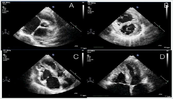
A 12-year-old young woman with familiarity for hypertrophic cardiomyopathy (mother and mother’s twin with biventricular hypertrophic cardiomiopathy and MYH7 mutation) was hospitalized in our hospital for dyspnea after mild-moderate efforts and reduced functional capacity (NYHA Class II). Mother and aunt of the patient were asymptomatic with good functional capacity. Patient had the same genetic mutation of mother and aunt (p.Asn696Ser heterozygosis MYH7) but with increased and poor phenotypic expression [4]. Echocardiography and cardiac magnetic resonance were performed showing a hypertrophic cardiomyopathy with right ventricular involvement. Precisely, cardiovascular imaging showed left ventricle asymmetric hypertrophy especially at the level of anterior and inferior wall (basal and mild anterior wall =14 mm, z score= 3,5; antero-lateral basal wall = 12 mm, z score 2,78; mild inferior wall = 14 mm and apical inferior wall = 12 mm) with normal ejection fraction (FE = 62% at CMR) and moderate diastolic dysfunction (panel B and D). In addiction wall thickness of right ventricle outflow and basal-mild free wall were increased (= 13 mm) with apical obstruction and development of maximum gradient of 10 mmHg (PANEL A and C) [5,6] (Figure 1).
Figure 1.
Lupinepublishers-openaccess-cardiology
The function of right ventricle was at inferior limits (FE = 51% at CMR, TAPSE = 16 mm at echocardiography). Thus the patient had an interesting right ventricle involvement and moderate diastolic dysfunction of left ventricle. She had not arrhythmia at ECG-Holter but she had reduced functional capacity. also demonstrated at stress test. Stress test was suspended at 6 min (Bruce Protocol) after pre-syncopal symptoms: lack of adaptation of the blood pressure to the effort was observed. In addition, from several months she had pre-syncopal episodes at the peak of the effort. ECG showed left ventricular hypertrophy and biatrial enlargement. Pro BNP was increased = 5841 pg/ml. Considering clinical situation, we decided to start medical treatment with betablockers (bisoprolol) but the patient didn’t tolerate medical treatment. Thus, we decided to start low dose of captopril without improvement of symptomatology. Also, treatment with diuretic was not tolerate by patient [7,8]. Therefore, considering symptom refractory to medical therapy, the poor prognosis and the impossibility to optimize medical treatment, we decided to plan cardiac transplantation, the only option possible at this moment.
Thus right catheterization was performed and patient was inserted in heart transplantation list. ICD implantation was not considered in the absence of ventricular arrhythmia and other factors. Discussion: hypertrophic cardiomyopathy associated with MYH7 mutation and right ventricle involvement seems to have poor prognosis, especially if right ventricle hypertrophy is severe [9]. In effect the young patient had a greater right ventricular hypertrophy compared than mother and aunt. In these cases, after medical treatment failure, heart transplantation seems to be the only strategy to improve symptomatology and quality of the life of the patient. Especially in pediatric population, it is not always easy the management of hypertrophic cardiomiopathy after medical treatment failure and heart transplantation seems to be the only one therapeutic option. Other study are needed to study some variants of HCM with right ventricle hypertrophy, their treatment and prognosis.

Monday, 24 February 2020

Lupine Publishers - A Unique Challenge: Laparoscopic Management of anAde...

Lupine Publishers | We Hear With our Brain as the same as our Ears?

Lupine Publishers | Journal of Otolaryngology Research Impact Factor


Abstract

There are some studies which confirmed that dysfunction in Central Nervous System (CNS) may cause a malfunction in the Peripheral Auditory system (Cochlea_ Auditory Nerve, Auditory Neuropathy), but the question is could Brain Disorder without any lesion in Cochlea and/or Auditory nerve cause Sensorineural Hearing Loss? It seems that there are a lot of Sensorineural hearing loss which they have neither Sensory nor Neural lesion, Brain is involved causing them. We deal with this subject in this paper and we propose a new theory that External Ear Canal is not the only input of Auditory Signals, Sounds could receive by the head and Cerebral Cortex and approach to the Cochlea (Backward Auditory input of Sounds).

Discussion

There are some studies that verified Otosclerosis and Meniere Diseases which are Peripheral pathologies initiated from CNS [1,2]. Increasing Cortisol Level and lack of Dopamine in Hypothalamus is involved in Meniere Disease and in Otosclerosis infection of CNS by Measles Virus [1,2]. According to these studies medications such as Ribavirin, Interferon, Inosine Pranobex with or without other medications such as, bisphosphonates, Sodium Fluoride with Calcium may be a cure or treatment of Otosclerosis and Dopamine Agonist Medication may be helpful for the treatment of Meniere Disease, further studies need to be done to find out the usefulness of these medications for these disorders and side effect, interaction and so on [3,4]. There are some papers which indicated Auditory Neuropathy occurred following Inferior Colliculus Disorder and Sudden Deafness following Primary Auditory Cortex infarction so it seems Central Auditory processing disorders may cause damage in Peripheral Auditory system, however, there are some patients with normal Audiogram(normal peripheral hearing) but they suffer from Central Auditory Processing Disorder(CAPD) such as Autism spectrum, therefore, malfunction in CNS may cause peripheral hearing loss but it depends on the site and degree of Lesion in the CNS, probably mild lesion in some part of CNS does not affect peripheral Auditory system [5-13]. We had a case who suffered from unilateral sudden deafness MRI showed Tumor on Occipital Lobe which is a Visual Central System, not an Auditory section perhaps because two Lobes are near to each other and because of correlation between them this occurred, so not only disorder in Central Auditory system may cause peripheral hearing loss malfunction in other parts of Brain may involve in causing hearing loss [3]. Neurofibromatosis type II is a genetic condition which may be inherited or may arise spontaneously. The main manifestation of the condition is the development of symmetric, benign brain tumors in the region of the cranial nerve VIII, which is the “auditory-vestibular nerve” that transmits sensory information from the inner ear to the brain and Auditory Brainstem Implant (ABI) is a solution for individuals with hearing loss due to a nonfunctioning auditory nerve (Neurofibromatosis Type 2). Bypassing both the inner ear and the auditory nerve which stimulates the cochlear nucleus (CN) and provides users with a variety of hearing sensations to assist with sound awareness and communication [14]. There are some case studies which verified that non- tumoral cases such as cochlear hypoplasia, Mondini dysplasia who underwent Auditory Brainstem Implant could even understand speech with phone so if hearing without Cochlea and/or Auditory Nerve is possible it is obvious that hearing loss because of lesions in some part of brain without any lesion in Cochlea and/or Auditory Nerve is possible [3,14].
We have seen a lot of patients with Sensory Neural hearing loss and normal Otoacoustic emissions this could be because of Auditory Neuropathy, lack of cooperation of patient during the Audiometry test, neural lesion, the collapse of the ear canal, but it is not always the case. There are some studies which showed normal or near normal OAE and compound action potential in those who suffer from the sensorineural hearing loss which indicated Cochlea and the Auditory nerve is intact therefore the reason of hearing loss could be because of lesions in the brain with or without mild lesion in Cochlea and/or Auditory nerve [3,15] FMRI and PETSCAN show that Prefrontal and Temporal Lobes are more responsible for High-Frequency Auditory system and Cerebellum and brainstem typically subcortical areas are more in charge of understanding Low-Frequency Signals [5,8,13,16]. Reticular Formation and Prefrontal Lobe has a tremendous role in the hearing system typically in Speech recognition and selective hearing and the correlation between these part and other parts of the brain such as whole Cortex and Hippocampus are involved in speech recognition and Listening [16,17].

Backward auditory input theory

It is impossible that our small Pinna and our tiny external ear canal is the only input to the auditory system, Cerebral Cortex and Brain could receive the sound from the Head [18]. It seems that there are two paths, Forward Auditory input signals which are the information from the external ear canal and the other one Backward Auditory input signals that is the information of signals which receive by Cerebral Cortex from the head [3]. Information from Forwarding Auditory input system reach to the Cochlear which is more Low frequencies and information from Backward Auditory input system comes down from the brain to approach to the Cochlear, typically more high-frequency information received by Backward Auditory input system [3,19]. The cochlea is the rendezvous between signals which come from Forwarding Auditory input, more low frequencies and signals which come from the Backward Auditory input, more high Frequencies and all information will be combined and organized and coding by Cochlea and will be sent to the Brain for final processing [3,18,19]. There are some examples which can confirm Backward Auditory input signals.
The First Maximum Conductive Hearing loss is 50 to 60 Decibels and not more than that and still we can hear [3]! Second studies showed that earmuff and ear plugs could not protect our ears from noise-induced hearing loss entirely [20]. Third, Spontaneous Otoacoustic Emissions (SOAE) s are sounds that are emitted from the ear with any external stimulation and are measurable with sensitive microphones in the external ear canal. If SOAEs is only because of Cochlea, why there are some studies which verified normal SOAE despite lesion in Cochlea? [21]. There is a possibility that SOAEs are because of backward Auditory input signals because there is a link between Tinnitus and SOAEs, nowadays we know that Tinnitus is more related to CNS, Limbic system and less related to Auditory system, so all these could confirm Backward Auditory input of sounds theory and the fact that our external ear canal is not the only input of sounds [22].

Conclusion


Cochlear Implant is a successful prosthesis not only because of Cochlear role in the hearing system because of transmission of sounds to the Brain and intensifies the Backward Auditory input system. Some of the contraindications of Cochlear Implant are Psychosis, Autism spectrum, patients who suffer from Central Auditory Processing Disorder (CAPD), so it means that hearing without CNS is impossible. Sensorineural hearing loss without lesions in Cochlea and/or Auditory Nerve is possible. Top-Down Auditory system disorders may cause disruption in Backward Auditory input system, receiving the sounds by Brain and cause hearing loss. For preventing Noise-Induced Hearing Loss new protections should design which can cover entire head and Auricles. There are a lot of similar sensorineural hearing losses which are the same in type and degree, but in inside the site of lesions may different.

Read More Lupine Publishers Otolaryngology Journal Articles :
https://lupine-publishers-otolaryngology.blogspot.com/
Read More Lupine Publishers blogger Articles : https://lupinepublishers.blogspot.com