Showing posts with label Journal of Otolaryngology Ent Research. Show all posts

Tuesday, 22 November 2022

Lupine Publishers| Nasolabial Cyst: Report of 2 Cases and Literature Review

 Lupine Publishers| Journal of Otolaryngology


Abstract

Introduction: Nasolabial cysts are a rare, ectodermal development cyst, presenting as a fullness of canine fossa, nasal ala or vestibule of the nose. They are usually asymptomatic but may become infected. Treatment is complete surgical excision by sublabial approach, or transanal endoscopic marsupialization.

Case Report: Description of two cases, one female presenting as nasal deformity due to progressive growth of unilateral nasolabial cyst, and a healthy young male presenting severe facial cellulitis, with a CT showing bilateral nasolabial cysts.

Discussion: Nasolabial cyst is a rare condition, but diagnosis and treatment are simple. Nasolabial cyst should be incorporated by ENT in the differential diagnosis of nose deformities and facial swelling.

Introduction

Nasolabial cysts are also described as nasoalveolar cyst or Klestadt cyst and were first described by Zuckerkandl [1,2]. They are rare, affecting 1,6 per 100.000 persons per year, more frequently in females (4:1 ratio), especially among African Americans, in the fourth and fifth decades of life. 90% are unilateral, and often underdiagnosed [3,4]. Nasolabial cysts are non-odontogenic cysts that develop lateral to the midline of the maxillary lip and alar base. They usually present as a swelling in the nasolabial fold, causing alar nose elevation and upper lip projection. They may grow slowly and painlessly over several years. Because of its close anatomical relation to the nasal cavity and teeth, they may become infected easily, rapidly growing and being painful [1,5]. Diagnosis is made by clinical examination, imaging tests, and is confirmed by histopathologic study. The cyst can be palpated bimanually with one finger in the floor of the nasal vestibule and another in the labial sulcus. Computed tomography (CT) and magnetic resonance (MR) can be useful. Differential diagnosis includes cysts of the nasopalatine duct, periapical inflammatory lesions (granuloma cyst, abscess), and epidermoid cysts [1,6]. Complete surgical excision of the nasolabial cyst is the best treatment, and sublabial approach is most used. Other authors propose transanal marsupialization as an easier and shorter procedure, with lesser complications, but recurrence may be a problem [3,5].

Case Report 1

Figure 1(a).

lupinepublishers-openaccess-journal-otolaryngology

Y=372.256 + 6.8856. X1+residuals

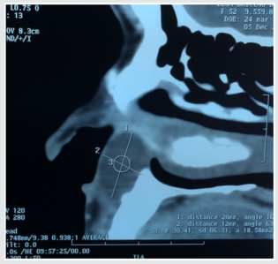
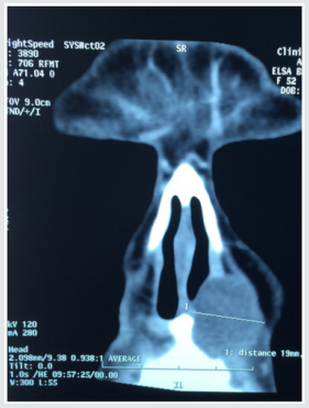
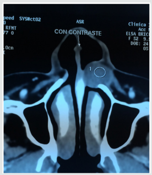
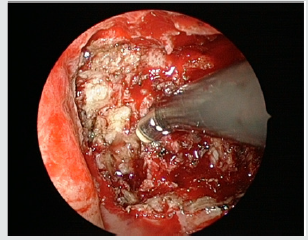
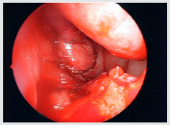
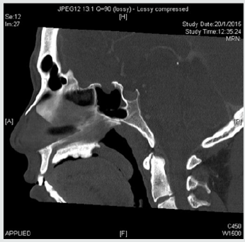
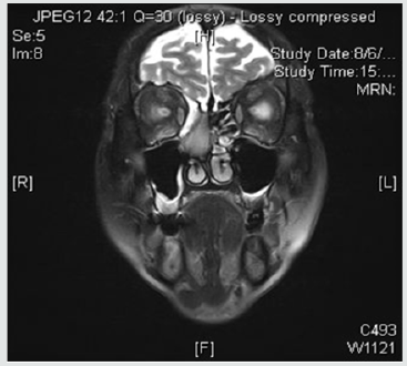
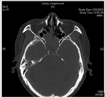
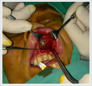
52-year-old female patient with a 3-year history of left floor of nose intermittent painful swelling, and progressive deformity of this area. Computed tomography (CT) study showed a 3,2 cm rounded lesion in left pyriform aperture compatible with nasoalveolar cyst (Figure 1). A sublabial approach was performed, and complete excision of the cyst was obtained (Figure 2). Histological study confirmed suspected diagnosis. Patient evolved in excellent conditions, with no complications or recurrence in a 2 year follow up period.

Figure 1(b).

lupinepublishers-openaccess-journal-otolaryngology

Figure 1(c).

lupinepublishers-openaccess-journal-otolaryngology

Figure 2. Intraoperative view of sublabial access to left nasolabial cyst in Case 1.


Case Report 2

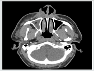
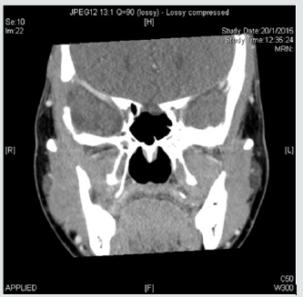
Figure 3. Computed tomography of bilateral nasoalveolar cysts. Case 2.

lupinepublishers-openaccess-journal-otolaryngology

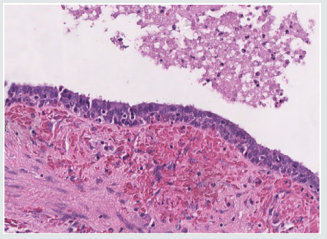
Figure 4. Pathology H-E study of Case 2 showing cyst wall with pseudostratified columnar cells.

lupinepublishers-openaccess-journal-otolaryngology

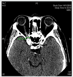
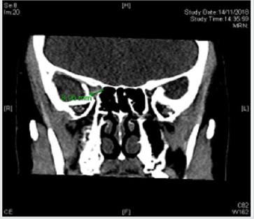
37-year-old male patient, with no prior medical record, developed a facial cellulitis with no initial response to oral antibiotics, requiring hospitalization and intravenous antibiotic treatment. Maxillary and orbital CT study showed bilateral nasoalveolar cysts, with signs of active infection in the right cyst (Figure 3). Antibiotic treatment was completed, and cyst resection was programmed a month later. An extensa sublabial approach was performed, and bilateral cysts were removed. Histological findings confirmed diagnosis (Figure 4). Patient did not have any other episode of facial cellulitis or cyst recurrence in a 14 month follow up

Discussion

Origin of nasolabial cysts is controversial. Most accepted theories are that cysts derive from remnants of the nasolacrimal duct or inclusion cysts of mesenchymal cells during the fusion of medial and lateral nasal prominences to the maxillary prominence during facial skeletal formation [2]. Histology usually present pseudostratified columnar epithelium. Su et al noticed with electronic microscopy that that cysts had a highly placated mucosa, of non-ciliated stratified columnar epithelium, differing from the ciliated columnar epithelium of the paranasal and nasal sinuses [7]. The treatment of nasolabial cysts consists on complete removal of the lesion, with the objective of prevention of infectious complications, histologic diagnosis, and aesthetic improvement. Fine needle aspiration can help in diagnosis, and relieve of symptoms, but recurrence is high [1]. Surgery is usually done by sublabial approach, creating a mucosal flap of gingiva to allow access to the pyriform aperture and to the cyst, which can be resected carefully to avoid rupture, especially to the floor of the nose mucosa, and complete excision is mandatory to avoid recurrence. After the intervention, the gingival mucosal flap is fixed in its original position with absorbable sutures. Potential complications are uncommon, including facial swelling, insensitive gingiva, teeth numbness, and surgical site infection.

Patients must use a toothbrush on the surgical site. Diet should be soft for the first week, then normal. Dental prostheses can be used immediately after surgery [1]. Lee et al published in 2009 a comparative study between surgical techniques, and strongly suggested endoscopic trans nasal marsupialization as a simpler, shorter and safer procedure, and in most cases, it could be done under local anesthesia [3]. In our two cases we used sublabial approach with general anesthesia because is the usual technique used in our practice, but we are interested to use endoscopic marsupialization in next cases. We did not have any complications or recurrences in follow up of both cases.

Conclusion

Nasolabial cysts are rare but must be considered in the differential diagnosis of floor of the nose deformities and facial swelling. Sublabial approach is the most common procedure, but probably be replaced by endoscopic marsupialization in the future.

Read More About Lupine Publishers Journal of Otolaryngology Please Click on Below Link: https://lupine-publishers-otolaryngology.blogspot.com/

Saturday, 6 February 2021

Lupine Publishers | Categorical Loudness Scaling in the Fitting of Cochlear Implanted Children

 Lupine Publishers | Journal of Otolaryngology


Abstract

The correct - optimal - fitting of speech processor determines the best result of rehabilitation. The optimal setting of most comfortable level (MCL) is achieved by accurate patient’s assessment of electrical stimuli loudness. Unfortunately, small children can’t give a reliable report about their feelings. How to determine the MCLs in every channel of children implant? Objective methods don’t give the final (optimal) comfort levels of the child’s working program. Therefore, we need subjective assessments. We tried to use a method of categorical loudness scaling (CLS). This article is a guide how to teach CI children to evaluate loudness. Good results of the CLS were observed.

Keywords: Cochlear Implant; Fitting, Categorical Loudness Scaling; C – Levels; Most Comfortable Levels (MCLS); Threshold Discomfort Levels

Introduction

The correct (optimal) fitting of the processor determines the best result of rehabilitation [1]. The optimal settings of C-levels are achieved by accurate subjective patient’s assessments of the electrical stimuli loudness. Unfortunately, young children cannot give a verbal report about their feelings. How to determine the maximum comfortable levels (MCLs) in every channel of an implant in children, i.e. to find threshold discomfort levels? For example, an objective method – reflexometry (registration of stapedial reflex) – is used for fitting of children. But the program in which MCLs are equal to the reflex threshold levels is very rarely optimal one [2].

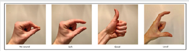
Therefore, subjective estimates of loudness are necessary. There are studies of categorical loudness scaling (CLS) in cochlear implant recipients [3] in which adult subjects participated. Results were reliable ones. What to do with children? We tried to use CLS for assessment of the loudness in cochlear implanted children. The aim of our study is how to find the equal loud C-levels in all channels and using these C-levels to create program with equal loud C-levels. Our study has a practical purpose, so we did not estimate the loudness function. We will not discuss the individual electrical levels of discomfort due to the large differences of these current values between listeners. The CLS is started when we had done reflexometry and parents selected an optimal program. We use our four pictures corresponding to categories “NO SOUND”, “SOFT”, “GOOD” and “LOUD” as a function of the electrical stimulus level (Figure 1).

Figure 1: Four categories of loudness

Lupinepublishers-openaccess-otolaryngology-journal

What reference electrical levels do we use?

Soft Levels

As we wrote earlier, at first day of fitting we approximately defined comfort levels (C-levels) which a child hears as quiet sounds [4].These stimulation levels had been recorded as C-levels in MAP3 of the first configuration. N.B. It is possible that the child did not hear a sound in some channels. Since by the time CLS starts, the child has some experience assessing the loudness of sounds, we can try to clarify these quiet C-levels. We use the first two pictures. This is a part of the CSL already. We stimulate, in our opinion, a quiet signal and “ask”. If child does not hear, we show picture “NO SOUND”. If child hears we show picture “SOFT”. Further, we reduce C-level below the threshold of hearing (2 presses on “Pg down”) and stimulate. We show squeezed fingers – “NO SOUND”. We repeat stimulation on different electrical levels involving the child in this game-study.

Loud Levels

By the time the CLS will be performed at relatively loud С-levels, we had done a reflexometry. How it is performed was described in our article [5]. N.B. In the case of absence of intraoperative ipsilateral reflex, it is impossible to exclude the presence of the contralateral reflex. We created a reflex-program with C-levels equal to the threshold levels of the reflex and recorded it as MAP2 of new configuration. It is known that different children can hear the different intensity of the sounds (from not too loud to loud) at the program where C-levels are equal to reflex threshold levels (our MAP 2). From this reflex-program we had created 3 programs and write them into the configuration. The first MAP is 3 steps lower, and 3-rd and 4-th maps are 3 and 6 steps louder correspondingly. In accordance with our instruction-explanation (“Program is optimal one if your child sometimes hears loud sounds” [6] speech therapists and parents selected optimal (working) program. It should be noted that almost all patients successfully use programs with C-levels above the reflex threshold levels by 3-6 steps (MAP3 and MAP4) [7] and even more. This is normal physiological phenomenon.

When we fit the patient without intraoperative reflex we work in accordance with the standard algorithm of fitting [8]. We gradually increase (in parallel) C-levels at all the electrodes until the parents and we’ll see that some program is loud one. Below this program is a working program (the optimal). Threshold levels are set to 10 percent of C-levels. Using this working program children respond well to all sounds and do not display negative reaction when surrounding sounds are the loud ones. Children can use a louder program in a quiet environment, but a child does not like it in a loud environment (according to parents’ comment). For the purposes of the CLS we use the test program, in which C-levels are less than levels of working program by 3 steps.

Why do we use a program less than optimal one for CLS? The child uses the optimal program in everyday life without problems, but when he will hear a sequence of long-term (300 ms) stimuli with an interval of 300 ms (SWEEP mode) on the one channel at maximal C-levels, the sensation can be unpleasant. N.B. Before CLS, it is necessary to visually assess the child’s reaction to the presentation of single and SWEEP stimuli at the C - levels of the test program - is there any negative reaction? Quite possible that child will not like the maximum C-levels of test program on some channels. C-levels in such channels must be corrected. Corrected C-levels of this test program will be used as loud sounds of the “LOUD” picture.

So. What do we have for our research?

A child hears and orients in sounds in accordance with an information of speech therapists and parents. Parents and teachers have identified a working program that the child uses without problems in all sound environments. Its C-levels are equal (very very rarely) or higher (almost all patients) than threshold levels of stapedial reflex. A child uses CI readily, in the morning the child asks to wear CI himself. A child indicates that the optimal program is the best one. (We asked parents to switch on processor at the first program –the child indicated to change the program). We know approximate loudness of some electrical levels. We know electrical levels, where the child hears quietly. We know where the child hears loudly - at C-levels of the test program. We know where the child hears well- in the area of the third quarter of the dynamic range of the audible current. The child has some experience of distinction between “SOFT” levels and “NO SOUND”. We can start the Categorical Loudness Scaling.

Methodics

How Do We Perform Categorical Loudness Scaling?

We use SWEEP stimulation, i.e. we provide a sequence of identical stimuli of the same amplitude on one channel. The duration of stimuli is 300 ms, the interval between them is 300 ms. SWEEP stimulation is started by pressing down the “Enter” button. The duration of the stimulation is determined by the duration of pressing the “Enter” key and the reaction of the child. We start CLS with a channel with a central frequency in the area of 800-1000Hz. We use categories “NO SOUND”, “SOFT”, “GOOD” and “LOUD” as a function of the electrical stimulus level. At first, we show our fig. 1 to the child. Child already has some experience in categories “NO SOUND” and SOFT”. We show signs with our fingers and explain what the volumes of the sound the child will hear in his (her) head (ear). Owing to our practice, we think that our pictures are more understandable and natural signs for description of child’s own sensation than a circle, squares, cubes etc. These signs are easier to repeat by children. Children may understand meaning of these signs from the birth.

We explain to the child that now we will stimulate, and he will hear a sound in the head (ear). We show the second picture, repeat the sign with our fingers and transmit quiet SWEEP-stimuli. We “ask” the child. If child agrees that he hears a quiet sound, we invite him to show it in the picture or with his fingers. Switching off stimulation, we squeeze fingers, show the first picture, that now there is no signal. Next, we show that we are going to increase the sound. We increase C-levels to 60% of the C-level of test program. We show the “GOOD” picture, raise our thumb and send SWEEP-stimuli. “Ask”. If child agrees, we invite him to show it in the picture or with his fingers. Switching off stimulation, we are clenching fingers, show that now there is no signal. The child agrees. Changing the electrical levels up-down we show with our fingers and on the corresponding picture how loud the signal or no signal will be heard. We invite him to show by his fingers or at appropriate picture. That’s how we perform the CLS training. After some training, a child begins to navigate in their feelings and to give real answers. When we reduce the level of stimulation the child brings own fingers closer, when we increase-move apart. Or they show the corresponding picture. He should be praised. Now we can go to the loudness estimations of the stimuli from the third quarter of the dynamic current range. When a child is assessing of the sound as “GOOD” we “ask” him if it is possible to increase the level of stimulus a little. Waiting for consent or refusal. Many children agree of our offer to slightly increase the stimulation. Then we increase C-level by 1-2 steps, stimulate and look how child displays this increase. Or by fingers, or on pictures. So, we move to the maximal C-levels of our test program where child will hear loudly i.e.to the fourth picture. Closely observe a behavior of the children during the CLS and involve them in the process!

It is curious to note that if some children show estimations of loudness not with their fingers, but with the pictures, they can show their ratings between pictures. For this reason, all four pictures must be placed in one line. Some children begin to show their estimations by the distance between palms. If the child is a contact one and cooperates with the audiologist, you can propose him to increase levels in order to gently touch the threshold discomfort levels. For this purpose, it is necessary to increase a C-levels of test program. But this is the best result. Repeated CLS measurements were done using single-electrode stimuli at a few electrode positions (sometimes all). C-levels at unmeasured electrodes were interpolated. The results were recorded. At the end of the CLS, we set equal-loud C-levels at all channels and make a program. Further, we compare these C-levels with the C-levels of working program defined by parents and teachers. We create new program with C-levels close to C-levels of working program. Since the C-levels of the created program and the working one are not the same ones, we check new program vootiue (on the child’s own ear) and create one program of 3 steps lower and 2 programs by 3 and 6 steps higher. Parents choose an optimal program in accordance with our instruction-explanation [8].

During the CLS we “communicate” with the child, “ask” and “explain”. Naturally, by signs: gestures, fingers, touching, facial expression and praise. We think that such a relationship is interesting to the child - child cooperates with the audiologist, we praise him for his work, correct mistakes, rejoices for the correct answer. Children tend to participate in this “research-game” with interest. We think that children are interested in judging the volume of sounds of different intensity and frequency, that’s why they willingly participate in the CLS.

Discussion

Children work in the CLS successfully. But loudness is a subjective evaluation. Naturally, for example, the same “LOUD” ratings of different patients will be different if they are measured in the terms of SPLs. We believe that the child himself chooses some criteria for assessing the loudness of sound and relies on it for all channels. It is quite natural for each patient to have his criterion, but we hope it is the same one for each child. Somehow it is used in repeated measurements on the different electrodes. Stable repeatable estimates are confirmation of this thesis. Every child adjusted all channels in accordance with own volume criterion. It is clear that adults also have their own criteria too, based on which they assess the loudness of the stimuli. But adult participants themselves noted difficulties in assessing the loudness of singlechannel stimuli of different spectral color. During the development of the fitting program SHCHUP [9] in which the stepped noises are used, adult patients themselves said that the estimation of the loudness of the stepped noises is easier than the loudness estimation of single-channel stimuli. Of course, children have the same difficulties. So, it is clear that the results of CLS on separate channels are not the completion of the fitting of children. The results of the detection equal loud(!) C-levels are important to configure the same equal loud levels in all channels and create a program. Despite the successful mastering of CLS by children, the last step of the fitting is the SHCHUP [9] . SHCHUP is the definition of comfortable SPLs of the stepped noises. The estimation of loudness of the stepped noises is a simpler task for experienced in CLS children too. On the base of SHCHUP’s results, we create four programs in new configuration.

The last step of fitting is the parents’ evaluation of the child’s perception of these programs in different sound environments and the definition of the optimal program in accordance with our instruction-explanation (Petrov & Tsjuk, 2015). Several hundred children (I did not count) participated in procedure of CLS, and I can surely say that the categorical loudness scaling in the fitting of cochlear implant children works successfully. We are sure that CLS is interesting game-procedure for the implanted children and useful method for an audiologist in order to fit children successfully. The CLS is a good encouraging and illustrative program for parents too. For example, we increased the level of stimuli and said mother that sound will be louder now. We stimulate and she sees that her child moves his fingers wider or moves his finger on the fig. 1 to the right. We reduce the level of stimulus and tell mother that sound will be quieter now. We stimulate and she sees that her child brings fingers closer or moves a finger on the fig. 1 to the left. At zero level, child squeezes his fingers together. So CLS is interesting and encouraging procedure for parents - mother sees the coherence of our words about changing of intensity (up or down) and the child response. Mothers are glad that her child correctly assesses the volume of sounds. This article describes General guidelines for performing of CLS. The main aim of this article is to guide how to teach the CI child to assess the loudness of sounds that is very important in the fitting process. Naturally, each child needs his own approach and this CLS, of course, is not done immediately. Speech therapists can use these pictures in their job with implanted patients and hard of hearing children too. Perhaps this method of the CLS can be patented.

Read More Lupine Publishers Otolaryngology Journal Articles:
https://lupine-publishers-otolaryngology.blogspot.com/

Thursday, 28 January 2021

Lupine Publishers | Review of Ten Years’ Experience of Endoscopic Skull Base Surgery in A Regional Hospital ENT Department - Queen Elizabeth Hospital

 Lupine Publishers | Journal of Otolaryngology


Abstract

Endoscopic skull base surgery is a relative new approach in ENT specialty. ENT Department of Queen Elizabeth Hospital started to do some ten years ago. It is a review of the types and experience of that endoscopic transnasal approach to manage skull base disease. 63 procedures in four types of surgery is recorded in a ten years period in that regional hospital in Hong Kong. The largest number being done is endoscopic nasopharyngectomy which is more common in southern part of China.

Introduction

The endonasal and endoscopic approach to the skull base provides a minimally invasive way to remove tumors that would otherwise require either a large cranial opening and brain retraction or disfiguring facial scars. Instead, an endoscope is advanced through the natural opening of the nasal cavity. A variety of tumors can be removed in this fashion, in various locations, with different maneuver of the endoscope to the desired location. This approach was first developed by endoscopic sinus surgeons but is now being used as minimally invasive skull base surgery to treat tumors as well. A ten years review of the cases being done in a regional hospital in Hong Kong gives some information about the possible future development of this field of surgery.

Method

Cases review of hospital notes and operative records from 2010 to 2019 in ENT Department of Queen Elizabeth Hospital. Different surgery involved skull base lesion done with endoscopic endonasal approach were studied.

Result

Four types of endoscopic endonasal skull base surgery were being done

a) Endoscopic nasopharyngectomy about 54 procedures done for recurrent NPC and result like open surgery.

b) Five procedures done for CSF leak & one is repeat procedure.

c) Two intranasal neuroblastoma & 1 sphenoid sinus procedures done for inverted papilloma.

d) Two intraorbital and intraconal haemangioma removal procedures.

Discussion

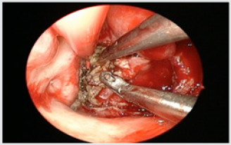
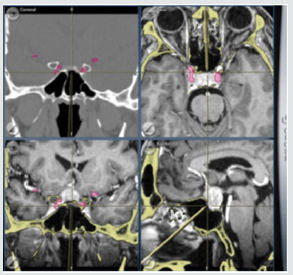
The development of navigation system and other power instrument used endoscopically helped the development of endoscopic skull base surgery to be done more safely and effectively. Example of endoscopic nasopharyngectomy was done first in ENT department QEH in 2010 for a post radiotherapy nasopharyngeal carcinoma patient with primary adenocarcinoma. It was done with two surgery four hands technique (Figure 1) and a complete excision was achieved. Subsequent endoscopic nasopharyngectomy were done for recurrent NPC patients with aid of power instrument of coblator which give a thin cut layer by layer when approaching the dangerous area of carotid and skull base (Figures 2&3) A nasoseptal flap were created to cover the nasopharyngectomy wound and help patient recovery faster (Figure 4). The overall longterm result is about the same as open surgery. The second types of surgery done for skull base lesion is cerebrospinal fluid leak which either resulted from iatrogenic causes or spontaneous causes or from meningocele. Dura seal and fibrin glue and cauterization of meningocele were used to stop the CSF leak. Lumbar drain was used by neurosurgeon in these cases when two specialty worked together (Figures 6 & 7).

Figure 1: Two surgeon four hands technique.

lupinepublishers-openaccess-journal-otolaryngology

Figure 2: Navigation system.

lupinepublishers-openaccess-journal-otolaryngology

Figure 3: Use of coblator.

lupinepublishers-openaccess-journal-otolaryngology

Figure 4: Nasoseptal flap covering nasopharyngectomy wound.

lupinepublishers-openaccess-journal-otolaryngology

Figure 5: Meningocele.

lupinepublishers-openaccess-journal-otolaryngology

Figure 6:

lupinepublishers-openaccess-journal-otolaryngology

Figure 7: Extracranial neuroblastoma.

lupinepublishers-openaccess-journal-otolaryngology

Figure 8: Sphenoid sinus inverted papillom

lupinepublishers-openaccess-journal-otolaryngology

The third type of surgery is for tumor of neuroblastoma and inverted papilloma involving skull base. Endoscopic approach with post operation radiotherapy is used for a complete control of two cases of neuroblastoma with no intracranial extension. One inverted papilloma occupied and expands the sphenoid sinus and removed endoscopically (Figures 8 & 9). The last type of surgery is endoscopic removal of intraconal intraorbital tumor. It was done together with eye doctor to debulk and excise one cavernous haemangioma and one schwannoma (Figure 10). Lastly the degree of difficulty of endoscopic skull base surgery can be classified into different ladder according to some expert (e.g. Prof. Richard Carrau) It starts with level 1 to level 5. Level include sinonasal surgery, level 2 Pituitary gland surgery, CSF leak. Level 3 is extradural surgery of transcribriform to transodontoid etc. Level 4 is intradural and level 6 in cerebrovascular surgery. It takes a great effort to learn the technique in advanced level and a high-risk surgery. A teamwork with neurosurgeon is essential when intradural pathology is managed and therefore not every center even with neurosurgery support can do these surgeries especially with inadequate caseload experience.

Figure 9:

lupinepublishers-openaccess-journal-otolaryngology

Figure 10:

lupinepublishers-openaccess-journal-otolaryngology


Friday, 22 January 2021

Lupine Publishers | Nasolabial Cyst: Report of 2 Cases and Literature Review

 Lupine Publishers | Journal of Otolaryngology


Abstract

Introduction: Nasolabial cysts are a rare, ectodermal development cyst, presenting as a fullness of canine fossa, nasal ala or vestibule of the nose. They are usually asymptomatic but may become infected. Treatment is complete surgical excision by sublabial approach, or transanal endoscopic marsupialization.

Case Report: Description of two cases, one female presenting as nasal deformity due to progressive growth of unilateral nasolabial cyst, and a healthy young male presenting severe facial cellulitis, with a CT showing bilateral nasolabial cysts.

Discussion: Nasolabial cyst is a rare condition, but diagnosis and treatment are simple. Nasolabial cyst should be incorporated by ENT in the differential diagnosis of nose deformities and facial swelling.

Introduction

Nasolabial cysts are also described as nasoalveolar cyst or Klestadt cyst and were first described by Zuckerkandl [1,2]. They are rare, affecting 1,6 per 100.000 persons per year, more frequently in females (4:1 ratio), especially among African Americans, in the fourth and fifth decades of life. 90% are unilateral, and often underdiagnosed [3,4]. Nasolabial cysts are non-odontogenic cysts that develop lateral to the midline of the maxillary lip and alar base. They usually present as a swelling in the nasolabial fold, causing alar nose elevation and upper lip projection. They may grow slowly and painlessly over several years. Because of its close anatomical relation to the nasal cavity and teeth, they may become infected easily, rapidly growing and being painful [1,5]. Diagnosis is made by clinical examination, imaging tests, and is confirmed by histopathologic study. The cyst can be palpated bimanually with one finger in the floor of the nasal vestibule and another in the labial sulcus. Computed tomography (CT) and magnetic resonance (MR) can be useful. Differential diagnosis includes cysts of the nasopalatine duct, periapical inflammatory lesions (granuloma cyst, abscess), and epidermoid cysts [1,6]. Complete surgical excision of the nasolabial cyst is the best treatment, and sublabial approach is most used. Other authors propose transanal marsupialization as an easier and shorter procedure, with lesser complications, but recurrence may be a problem [3,5].

Case Report 1

Figure 1(a).

lupinepublishers-openaccess-journal-otolaryngology

Y=372.256 + 6.8856. X1+residuals

52-year-old female patient with a 3-year history of left floor of nose intermittent painful swelling, and progressive deformity of this area. Computed tomography (CT) study showed a 3,2 cm rounded lesion in left pyriform aperture compatible with nasoalveolar cyst (Figure 1). A sublabial approach was performed, and complete excision of the cyst was obtained (Figure 2). Histological study confirmed suspected diagnosis. Patient evolved in excellent conditions, with no complications or recurrence in a 2 year follow up period.

Figure 1(b).

lupinepublishers-openaccess-journal-otolaryngology

Figure 1(c).

lupinepublishers-openaccess-journal-otolaryngology

Figure 2. Intraoperative view of sublabial access to left nasolabial cyst in Case 1.

lupinepublishers-openaccess-journal-otolaryngology

Case Report 2

Figure 3. Computed tomography of bilateral nasoalveolar cysts. Case 2.

lupinepublishers-openaccess-journal-otolaryngology

Figure 4. Pathology H-E study of Case 2 showing cyst wall with pseudostratified columnar cells.

lupinepublishers-openaccess-journal-otolaryngology

37-year-old male patient, with no prior medical record, developed a facial cellulitis with no initial response to oral antibiotics, requiring hospitalization and intravenous antibiotic treatment. Maxillary and orbital CT study showed bilateral nasoalveolar cysts, with signs of active infection in the right cyst (Figure 3). Antibiotic treatment was completed, and cyst resection was programmed a month later. An extensa sublabial approach was performed, and bilateral cysts were removed. Histological findings confirmed diagnosis (Figure 4). Patient did not have any other episode of facial cellulitis or cyst recurrence in a 14 month follow up

Discussion

Origin of nasolabial cysts is controversial. Most accepted theories are that cysts derive from remnants of the nasolacrimal duct or inclusion cysts of mesenchymal cells during the fusion of medial and lateral nasal prominences to the maxillary prominence during facial skeletal formation [2]. Histology usually present pseudostratified columnar epithelium. Su et al noticed with electronic microscopy that that cysts had a highly placated mucosa, of non-ciliated stratified columnar epithelium, differing from the ciliated columnar epithelium of the paranasal and nasal sinuses [7]. The treatment of nasolabial cysts consists on complete removal of the lesion, with the objective of prevention of infectious complications, histologic diagnosis, and aesthetic improvement. Fine needle aspiration can help in diagnosis, and relieve of symptoms, but recurrence is high [1]. Surgery is usually done by sublabial approach, creating a mucosal flap of gingiva to allow access to the pyriform aperture and to the cyst, which can be resected carefully to avoid rupture, especially to the floor of the nose mucosa, and complete excision is mandatory to avoid recurrence. After the intervention, the gingival mucosal flap is fixed in its original position with absorbable sutures. Potential complications are uncommon, including facial swelling, insensitive gingiva, teeth numbness, and surgical site infection.

Patients must use a toothbrush on the surgical site. Diet should be soft for the first week, then normal. Dental prostheses can be used immediately after surgery [1]. Lee et al published in 2009 a comparative study between surgical techniques, and strongly suggested endoscopic trans nasal marsupialization as a simpler, shorter and safer procedure, and in most cases, it could be done under local anesthesia [3]. In our two cases we used sublabial approach with general anesthesia because is the usual technique used in our practice, but we are interested to use endoscopic marsupialization in next cases. We did not have any complications or recurrences in follow up of both cases.

Conclusion

Nasolabial cysts are rare but must be considered in the differential diagnosis of floor of the nose deformities and facial swelling. Sublabial approach is the most common procedure, but probably be replaced by endoscopic marsupialization in the future.

Read More Lupine Publishers Otolaryngology Journal Articles:
https://lupine-publishers-otolaryngology.blogspot.com/

Friday, 15 January 2021

Lupine Publishers | Endoscopic Recording (or its Absence): A Disappointment

 Lupine Publishers | Journal of Otolaryngology


Short Communication

One of the advantages of being old and seeing science and clinical practice advance is that as time passes, we can observe those things that succeed and those that fail if we are perfectly honest in our assessments. Moreover, we can determine if practices we thought were transferable to our colleagues simply by describing them in the literature have often proven to be false hopes that leave us scratching our heads in order to determine just what went wrong. Such is the case with endoscopic procedures designed to assess the upper airway anatomy and physiology [1]. Between the two of us, we have more than 75 years of combined experience with flexible fiberoptic nasopharyngoscopy of the upper airway, literally thousands of cases examined both individually and together, and between us, we have published approximately 200 articles, chapters and books that directly or indirectly report on the use of flexible fiberoptic nasopharyngoscopy in people with upper airway disorders, respiratory obstruction, speech disorders, and voice disorders. In other words, time has afforded us the opportunity to see the application of endoscopic procedures from their beginnings to the current state of the art practice [2,3]. What we see disturbs us mightily.

The first author was introduced to endoscopic procedures for assessing velopharyngeal insufficiency in 1974 when he met and became familiar with the work of two pioneers in imaging procedures who were on opposite ends of the planet, specifically the United Kingdom and Japan. Ron Pigott, a plastic surgeon at Frenchay Hospital in Bristol, UK, and Tadashi Miyazaki, head of the Department of Maxillofacial Surgery at Osaka University Dental School in Japan published and presented papers in the late 1960s and early 1970s describing the application of nasopharyngoscopy to assess velopharyngeal closure [4]. Mr. Pigott published papers describing the use of both flexible fiberoptic endoscopes and rigid endoscopes in 1969. Dr. Miyazaki and his colleagues, Drs. Matsuya and Yamaoka, described the use of a side-viewing flexible fiberoptic endoscope of what at that time was a narrow diameter just above 3mm. We were introduced to narrow diameter endviewing pediatric fiberoptic endoscopes several years later and immediately applied them in day-to-day clinical use and research, both for children and adults. With flexible nasopharyngoscopy applicable to assessing speech, voice, respiration, and swallowing being available in the Americas since the 1970s, the market for the instruments expanded dramatically and smaller, narrow diameter endoscopes with improved optics and light transmission characteristics were rapidly developed by a number of companies. The introduction of recording the examinations with video were introduced by both the team in Osaka and Mr. Pigott in Bristol nearly simultaneously. In our own institutions, the ability to record the examinations and share them with our interdisciplinary cleft palate teams advanced our treatment techniques dramatically and increased a transdisciplinary appreciation for the complexity of normal speech production.

Today, flexible fiberoptic nasopharynges laryngoscopes are ubiquitous in the offices of otolaryngologists worldwide. High definition video/sound recordings are easy to accomplish and should be inexpensive and easy, whether equipment is purchased as a package from commercial medical instrument companies or rigged by clinicians who have some knowledge of digital recording of endoscopic images. The process is not difficult. Because the information is used often in the assessment of speech and vocal production, the recording of speech synchronous to the video recording is essential. Data storage is inexpensive with digital recording devices storing four terabytes of information available for as little as $100. Four terabytes provide even busy clinicians enough storage space for years. Nonetheless, many clinicians do not record office endoscopies, or they record video without sound thereby rendering later review impossible. Modern computer equipment is the perfect platform for recording endoscopic studies with sound and high quality (high definition video with 1080p or higher resolution) digital images. The first author on this paper put together his own system consisting of a Mac computer (any Mac computer would do) with video recording software (lowest price would be approximately $1,000), an endoscopic video camera (available for approximately $1500 and higher), a microphone ($50) and digital storage ($100). Thus, an entire system to allow recording and storage of digital video and sound would be well under $3,000, the cost of which would be amortized within a very short period.

Why would it be essential to record endoscopic examinations? That the question has to be asked is disappointing. Let’s develop an imaginary scenario. You, the reader, have a complaint of difficulty catching your breath, coughing frequently, a fever, and malaise. The doctor who sees you thinks that you may have pneumonia, so the doctor orders a radiographic procedure to look at your lungs. The doctor does a fluoroscopy but does not record it or print a picture from the study. You go to a second doctor who believes you simply have a severe allergic reaction to pollen and wonders why the other doctor thought you had pneumonia. The second asks the first doctor for copies of the x-ray study. The first doctor says, “I didn’t put it on film, but I saw evidence of pneumonia.” The second doctor indicates his displeasure because the first doctor’s findings disagree with the second doctor’s. Would you trust a verbal report of a radiographic procedure that only one person has seen? A rhetorical question, I hope. Moreover, the interpretation of endoscopic studies is variable leading to possible false positive/ negative findings affecting treatment recommendations. If not recorded, the value of the study is questionable. Reviewing 1,525 consecutive nasopharyngoscopic examinations with recorded video and sound resulted in 598 surgical recommendations including velopharyngeal reconstruction, adenoidectomy, and/ or tonsillectomy and surgical airway management. In all cases, surgery was based on review of recorded studies by the surgeon and endoscopist, and often by other clinicians and students. In a subset of 200 consecutive reviewed by three or more clinicians, we found that interpretation disagreements occurred in 48% of cases. Disagreements were relevant to treatment decisions in 94%. Group reviews eventually resulted in consensus agreement. Efficacy was measured by surgical outcomes for velopharyngeal insufficiency and obstructive airway procedures (94% and 95% respectively). Because we currently consult on cases from the U.S. and overseas, we often request copies of endoscopic videos; 296 requests have been made since 2012, 221 from the US, 75 from Europe Of these, 48% from the U.S. and 39% from overseas had available video recordings with less than one-third of the videos having sound [5]. The inability to review completed studies may lead to treatment error, unacceptable for any imaging procedure. It is time that clinicians stop doing imaging procedures that cannot be reviewed by more than one person. An endoscopic study without video and sound recording is archaic, inadequate, and not to be trusted.

Read More Lupine Publishers Otolaryngology Journal Articles:
https://lupine-publishers-otolaryngology.blogspot.com/

Wednesday, 30 December 2020

Lupine Publishers | Help! Our Tinnitus Patients Want A Drug?

 Lupine Publishers | Journal of Otolaryngology


Abstract

Tinnitus is common and can severely affect life quality in many. There are no FDA approved drugs for tinnitus treatment and no surgery for sensorineural tinnitus. Up till now, there are no medications or supplements approved for tinnitus; our impression is that they are widely used. Therefore, we believe it is helpful to review the literature and provide some suggestions for clinicians and sufferers. We review studies which describe current and emerging pharmacotherapies. In addition, we describe recent advances in the tinnitus field which may help overcome obstacles faced in the pharmacological treatment of tinnitus.

Introduction

Tinnitus is a common complaint and can be very debilitating [1]. The prevalence of tinnitus in adult populations ranges from 7 % to 19 %. It increases with age [2]. In up to 5% of the adult population, tinnitus interferes negatively with the ability to lead a normal daily life; affecting many activities of daily life; including (a) thought and emotions, (b) hearing, (c) sleep, and (d) concentration. Each person is affected differently [3]. Several counseling and sound therapies are used and are helpful to many patients Tyler [4]. The Psychological Model of tinnitus considered as an important distinction between the tinnitus from reactions to the tinnitus [5]. The counseling and sound therapy help with the reactions, but tinnitus suffers often prefer pill [6]. Medications can be used to treat patients with depression and anxiety and to help with sleep problems, but do not directly change the tinnitus. Despite the significant clinical need for effective treatment of tinnitus, there is currently no single U.S Food and Drug (FDA)-approved medication for treatment of   tinnitus. Various drug regimens have been tried and a few of them had shown some favorable results, but most did not result in significant benefits [7]. As with any bothersome, common disorder which lacks understanding and effective treatments, tinnitus considered as an easy target for scams [8]. Thousands of purported cures can be found on the internet and in stores providing ‘over the counter’ pills.  People waste billions of dollars on tinnitus treatment yearly. Some scams are blatant.  Others are subtle.

Drugs for Tinnitus Treatment

It seems unlikely that there will be one single drug which will cure all forms of tinnitus. It is likely that many subtypes of tinnitus exist and that each will require a different form of treatment. Thus, one of the pressing needs in tinnitus research is a scheme to classify patients into subtypes that might respond positively to a specific drug treatment. Indeed, some drugs have been reported to provide some kind of symptoms relief in a subset of patients but in others not [9]. We will review some of these drugs which might help in tinnitus symptoms relief.

Anti-Arrhythmic

a) Lidocaine

In 1935, lidocaine was suggested and used to suppress tinnitus through nasal administration and since then many clinical studies confirmed transient suppression of tinnitus through lidocaine administration [10].  Its effect seems to be dose dependent with suppression of tinnitus occurring at free arterial plasma concentrations from 1.75 to 3.5 μmol/L.  However, it was found that concentrations more than 3.5 μmol/L might induce tinnitus [11]. The mechanism of action by which intravenous lidocaine might suppresses tinnitus is incompletely understood, but there is evidence that it affects both the cochlea and the central nervous system [7]. It is used as local anesthetic and anti-arrhythmic, it acts mainly through binding to fast voltage-gated sodium channels, reducing the magnitude of the sodium current during depolarization [12]. It also affects calcium, potassium, and glycine-evoked chloride currents [13]. Since the effects of lidocaine are short lived and due to its potentially life-threatening side effects, like cardiac arrhythmia, drowsiness, dizziness, confusion, and restlessness; lidocaine is not an option for long-term treatment. Oral analogues of lidocaine such as tocainide have been evaluated as a potential long-term therapy for tinnitus. However, several randomized, controlled studies found that tocainide had little benefit for tinnitus [14]. A nonsterile patch (Lido PAIN TV, EPI Cept) has been developed for delivery of lidocaine applied over preauricular skin. Its clinical efficacy is under clinical trial. It is produced under the pharmaceutical company EPI Cept which has less systemic concentrations than intravenous lidocaine which is needed to suppress tinnitus [5

Anti-Depressants

The tricyclic antidepressants nortriptyline, amitriptyline and trimipramine have been investigated for the treatment of tinnitus. Beside their antidepressant properties, tricyclics have been shown to be highly efficient for the treatment of chronic pain, which is of interest in view of the proposed etiological similarities between tinnitus and neuropathic pain [16]. Examples of tricyclic antidepressant which can be used for tinnitus include (amitriptyline, trimipramine, and nortriptyline), the latter found to inhibits mainly the reuptake of norepinephrine and to a lesser extent serotonin, whereas amitriptyline inhibits the reuptake of serotonin and noradrenaline almost equally, on the other hand trimipramine differs in its mode of action from other tricyclic antidepressants in that it blocks postsynaptic dopamine and serotonin receptors [17]. From all tricyclic antidepressants, nortriptyline is worth noting. Sullivan et al. [18] reported small-scale, single blind placebo-washout study involving patients with severe tinnitus and major depression, nortriptyline significantly reduced depression and tinnitus loudness (10 dB reduction) in some. In a follow-up double blind placebo- controlled study involving subjects with severe tinnitus and severe depression or depressive symptoms, nortriptyline significantly reduced depression scores, tinnitus disability scores, and tinnitus loudness (6.4 dB reduction) relative to a placebo [19].  They concluded that there was a significant correlation between the reduction in tinnitus disability scores and depression scores, suggesting that nortriptyline is effective in reducing tinnitus loudness and severity in severely depressed tinnitus patients.  There was less benefit in non-depressed individuals [20].

a) Amitriptyline

Bayar et al. [21] reported a study (n= 37) to compare amitriptyline with placebo and found after 6 weeks of 100 mg amitriptyline, a significant reduction of tinnitus complaints and tinnitus loudness compared to the placebo group. Podoshin et al. [22] reported another study (n=225), where amitriptyline was compared with biofeedback, and 27.5% of patients reported improvement with amitriptyline while 43.5% reported improvement with biofeedback revealing the superiority of biofeedback.

b) Trimipramine

Mihail et al. [23] evaluated Trimipramine small double-blind placebo cross-over study (n=19) and did not demonstrate any difference between trimipramine and placebo treatment.

c) Selective serotonin reuptake inhibitors (SSRI)

SSRI such as sertraline have been tested. In a randomized double-blind placebo-controlled study (n=76), patients without severe hearing loss, but with depression, anxiety, and a high risk for developing severe tinnitus, sertraline was significantly more effective than placebo in reducing tinnitus loudness and tinnitus severity [24].

On the other hand, paroxetine was evaluated in a double-blind, placebo-controlled study involving chronic tinnitus patients without comorbid depression, the paroxetine group showed little difference from placebo on tinnitus loudness, Tinnitus Handicap Questionnaire (THQ) scores, and other measures [25]. Collectively, the results suggest that tinnitus patients with depression and anxiety may benefit from antidepressants [25]. However, this effect may not to be a direct effect of antidepressants on tinnitus severity, but rather due to the beneficial effect of antidepressants on comorbid depression and anxiety. Dose of antidepressants for the treatment of tinnitus is in a similar range as that used in the treatment of depression. In general, a low starting dose and slow increase of the dosage reduce side effects. Since beneficial effects do not occur immediately, minimum treatment duration of 6–12 weeks at the effective dose is recommended. If treatment effects are unsatisfactory and the decision is made to discontinue or change treatment, dosage should be reduced slowly. If a patient experiences beneficial effects, treatment should be continued at a stable effective dose for about 6 months, then the dose can be reduced over the course of weeks to months. Should the tinnitus get worse during a reduction of the dose, it is recommended to keep the dosage at the minimum providing relief.

Anxiolytics

Since tinnitus is thought to be the result of an imbalance between excitatory and inhibitory neurotransmission toward the GABA receptor [26], and benzodiazepines are allosteric potentiators of the GABAA receptor so, they have a positive effect on tinnitus by increasing inhibitory neurotransmission. Furthermore, due to their anxiolytic and sleep-inducing properties, benzodiazepines should have beneficial effects on comorbid anxiety and insomnia, and thus may help patients cope with their tinnitus.

a) Alprazolam

Johnson et al. [27] reported double-blind placebo-controlled study(n=36), 12 weeks of alprazolam administration at an individually adjusted dosage reduced tinnitus loudness in 76% of subjects  measured with a tinnitus synthesizer and a visual analog scale – whereas only 5% showed a reduction in tinnitus loudness in the control group. Although the strong positive effects of alprazolam are encouraging, the study has been criticized because of the small sample size, drug dosing method, and failure to assess emotional effect.

b) Clonazepam

Bahmad et al.  [28] Stated in their retrospective study of medical records from over 30patients taking clonazepam (0.5–1 mg/day, 60–180 days) for vestibular or cochleovestibular disorders, that 32% of patients reported an improvement in their tinnitus. Due to their immediate effects, short-acting benzodiazepines such as lorazepam or alprazolam are widely used for acute treatment of anxiety, agitation, and insomnia, symptoms that frequently occur with tinnitus. The longer acting clonazepam provides some relief in a considerable subgroup of patients. The use of benzodiazepines should be restricted to short periods of time due to the risk of drug dependency. Moreover, caution is warranted since protracted tinnitus has been reported after discontinuation of benzodiazepines [29].

Anticonvulsants

 Anticonvulsants are increasingly used in the treatment of several non-epileptic conditions, including various psychiatric disorders and pain syndromes [30].  Some of them have also been investigated for the treatment of tinnitus. As this anticonvulsant act on voltage gated Sodium and calcium channels, and on synaptic transmission mainly mediated by gamma amino butyric acid type A (GABAA) receptors [31] with resultant reduction in neuronal excitability, thus they should be beneficial for the treatment of tinnitus.

a) Carbamazepine

Carbamazepine binds to voltage-gated sodium channels and stabilizes the sodium inactivation state, thereby reducing neural firing [32], it had been investigated for tinnitus and results were mixed. Based on the assumption that carbamazepine resembles lidocaine in its mechanism of action, studies investigated the effect of carbamazepine in tinnitus patients who previously had responded to intravenous lidocaine [33]. About half of these patients had a positive response to carbamazepine (600–1,000 mg daily). A significant benefit from carbamazepine has been reported for a rare group of patients who have intermittent tinnitus that sounds like a typewriter, popcorn, or ear clicking, and which is caused by a neurovascular conflict [34].

b) Gabapentin

The anticonvulsant gabapentin acts on voltage gated calcium channels and is also used for the treatment of seizures, neuropathic pain, and migraine [35]. Results with gabapentin for the treatment of tinnitus are contradictory; one controlled trial (n=39) has shown a significant improvement in tinnitus annoyance and loudness for a subgroup of participants with tinnitus related to acoustic trauma [36], other study (n=76) did not detect any improvement in tinnitus handicap, but did report a significant improvement in tinnitus annoyance when compared to placebo [37].

Anti-Glutamatergic Agents

Glutamate receptor antagonists have been tried in tinnitus sufferers. The rationale behind it is that imbalance between inhibitory versus excitatory neurotransmission which was observed in several regions of the auditory pathway in tinnitus development thus blocking glutamatergic neurotransmission could also exert neuro protectant effects [38].

a) Caroverine

Caroverine, a spasmolytic drug, which is an antagonist of non- N-methyl-D-aspartate (NMDA) and NMDA glutamate receptors [39], has been investigated in patients with putative cochlear tinnitus. Patients were randomized to receive either caroverine (I.V., maximum dose 160 mg) or placebo. Within the caroverine group, 63% showed a reduction in loudness immediately after treatment and 43 % still showed improvement one week later, whereas there was no improvement in the placebo group [40].

b) Acamprosate

Acamprosate is non-selective NMDA receptor which had been tried in a double-blind study (n=50), Patients received placebo or acamprosate (333 mg, three times per day) and rated the loudness and annoyance of their tinnitus before and at monthly intervals of treatment. Acamprosate had no beneficial effects after 30 days of treatment, a modest benefit at 60 days, and a significant effect at 90 days. Approximately 87% of the subjects in the acamprosate group showed some improvement, including three subjects in which tinnitus disappeared, compared to 44% in the placebo group [41]. This study has not been published but the main author said it had no effect on tinnitus. A larger clinical trial is currently underway to analyze the encouraging results from this preliminary study (http://clinicaltrials.gov/ct2/show/NCT00596531).

Dopaminergic–Antidopaminergic Drugs

Dopaminergic pathways in limbic and prefrontal areas found to be involved in mediating emotional aspects of tinnitus so both dopaminergic and antidopaminergic drugs have been proposed for treating tinnitus.

a) Sulpiride

An antipsychotic drug that selectively blocks dopamineD2 receptors [42], significantly reduced subjective ratings of tinnitus and tinnitus visual analogue scores in one double-blind, placebo-controlled study. Further studies investigated combinations of sulpiride with melatonin, A prospective, randomized, double-blinded, placebo-controlled study was done (n=120) patients consulted for subjective tinnitus. They were included randomly in four groups of 30. One group took sulpiride (50 mg/8 h) alone, the second group took melatonin (3 mg/24 h), the third group took the same doses of sulpiride (50 mg/8 h) plus melatonin (3 mg/24 h), and the fourth group took placebo (lactose 50 mg/8 h), all for 1 month, Subjective grading of tinnitus perception and a visual analogue scale (0-10) were done for evaluation of results. Based on the subjective grading, tinnitus perception diminished by 56% in patients treated with sulpiride, by 40% in patients treated with melatonin, by 81% in patients treated with sulpiride plus melatonin, and by 22% in patients treated with placebo, concluding that Sulpiride and melatonin reduce tinnitus perception, decreasing dopamine activity. The tinnitus auditor-limbic dopaminergic pathway has broad therapeutic implications [43].

b) Piribedil

Piribedil was investigated recently in a double-blind placebo-controlled cross-over study (n=56). Results showed that piribedil was not superior to placebo; however, a post-hoc analysis suggested that a subgroup of patients with specific findings in electrocochleography may benefit from piribedil [44].

Other drugs

a) Misoprostol: is a synthetic prostaglandin E1 analogue which is primarily used to prevent gastric ulcers induced by non-steroidal anti-inflammatory drugs [45]. In a small, placebo-controlled cross-over study, tinnitus severity improved in 33% of subjects during misoprostol treatment (escalating to 800 mg/day), while none improved with placebo [46]. A subsequent double-blind placebo-controlled study (n=40) has shown a significant reduction of tinnitus loudness with misoprostol treatment, but no differences in subjective measures of tinnitus severity [47]. A further study has shown efficacy of misoprostol in the treatment for chronic tinnitus in hypertensive and/or diabetic patients [48].

b

b) Cyclandelate, a vasodilator used in the treatment of cerebrovascular and peripheral vascular disorders, that is believed to act by blocking calcium influx [49], has been investigated for the treatment of tinnitus based on the assumption that some forms of tinnitus may arise from cerebrovascular insufficiency. In an open multicentric clinical trial of patients with tinnitus, vertigo, and visual disturbances, 90-day treatment with cyclandelate reduced the severity and frequency of these symptoms with minimal side effects [50]. However, in a subsequent placebo-controlled double-blind study, cyclandelate did not significantly change audiometric measures of tinnitus loudness and pitch and caused side effects in many patients [51].

c) Furosemide is a loop inhibiting diuretic used to treat congestive heart failure and edema; it inhibits the Na-K-2Cl cotransporter [52], which is expressed in the inner ear as well as in the brain [53]. Furosemide has been proposed as a treatment for tinnitus of cochlear origin because it strongly suppresses the endolymphatic potential and other cochlear responses [54]. In one study, approximately 50% of patients reported a reduction of tinnitus symptoms following intravenous furosemide treatment. Furosemide has also been found to suppress tinnitus in approximately 40% of patients with Meniere’s disease [55]. In contrast to these positive findings, high doses of furosemide have also been found to induce temporary hearing loss and tinnitus [56].

d) Nimodipine, a calcium antagonist, which crosses the blood - brain barrier and blocks L-type calcium channels [57], is primarily used in the treatment of subarachnoid hemorrhage. Pilot studies also suggest beneficial effects in mood disorders [58]. In animal studies nimodipine significantly reduced tinnitus related behavior caused by high doses of quinine or sodium salicylate [59]. The first open clinical trial suggested positive effects of nimodipine on tinnitus in some patients [60]. However, these results could not be confirmed in a second open clinical trial [61].

e) Cyclobenzaprine a centrally acting muscle relaxant with pharmacological properties similar to amitriptyline, used in the management of fibromyalgia, low back pain, neck pain and temporomandibular disorders. Two open preliminary studies evaluated the action of cyclobenzaprine in the treatment of tinnitus have shown evidences that this drug presents a clear action in reducing tinnitus magnitude as well as in tinnitus discomfort [62,63]. Another study conducted in animal model found that cyclobenzaprine was effective in the transient suppression of noise-induced tinnitus in rats. Cyclobenzaprine is a promising drug to treat tinnitus and preliminary results need to be validated in controlled clinical trials [64].

f) Other drugs that have been tested with either limited efficacy or are in need of further controlled trials include the HMG-CoA reductase atorvastatin [65], betahistine [66], Deanxit [67], oxytocin [68], naltrexone [69], ondansetron [70], the phosphodiesterase inhibitors cilostazol [71]and cannabinoids [72].

Drugs in the pipeline for patients with tinnitus

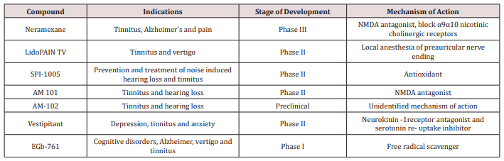
a) Neramexane: acts as a non-competitive, voltage dependent NMDA antagonist and also blocks α9α10 nicotinic cholinergic receptors which are expressed on hair cells in the inner ear [73]. After obtaining positive results from a Phase II trial. (http://clinicaltrials.gov/ct2/show/NCT00405886), Merz Pharmaceuticals is currently conducting Phase III multi-center clinical trials to determine the efficacy, safety and tolerability of Neramexane for treating tinnitus.

b) LidoPAIN TV: is a non-sterile patch delivering lidocaine, which is applied to the periauricular skin region. According to the company’s web page, it demonstrated efficacy in a clinical proof of- concept study and has been in phase II trials for tinnitus [15].

c) SPI-1005: contains ebselen which acts as an antioxidant by stimulating glutathione peroxidase. It has completed a phase I trial and phase II trials are planned for the treatment of noise-induced hearing loss and tinnitus [9].

d) AM 101: is an NMDA antagonist, which is applied topically to the cochlea by round window Injection for the treatment of acute tinnitus. In a randomized, double-blind placebo-controlled German Phase I/II trial in 24 patients with acute tinnitus following noise trauma or sudden deafness, single-dose AM-101 intratympanic injection was well tolerated. It also had a positive effect on tinnitus loudness. Currently a double-blind, randomized, placebo-controlled Phase II trial with cochlear application of AM-101 is being carried out. The study involves patients with acute (<3 months) noise-induced tinnitus that have not responded to glucocorticoid treatment. he same company has also a second compound under development (AM-102). This compound is of unidentified pharmacologic activity and is also delivered by intratympanic application. It is currently in preclinical test [9].

e) Vestipitant: is a novel antagonist of the neurokinin-1 (NK-1) receptor which binds substance P. Neurokinin receptors are present in the inner ear and therefore represent a potential therapeutic target for tinnitus [74]. Vestipitant and the combination of vestipitant and paroxetine are currently undergoing a phase II clinical trial for the treatment of tinnitus [75].

f) EGb-761: is a concentrated extract of Ginkgo biloba, enriched in flavonoids and terpenes, which has a broad spectrum of pharmacologic actions, including a free-radical scavenger effect and which has shown efficacy for tinnitus in a phase I trial (Table 1).

Table 1: Drugs under development for tinnitus.
lupinepublishers-openaccess-journal-otolaryngology

Conclusion

The use of drugs to treat tinnitus is widespread. Some subjects report benefits from drugs, while others do not. There is likely a placebo effect influencing those results. It is also noteworthy that many patients develop side effects. Patients using drugs might not be aware of the side effects, even though warnings might be present on labels. Tyler et al. [76] stated that there are many subgroups of tinnitus and it might be that different drugs help some subtypes tinnitus sufferers, but not all tinnitus sufferers. Despite the huge potential market, we still lack approved pharmacological treatments for tinnitus. Despite the significant unmet clinical need for a safe and effective drug targeting tinnitus relief, there is currently no single FDA approved drug on the market. The empirical approach, in combination with new developments in information technology such as the advent of big data approaches in medicine, might facilitate the identification of promising drugs. We have also stressed the importance of examining individual results.

Read More Lupine Publishers Otolaryngology Journal Articles:
ps://lupine-publishers-otolaryngology.blogspot.com/

Friday, 25 December 2020

Lupine Publishers | An Actual Statistical Problem with Model Selection My Solution

 Lupine Publishers | Journal of Otolaryngology


Introduction

This manuscript is a follow-up of my last one in SJO where I promised to show you my solution of a client problem. This case has been transformed such, that the logic of the problem remained unchanged and the confidentiality of my client’s data is warranted, however. In the last manuscript the results of the commonly found methods of modelling were shown and discussed. Now we will show my results and a discussion of them. Mathematically speaking the mainstream models already shown could be summarized as two univariate approximations by straight lines or a two-dimensional fit of a plane, describing the dependent variable by an approximation based simultaneously by a constant and two linear terms. I was fully aware of the limitations of the very small sample size of the mainstream models and I hope to successfully use this example to convince my readers about the economic benefits of discussions with professional mathematicians/statisticians instead of users of statistical software as I have classified them.

Methods

As I was blinded to the actual meaning of the variables X1, X2 and Y my experience indicated that I should try a second order polynomial fit as this would be the simplest possible model extension as compared to the mainstream linear models. The similarity to the considerations of Occam’s razor (see Wikipedia) are also well based on my personal professional experience. My model equation used is displayed below:

Y (X1; X2) =a0 + a1. X1 + a2. X2 + a3. X12 + a4. X1. X2 + a5 .X22 + error term (equ 1)

The above equation contains prior regression analysis the coefficients a0, a1 and a2 for the linear terms and a3, a4 and a5 for second order polynomial terms which must be estimated from the data by means of linear regression based on the method of least squares. The error term must fulfil the assumption that the data points represent statistically independent observations with constant variance in the domain of data points and an approximate Gaussian distribution. The most important data requirement is a continuous and metric measurement scale of the data and based on my long- term experience in medicine and other statistical applications, if fulfilled, the basis for a highly robust behavior of the regression analyses based on least squares. Finally, enough data points must be available. This is a problem in the determination of the sample size, which, in my opinion, requires professional statistical assessment.

Result

The numerical details are shown in Table 1 below with additional information necessary in the Excel data analysis software as the input for Excel’s regression routine:

a) Note 1: X1 and X2 and Y refer to the client provided original data. The author intended to look at a standard polynomial of degree 2 and the calculated data columns indicate all second order terms necessary from Excel logic for that purpose. The contents after the provided Y in the brackets are a help to understand that Y (as provided from my client) is the dependent variable of X1 and X2 in this very model. For physicians unfamiliar with exponential floating-point formatted numbers reading of the Excel online documentation is recommended.

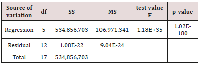
b) Note 2: There are three lines in Table 2. The descriptions in column one show regression in the first, residual in the second and total in the third line. The total in line 3 displays the SS of all data against the grand mean. The residual in line 2 shows the sum of squares of the differences between data and calculated Y values using the coefficients a0, a1, …, a5 and the line 1 described as regression provides us with the information of the explained variation by the calculated regression coefficients. In view of the raw Y data shown in Table 1 we observed therefore a residual variance - in the magnitude of 9,0403. 10-28 which – for practical purposes – might be judged as zero. The mathematical interpretation in everyday language is there is an interpolation problem or a perfect fit between the raw Y data and the regression equation with the calculated coefficients shown in

Table 1:

lupinepublishers-openaccess-otolaryngology-journal

Table 2: ANOVA analysis of variance table

lupinepublishers-openaccess-otolaryngology-journal

df: degrees of freedom

SS: sum of squares

MS: mean squares (represent the variances which are the squared

standard deviations).

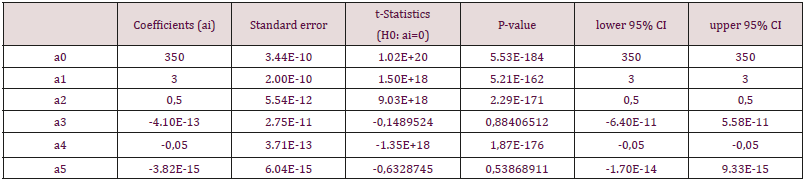
c) Note 3: The coefficients ai refer to the equation (1). Please note that i = 0, 1, 2, …, 5 in column one and a0 is frequently assigned the name intercept. We follow the frequently engaged standard statistical practice of setting not statistically significant coefficients to zero and have the solution of our model (from equation (1)) in equation (2) below:

Y (X1; X2) = 350 + 3. X1 + 0,5. X2 - 0,05. X1. X2 (equ 2)

The inevitable rounding errors which are present in all common computers are reflected in the Excel documentation which states that about ten to twelve digits in decimal results should be reliably exact. Therefore, it seems not to be a problem that 95% confidence intervals cover zero and actual numbers of digits of the raw data in Table 3 justify this decision. We analyzed in addition the model of equation 2 and for practical purposes we concluded that there were perfectly consistent results (data on file but not shown here). You might consider this fact as a simple way to be on the safe side with our conclusions about this data set. Our verbal comment to equation (2) is that the available data set very strongly indicates that a perfect functional relationship between X1, X2 and Y exists. In view of the relatively small sample size of the evaluated data here, it is strongly recommended to collect substantially larger data sets in the next future and only if results could be reproduced within the sampling error limits then an application for the Nobel Price could be envisaged in case our data originated from medical data.

Table 3:

lupinepublishers-openaccess-otolaryngology-journal

Conclusion/Discussion

The reader should consider several aspects of our example: First, finding practical interpolation from data sets could have apart from chances for a successful Nobel price application and small sample sizes other causes, e. g. that the Y-data is already a derived data item calculated from X1 and X2 actually. The originator of the data set could be consulted, and this issue might sometimes be clarified quickly. Second, a review of the selection criteria might shed additional aspects and one of the most likely finding might be that the data were collected from young, healthy volunteering soldiers instead of a larger sample with males and females in about 1:1 relation. Many other explanations for such a result might be presented here, but I think that an experienced statistician would likely be a valuable contributor to such – admittedly very rare – events. I think under all circumstances the plan for a follow-up study could be quite a challenge for the responsible physician as well. I’d like to emphasize that from a mathematical viewpoint a real and strong and simple functional relationship (interpolation) is likely to be considered as a very strong scientific revelation, finally. Another important consideration was in the results’ section mentioned and I’d like to address it here: In case of a polynomial of degree k with a sample n=k+1 there will be always an interpolation solution, which is just due to lack of sample size and as such not informative at all. My personal experience indicates very strongly that in cases where n-k coefficients are estimated and two k is at least contained in n-k several times then degenerate interpolation could safely be excluded, however.

In my early professional work life I was once confronted to a study to assess the effect of a substance on the blood pressure and heart rate which did not contain blood pressure as a selection criterion. It seemed to everybody as highly representative for the selected patients. Based on some 150 patients the baseline data showed certain, quite considerably big percentages of hypotonic, normotonic and hypertonic patients. The evaluation of baseline to end of treatment differences showed only a very weak linear trend for the changes of systolic, diastolic blood pressure and heart rate. A second order polynomial showed a clear, statistically highly significant quadratic trend: The hypotonic patients showed increased blood pressure data, normotonic had just data varying around zero and hypertonic patients showed statistically highly significant blood pressure reductions. Sponsor’s headquarter asked me to provide the average blood pressures from the full sample and as I assume - the international medical director - decided not to pursue this substance as the pooled average across hypotensive, normotensive and hypertensive patients was medically relatively small compared to the established hypertensive drugs of this pharmaceutical giant. It is no surprise at all, that a subgroup evaluation of the three blood pressure subgroups clearly indicated that young and middle-aged patients revealed quite small shares of hypertensive patients and patients aged over 60 years had considerable shares of hypertensive patients consistent with published literature of epidemiology. Today, I still judge this as a mistake based on the omnipresent linear thinking of the very company’s headquarter. Finally, I think the examples discussed here are at least some evidence that non-linearity can be present in medical data and the consequences could cause major detrimental damages to financial operations of corporations and by withholding potentially interesting drugs from patients’ unnecessary burden of disease(s).

Read More Lupine Publishers Otolaryngology Journal Articles:
https://lupine-publishers-otolaryngology.blogspot.com/