Showing posts with label journal of veterinary science impact factor. Show all posts

Friday, 16 September 2022

Lupine Publishers | Haematology and Serum Biochemistry in the Agouti (Dasyprocta spp.): A Neo-Tropical Rodent with the Potential for Domestication

 Lupine Publishers | Journal of Dairy & Veterinary Sciences


Abstract

This review serves to summarize blood cell and serum biochemical values of the agouti (Dasyprocta spp.). The majority of the researches on the blood and biochemical profiles on the agouti were done in many countries, namely Columbia, Brazil, Peru, Trinidad and Tobago. The literature collected for this review spanned over twenty (20) years from 1997 to 2019. The haematological and biochemical values recorded at various locations were tabulated to give a composite reference range for Dasyprocta spp. The blood cells of the agouti were similar to those found in rodents and human with no differences seen in cell types seen between agoutis of different ages or sexes. The majority of articles showed that there was no difference in blood and biochemical values between ages or sexes. However, there were differences seen in calcium and phosphorus levels of pregnant animals and non-pregnant female animals.

Keywords: Brazil; Peru; Columbia; Trinidad and Tobago; Non-Pregnant; Calcium; Phosphorus

Introduction

The agouti a Neo-tropical rodent of the Dasyproctidae family Emmons & Feer [1]. The agouti is utilized for its meat however captive rearing of these animals throughout the neo-tropics are currently being developed. The agouti practiced scatter hoarding which made it essential for the forest environment Silvius & Fragoso [2]. The agouti is one of six neo-tropical species with the potential for domestication Brown-Uddenberg et al. [3]. Recently biological information about the agouti has been reported in the Caribbean and South America. Observation of the diet, nutrition and digestive anatomy has been done. The agouti was reported to be a frugivorous animal which practices caecotrphy, with a preferred food particle size of 6.25mm x 1.25mm in captivity Garcia et al. [4]; Lall et al. [5]; Dookie et al. [6]. However, recently the agouti was described as being an omnivore where observers recorded the animal consuming eggs and animal matter Jones et al. [7]. Infectious pathogens the agouti can harbour have been reviewed but not no clinical effects have been reported Lall et al. [8]. Endoparasites in the wild population of agouti in Trinidad have been investigated Suepaul, et al. [9], in Brazil Lainson et al. [10]. Endoparasities of captive reared populations in Trinidad have been reported Jones & Garcia [11,12]. Morphological studies of the peripheral blood cells of the agouti (Dasyprocta prymnolopha) in captivity were done. Blood cell types found were erythrocytes, reticulocytes, lymphocytes, eosinophils, neutrophils, monocytes, basophils and platelets. The cellular morphology was similar to other rodents and humans Conde Junior et al. [13]. Therefore, the management and monitoring of the health of the agouti in captivity has become important. The use of reference haematological and biochemical data in Dasyprocta spp. will be a valuable tool in the identification of sub-clinical diseases. The objective of this document was to give a comprehensive summary of haematological and serum biochemical reference values

Haematological analysis of Dasyprocta spp.

Peripheral blood cells of the agoutis in captivity were analysed in Brazil. Results showed that there was no difference in the morphology and morphometry of blood cells between sexes. Erythrocytes had an elliptical form without a nucleus; lymphocytes were spherical with scarce cytoplasm having a dense centralized round nucleus Conde Junior et al. [13]. Monocytes were slightly basophilic having a spherical nucleus with central constriction. Neutrophils and eosinophils were spherical with a polymorphic lobulated nucleus. Basophils were observed to have an abundance of cytoplasmic granules Conde Junior et al. [13]. De Aquinos et al. [14] found that haematocrit values were higher in adults, platelets were superior in young females but there was no difference in white blood cells values between age groups and sexes. In contrast, Andrade et al. (2011) found that there was no significant difference in the blood parameter for age and sex in the agoutis (Dasyprocta fuliginosa) sampled. Further work was done in Brazil on the agouti and results showed that there was no significant difference in blood parameters for age or sex, but lymphocyte values were higher for older animals Ribiero et al. [15]. Haematological reference values for Dasyprocta spp. was summarized using information from published work and presented in Table 1.

Table 1: Reference Haematological values of adult male agoutis (Dasyprocta spp.).

Lupinepublishers-openaccess-Dairy-Veterinaryscience

Serum Biochemical analysis of Dasyprocta spp.

Dasyprocta spp. were sampled in Brazil and biochemical analysis found there was no difference in parameters based on age or sex of the animal Ribiero et al. [15,16]. Male agoutis (Dasyprocta leporina) captive reared in Trinidad and Tobago were analysed with similar results as described in Brazil Jones et al. [17]. Nunes et al. [18] examined blood glucose, cholesterol, triglycerides, creatinine and total proteins of adult and young male agoutis in Columbia. Nunes et al. [18] values for glucose, creatinine and total proteins were similar to results obtained in Trinidad and Tobago by Jones et al. [17]. However, serum cholesterol and triglycerides were not recorded for the male agouti in Trinidad and Tobago Jones et al. [17]. Serum biochemistry of agoutis (Dasyprocta prymnolopha) was analysed during pregnancy. Finding showed that protein, globulin, urea, creatinine, alanine aminotransferase, glucose, aspartate aminotransferase, and total bilirubin of the pregnant and non-pregnant animals were not significantly different. Calcium and phosphorus were significantly different for pregnant and non-pregnant animal De Carvalho et al. [19]. However, the level of calcium and phosphorus during the entire pregnancy were constant De Carvalho et al. [19]. Serum biochemistry values are seen in Table 2 which was collected from captive reared and wild populations of Dasyprocta spp. with similar results seen at different locations [20].

Table 2: Reference serum biochemical values of agoutis (Dasyprocta spp.).

Lupinepublishers-openaccess-Dairy-Veterinaryscience

Sources: Jones et al. [7]; Ribiero et al. [15]; Nunes et al. [18].

Conclusion

Haematological parameters were similar for different ages and sexes in the Agouti (Dasyprocta spp.). Serum biochemical parameters were also similar for different ages and sexes with the only difference seen in pregnant animals. Calcium and phosphorus were different in pregnant animals as compared to non-pregnant animals but during pregnancy there was no difference seen in the blood calcium and phosphorus levels.

Declaration of Conflict of Interest

The authors declare that there was no conflict of interest when constructing this document

Authors Contributions

KRJ searched the databases for relevant in the construction of the review. KRJ formally wrote the draft document. KRJ and GWG revised the draft manuscript. GWG supervised the entire project.

Read More About Lupine Publishers Journal of Dairy Please Click on Below Link:
https://lupine-publishers-dairy-veterinary.blogspot.com/

Friday, 1 April 2022

Lupine Publishers | Use of Dietary Yeast and its Products in the Feeding Regime of Meat Type Goats

 Lupine Publishers | Journal of Dairy & Veterinary Sciences


Abstract

All around the world, sheeps and goats play an important role in small scale farming systems. Goat farming is very beneficial from economic point of view. It provides many products but meat and milk are the major products. Goat meat has low level of calorie, fat and cholesterol so it is a healthy substitute to beef and lamb. Moreover goats also use extensively to provide milk for human consumption. It is easier to digest than cow’s milk because it has smaller fat globules than cow’s milk. In order to support metabolic process all living organisms require essential nutrients, to keep themselves alive so variations in animal diets may improve both the quantity and quality of the final products. In recent years, yeasts are gaining popularity in fattening system as a probiotics.

Since yeast is robust with high viability under a range of environmental conditions and can be culture very easily so yeast cultures are more commonly used as a feed supplements in livestock feeding systems. These cultures have positive impact on microbial population in gastrointestinal tract and they increase the beneficial activities associated with these microorganisms that has indirect impact on animal performance. Saccharomyces cerevisiae and Aspergillusoryza are the most important yeast products and they are very significant for the manipulation of rumen metabolism.

It is stated by most of the researchers that yeast culture supplementation has positive impact on carcass characteristics, nutrient digestibility, feed intake and the growth performance of the goats in cost effective way but on the other hand some of the scientists do not support that results and they concluded that yeast supplementation in the diet of goats and other ruminants do not have any significant influences on animal performance, carcass characteristics and other features. Step by step, this paper will make the detail evaluation of the use of dietary yeast and its product in the feeding regime of meat type goats, impact of yeast on goat physiognomic features such as growth performance, feed efficiency, digestibility and meat quality.

Keywords: Goat farming; Feeding systems; Yeast products; Yeast culture; Nutrient digestibility; Growth performance; Carcass traits; Meat quality

Introduction

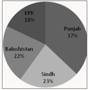
Agriculture is the backbone of Pakistan’s economy and improvement in this sector is essential for the development of country. This sector contributes 19.82 percent to the Gross Primary Production (GDP), provides employment to 42.3 percent people in the country, provide food to the population and many other products. This sector includes many sub-sectors but main sub-sector is livestock in this sector. About 58.55 percent is the contribution of this sub-sector to agriculture value added and to national GDP its share is 11.8 percent and gross value addition increased from Rs.778.3 billion to Rs. 801.3 billion. Livestock animals produce milk, meat, wool, bones, fat, skin etc. But milk and meat are the major harvests [1]. About 30-35 M people in the country are associated with livestock related activities and they earn 30-40 percent of their income from this sub-sector [2]. In livestock, Small ruminants (goats and sheep) preferred over large ruminants because of their high productive potential and small size [3].Sheeps and goats are the earliest animals to be domesticated. They can survive on pastures that cannot be used by other livestock and they can tolerate the period of drought better than any other livestock. Especially, goats are capable to tolerate the water deficiency and even limited amount of fodder is enough or them to survive [4]. After china and India, our country is the third largest goat keeping country in the world [5]. There is total 68.4 million goat population in country and Punjab is the province that has highest goat population as given in Figure 1 [6]. Goats in the country produce 845 million liters of milk and have share to 671 thousand tonnes of mutton produce by small ruminants [1]. More than 25 recognized goat breeds exist in Pakistan [7] along with two wild relatives of goats that are Markhor and Ibex [8].

Figure 1: Population of goats in provinces of Pakistan. Source: ACO (2006)

Lupinepublishers-openaccess-Dairy-Veterinaryscience

Goat farming and its products

All around the world, sheeps and goats play an important role in small scale farming systems. Goat farming is very beneficial from economic point of view. It provides many products but meat and milk are the major products. Food is necessary to stay alive. Among the humans religion is the major factor that influences the choices of food. Muslims and Jews are very careful and they only allowed eating halal food such as halal meat and drinks. Halal meat is a meat that obtained from the animals such as goats, sheeps, cattle and chicken that slaughtered under the rules provided by Islam and halal meat do not contain any ingredient from haram animals such as pig etc. [9].

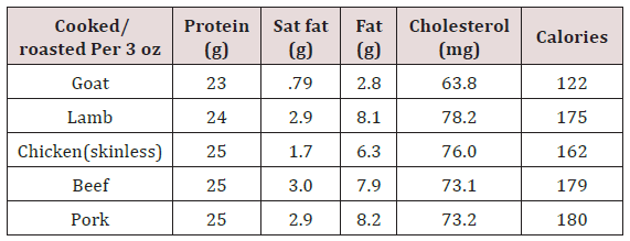
Goat meat is highly nutritious, internationally it is known as lean red meat [10-12]. In most of the developing countries goat meat is very important source of protein [13]. It is one of the staple meats in human diet. It comprises 63 percent of all red meat that consumed worldwide [14]. Goat meat is healthy and better substitute as compared to other red meats. It contains low level of calorie, fat, cholesterol (Table 1) [15] buthas high levels of iron and it becomes more popular widely among general population so its demand (as alternative low fat meat) increasing continuously [16]. However, goat meat has necessary fatty acids this is because of the fact that goats deposit greater level of polyunsaturated fatty acids than other ruminants [17,18]. Moreover, goats extensively use for the provision human consumable milk [19]. Pakistan is the fourth largest milk producing country in the world.

Table 1: nutritive comparison of goat meat with other meat.

Lupinepublishers-openaccess-Dairy-Veterinary-Sciences

Source: USDA (2001)

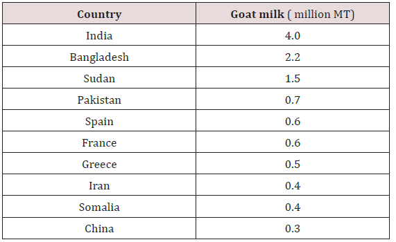
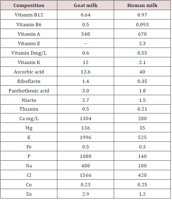
Approximately 0.7 million tonnes of the milk is produced by goats in Pakistan which is Only 0.2 percent of the total milk produced in the country as shown in Table 2 [20]. Scanty information is available regarding the physiological and chemical facts about the unique quality of goat milk. It contains greater level of short and medium chain fatty acids; these fatty acids are remedial against many diseases and disorders [21]. Goat milk is easier to digest than cow’s milk because it possesses smaller fat globules than cow’s milk [22]. Goat’s milk helps to prevent iron deficiency and bone demineralization because it contains higher vitamins and mineral contents than human milk as shown in Table 3 [23].

Table 2: Amount of milk produced in top ten countries.

Lupinepublishers-openaccess-Dairy-Veterinary-Sciences

Source: FAOSTAT (2008)

Table 3: Comparison of vitamins and minerals in human and goat milk.

Lupinepublishers-openaccess-Dairy-Veterinary-Sciences

Animal hair fibers are more soft and fine than other fibers but they are scarce. Factors such as genetic factors and climatic conditions make it difficult to produce these fibers on large scale. Hair fiber is another very important product of goat farming especially pashmina. All over the world the finest fiber produced by the animals is pashmina that produce in large quantity. It is formed by the hair of Asian native domesticated goat named as Capra hircus [24]. China, Mongolia, Iran, Afghanistan, Pakistan, Nepal and India are the chief fiber producing states. Among all these countries china is the major pashmina producer that produce about 70 percent of the total pashmina production. Mostly, pashmina used to make shawls, scarves, gloves, blankets and other fabrics that manufactured manually or are hand spun [25]. It is protenacious in nature which have 18 amino acids containing polyamide polymer and just like wool α- keratins are arranged helically that is why it is chemically similar to wool and mohair fibers [26].

Yeast cultures as a feed supplements in livestock

In order to support metabolic process all living organisms require essential nutrients, to keep themselves alive. In recent years, use of live microorganism and their products as a feed supplements for ruminants is very common. In many types of production systems microbial supplementations have beneficial impact on animal performance. Outcomes of many experiments indicated that the addition of yeast in the diet of goats and cattle is advantageous [27-30].

According to Putnam et al. [29] supplementation of yeast culture in the diet of dairy cows results in the rise in dry matter intake that cause further increase in the milk yield and the amount of milk protein in these animals. Yeast cultures (YC) contain dried yeast cells and these are actually microbial supplements. From many years these cultures use to increase the growth performance of animals. These cultures has positive impact on microbial population in gastrointestinal tract and these cultures increase the beneficial activities associated with these microorganisms that has indirect impact on animal performance [31].

Yeast varieties and its products in small ruminants

In many parts of the world there is a trend to use the yeast products (YP) as a feed additive in the diet of ruminants. Different yeast products are produced by many companies with variable trade names but a limited number of YP have been evaluated under controlled research studies. Saccharomyces cerevisiae and Aspergillusoryza are the yeast products and they are very important for the manipulation of rumen metabolism. Agarwal et al. [32] performed an experiment in which they use different strains of S. cerevisiae as a microbial feed additive and they reported that S. cerevisiae strain NCDC 49 is the best strain because it can tolerate the stressed conditions in gastrointestinal tract of animal. Yeast strains differ in their ability to stimulate the ruminal bacteria. It was reported by Dawson and Hopkins [33] that out of 50 only 7 strains are capable to stimulate the growth of bacteria that digest fiber from the rumen so care should be taken to select S.cerevisiae strains for use in yeast culture preparation of ruminants. Baker’s yeast strains and Brewer’s yeast strains vary in their capabilities to stimulate the critical groups of ruminal microorganisms and former strain has limited capability in this kind of stimulation [34]. According to Zelenak et al. [35] ruminal hemicellulose digestibility increase by the addition of live yeast product Yea-Sacc to the diet when substrate contains 20 percent and 50 percent barely in vitro.

Feeding system and yeast addition

Feed is the essential component in livestock farming and different experiments have been conducted to improve the quality of feeds given to the ruminants. Feed additives has positive influence on digestion rate and leads to the better growth performance, this is because of the fact that feed additives increase gut health of the animals [36]. As probiotics, yeasts are gaining popularity in fattening system. It is robust with high viability under a range of environmental conditions and can be culture very easily [37]. Many researchers proposed that supplementation of yeast culture in the diet up surge feed intake [38,39], nutrient digestibility [40] and the rate of growth [41] in cost effective way [42]. According to some investigators addition of yeast in the feed of meat producing ruminants leads to the variable growth responses that vary from no significant rise in average daily gain to an increase of greater than 20 percent. In this case the average daily gain was 8.7 percent [43].

There are various feeding systems such as high input feeding system, extensive feeding system, intensive feeding system, fodder based feeding system and grain based feeding system, use of yeast as a probiotic in these systems results in variable responses for example Shankpal et al. [44] conducted an experiment in India on Surti goat Kids. In their experiment, these scientists raised their goats under total mixed ration based feeding system with the supplementation of yeast to access its effect on growth performance, digestibility, feed conversion efficiency and feed intake of these kids. Total mixed ration (TMR) based feeding system actually involve mixing of all feedstuffs in an equal ration with respect to their nutritive value so this system supplies an ample amount of nutrition and energy to the animals which enable them to attain maximum performance. These Indian scientists concluded that this inclusion of yeast in feeding system had positive impact on all the 4/8 Citation: Memoona N, Kashif I. Use of Dietary Yeast and its Products in the Feeding Regime of Meat Type Goats. Con Dai & Vet Sci 1(2)- 2018. CDVS.MS.ID.000108. described parameters and also results in 6.66 percent lower feed cost in the experimental group than control group [44].

Furthermore, many scientists conducted an experiments on different animals raised under intensive feeding system for example Kawas et al. conducted an experiment on Pelibuey male lambs in mexico. These lambs were raised under intensive feeding system with the supplementation of yeast culture and sodium bicarbonate in their finishing diets to check its effect on growth rate and other parameters. At the end of the experiment these scientists concluded that supplementation of yeast in the finishing diet of intensively fed lambs has no significant impact on growth rate, digestibility and rumen pH [45]. Outcomes of this study were similar with the work of some other scientists who concluded that the addition of yeast in the finishing diets of intensively fed bulls has no impact on their growth rate [46-48]. Rearing of the kids under grain based feeding system is helpful to fulfill their nutritional requirements which have positive impact on their growth. However supplementation of diet with yeast enhances these beneficial impacts. According to Nisbet et al. [49] for the fattening ruminants addition of malate and yeast in grain based diets has much more beneficial influences and important for animal health [49].

Effect of yeast on the digestibility and feed intake

Supplementation of yeast in feed improves the digestibility of nutrients and results in higher production of carboxymethyl cellulase activity in rumen [50,51]. Yeast culture leads to the beneficial changes in the digestion by positively influencing the ruminal fermentation. In India it is demonstrated by Kumar et al. [52] that yeast culture has positive impact on feed intake and process of digestion in rumen. On the other hand, some scientists conducted an experiment in Turkey to demonstrate the variations in ruminal fermentation and nutrient degradability of some feedstuffs due to the supplementation of live yeast culture in diet of yearling lambs. In their study they use corn DDGS as a protein source, barley grain as an energy source and wheat straw as roughage and they concluded that live yeast addition in diet has no impact on dry matter (DM) degradability but it reduce the degradability of corn dried distillers grains with soluble (DDSG) in rumen.

Better feed intake has positive impact on animal production [53]. Within 6-8 hrs after meal yeast culture supplementation stimulate the higher degradation of solid feeds and higher intake of dry matter. Dry matter intake is the part of initial rate of fiber digestion. It is reported that yeast culture supplementation increase dry matter intake in some studies [40,54] but not in other studies [55,56]. Dawson et al. [57] demonstrated that yeast culture supplementation has pivotal role in digestion in those animals that maintained at high forage diets In South Brazil, Lima et al. [58] analyze the impact of inactive dry yeast (S. cerevisiae) from sugar cane as protein source on dairy goat performance and they reported that dry yeast supplementation in goat’s diet cause lower intake of dry matter and organic matter intake as compare to the soybean + dry yeast diet. This is because of the fact that dry yeast has very fine texture and unusual aroma from sugar cane that cause decrease in the intake of dry matter. Williams [59] confirmed that the quantity of rumen cellulolytic bacteria was amplified by yeast supplementation, exclusively in high concentrate diets.

Effect of yeast addition on growth performance

Many studied demonstrate that diet supplementation with yeast improves the growth performance in finishing lambs [60], calves [61] and bulls [46,47]. According to Haddad et al. [62] yeast supplementation in fattening sheep diet increase the weight gain. In Turkey, Bugdayci et al. [63] conducted an experiment on Saanen male kids and they reported that live yeast supplementation do not influence the live weight gain in goats but some researches do not support this idea [64,65]. However, Mikulecet al. [66] stated that there is no influence of live yeast supplementation on weight and weight gain in lambs. Titi et al. [67] performed an experiment in Jordanand demonstrated that live weight gain and live weight were not affected by the yeast culture supplementation in both awassi lambs and Shami goat kids.

Abou-ward [68] conducted an experiment on lambs and he find out that in average daily gain and dry matter intake significant upsurge occur in lambs as a result of feeding on the ration that contain at least 0.1 percent yeast culture. Rise in dry matter intake was because of the stimulation by cellulolytic bacteria [29,30,54,69]. Some Brazilian investigators reported that supplementation of live yeast never influence lamb performance such a dry matter intake, daily live weight gain, total live weight gain and final live weight [70]. However, it was also observed that the addition of 2.5 to 5g of commercial yeast in the diet of Nubian goats fed sorghum (whole plant) has significant impact on daily live weight gain [71].

Effect of yeast addition on slaughter and carcass traits

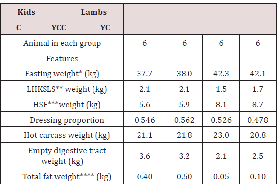
Particularly with reference to small ruminants scarce work had been published regarding the impact of yeast culture supplementation on carcass characteristics. Titi et al. [67] reported that yeast culture supplementation has no impact on cold dressing proportion and hot carcass weight of Shami goat kids but yeast culture supplementation decrease the cold dressing proportion, overall muscle/bone ratio and hot carcass weight in lambs. Carcass weight reduced in lambs because of the enlargement of the digestive tract due to higher dry matter intake (Table 4). It is demonstrated by kawas et al. [60] that in the lambs that were fed with high grain finishing diet, there is no impact of yeast culture supplementation on dressing proportion and chilled carcass weight of these lambs.

Table 4: Carcass characteristics of Awassi lambs and Shami goat kids as influenced by yeast culture (YC) addition in diet.

Lupinepublishers-openaccess-Dairy-Veterinary-Sciences

C: control group (without yeast addition).

YC: treatment group (with yeast culture supplementation).

*Average weight of 6 animals.

**LHKSLS: weight of liver+heart+kidneys+spleen+lungs+sweetbread.

***HSF: weight of head+skin+feet.

**** fat of heart+kidney+pelvic.

Source: O’Connor (1994)

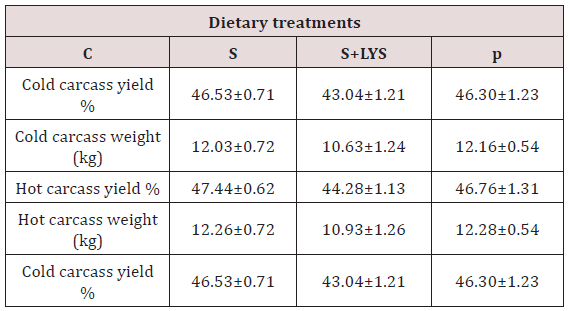
Similarly, Freitas et al. [72] reported that in different breeds of goats inclusion of dry yeast has no influence on hot and cold carcass weight, true carcass yield and carcass compactness index. According to Bugdayci et al. [63] live yeast supplementation in diet has no influence on hot and cold carcass yield in goats as shown in (Table 5) [63]. Similarly, investigators failed to find out any positive impact of yeast culture supplementation on carcass characteristics and composition in bulls or steers [73]. In the same way, some scientists reported that supplementation of S.cerevisiae results in the improvement of lamb fattening performance without any significant alteration in carcass traits [74].

Table 5: Impact of yeast supplementation on Saanen male kid’s carcass yields and characteristics.

Lupinepublishers-openaccess-Dairy-Veterinary-Sciences

p<0.05

C: Control group

S: Group served with sucrose added diet

S+LYS: Group served with sucrose and live yeast culture added diet

Source: Bugdayci et al. (2016)

Effect of yeast addition on meat quality

Growth rate of goat population is high (3.5 percent per annum) because of preference of goat meat [75]. Meat quality is the standard term that uses to describe the properties and perceptions of meat. Goat meat is highly nutritious internationally it is known as lean red meat [10-12]. It has course texture, dark red colour and as compare to lamb meat it has different flavor and aroma, characteristically [76]. Sethy et al. [77] observed that redness and tenderness of meat increase but shear force value decrease as a result of addition of 0.3 mg Seas selenium yeast and 100 IU vitamin E in goat diet. Similarly it was proposed by Morrissey et al. [78] that supplementation of diet with selenium yeast results in the rise of α-tocopherol in muscles but decrease the chances of lipid oxidation in muscles.

Likewise, Rufino et al. [79] reported that the addition higher level of inactive dry yeast in the diet of lambs improves the meat quality and carcass characteristics by decreasing the level of intramuscular and subcutaneous fat but by increasing the level of meat crude protein and ash concentration. Dry yeast addition in the diet of different goat breed does not influence the values of cooking loss and shear force value [72]. Some investigators concluded that quality of meat improved and shelf life extend as a result of supplementation of diet with Selenium yeast and vitamin E because it enhanced antioxidant status in the muscle [80]. Effect of yeast supplementation on lamb meat quality has also been demonstrated by Titi et al. [67] who concluded that the addition of live cultures of S.cerevisiae yeast in diet leads to the significant change in the chemical composition of meat.

Conclusion

All the yeast culture supplementation strategies are not identical in their efficiency because the influences of yeast culture supplementations on animal efficiency are strain dependent so the results of supplementations may vary considerably. However, it is concluded that the addition of yeast in the diet of goats and other ruminants is beneficial. Yeast culture supplementation is very convenient strategy that positively modifies the microbial actions, digestive tasks in rumen, meat quality and fattening performance in goats but still it is prerequisite to conduct more experiments under different feeding circumstances and in extended fattening episodes to clarify the beneficial influences of yeast addition in the diet of goats.

Read More About Lupine Publishers Journal of Dairy & Veterinary Sciences Please Click on Below Link:
https://lupine-publishers-dairy-veterinary.blogspot.com/

Saturday, 11 December 2021

Lupine Publishers | Pressure of Climatic Factors on Sheep Bluetongue Epidemics in Tamil Nadu

 Lupine Publishers | Journal of Dairy & Veterinary Sciences


Abstract

This research correlates the effect of climatic factors on bluetongue epidemiology in sheep of delta districts in Tamilnadu based on merteriological data collected from 2014 to 2016. There are a total of eighty two villages of delta districts were affected with Blue tongue virus (BTV). We used multiple linear regression analysis to assess the effect of climatic factors on the prevalence of bluetongue. Our results showed that maximum temperature, rain fall and relative humidity positively correlates with the number of BTV epidemic in sheep flocks. These findings suggest that the risk of BTV infections in sheep increased in warm and humid environments. Moreover, climatic factors can be used to forecast the possible epidemic in this area and other regions characterized by similar climatic conditions.

Keywords: Epidemiology; Blue Tongue Virus; Culicoides Spp; Laminitis; Coronitis; Facial Edema; Necrosis of Cardiac; Vectorial Capacity; Multiple Linear Regression Analysis Model; Semi Intensive Livestock; Antioxidant System

Introduction

Bluetongue, is caused by the bluetongue virus (BTV) and transmitted by Culicoides spp. [1,2]. Bluetongue virus was pandemic in nature with areas between 40◦N and 35◦S in the world [3,4] . Symptoms of this disease are usually characterized by laminitis, coronitis, and facial edema. In serious cases, pulmonary edema, pleural effusion and necrosis of cardiac and skeletal muscle can be observed [5].

Many researchers have evaluated the correlation between meteorological factors and the distribution of the Blue tongue virus [9,10]. However, few researchers have specifically attempted to investigate the influence of climatic factors on bluetongue epidemic. In the present study, we have investigated the effect of climatic factors on the epidemics of bluetongue, by means of multiple linear regression analysis model.We designed to assess the influence of climatic factors on the epidemiology of bluetongue in sheep in Tamilnadu, in order to provide accurate information to design the more effective and economical surveillance and control systems for bluetongue.

Materials and Methods

Sheep farming of Delta Districts

In delta districts of Tamilnadu, two types of sheep farms were observed. One is semi intensive livestock farms, which must meet a moderate standard regarding production management and epidemic prevention systems. The other one is high number holdings with trans humane methods, where sheep kept as a freerange system.

Location

This research work was carried out in the delta region of Tamilnadu which includes Thanjavur, Thiruvarur, Nagapattinam and Pudukottai districts during 2014 to 2016.

Data collection

Metrological data were collected from government agencies like District Collector Office and disaster management centre. Data were also obtained from research institutes like Tamilnadu Agricultural University, Thanjavur.

Data analysis

Multiple linear regression analysis model was used to assess the effect of climatic factors on the epidemics of bluetongue.

Results

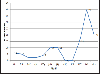
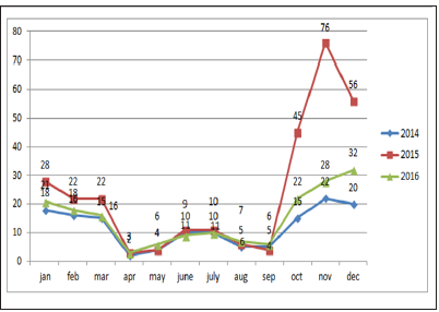
Sums of 608 bluetongue epidemics have been reported in delta districts during the period of study. Then we have analysed the change trend of bluetongue epidemic and the annual variation of meteorological data (Figure1). The results indicated the change trend and the annual variation of the disease epidemics, monthly maximum temperatures, monthly minimum temperature, wind speed and rainfall in delta districts of Tamilnadu from 2014 to 2016. Analyses of time series revealed that the epidemic of bluetongue of a determined month was changed by case numbers registered during the previous years. The epidemic trend was changing with the months were also recorded (Figure 2).

Figure 1: Standard epidemic of Sheep bluetongue in Tamilnadu.

Lupinepublishers-openaccess-Dairy-Veterinaryscience

Figure 2: Changing epidemic of Sheep blue tongue in Tamilnadu.

Lupinepublishers-openaccess-Dairy-Veterinaryscience

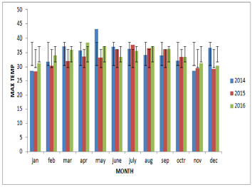
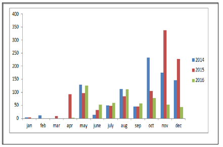
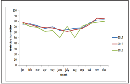
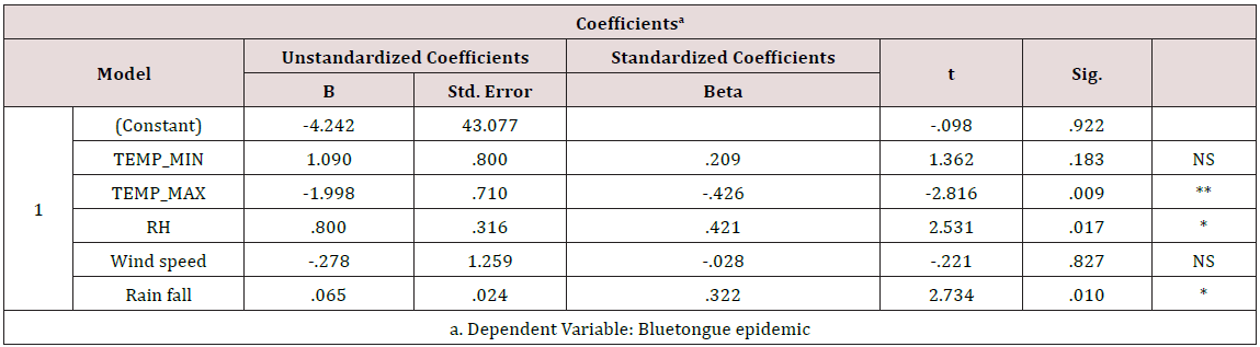
From the year analysis, a seasonal distribution and change in the disease cases from 2014 to 2016 were noticed, which correlates with the climatic factors of our investigation results from the results of data analysis, we found that temperature is the main risk factors which can influence the epidemic of bluetongue (Figure 3). In delta region maximum temperature at the tune of 330C will have higher epidemics during 2015 (288) than in 2013 (142) and in 2016 (178). Rainfall was also showed a influence on the occurrence of disease, a rise in mean monthly rainfall is related to increase in the monthly number of disease cases (Figure 4). High relative humidity positively related to the epidemic of bluetongue (Figure 5). Clinical signs like cutaneous ulcers (Figure 5) and bilateral purulent nasal discharge also evident. Under multiple linear regression model analysis maximum temperature, relative humidity and rainfall were statistically significant (P< 0.05) with the epidemiology of bluetongue (Table 1).

Figure 3: Maximum temperature (0C) data in the study area during 2014 to 2016.

Lupinepublishers-openaccess-Dairy-Veterinaryscience

Figure 4: Trend line of Rain fall (mm) in the study area.

Lupinepublishers-openaccess-Dairy-Veterinaryscience

Figure 5: Relative humidity (%) trend in the study population.

Lupinepublishers-openaccess-Dairy-Veterinaryscience

Table 1:Multiple linear regression model on climatic factor with bluetongue epidemic (P < 0.05).

Lupinepublishers-openaccess-Dairy-Veterinaryscience

Discussion

In this study, we aimed to prove the relationship between climatic factors and the epidemiology of bluetongue in delta districts of Tamilnadu. We found that a positive relationship between rainfall, monthly maximum temperature and relative humidity in the epidemic of Blue tongue in Sheep (Figure 6).

Figure 6: Relative humidity (%) trend in the study population.

Lupinepublishers-openaccess-Dairy-Veterinaryscience

The positive effect of temperature and relative humidity on blue tongue infections is comparable to previous research [11-13]. It has been proven that a hot and humid environment can interfere with the antioxidant system of the animal and increases the synthesis of lipid peroxides, which may cause peroxide damage [14-16] and increases the serum levels of epinephrine and norepinephrine will negatively affect humoral and cell-mediated immune responses and make animals more susceptible to infectious diseases like bluetongue.

Maximum temperature in our study helps in the epidemic of bluetongue because temperature also significantly influences the activity rates of adult insects. The scientific reason behind is adult culicoides are more active than on cooler days and their reproduction will be inhibited by low temperature [17]. However, BTV has a strong environmental resistance and will not lose its infectivity in seven days under 56 oC [18]. Thus, the effect of temperature may be limited in the normal / lower temperature conditions (Figure 7). High Rainfall showed increased cases of bluetongue because of contamination of soil water and that can influence the life cycle of insects. Moreover at the right level, rainfall can create humid microhabitats which promote the activities of [19] and can regulate the growth and activity of Culicoides spp. populations [20,21]. Even though this research found that wind velocity did not favors the spread of Bluetongue virus but, long distance movement of Culicoides is mediated by the prevailing winds [21].

Figure 7: Relative humidity (%) trend in the study population.

Lupinepublishers-openaccess-Dairy-Veterinaryscience

As a final point, mechanisms of transmission of bluetongue are more complex and climate factors only represent some components of the epidemic of blue tongue. Many other risk factors like age, immunization calendar and management practices may also have strong impact on the incidence of the disease. In this paper, we only studied the effect of climatic factors on the epidemiology of bluetongue. This research work can able to help to design in the control of blue tongue in sheep by adopting timely vaccination methods, insects management program and healthy ecosystem to make the flock more positive energy and protein balance which will augment the immunity of the herd sustainably.

Acknowledgement

The authors sincerely thanks the sheep farmers, Director of Animal health studies, TANUVAS, Chennai and The Dean, Veterinary college and research institute, Orathanadu, Tamilnadu for their support.

Read More About Lupine Publishers Journal of Dairy & Veterinary Sciences Please Click on Below Link:
https://lupine-publishers-dairy-veterinary.blogspot.com/

Friday, 30 July 2021

Lupine Publishers | Microbial Source Tracking Markers for Detection of Faecal Contamination in Environmental Waters

 Lupine Publishers | Journal of Dairy & Veterinary Sciences


Mini Review

Microbial source tracking (MST) describes a suite of methods and an investigative strategy for determination of faecal pollution sources in environmental waters. They rely on the association of certain fecal microorganisms with a particular host, that ranges from human [1] to agricultural animals [2,3] to pets [4] and wild animals such as gulls [5]. MST is used to appraise recreational water quality and to correlate with human health risk.

Many MST publications fixate mostly on human source contamination, as this has been an issue of concern for managers and regulators. Human sewage pollution is among the greatest concerns especially in India [7,8] for human health due to [1] the known risk of exposure to human waste and [2] the public and regulatory will to reduce sewage pollution. However, methods to identify animal sources are receiving increasing attention as our understanding of zoonotic disease potential improves.

Numerous epidemiological studies of waterborne illness in countries like India indicate that the common aetiological agents are bacteria, viruses and parasitic protozoa and the coliform presence is an important parameter for determining water pollution levels. But the presence of coliforms of soil and litter origin had raised doubt about their reliability as an indicator of the pathogen [6]. Also, the method used for coliform estimation is not precise. Hence CPCB believes that BOD is the single most reliable parameter. Based on this approach the CPCB estimated that 67% of river stretches in India is relatively clean with BOD less than 3 mg/litre. Also, poor correlations have been reported between waterborne human viruses or protozoa and thermotolerant coliforms [6,9,10]. In contrast to MST markers, faecal coliforms provided a poor metric to assess risks of exposure to faecal contamination of human origin in the rural setting in India [9]. Such a situation is critical to understand, as evident from recent drinking water outbreaks where coliform standards were met [11] and in India most of the outbreaks go unnoticed. FIB concentrations have not been well correlated with pathogens in many studies [10].

Furthermore, many epidemiology studies have failed to find a correlation between human health outcomes and FIB levels, particularly when the pollution is not from a known point source such as a wastewater treatment plant (WWTP) [14,15]. Nonetheless, water regulatory agencies have yet to come to terms with the inherent problems resulting from reliance on faecal indicator bacteria as currently determined. Fortunately, new index organisms for some pathogens look promising like C. perfringens, phages and viruses like Adenoviruses. As these index organisms are relatively untested worldwide, extensive trials are necessary before their general acceptance in microbial risk assessment. It should be noted that useful index organisms in one system are not necessarily of value in a different environment.

Use of multiple tests increases the surety of presence or absence of water pollution. In particular, it has been reported that at least two parameters are needed to accurately differentiate between two distinct faecal pollution sources: one specific indicator that identifies the source and another, a universal indicator that provides information on the faecal load [12]. In consequence, the indicators should be carefully selected based on appropriate statistical analyses. Persistence studies are needed to provide complementary information, addressing the effects of environmental aspects like temperature, solar radiation, salinity, pH, chemical pollutants, water filtration, turbidity, starvation, predation and presence of heavy metals, among others.

Other relevant issues are the use of methods based either on genomic targets or in the quantification of new microbial tracers through library-independent methods, the combination of methods, and comparative and integrated studies between research groups with standardised procedures to avoid differences in implementation. Also, in order to increase the validity of MST methods, it is necessary to consider temporal variability when designing the sampling scheme of the source material and constructing source libraries and increase the specificity and field testing of DNA-based markers. The number and range of potential host sources included in MST studies must be deliberately chosen to suit the water body and particular questions associated with it [13].

For More About Lupine Publishers Journal of Dairy & Veterinary Sciences Please Click on Below Link:
https://lupine-publishers-dairy-veterinary.blogspot.com/

Friday, 28 May 2021

Lupine Publishers | Molecular Typing Of Capsular Polysaccharides of Staphylococcus Aureus Isolated From Cases of Bovine Mastitis by PCR

 Lupine Publishers | Journal of Dairy & Veterinary Sciences

Abstract

Forty five Staphylococcus aureus isolated from cases of bovine mastitis were subjected to Molecular typing by Polymerase chain reaction to determine their capsular polysaccaharide type. Of the 45 isolates, 33 were confirmed to carry a cap5 locus and cap8 locus was detected in remaining 12 isolates. To the best of our knowledge this is the first report of capsular polysaccharide typing of S.aureus isolates from India

Keywords: Staphylococcus aureus; Polymerase chain reaction and capsular polysaccaharide type

Introduction

S. aureus produces a variety of extracellular and cell wall associated components which are involved in the pathogenesis of bovine, ovine and caprine mastitis [1]. S. aureus strains produce capsular polysaccharide (CP) in-vivo [2] or under defined culture conditions [3]. Although capsule production of staphylococci was first recognized in 1930 [4], the prevalence of encapsulation among S. aureus has been appreciated only recently. Eleven capsular polysaccharide serotypes have been proposed on the basis of agglutinating reactivity with adsorbed rabbit antiserum and precipitation in double immuno diffusion [5,6]. Of these capsular serotypes 5 and 8 are the most predominant serotypes in human and animal S. aureus infections.

Studies on the prevalence of encapsulated strains in bovines shows the considerable variability that exist in the prevalence of serotype 5 and 8 capsules among bovine mammary isolates of S. aureus from different countries (Tollerstud et al., 2000). Moreover, the presence of S. aureus in raw milk is a public health problem, because it was reported that 95% of S. aureus isolates from bovine mastitis were either CP5 or CP8 in Norway [7]. For effective control of bovine mastitis caused by S. aureus in a particular geographical location, a careful characterization of the prevalent strains in the target population is essential [6]. Studies on capsular serotyping of isolates are important for the rational design of mastitis vaccines, containing staphylococcal capsular antigens. If improved vaccines against bovine mastitis are to be generated, more studies are required to elucidate the role of these polysaccharides in the pathogenesis of bovine mastitis [7].

However, capsular serotyping employing conventional techniques fails to identify non encapsulated strains of S. aureus. Hence DNA based technique for differentiation of serotypes provide an alternative to conventional serotyping and has a potential to overcome the problems associated with the current serotyping techniques which relay on inconsistent expression of phenotypic traits [7-9]. No data regarding the prevalence of capsular serotypes of S. aureus causing bovine mastitis is available in India. The proposed study would help in understanding the prevalence of capsular serotypes of S. aureus in Puducherry, India. This data would help in formulating vaccine based strategies for control of mastitis.

Materials and Methods

Cultures used in the study. Forty five Staphylococcus aureus obtained from the milk of dairy cattle with clinical and subclinical mastitis in and around Puducherry, India and S. aureus strain Reynolds (Capsular polysaccharide type 5) and S. aureus strain Wrights (Capsular polysaccharide type 8) were used as standard reference for identification of Capsular polysaccharide types of S. aureus by PCR Identification of S. aureus isolates [10]. The S. aureus isolates were initially selected on the basis of colony appearance and a positive tube coagulase test and their identity was verified by [8] and Garrity et al. [9]. Their identity was confirmed by PCR using the primer pairs targeting the nuc gene of S. aureus. DNA extraction. Strains were grown on Luria broth 37 °C overnight. Genomic DNA was extracted with a standard phenol-chloroform procedure as described elsewhere [10]. Detection of capsular genotype by PCR. The PCR assay for typing of capsular polysaccharide S.aureus was carried out as described by Verdier et al [11]. Genomic DNA was used as a template for PCR amplification with the primers Cap5 k1 (5’-GTCAAAGATTATGTGATGCTACTGAG-3’) and Cap5 k2 (5’-ACTTCGAATATAAACTTGAATCAATGTTATACAG-3’) located in cap5k for capsular type 5 and the primers Capsule 8 k1 (5'GCCTTATGTTAGGTGATAAACC-3') and Capsule 8 k2 (5'-GGAAAAACACTATCATAGCAGG-3') located in cap8I for capsular type 8. The PCR amplification was carried out in an automated thermal cycler (Eppendorf mastercycler, Germany) according to the following programme. initial denaturation at 94 OC for 4min followed by 25 cycles of denaturation at 94 OC for 30seconds , annealing at 55 OC for 30sec and extension at 72 OC for 1min and final extension at 72 OC for 5min. The amplified products were analysed on agarose gels along with positive control, negative control and molecular size marker (100bp la).

Results and Discussion

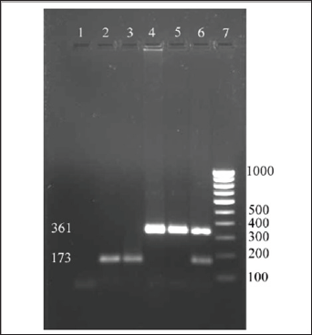
All the 45 isolates S. aureus were subjected to molecular typing using the primer pairs targeting the cap5 locus and cap8 locus of capsular polysaccharide of S. aureus. The primer pairs successfully amplified the DNA prepared from the field isolates of S. aureus as well as the DNA prepared from the reference cultures used in the study. The sizes of the amplicons were 361bp for capsular type 5 and 173bp for capsular type 8 (Figure 1). Among the 45 S. aureus isolates subjected for PCR with CP5 and CP8 primers, 33 isolates were confirmed to be CP5 strain and 12 isolates were confirmed to be CP8 strain. Out of the 45 isolates, 73.33% were found to carry the cap5 locus and 26.66 % were found to carry the cap8 locus. Poutrel et al. [1] reported that a majority of S. aureus from cases of mastitis strains belong to serotype 5 (CP5) and 8 (CP8). In their study they used monoclonal antibodies to S. aureus capsular polysaccharide types 5 and 8 to serotype the isolates by enzyme- linked immunosorbent assay and showed that 69% of 212 isolates recovered from cow's milk in France were serotype 5 (51%) and serotype 8 (18%) and 30.6% were non-typeable [12].

Figure 1: Screening field isolates of S.aureus for capsular types.
Lupinepublishers-openaccess-Dairy-Veterinaryscience

Lupinepublishers-openaccess-Dairy-Veterinaryscience

Tollersud et al. (2000) have showed the variability in prevalence of serotype 5 and 8 capsules among bovine mammary isolates of S. aureus from different countries. They performed immunoblot assay using CP5 and CP8 antibodies and isolates that consistently giving weak reactions with antibodies to CP5 and CP8 were further evaluated by immunodiffusion or ELISA. Capsular serotyping of 274 bovine mastitis isolates of S. aureus from Europe, showed that the majority of isolates from Denmark (23 out of 39 isolates), Sweden (29 out of 38 isolates) and Ireland (62 out of 101 isolates), were of serotype 8 [13]. Isolates from Iceland showed an equal distribution of serotype 5 (10 isolates), serotype 8 (13 isolates) and non-typeable isolates (11 isolates), whereas in Finland half of the isolates (32 out of 62 isolates) tested were non-typeable. Serotyping of the U.S. isolates revealed that only 42% of 362 isolates from seven different states were typeable with the available antisera and showed 27 % of the isolates were serotype 8 strains and 15 % were serotype 5 strains, but the majority (58%) of U.S. isolates were non-typeable.

Strains of S. aureus that do not react with antibodies to CP5 or CP8 are referred to as non-typeable (NT) by conventional serotyping. Karakawa et al. [12] and Lee et al. [13] reported that these NT strains also fail to react with specific antibodies to serotype 1 or 2 CP. This is one of the problems encountered in the conventional serotyping of S.aureus. Han et al. [13] reported the usefulness of monoclonal antibodies reactive with the type 5 and 8 CP in characterizing S. aureus from clinical isolates that monoclonal antibodies have been described, and has also been demonstrated. Monoclonal antibodies for CP5, CP 8 and 336 were used to characterize 107 isolates of S. aureus [14-15]. Forty six per cent of them were typed as 336, while serotype 5 and 8 accounted for 12.1% each. The rest were declared as non-typeable. However O'Brien et al. [14] and Ma et al. [15] reported that Type 336 isolates do not express capsule but do express cell surface polysaccharide or the 336 polysaccharide (336PS), which resembles S. aureus cell wall teichoic acid and hence not a true capsular type. In order to avoid the problems encountered in the conventional serotyping, a PCR method was developed by Verdier et al. [11] to detect capsular types of S. aureus. In their study using the rabbit polyclonal antibodies specific to capsular polysaccharide types 5 and 8, 81 of the 195 isolates were capsular serotype 5 (T5) (42%), 88 were capsular serotype 8 (T8) (45%), and 26 (13%) were non-typeable. A PCR method was developed to detect capsular type of S. aureus isolates since serotyping method allowed typing of only 87% of strains (169 of 195). All strains included in the study have been investigated by PCR. But PCR method allowed genotyping of 100% strains [16-18].

Their study revealed that all S. aureus clinical isolates included in the study carried either the cap5 (46% of cases) or the cap8 (54% of cases) locus by PCR method, and demonstrated that the capsular phenotype that was determined by conventional serotyping method was confirmed by PCR. However, all 336 serotype strains that reacted specifically with 336 antibodies but not with capsular polysaccharide type 5 or 8 antibodies, carried the cap8 or cap5 genes (cap8 18 and 8 cap5 isolates). This study revealed the predominate capsular polysaccharide types prevailing among the bovine S.aureus isolates was the CP5 compared to CP8. Data on the S. aureus capsular polysaccharide types will help in formulating vaccine based strategies for the effective control of bovine mastitis due to S. aureus.

Read More About Lupine Publishers Journal of Dairy & Veterinary Science Please Click on Below Link:
https://lupine-publishers-dairy-veterinary.blogspot.com/

Friday, 5 March 2021

Lupine Publishers | Survey on Pathological Lesion and Its Financial Losses in Ovine Slaughtered at Jimma Municipal llll Abattoir, Jimma, Ethiopia

 Lupine Publishers | Journal of  Diary & Veterinary Sciences


Abstract

A cross-sectional study was conducted from October 2016 to July 2017 on ovine slaughtered at Jimma municipal abattoir with the aim of identifying main pathological lesions causing organs and carcass condemnation, and associated direct financial losses. In this, 384 sheep were recruited to the study using systematic random sampling and standard antemortem (AM), and postmortem inspection (PMI) procedures were employed. Nasal discharge, tick infestation, coughing, lameness, emaciation, depression and salivation are recorded as the major AM findings of the current study. Accordingly, 47 (12.2%) sheep showed signs of diseases and abnormalities; of which 23(6%) were conditionally approved whereas 11 (2.9%) sheep were unfit and judged to be detained and rejected. In the present study age, body conditions and geographic origin of the animals were considered as study variables, and the results showed BCS and age groups had statistically higher (p ≤ 0.05) rejection probabilities. PM examination findings indicated a total of 192 lesions were encountered, of which 48.9%, 29.2%, 6.8%, and 5.7% lesions were recorded from livers, lungs, GIT, and hearts and kidneys, respectively. During the PM inspection, both total and partial condemnation judgments were passed on organs and carcass appeared with a sign of abnormality. C. teniculosis 58 (30.2%), calcification 36(18.7%), abscess 31(16.2%), hydatid cyst 23 (12%), hepatitis 10 (5.2%), pericarditis 4(2%), bruise 3(1.6%) and nephritis 2(1%) were found to be the major pathological lesions recorded. In two sheep all organs and carcass were totally condemned as their entire bodies appeared yellowish. The direct loss due to the condemnation of organs and carcasses at Jimma municipal abattoir was also investigated in slaughtered sheep, and there were about 56,576 USD losses per year. In conclusion, this study has identified the pathological lesions affecting edible organs and meat, and then rendering them unfit for human consumption. The study also estimated pathological lesions associated direct financial losses at Jimma abattoir. Therefore, further studies focusing on the primary causes of the abnormalities were recommended in the study area.

Keywords: Abattoir; Financial loss; Lesion; Organ condemnation; PMI; Sheep; Jimma

Introduction

The livestock sector globally is highly dynamic, contributes 40% of the global value of agricultural output and support the livelihoods and food security of almost a billion people (Thornton [1]). Within African society, small ruminant comprises a greater proportion of the total wealth of the rural families, because of the low input requirements such as low initial capital, fewer resources and maintenance cost. They are also able to produce milk and meat in readily usable quantities using marginal lands and poor pasture and crop residues. Furthermore, their production cycle makes them need only short periods to reconstitute flocks after a disaster and respond quickly to the demand (Getenby, [2]). Ethiopia is the leading African country in livestock population, having around 34-40 million TLU (Tropical livestock unit) out of which 17% and 12% cattle and small ruminants, respectively, are found in Ethiopia (Ministry of information (MOI) [3]. According to (Development, pp Statis, [4]), the population of sheep and goats in Ethiopia is estimated to be 26.1 and 21.7 million respectively. It was the third largest number of sheep and goat among African nations and rank eighth in the world (Alemu and Merkel, [5]).

They generate cash income from export of meat, edible organs, skins and live animals (Ibrahim, 1998). There is also a high domestic meat demand from these animals, particularly during religious festivals. Even though this sub-sector contributes much to the national economy, its development is hampered by various constraints. These include endemic animal diseases, insufficient nutrition, poor husbandry, and lack of sufficient infrastructure, trained labor and government policies (PACE, [6]). Each year a large loss results from the death of animals and weight loss during transportation; and condemnation of edible organs and carcasses at slaughter.

Abattoir meat inspection is essential to remove gross abnormalities from meat and its products, to prevent the distribution of contaminated meat and to assist detecting and eradication of certain livestock diseases. More specifically, antemortem inspection attempts to avoid introduction of clinically diseased animals into slaughter house and also serves to obtain information that will be useful in making sound post mortem inspection. Likewise, postmortem inspection is the center around which meat hygiene revolves since it provides information essential for evaluation of clinical signs and pathological process that affect the wholesomeness of meat (Herenda, et al. [7]).

As the meat is the sources of protein to a human being, it should be clean and free from diseases of particular importance to the public such as tuberculosis and cysticercosis. Meat is also condemned at slaughterhouse to break the chain of some zoonoses which are not transmitted to man directly via meat like hydatidosis and other important diseases of animals such as fasciolosis (Arbabi and Hooshyr [8]; Fufa et al. [9]).

Each year a significant economic loss results from mortality, poor weight gain, condemnation of edible organs and carcasses at slaughter. This production loss in the livestock industry is estimated at more than 900 million USD annually (Jacob, [10]; Abebe, [11]; Jobre et al. [12]). The major causes of pathological lesion during PMI of slaughtered ovine at abattoir are the disease caused by parasites, bacterial and other abnormalities. The final judgment as to action to be taken with an organ, the carcass or part of a carcass is based on the total evidence produced by the visual observation, palpation and incision (Teka, [13]). Abattoir data is an important option for observing the diseases of both economic and public health importance (Arbabi and Hooshyr, [8]; Fufa, et al. [9]). Nowadays, several modern abattoirs like: HELMEX, ELFORA, Metehara, Modjo and Luna are established in Ethiopia. This increase in a number of slaughterhouse shows that increase in demand for meat supply, but the provisions have been challenging due to diseases, production problems and other factors. Given this, proper evaluation of financial losses due to organ condemnation resulting from various diseases at abattoirs is needed (Ezana, [14]). It is necessary to have enough information on a pathological lesion that causes organs and carcass condemnation at the abattoir. Hence, having information on where and how to reduce the losses that may be caused by the various abnormalities (lesions/pathology). Various studies (Jembere, [15]; Yimam, [16]; Aseffa, [17]; Getachew, [18]; Regessa et al. [19]) were carried out in the country in this regard to know the causes and losses associated. However, in Jimma there are no recorded studies conducted on major causes and financial losses associated with organs and carcass condemnation along with survey on pathological lesions. Therefore, the objectives of this study were to:

    a) Identify major pathological lesions causing organs and carcass condemnation in slaughtered sheep at Jimma municipal abattoir

    b) Estimate the direct financial losses attributed to condemned organs and carcass in sheep

Materials and Methods

Study Area

The study was conducted from November 2016 to July 2017 at Jimma municipal abattoir in Jimma zone. Jimma two is found in Oromia region south-western part of Ethiopia at a distance of 346km away from Addis Ababa and lies between 36°50'E longitude and 7°40'N latitude atan average elevation of 1750 meter above sea level. Jimma is the largest city in south-western Ethiopia. It is special zone of the Oromia Regional state and is surrounded by different Jimma woreda. The climate of the area is characterized by humid tropical with bimodal heavy rainfall which is uniform in amount and distribution, ranging from 1200 to 2800mm per year, with short and main seasons occurring from mid-February to May and June to September, respectively. The rainy season extends from mid-February to early October. Temperatures at Jimma are in a comfortable range, with the daily mean staying between 20 °C and 25 °C year-round. The total human populations of Jimma town was about 174, 446(88, 766 males and 85, 680 females). The livestock population of the area was reported to be about 2, 016, 823cattle, 942, 908 sheep, 288, 411 goats, 74, 574 horses, 49, 489 donkey, 28, 371 mules, 1, 139, 735 poultry and 418, 831bee hives (GOR, [20]).

Study Population

The study animals were sheep brought Jimma municipal abattoir and destined for slaughter. All animals were male and belonged to indigenous breeds kept under extensive management system. Sheep destined for slaughter had come from different parts of the weredas in the Jimma zone such as Dedo, Serbo, Saqa and Bilida inspected by standard AM, and PMI.

Study Design

A cross-sectional study using systemic random sampling technique was conducted from December 2016 to April 2017 to determine the pathological lesion that causes organs and carcass condemnation and to estimate the magnitude of direct financial loss attributed in sheep slaughtered at Jimma abattoir.

Sampling Method

Sample size determination

In this study, systematic random sampling method was applied to include study animals, and study animals were grouped into young (under 1year and three months) and adult above this based on the eruption of one or more incisor teeth according to Vatta et al. [21]. Since there was no published work on lesion survey from Jimma abattoir, 50% expected prevalence is considered to calculate the total sample size with 95% CI, 5% level of precision (Thrushfield, [22]). The sample size was 384 and determined using the formula given by

Lupinepublishers-openaccess-Dairy-Veterinaryscience

Where N= required sample size, Pexp= expected prevalence and d is desired absolute precision. Accordingly, the total sheep included in the study were 384.

Abattoir Survey

In the cross-sectional study of active abattoir survey, both AM and PMI were carried out in accordance by the procedures of Ethiopian Meat Inspection Regulation (Belina and Melese, [23]).

Ante-mortem Inspection (AMI)

The AMI (pre-slaughter examinations) of ovine was conducted at lairage both in motion and at rest and information related to study variables such as the behavior of an animal, age, BCS and origin and were recorded. At the same time, various signs of diseases and abnormalities were inspected with physical animal examination and its judgment were approved, conditionally approved, detained and rejected. Study animals were grouped into young and adult age groups according to standard dentation method (Vatta et al.[21]).

Post Mortem inspection (PMI)

During PMI all internal organs (liver, lungs, heart, kidney, gastro-intestinal tract), and carcasses were thoroughly inspected by visualization, palpation and making systematic incisions for the presence of cysts, parasites and other abnormalities. Pathological lesions were differentiated and judged according to guidelines on meat inspection for developing countries as totally fit for human consumption, and conditionally approved, and totally or partially condemned when unfit for human consumption (FAO, [24]).

Assessment of direct Financial Loss

In the current study, the total financial loss due to organs and carcass condemnation was computed by considering the condemnation rate or percentage of each edible organ and carcass, average number of animals slaughtered in the abattoir per year from retrospective data of the abattoir. The average weight of each organ and carcasses in kg, average current local market price of major organs and carcass, and each condemned organ was counted to estimate the financial loss. The average current local market price of each organ and mutton was collected by questionnaire from the butcheries in Jimma town for ease of computing the loss.

The retail average market prices obtained from butcher shops found in Jimma town in ETB were: Liver=30, lung=20, kidney=15, heart=18, GIT=90,whole carcass=4000 and mutton =150ETB/kg. In the case when there was whole carcass plus organs (whole body) rejection at PM, the average price of sheep came for slaughter was considered (4,000ETB). The direct loss is calculated according to the procedures described by Ogurinade and Ogunrinade [25], and the formula:

Lupinepublishers-openaccess-Dairy-Veterinaryscience

LOS is direct annual financial loss due to organs and carcass loss, MAK is annual average number of sheep slaughtered at Jimma abattoir, PL is overall prevalence of lesion, Pi is prevalence of each organ and carcass condemned, Ci is average market price of each organ and 1kg mutton at butcher shops of the Jimma town. The direct financial loss was expressed in.com Dollar ($) based on the current currency exchange rate of 1 USD = 22.5 Ethiopian Birr (ETB).

Data Analysis

The active abattoir data, and questionnaire survey were entered into Microsoft Excel- 2016 spread sheet and the process of coding, cleaning and validating was done on this sheet and analyzed using SPSS version 20. For the data from PMI, descriptive statistics were used to determine organ and carcass condemnation rates, defined as the proportion of organs and carcasses condemned to the total number of organs and carcasses examined. Each financial loss was also calculated. Possible variation between rejection rates of specific organs, age groups and origin, were taken into consideration.

Results

Abattoir Survey

Antemortem Inspection (AMI)

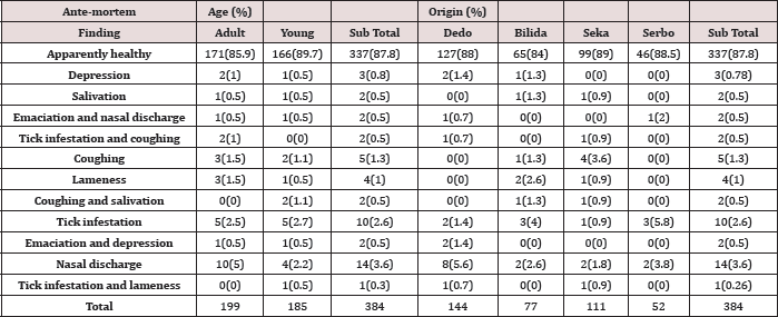
Detail AMI was conducted on a total of 384 sheep destined for slaughter at Jimma municipal abattoir and 47 (12.2%) of ovine were found to have different abnormalities. Nasal discharge, coughing, tick infestation, depression, emaciation, and lameness were those frequently observed among signs of diseases encountered in both age groups. The result also showed 6% (23/384) animals were conditionally passed for slaughter because of abnormalities such as lameness, respiratory problem, and their collection with tick infestation. On the other hand2% (8/384) sheep were unfit for human consumption and rejected during AMI. Since, they showed two and more signs of diseases such as emaciation with nasal discharge and depression 1% (4/384), salivation and salivation with coughing 1%(4/384) were the major cause of rejection (Table 1).

Table 1: Abnormalities encountered during AM inspection within Age groups and Origin of the animals.
Lupinepublishers-openaccess-Dairy-Veterinaryscience

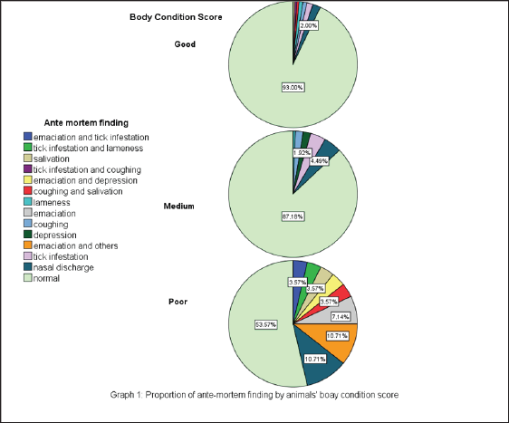
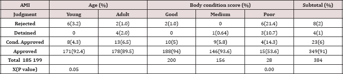
The AMI result also depicted of 28 sheep with poor body conditions 46.43% were found to have one or more sign/s of illness whereas in those with good BCS only 7% of the sheep were showed sign/s of diseases (Graph 1). In the current study, four different decisions were passed as AM judgments where apparently healthy sheep were passed for slaughter (91%), and others showed mild sign of illness and conditionally approved (6%) whereas 2.9% of sheep were detained and rejected as unfit for slaughter. Rejection rate was significantly higher (p ≤ 0.05) in young animals with poor BCS than in adult animals with good and medium BCS (Table 2).

Figure 1: proportion of ante-mortem finding by animals'boay condition score.
Lupinepublishers-openaccess-Dairy-Veterinaryscience

Table 2: Proportion of animals rejected and passed the AMI judgments by age group and BCS at the abattoirs.
Lupinepublishers-openaccess-Dairy-Veterinaryscience

Postmortem Inspection (PMI)

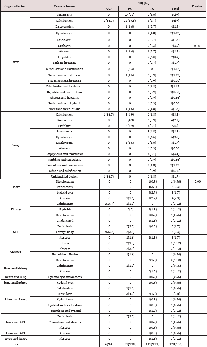
Among animals that had been examined during AMI 373 were slaughtered and subjected to through PMI following standard postmortem procedure and a total of192gross pathological lesion leading to partial and total condemnation of organs and carcasses were recorded. Among these abnormalities, lesions were frequently encountered from liver; and of which Cyst cercus teniculosis (34%) and calcification accounted 25.5%. These followed by abscess (13.8%), hepatitis (10.6%), cirrhosis (7.4%) and hydatid cyst (7.4%), fasciolosis (5.3%) and stelezia hepatica (3.2%).

Table 3: Relative percentages of pathological lesions resulted in condemnations of organs and or carcasses at the abattoir.
Lupinepublishers-openaccess-Dairy-Veterinaryscience

*AP=organs conditionally passed for human consumption

A total of 56 lungs were also condemned as they were affected abscess (3.6%) and 5% with unidentified lesions. In this study by teniculosis (35.7%), Hydatid cyst (21.4%), marbling lesion abscessation was also inspected in other organs like heart, kidney (17.8%), Emphysema and calcification (16%), pneumonia (12.5%), and GIT, and carcass (Table 3).

Table 4: Summary of direct financial losses and organs and carcass condemned at abattoir.

Lupinepublishers-openaccess-Dairy-Veterinaryscience

There was a condition of examining, a single to multiple lesions per organ e.g., we examined teniculosis and Calcification from a single liver; and all are recorded as different lesions.

*=Whole carcass plus organs totally rejected at PM; except in carcass, PC indicates 50% loss.

Out of a total of 11 hearts condemned (Table 4), hydatid cyst Renal problems were observed in 11 kidney examined and 54.5% and pericarditis recorded as major causes contributing 45.5% and found to be caused by abscess whereas 27.3% and 18.2% were 36.4% which followed by abscess (18.2%) and discoloration (0.9%). due to calcification and nephritis and other unidentified causes, respectively.13 GITs were also encounter as with abnormalities likecyst cercus teniculosis and abscess and foreign bodies, and they were subjected to total and partial condemnation accordingly.

The major pathological conditions for carcass rejection from local market were bruising accounting for 42.9%. Out of 7 rejections judgments2 were total and the rest 5 were partial (Table 3&4). There was a condition of examining, a single to multiple lesions per organ e.g., we examined teniculosis and Calcification from a single liver; and all are recorded as different lesions.

*=Whole carcass plus organs totally rejected at PM; except in carcass, PC indicates 50% loss.

Assessment of Direct Financial Losses

The direct financial loss was computed based on average cost/ price of individual condemned organs and carcasses during the study period, applying the formula given by Ogurinade and Ogunrinade [25]. The study indicated that there had been a total loss of 12,729,960 ETB which is 56,576 USD due to a partial and total condemnation of organs and carcass at slaughterhouse annually. The study result also indicated there was a total condemnation of 2(1.3%) whole carcasses (carcass plus organs) (Table 4). For the calculation, alive price of one sheep is considered as 4000ETB for total carcass condemnation.

Discussion

The AM and PM inspections were conducted in the abattoir for the purpose of identifying an abnormality and removing animals' products with pathological lesions which were unsafe for human consumption and having poor aesthetic values (Van L logtestijn, [26]; Gracey et al. [27]). In this research, out of 384 study animals 11(2.9%) were rejected and detained as unfit for human consumption suspecting different zoonotic diseases such as rabies in the case of salivation with high fever and tuberculosis in animals with a sign of emaciation with high coughing, depression and nasal discharge. Similarly, suspecting rabies in the case of salivation and bovine TB in animals with sign of emaciation with high depression and coughing was reported by Belina and Melese [23]. Radostits et al. [28] stated that in domestic ruminants, cobalt deficiency results in appetence and loss of body weight, emaciation, weakness, decreased growth, unthrifty appearance, diarrhea, and anemia. Again during the AMI, 23(6%) of sheep were found to be showing signs of abnormalities such as lameness, tick infestation along with coughing and nasal discharge frequently encountered and passed with judgments of conditional approval where due attention was given for whole body part and specific organ at postmortem examinations.

One of the causes of lameness was trauma caused by hitting with a thick stick during driving to abattoir on foot and inappropriate vehicles and loading and off-loading negligence during transportation to marketplaces and to the abattoir. During the AM examinations, it was found that respiratory disorders were higher than other abnormalities encountered during the AMI 14(3.7%) nasal discharge and 5(1.3%) coughing. The respiratory signs such as the presence of nasal discharge and coughing were most probably related to stress due to lack of feed and water that may lead to immune suppression enhancing opportunistic pathogens. On the other hand, overcrowding during transportation is also a source of stress (Getachew, [18]). In agreement to the current study, coughing, depression and lameness are frequently observed abnormalities encountered during AMI (Mandefro et al. [29] ) at Elfora Export Abattoir, Ethiopia.

The rejection rate was significantly higher (p<0.05) for those poor body conditions than good and medium body conditions (Table 2). Because of poor BC by itself may be due to unidentified abnormalities that increase rejection probability. Jibat et al. [30] studied and determined the rate of organs and carcasses condemned and the associated annual financial loss at HELMEX abattoir in Ethiopia and they reported out of 2688 sheep and goats examined 188 (7%) carcasses were condemned due to poor body condition cases.

On the other hand, there was a significant difference (p=0.051) within the age groups of animals in rejection at AM more young than the adult which were 3.2% (6/185) and 1% (2/199) respectively. It may due to difficulty in protection from stress, shortage of feed and water, not getting enough rest. Herenda et al. [31] stated that leanness (Poorness) is often observed in case of poor quality pasture and young growing animals which have had protein-deficient diet.

In the present study, organ condemnation rate showed that, liver and lung were the most frequently affected organs with the highest condemnation rate followed by GIT, kidney and heart and carcass significantly (p=00), which is 94(48.9%), 56(29.2%), 13(6.8%), 11(5.7%), 11(5.7%) and 7(3.6%) respectively. This finding is in agreement with reports of Cadmus and Adesokan [32] who recorded that lungs (45.7%) and the liver (32.9%) were the most affected organs with the kidney (0.02%) and the heart (0.01%) being the least. The current study introduced that parasites are the major causes of organ condemnations. Parasitic causes like, Cyst cercus teniculosis, hydatidosis, fasciolosis and Stelezia hepatica were found to be the major parasitic conditions responsible for organ condemnation. There was no statistical difference in the rate of organ and carcass condemnation from parasitic infestation considering the age and origin of animals. This shows that parasitic diseases of sheep are widely spread in all age groups and everywhere in the country.

The presence of small ruminant hydatidosis at slaughterhouse has been documented in Ethiopia. (Bekele et al., [33]) reported a prevalence rate of 16.4% in sheep which is higher than the finding in this study (7.4%). Similarly (Jobre et al. [12]) reported prevalence rate of 11% and 6% from South Omo and Debrezeit slaughterhouses, respectively in sheep and goats. In present finding, hydatid cysts were more frequently observed in lungs than liver of sheep (6.3%) and (3.6%) respectively. Additionally, similar findings were also reported by different authors (Khan et al. [34], Dalimai et al. [35] and Daryani et al. [36]). However, the most common site for hydatid cyst was the liver followed by the lungs in the Middle East (Kamhawi et al. [37]). Lungs are most commonly affected by hydatidosis because at old age the liver capillaries are dilated, and most cysts passed directly to the lung. Secondly, the cyst passes to the lung via the thoracic duct without involving the liver (Gracey, [38]). And also, many researchers reported that liver and lung are the most commonly affected organs by hydatid cyst (Abunna and Hordofa, [39]; Denbarga, [40]; Jobre, [12]). The reason being that lung and liver contain highest capillary bed in the body and therefore, the majority of the oncospheres were filtered out and trapped in the fine blood capillaries and only small number of oncospheres reaches the remaining organs (Gracey, [38]). In present study also lungs and liver 12(6.3%) and 7(3.6%) respectively, were affected by hydatidosis.

Out of 94 condemned liver teniculosis is the most frequent cause of organ lesion (34%) followed by calcification (25.5%), abscess 13(13.8%), hepatitis 10(10.6%), cirrhosis 7(7.4%), discoloration 4(4.3%), more than 3 lesion on liver 3(3.2%) and parasites like teniculosis, Stelezia hepatica, Fasciola species and hydatid cyst were found to be the major causes that rendered liver rejection from the local market (Table 3). Fascioliasis constitutes both economic and public health constrains to ruminant production. It is caused by two trematode species, Fasciola hepatica and F.gigantica, which develop in different livestock species mainly sheep and cattle; but, also in many other domestic herbivores (Gracey, and Collins, [41]). The reported prevalence of Fasciolaspp. (5.3%) was lower than other studies in bovine, like Belina and Melese [23] study result showed fasciolosis and hydatidosis alone contributed 690(35.1%) gross pathological lesions.

Previous studies have indicated a higher economic loss resulting from a condemnation of edible organs and carcasses due to parasitic causes (Negategize et al. [42]; Jembere, [15]; Jibat, [43]). In the current study, these parasitic causes of liver lesion might be due to improper wasting of condemned organ and the stray dog feed it at abattoir and selling of infected offal for dog which is final host for teniculosis and hydatidosis and stay them. Sissay et al. [44] studied the prevalence and seasonal incidence of cestodeparasite infections of sheep in Eastern Ethiopia for two years (2003-2005). During this period, viscera including liver, lungs, heart, kidneys and the gastro-intestinal tract were collected from 655 sheep slaughtered at four abattoirs. One of the most prevalent metacestodes was C. teniculosis. In sheep, the overall prevalence was 79% for C.teniculosis.

The causes for calcification abscess, hepatitis, cirrhosis and discoloration were difficult to identify grossly and it may be due to systemic infectious diseases. Calcification is also another lesion that we encountered; it can be caused by injury, infection, and autoimmune disorders. Large-scale tissue damage is associated with extensive loss of cells, a situation referred to as tissue necrosis. The death of tissue in a specific area of the body leads to the release of signaling factors that attracts cells to clean up and heals the dead tissue. This process, known as an inflammatory response, attracts calcium into the damaged area as it heals (Carne, [45]). This study indicates 24(12.5%), 9(4.7%) and 3(1.6%) of Liver, lung, and kidney, respectively were affected by calcification.

Abscess was also apathological condition; which is a collection of .com circumscribed by fibrous tissues. It occurs with great frequency throughout many organs and the carcasses of the meat animals and may be associated with a general condition or be found as isolated lesions (Libby, [46]). In present study 13(6.8%), 6(3.1%), 6(3.1%), 2(1%) and 2(1%) of liver, GIT, kidney, heart and carcass were affected by abscess. In agreement with (FSIS, [47]) stated that caseous lymphadenitis is a disease of sheep and goats caused by the C.Pseudotuberculosis. Postmortem findings may include, enlarged abscessed lymph nodes with greenish white- yellow caseous exudate, which tends to become dry and granular, cross-sections of lesions contain remnants of connective tissue capsules (resembles the concentric rings seen on the cut surface of an onion). Lesions found in many lymph nodes, especially the subiliac, superficial cervical, deep popliteal, tracheobronchial, and mediastinal lymph nodes, as well as lungs, heart, liver, spleen, and kidneys. (Asrat, [48]) stated that occasionally the worms penetrate the bile duct wall into the liver parenchyma causing liver abscesses.

The study conducted in Gondar abattoir (Mesele et al. [49]) and Nekemte (Moje et al. [50]) also revealed that livers and lungs are the most rejected organs by PM inspection and fasciolosis and hydatidosis are the major causes of rejections. However, in the current study different calcifications, cirrhosis, hepatitis, abscessations, emphysema, pneumonic lesions, marbling (contagious caprine pleuropneumonia (CCPP)) lesion, nephritis, foreign body, traumatic lesions and others non parasitic abnormalities and unidentified lesion contributed to a condemnation of organs and carcasses were investigated. Lungs were condemned because of C.teniculosis, hydatid cysts, marbling, emphysema, calcification, pneumonia, other unknown caused lesion and abscess which were (35.7%), (21.4%), (17.8%),(16%), (16%), (12.5%), (5%) and (3.6%) respectively. C.teniculosis accounts for 35.7% as a principal cause of lung condemnation in sheep This might because of increased number of a stray dog in the area, the principal cause of lung condemnation was parasitic. However, the report observed during a retrospective study (Regassa et al., [19]) reported pneumonia as a principal cause of lung condemnation in central Ethiopia accounting for 42.1% (Getachew, [18]). In current study, from the total lungs inspected higher 56 (29.2%) lungs were condemned. It may because of the animals unable to resist stress within a short period of time during transportation along way on foot, shortage of feed and water, stress due to hitting of animal by personnel who driving animal to market from the farmer and to abattoir and does not getting sufficient amount of rest at lairage may causes this respiratory problem.

FSIS [47] reported that pneumonia is an inflammatory condition of the lungs that maybe caused by infectious agents, parasites, physical trauma, or foreign material inhalation. In similar reports pneumonia might also be as a result of endemic diseases of sheep and goats such as pasteurellosis, which is triggered by stress, contagious caprine pleuropneumonia (Radiostitis et al. [51]). The other cause was marbled appearance (CCPP) lesions: CCPP is a disease peculiar to shoat and takes the form of a chronic inflammation of the lungs and pleura. It is not communicable to man and the carcass and the lungs found to be positive for CCPP can be passed for human consumption after a partial condemnation of the diseased part Gracey and Collins [41]. On the other hand, Emphysema is an abnormal and permanent enlargement of air spaces distal to terminal bronchioles with destruction of their alveolar walls, whereas oedema is a nonspecific lesion in which interstitium and alveoli are accumulated with fluid (Carne, [45]).

Also, different lesions of infectious and noninfectious causes like abscess, pericarditis, nephritis, and discoloration were found to be important causes for the condemnation of edible organs like liver, heart, and kidney. Similarly the same causes were found at central Ethiopia (Getachew, [18]; Regassa et al. [19]) and in goats slaughtered at Nigeria (Ojo, [52]).

In the present study, out of the 11 (5.7%) kidneys condemned abscess 6 (54.5%) account, whereas calcification accounts 3(27.2%), nephritis 2(18.2%) and other unidentified causes 2(18.2%) (Table 3). In this study abscess was a principal cause of kidney condemnation, however, the result in (Dejene et al. [53]) study revealed out of the 57 (6.71%) kidneys condemned Nephritis 20 (2.35%) accounting for 11 (2.59%) and 9 (2.12%) kidneys in Ovine and Caprine respectively, was the principal cause of condemnation. Radostitis et al., [28] stated that embolic nephritis occurs after septicemia or bacteremia when bacteria lodge in renal tissue.

The major causes of heart condemnation were found to be pericarditis, hydatid cyst and abscess. Out of the total of 11 (5.7%) hearts condemned due to gross abnormalities, pericarditis contributes about 4 (36.4%) and hydatid cyst also contribute 4(36.4%) and abscess 3(27.3%) out of condemned organs (Table 3). The main cause of lesion in GIT condemnation primarily parasitic C.teniculosis and abscess 6(46.2%) and foreign body 4(30.8%). As a septic lesion, whenever localized abscess is found, partial condemnation is recommended Gracey and Collins [41].

The main management practices that rendered organs and carcasses unfit for human consumption were bruising of the carcass mainly brought about by not proper handling of animals during transportation to the slaughterhouses by hitting the animal with thick stick and mechanical damage to organs due to faulty evisceration especially liver. Apart from affecting carcass value, bruising has also animal welfare implications as excessive use of sticks while driving to the abattoir, mishandling of animals during loading and unloading, improper transport vehicle and at slaughter could be responsible causes (Mungube et al. [54]). It is stated that bruising of animals during transport is the major source of economic loss in Africa and Asia (Mitchell and slough, [25]). In the present study out of 7 carcass condemnations, 2 (28.6%) whole carcass was also totally condemned due to the yellowish discoloration, suspecting liver disease which may toxicity, systemic disease causing prehepatic and hepatic jaundice. Herenda et al. (2000) stated that icterus is the result of an abnormal accumulation of bile pigment, bilirubin, or of hemoglobin in the blood. Jaundice is divided into three main categories. Prehepatic jaundice occurs following an excessive destruction of red blood cells. Tick-borne diseases such as Babesiaovis and Anaplasmosis cause this type of icterus. Hepatic jaundice occurs due to direct damage to liver cells as seen in liver cirrhosis, systemic infections, and in chemical and plant poisoning. In sheep, jaundice may have been caused by phytogenic chronic copper poisoning. Obstructive jaundice occurs when the drainage of the bile pigment bilirubin is blocked from entry into the intestine.

However, parasitic C.teniculosis and Stelezia hepatic have no public health importance; they are considered as the important cause of economic loss in the meat industry since viscera harboring them are rejected for aesthetic reasons. The threat these parasites pose to small ruminants' meat industry in Ethiopia is evident due to the present situation of improper disposal of offal at abattoirs and backyard slaughter. The presence of freely roaming stray dogs on grazing land together with livestock and the deeply-rooted habit of feeding dogs with offal, including sheep heads, are important risk factors. This may lead to the perpetuation of the life cycle between intermediate hosts (sheep) and the final hosts (dogs) for C.teniculosis and hydatidosis.

The financial loss in the abattoir was high, in this study analyzed those losses through condemnation of organs and carcass from local market. A total loss of (56,576 USD USD) was incurred in the abattoir. Carcass condemnation accounts highest part of the losses of the total direct losses whereas liver, lung, GIT, heart and kidney takes, respectively. The indirect losses from body weight gain, mortality at the farms, public health implications (cause of treatment for a human when diseased upon eating of the affected edible organ which is zoonotic) were not included in the analysis in this study. Thus, the total financial loss attributable to diseases of ovine and, hence, abattoir wastage could be much higher. The economic analysis of livestock diseases in Ethiopia is scarce and inadequate because of lack of information on the prevalence and partly by the complexity of the analysis. Negategize et al. [42] have reported a financial loss associated with a liver condemnation due to ovine fasciolosis alone in the central highlands of Ethiopia amounting to be 2.3 million Ethiopian Birr (460,000 USD). Similarly Jobre et al. [12] have estimated a total annual loss of 1.3 million Ethiopian Birr (260000 USD) resulting from offal condemnation and carcass weight loss [55,56].

Conclusion And Recommendations

The current study revealed that, during the study period different signs of diseases, and abnormalities leading to conditional approval, rejection and detain of animals were encountered at AM inspection. In lesion survey, a total of 192 gross pathological lesions resulting in partial and total condemnations of liver, lung, kidney, heart, GIT and carcass were investigated. Different calcifications, parasitic teniculosis, hydatidosis, fasciolosis, Stelezia hepatica, pneumonic lesions, abscess, cirrhosis, marbling (CCPP), emphysema, hepatitis, nephritis, pericarditis, bruising (mechanical damage), discoloration and foreign body were the main abnormalities recorded as causes of (56,576 USD) losses. There is perpetuation of the life cycle between intermediate hosts (sheep) and the final hosts (dogs) for C. teniculosis and hydatidosis by wasting the condemned organ near abattoir; and sometimes selling of affected organ. The results of the study showed that teniculosis and calcification were the two most frequently examined conditions, contributing 58(30.2%) of gross pathological lesions [57,58].

Based on this conclusion, the following recommendations are recommended:

    a) Awareness should be created for the animal attendants, farmers, customers, abattoir workers and butchers regarding the public health significance of diseases of animal origin and the related losses.

    b) The government must empower veterinarians and other meat inspector more in passing professional judgments and, avoid complains of investors working in meat industry sector on inspectors judgments.

    c) Immediate, safe and controlled elimination of all condemned abattoir materials and the sale of contaminated.