Lupine Publishers| Orthopedics and Sports Medicine Open Access Journal (OSMOAJ)
Abstract
The volar shearing distal radius fractures associated with subluxation or dislocation of the carpus were first described by Barton in 1838. To our knowledge there is no literature regarding spontaneous radiocarpal dislocation during an osteosynthesis recovery of a Barton's fracture. This article describes the treatment and outcome of a case of a patient submitted to radius osteosynthesis with an anatomic volar distal radius plate. After the cast removal he started to feel pain, with no history of associated trauma. Imaging showed a Dumontier type II dorsal radiocarpal dislocation. It was reduced through a double approach of the radius, and a radiocarpal fixation with 3 Kirschner wires was performed. There were no perioperative complications. The imobilization and the kirschner wires were maintained for 7 weeks. After removing the cast and the kirschner wires, the patient presented flexion of 20°and extension of 15°, which improved up to flexion 30° and extension 60°, supination 60°, with preserved pronation, 24 months after the trauma. At this time, the patient reports no pain, feeling of instability or signs of nerve compression. The DASH disability score result was 3, 1 and the Mayo Wrist Score was 93. The radiograph control did not show any relevant alteration. In the presence of a comminuted distal radius fracture two-dimensional and even three-dimensional computed tomographic scans may well provide important additional information preoperatively and in selected cases a temporary external fixation may prevent displacement of volar fragment.
Keywords: Radiocarpal; Dislocation; Radius
Introduction
The volar shearing distal radius fractures associated with subluxation or dislocation of the carpus were first described by Barton in 1838. They represent complex injuries, challenging to treat due to, in part, the anatomy of the volar surface of the distal part of the radius [1-4]. To our knowledge there is no literature regarding spontaneous radiocarpal dislocation during an osteosynthesis recovery of a Barton's fracture. The purpose of this report is to expose a case with the aforementioned characteristics.
Case Report
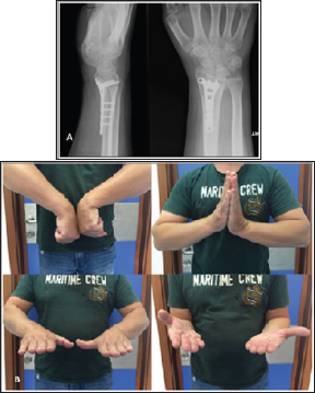
A 48 year-old male, caucasian, policeman, right handed, attended the emergency service due to right wrist pain and functional impotence. Five weeks earlier he had suffered a road accident resulting in a fracture of the distal right radius (AO subtype 23-B3) (Figure 1) and a tibial plafond fracture. At a foreign hospital that received him, he was submitted to radius osteosynthesis with an anatomic volar distal radius plate and in the lower right limb a temporary joint-bridging external fixation was placed during 1 week, and was posteriorly substituted by screws and plate fixation in the tibia and fibula. The wrist cast was removed 4 weeks after surgery in a private hospital in our country. After this episode he began to feel pain, without any history of associated trauma. He had no other previous history of interest. The physical examination revealed a "dinner fork” deformity, with a volar translation of the carpus. The skin was intact. There was no neurovascular deficit. Imaging showed a Dumontier type II volar radiocarpal dislocation (Figure 2A). At the emergency department we conducted closed reduction through traction after infiltration of a local intra- articular anesthetic, without success nevertheless (Figure 2B), so we decided to proceed to surgical treatment. Intraoperatively, through a volar approach of the radius, a bone fragment distal to the plate was visible. A dorsal approach was needed to reduce the dislocation. A radiocarpal fixation with three Kirschner wires was performed, two of them fixing the volar fragment of the distal radius, and one stabilizing the radiocarpal joint. One screw of the plate was removed due to its' intraarticular location. The distal radioulnar joint was evaluated under dynamic fluoroscopy and was apparently stable. Immediate postoperative radiographs confirmed a concentric reduction and stable fixation of the radiocarpal joint (Figure 3). Postoperatively, the patient was placed in an arm cast with free elbow and metacarpophalangeal (MP) joints. There were no perioperative complications. The immobilization and the kirschner wires were maintained for 7 weeks. After removing the cast and the kirschner wires, the patient presented flexion of 20°and extension of 15°, which improved up to flexion 30° and extension 60°, supination 60°, with preserved pronation, 24 months after the trauma. At this time, the patient reports no pain, feeling of instability or signs of nerve compression. The DASH disability score result was 3,1 and the Mayo Wrist Score was 93. The radiograph control did not show any relevant alteration (Figure 4).
Figure 1: Simple AP and lateral radiograph obtained upon arrival at the emergency service at the foreign hospital.
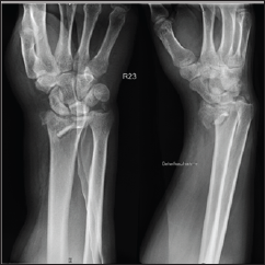
Figure 2: (A) Posteroanterior (PA) and lateral radiographs of the right wrist show volar dislocation of the carpus at our emergency department (B) Postreduction attempted failed PA and lateral radiographs of the right wrist.
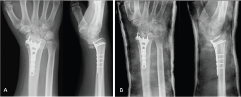
Figure 3: Postoperative PA and lateral radiographs of the right wrist show maintained reduction of the radiocarpal joint after radiocarpal fixation with 3 Kirschner wires.
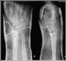
Figure 4: Radiographic and clinical results after 24 months. AP view and lateral view (A); Comparative photographs of both hand in flexion, extension, pronation, supination (B).
Discussion
Dislocations of the radiocarpal joint are high-energy injuries, extremely infrequent, with an incidence of 0.2% of all dislocations [5-6]. There are two classifications that are important in therapeutic decision: Moneim et al. [7] and Dumontier et al. [5]. The management of radiocarpal dislocations varies, since there are different therapeutic options and no long-term follow-up data. Most authors propose a surgical treatment, but nonoperative management has been successful and also reported positive functional results in several series [5].
Figure 5: Postoperative PA and lateral radiographs of the right wrist after surgery at the foreign hospital.
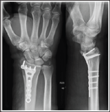
On the other hand, articular fractures can be associated with a spectrum of radiocarpal dislocation. Muller et al. classified articular fractures resulting from a shearing force to the wrist as type B. Jupiter subdivided these fractures in 3 subgroups, in which B3 is volar marginal articular fracture (Barton's fracture)[8]. Open reduction with internal fixation is a commonly used method for the treatment of these intra-articular injuries. Our patient had a 23-B3 distal radius fracture (Figure 1). At the foreign hospital that received him, he was submitted to radius osteosynthesis with an anatomic volar distal radius plate. Postoperative lateral radiographs of the right wrist, provided to.com, showed non- anatomical reduction of fragments after the fixation (Figure 5). Due to surgery to the right lower limb the patient was immobilized with an arm cast during four weeks. After it's removal he began to feel pain, with no history of associated trauma. Imaging showed a Dumontier type II volar radiocarpal dislocation (Figure 2A). There are few data reporting loss of fixation of volar shearing fractures of the distal part of the radius [2]. To our knowledge, no reports have documented spontaneous radiocarpal dislocation after distal radius osteosynthesis.
Some authors reported favorable results after distraction plating of comminuted distal radius fractures. In the published reports, the main indication for distraction plating over traditional internal fixation is the presence of articular or metaphyseal comminution, which makes anatomic fixation difficult, and for polytraumatized patients, where the injured upper limb is required to aid lower-extremity weight bearing [9-10]. We hypothesized that the non-anatomical reduction of fracture and the need for aid lower-extremity weight bearing resulted in loss of fixation and consequently caused radiocarpal dislocation. Our case had a volar dislocation of the radiocarpal joint. At first, we performed a closed reduction under the fluoroscopy, but we were not successful. We needed a double approach to reduce the dislocation, and the volar fragment. After that we perform the radiocarpal and the volar fragment fixation with kirschner wires. The distal radioulnar joint was evaluated under dynamic fluoroscopy and was felt to be stable. Using a simple surgical technique we obtained a good functional result after 24 months of follow-up.
This report brings attention when evaluating volar shearing fractures. In the presence of a comminuted fracture two-dimensional and even three-dimensional computed tomographic scans may well provide important additional information preoperatively and in selected cases a temporary external fixation may prevent displacement of volar fragment.
Read More About Lupine Publishers Orthopedics and Sports Medicine Open Access Journal (OSMOAJ) Please Click on Below Link: https://lupinepublishers-orthopedics.blogspot.com/

No comments:
Post a Comment
Note: only a member of this blog may post a comment.