Lupine Publishers | Journal of Urology & Nephrology
Short Communication
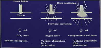
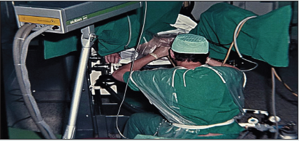
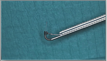
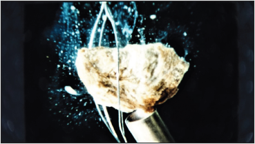
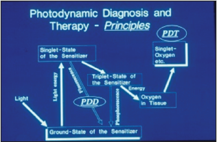
In 1970 I started to investigate the Laser-Technology for urologic surgery together with H. Müßiggang. First of all, it was to check the interacting of the different lasers with biological tissue. The result you can gather from the Figure 1 Decisive are the two factors: absorption and scattering of light into the tissue. The strong light absorption of the CO2 – laser leads to an excellent incision effect with low edema reaction. The light of the is mainly absorbed in the tissue by hemoglobin and pigment colorings and therefore suitable for the destruction of highly vascularized tumors or malformations. For achieving greater volume effects, - necessary for destruction of solid tumors, bilharzial bladder-lesions and inflamed areas in interstitial cystitis, - the Nd:YAG – laser was used by us since 1976 (Figure 2). Presupposed for the clinical application of lasers was the developing of a quartz glass fiber transmission system by Nath, a physicist from the Neuherberg Laser Labor (1973) and the developing of a special cystoscope insert, designed by my working-group (Staehler, Frank et al.) and constructed by the Storz Compagny/Tuttlingen/Germany (Figure 3). The next steps were the laser induced shock wave lithotripsy, developed between 1978 to 1986 (Munich/Lübeck) and the photodynamic procedures for early tumor diagnosis (Figures 5a & 5b).
Now is to ask – what survived?
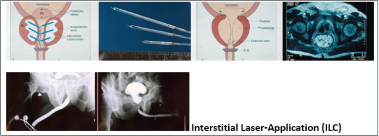
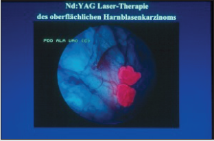
The laser-lithotripsy, especially in ureter, and after a long fight, the photodynamic diagnosis for bladder tumors and Cis, are on routine. Also in tumors of external genitalia, - e. c. condylomata acuminate, hemangiomas, penile cancer, - the ND:YAG- and Diodelasers are well established. Established are also the vaporization and enucleation of prostate tumors by laser. In contrast to these indications, the Nd: YAG-laser-application in bladder-tumors seems to be forgotten. I think this method is head and shoulders above the common TUR-resection because it generates a total homogenous necrosis with closing the blood- and lymphatic vessels simultaneously, it means, no bleeding, no tumor-cellspreading. You can perform the operation under excellent viewing, so that it`s possible to destroy also any satellite-tumors and the tumor vessels (Figures 6a, b, c, d, e). Histological examinations of the tumor tissue after Nd:YAG-laser-application have never been problematic according to our pathologist Dr. Keiditsch. It depends on the fact, that during the coagulation the temperature didn`t exceed 100 C°, it means, the cell-structure remains unchanged. I observed 5 patients, who refused the radical cystectomy, only treated by ND:YAG –laser, between 10 and 40 years. They all remain tumor free. Our photodynamic therapy of localized prostate cancer, published 1993, is still in the early stages. It seems, nobody is interested on this procedure. Forgotten seems to be also our interstitial laser coagulation (ILC) of prostate adenomas and bladder-neck-stenoses, although by this procedure the retrograde ejaculation can be avoided in the most cases (Figures 7a-f).
Figure 6 a-e: a/b: Tumor before and after Nd:YAG-Irradiation, c/d: Destruction of tumor-vessels, e: six weeks later.
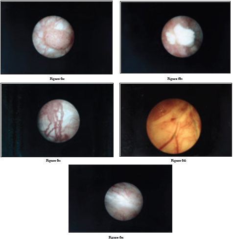
Figure : 7a= scheme, b= glasfibers with lightdistributors, c= scheme after laser application, d= CT after ILC, e = big adenoma of the prostate, f= state after ILC.
https://lupine-publishers-urology-nephrology.blogspot.com/

No comments:
Post a Comment
Note: only a member of this blog may post a comment.