Showing posts with label Jouranal of Urology. Show all posts

Friday, 28 April 2023

Lupine Publishers | Chronic Kidney Disease, Data from MIKD

 Lupine Publishers | Journal of Urology & Nephrology


Abstract

Chronic kidney disease (CKD) is a leaping up public health matter. It has an augmented effect on cardiovascular diseases and affects every system in body. In developed countries the extent of prevalence is available due to renal registries. In middle or low socio economic countries the proportion of this evolving health issue is not known. CKD is rising and a major contributor is soaring number of diabetes mellitus worldwide. Other significant addition in this cohort of CKD is by rising numbers of obese and hypertensive patients. In order to curtail this rising health matter immediate and intense measures both at preventive and curative levels are required.

Aim: Due to lack of formal renal registry we wanted to see spectrum of chronic kidney disease (CKD) in our catchment area and at what CKD stage they present to tertiary care hospital.

Methods: All patients who presented to Emergency department of Multan Institute of kidney disease from 01 Sep 2017 till Sep 2019 data were evaluated. Record of Emergency department patients were taken from electronic system of our hospital. Some patients had multiple visits and we took first visits kidney function in our analysis. eGFR was calculated from serum creatinine with help of CKD-EPI equation.

Results: Total 4303 patients were included in study. Males were 60% and females 40%. Age range from 13 years to 96 year old. 945 patients were excluded as they were not falling in chronic kidney disease category. Remaining 3358 patients had chronic kidney disease. Sub-analysis according to CKD stage showed 66.17% patients presented at CKD stage V.

Conclusion: Kidney disease is rising globally. Countries where renal registries are established provide incidence of chronic kidney disease ranging from 10 to 15%. Still a lot of countries worldwide do not have established system of data collection so true incidence is not established. Our work is first of its kind reported from this area. Drastic preventive strategies are need of time from health budget planners.

Introduction

Multan Institute of kidney diseases is a tertiary care nephrology and urology hospital located in Multan (Largest city) in South Punjab, Pakistan. Catchment area include Multan division, Dera ghazi khan division and Bahawalpur division. According to last census the total population of these 3 divisions is just over 34 million. All patients who presented to Emergency department of our institute from 01 September 2017 till 01 September 2019 were taken from electronic medical record of hospital.

Inclusion & Exclusion Criteria

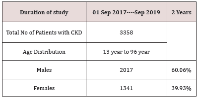
Total number of patients in our data are 4303. Patients from Age 13 year and above were taken in study. Less than age 13 were not included. Maximum age recorded was 96 year old. In case of multiple visits to Emergency department by same patient, only first presentation kidney functions were included in study and subsequent visits kidney function were excluded. 467 patients had normal kidney functions, normal urinalysis and no structural abnormality on renal imaging and were excluded from interpretation. 478 patients were found to have acute kidney injury as per acute kidney injury network (AKIN) classification. These patients were also excluded from analysis. Rest of the patients (n=3358) had chronic kidney disease [Table 1].

Table 1:

lupinepublishers-openaccess-journal-urology-nephrology

Kidney Function Measurement (eGFR)

Serum Creatinine was taken to measure estimated glomerular filtration rate (eGFR). Various methods of eGFR calculation are available, including the Cockcroft-Gault equation, MDRD (Modification of Diet in Renal Disease) Study equation, and the CKD-EPI (Chronic Kidney Disease Epidemiology Collaboration) equation. We took CKD-EPI for measuring eGFR. Patients were divided in CKD stages from 1 to 5.

CKD Stages and Prevalence

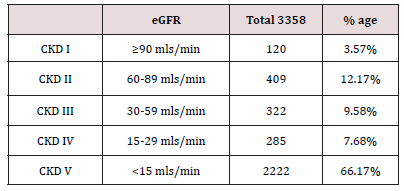
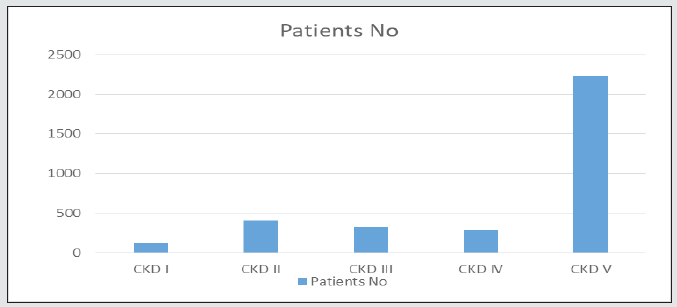
Total 3358 patients were identified with chronic kidney disease. Majority (n=2222) of patients presented when eGFR was <15mls/min for the first time to tertiary care facility. CKD stages with number of patients in each group are in following Table 2 [Figure 1].

Table 2:

lupinepublishers-openaccess-journal-urology-nephrology

Figure 1:

lupinepublishers-openaccess-journal-urology-nephrology

Causes of CKD

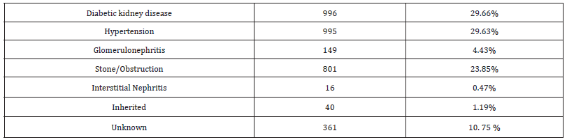
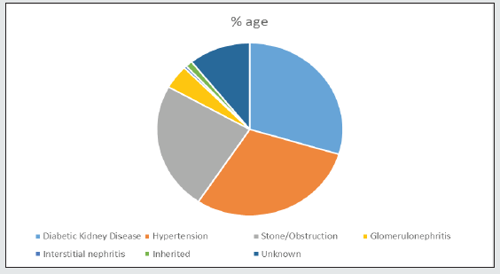
We analyzed our data to look into etiologies of chronic kidney disease. It showed 30 % patients had diabetic kidney disease and same proportion were hypertensive nephropathy patients. Next in line was obstructive nephropathy with 23.85%. Glomerulonephritis was about 4.5% and roughly 10% patients cause was not established as they presented late. Inherited diseases were just over 1% and interstitial nephritis was just 0.47% [Table 3]. Among the patient cohort diabetic kidney disease was separated based on presence of diabetes mellitus and one of the criteria (proteinuria, normal size kidneys, diabetic retinopathy on fundoscopy or >5 years duration of diabetes mellitus). Nearly 30% of total patients had diabetic kidney disease and more than 2/3rd of them had CKD-5 on presentation. A detailed analysis of CKD stages of diabetic group is in following table [Table 4] [Figure 2].

Table 3:

lupinepublishers-openaccess-journal-urology-nephrology

Table 4:

lupinepublishers-openaccess-journal-urology-nephrology

Figure 2:

lupinepublishers-openaccess-journal-urology-nephrology

Discussion

Chronic kidney disease is rising worldwide. It has big impact on health economics [1,2]. Developed countries have registries showing its prevalence which helps in health budget planning. Unfortunately under developed countries lack such planning. In Pakistan recently a national renal registry has started working and will give informative statistics regarding renal disease spectrum. This will help in health management planning and focusing on areas where rampant growing renal issues are focused.

Reviewing published data spectrum of renal disease is variable in different geographical areas [3, 4]. Ethnicity, dietary habits, use of herbal medicines, low birth weight & climate all have been postulated as contributing factors in prevalence of chronic kidney disease. According to a Meta-analysis, chronic kidney disease has prevalence of 11% to 13% globally [5]. In Pakistan studies published on chronic kidney disease prevalence are usually from tertiary hospital or region based [6]. Tertiary care centers are in urban cities so there is no good data from rural areas. South Punjab of Pakistan majority comprise of rural area. According to last census the population of south Punjab 3 divisions (Multan, Bahawalpur and Dera Ghazi Khan) is 34 million [7]. This is the catchment area served by Multan institute of kidney disease (MIKD). MIKD is an emerging kidney institute in this area. We analyzed all patients who presented to Emergency department of our institute. We analyzed presentation creatinine and used CKD-EPI Creatinine Equation for eGFR calculation as recommended by the National Kidney Foundation [8]. Chronic kidney disease in Pakistan is on rise and presentation to specialist center is late. Majority of patients present with CKD-stage 5 requiring dialysis. Our hospital’s 2 year record tells us that 66.17% patients who attended Emergency room were at CKD-5 and majority required renal replacement therapy. Diabetic kidney disease was alarmingly on 29.66 %. Further sub analysis of diabetic kidney disease patients revealed that majority patients (72%) were at CKD stage 5 when presented to hospital emergency department. We labelled diabetic kidney disease based on presence of one of the following criteria’s (established diabetic retinopathy and/or micro albuminuria and/or long duration (>5 years) of diabetes mellitus). Further stage of CKD was established based on CKD-EPI calculation. Diabetes mellitus is at tremendous rate in Pakistan. International diabetes federation data reveals that 17.1% rising adult population of Pakistan is now living with diabetes mellitus [9]. With such enormous rise in diabetics in last decade, the burden of diabetic nephropathy is on rise too. In rural areas a lot of people are undiagnosed and usually present to health facility with end organ damage. According to systematic analytic study done in 2016 prevalence of diabetes mellitus in Pakistan is more in males as compared to females and more common in urban areas [10]. The rising burden of diabetes is imputed to environmental and emotional changes. The main contributors are sedentary lifestyle including internet and TV usage, caloric rich diets leading to increasing obesity. Pakistan with very high numbers of prevalent diabetes needs a cost-effective population based approach for screening [11]. Obstruction leading to chronic kidney disease was immensely at 23.85% in our studied population. Obstructive nephropathy due to stone is highly prevalent in this area and reviewing published data the number is almost 2-3 times in comparison to other parts of world. Nephrolithiasis is a highly prevalent disease worldwide with rates ranging from 7 to 13% in North America, 5–9% in Europe, and 1–5% in Asia. Due to high rates of new and recurrent stones, management of stones is expensive and the disease has a high level of acute and chronic morbidity [12]. Epidemiologic studies have demonstrated that the stone risk incidence increases with Body Mass Index, through multiple pathways. Metabolic syndrome and diabetes are associated with an increased renal stones disease incidence [13, 14]. There is a known high incidence of stone disease in Pakistan as this country belongs to the so-called stone belt [15]. In summing up our chronic kidney disease burden we have identified Diabetic kidney disease, Hypertension and Kidney stones as our bulk. This helps in health budget planning and preventive strategies to cut down disease load. Lack of renal registry in Pakistan has been emphasized [16]. Recently at national level Pakistan renal data system (PKDRS) is established and will provide data from all parts of country in coming years [17]. Curtailing diabetes will have significant reduction in diabetic kidney disease and strategies have been proposed. Creating multidisciplinary team, primary preventive strategies and nationwide diabetic care programme are proposed steps to deal this impending pandemic [18].

Read More About Lupine Publishers Journal of Urology & Nephrology Please Click on Below Link:
https://lupine-publishers-urology-nephrology.blogspot.com/

Friday, 6 January 2023

Lupine Publishers | Efficacy and Safety of Prolonged Alfuzosin Treatment in Patients with Lower Urinary Tract Symptoms Associated with Benign Prostatic Hyperplasia: 7 Years of Observation

 Lupine Publishers | Journal of Urology & Nephrology


Abstract

Introduction: Amongst all of the medications prescribed to BPH patients undergoing conservative therapy, α1-blockers were used in 80% of cases. The question of optimal treatment length is one that has been constantly asked during the last years. The current study will encompass the data we have collected of a small study group of BPH patients treated with 10 mg of Alfuzosin daily, over a period of more than 7 years.

Material and Methods: From 2009 to 2011, 41 patients with mean age 67.7 years began medical therapy taking 10 mg of Alfuzosin per day. The mean treatment length was 7.6 years as of today. 16 patients (39%) with prostates exceeding 60 cm3 were additionally prescribed 5-ARIs.

Results: Overall, a positive dynamic was found in 85.4% of patients. Not a single patient chose to discontinue treatment. A very high level of satisfaction was reported by 88% of our patients. A statistically significant (P < 0.05) decrease in IPSS scores by 8.5 ± 6.1 (47.5%) was found. Mean QoL indices decreased from 3.7 ± 1.1 to 2.3 ± 1.1 over the period of observation. Mean Qmax values increased from 9.7 ± 0.52 mL/s to 14 ± 0.60 mL/s (an increase of 44.3%).

Conclusions: This study has demonstrated a high level of safety and efficacy when using Alfuzosin to treat voiding dysfunction in patients suffering from LUTS/BPH. Our study resolves the issue surrounding a prolonged course of α1-blockers, which, due to its widespread use, remains the gold standard for medical treatment of lower urinary tract symptoms associated with benign prostatic hyperplasia.

Keywords: LUTS, BPH, Alfuzosin, BOO

Introduction

It is well known that the prevalence of voiding dysfunction and patient age are in direct proportion and that prevalence is particularly high in patients over 50 years of age. Typical complaints associated with voiding dysfunction are termed lower urinary tract symptoms (LUTS) and have a wide variety of underlying causes. The most common aetiologies of LUTS are prostatitis, urethral strictures, cystitis, bladder stones, prostate cancer, and benign prostatic hyperplasia (BPH).

BPH is usually managed with surgical intervention, pharmacotherapy, or a strategy of watchful waiting, and numerous factors affect the decision of which method should be employed. Symptom severity is one of the most important subjective factors and is measured using the international prostate symptom score (IPSS) questionnaire. The nature of the symptoms (whether they fall under the category of obstructive or irritative) also influences the final decision. The primary objective factor can be considered to be the quality of voiding as measured by uroflowmetry, the interpretation of which should consist not only of the peak flow rate (Qmax), but also flow time, time to Qmax and many other indicators. Postvoid residual urine is an equally important value because it helps identify poor bladder contraction ability (detrusor underactivity) in the case of progressive intravesical obstruction. There are also those factors, no less important than both uroflowmetry and IPSS, which relate to the potential risk of BPH progression, and thus make their impact on the choice of treatment. Whilst the size of the prostate itself cannot be ignored, it is by no means a marker for surgical intervention when considered by itself, but rather a hallmark for potential future complications such as acute urinary retention (AUR) amongst other things. In recent years, prostate-specific antigen (PSA) levels have also become associated with BPH progression. Today, PSA levels are not only used to screen for prostate cancer, but also to reflect cellular proliferation rates of the prostate. Thus, high PSA levels increase the risk of rapid BPH progression. [1] The sum of the factors mentioned above influences the ultimate decision of which type of treatment should be employed in a specific patient.

Modern standards of medical practice dictate that pharmacotherapy is recommended in BPH patients with moderate LUTS without upper urinary tract complications, patients without obvious surgical indications, and patients who either decline surgical intervention, or are unable to commit to surgery in the nearby future. Watchful waiting is acceptable if a patient exhibits mild LUTS (IPSS ≤ 8), or if the patient’s symptoms do not significantly impact their quality of life (QoL). Patients who fall under this category should be made aware of the necessity to maintain an appropriate lifestyle as well as the importance of regular blood, urine, PSA, uroflowmetry and ultrasound testing. Surgical intervention is needed if a patient exhibits severe symptoms with upper urinary tract complications, or if there are sufficient reasons to suspect that medication will be ineffective[2,8].

In order to better illustrate modern tendencies in the treatment of BPH, we turn our attention to the TRIUMPH study. The TRIUMPH study recorded the treatment and outcomes of 2351 newly presenting LUTS/BPH patients in 6 European countries over a 1-year follow-up period. Out of the entire study population, 23.8% were managed with watchful waiting, 72.5% were prescribed medication, and only 2.7% underwent surgical treatment. These statistics accurately reflect the rise in medical management of LUTS as well as the decreasing need for surgical intervention during the past decade. It should be interesting to note that amongst all of the medications prescribed to BPH patients undergoing conservative therapy, α1-adrenergic receptor antagonists (α1-blockers) were used in 80% of cases [3].

The efficacy of α1-blockers in patients with BPH has been well documented in a large quantity of double-blind, placebo-controlled studies.[4,5,6,7] If it is possible to consider transurethral resection of the prostate (TURP) the method of choice for surgical intervention in BPH patients, then α1-blockers will fall under the same criterion for the medical management of BPH. It is important to note that the European Association of Urology considers α1-blockers the first line of therapy for the management of voiding dysfunction in patients suffering from BPH[8].

Whereas the rationale behind α1-blockers does not arouse suspicion in the majority of specialists, the question of optimal treatment length is one that has been constantly asked during the last 6 years. Solutions offered to address this problem range from a life-long course of medication, to one that only lasts a few months. There is no one correct way to approach this matter, but it is the duty of every qualified specialist to incorporate certain principles when making such a decision. Personal experience and knowledge gained from a wide variety of international studies should encompass the core of such precepts. It is arguable that the lack of a formulated strategy that applies to such clinical trials is hindering the formation of a unified point of view. If we turn to medical literature, we find that despite an abundance of studies indicating the effectiveness of α1-blockers in BPH patients, the studies lack the duration necessary to examine long-term effects and are mostly comprised of 3-6 month trials with only a few mentioning 1-3 year-long results. This has led many to assume that a 3-month course (being the most prominent study length) of α1-blockers is sufficient, whilst the actual reasoning behind the short length of most studies lies in the knowledge that the results achieved during that period of time will continue over the remaining course of treatment, however long it may be. Confirmation of this can only be found in a few currently available publications that demonstrate long-term results. Most articles cite results that were obtained using studies based on principles of Good Clinical Practice (GCP) in which the categories of patient inclusion/exclusion were so rigid, they created unrealistic expectations for the wide variety of patients that are actually treated. Only a few trials provide us with results that are based on a realistic study population. Such trials include those of Roehrborn C. et al., Elhilali M. et al. 2006 and Vallancien G. with Emberton M. et al. 2008[9,10,11].

During the early 21st century, the suitability of α1-blockers was no longer debated in the Russian Federation, whilst the question of long-term use was left unanswered even for us. Even now, considering the ageing population and the necessity of prolonged medical treatment, many specialists are hesitant to prescribe α1-blockers for more than a few years without consulting medical literature about the safety and efficacy of such a treatment.

Materialand Methods

The current study will encompass the data we have collected of a small study group of BPH patients treated with 10 mg of Alfuzosin daily, over a period of more than 7 years. We must mention that the data provided isn’t final, in the sense that patients are still undergoing treatment to this day. We had not originally planned for this to be a study with a specific design, but retrospective aspects make it similar to various non-comparative, real-life surveillance studies. Our goal is to introduce the reader to the results of a longterm course of α1-blockers that, in our opinion, are a safe and effective method of long-term medical therapy in patients suffering from BPH.
From 2000 to 2002, 41 patients with mean age 67.7 years (range 51– 83) began medical therapy. The mean treatment length was 7.6 years as of today. We would like to mention that this group of patients wasn’t specifically chosen as a study group at the time, nor were they the only patients that had been prescribed Alfuzosin. Our detailed observation of the isolated group in question is partially due to chance, and partially due to loss to follow-up. Patients began treatment taking 5 mg of Alfuzosin twice daily. From January 2005 onwards, all patients take 10 mg of Alfuzosin once daily at night due to numerous complaints of increased voiding problems during this time. Prostate volumes fluctuated between 25 cm3 and 100 cm3, and 16 patients (39%) with prostates exceeding 60 cm3 were additionally prescribed 5-alpha-reductase inhibitors (5-ARIs). During the early stages of treatment these patients were prescribed Finasteride, but during the last 2 years many have switched to Dutasteride in accordance with our recommendations.

Indications

Patients with LUTS/BPH

Contraindications

Patients with surgical indications
Patients known to have insufficient results from taking α1- blockers in the past
Patients with postural hypotension
Patients taking alternative medication for LUTS with good results
Patients with unstable angina (pectoris)
Patients with life-threatening comorbidities

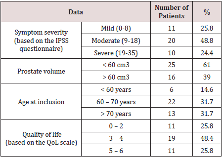
Table 1: Summary of initial patient data.

lupinepublishers-openaccess-journal-urology-nephrology

The mean duration of morbidity at the time of patient inclusion in the trial was 1.9±1.1 years. During ultrasound testing, 31.2% of patients had postvoid residual urine measuring 82 ± 41.7 mL. Mean PSA levels of all patients before the trial were 2.9 ± 2.8 ng/mL. The initial data of the patients included in this trial are summarized in Table 1.
Out of the 41 patients, 14 (34.1%) had hypertension 11 of which (26.8%) were receiving some form of hypotensive therapy, and 5 patients had suffered a myocardial infarction before inclusion into the trial.
The efficacy of the treatment was analysed using IPSS scores as well as QoL indices. Additional factors included Qmax and prostate volume in patients who were receiving combination therapy with 5-ARIs. The safety of the treatment was analyzed based on recorded incidents of adverse cardiac side effects (mainly hypotensive) during the trial. Vital-sign dynamics were also analyzed. Follow-ups were conducted biannually during the first 2 years, and no less than annually thereafter.
Two-sided hypothesis tests were conducted with a significance P-value cut-off of 0.05. Dynamics of safety and efficacy variables were analysed before and after the trial (absolute and relative change was considered). A paired Student’s t-test was used if statistics followed a normal distribution; a Wilkinson’s test was used if they did not.

Results

Not a single patient chose to discontinue treatment, notwithstanding the long period of observation. We must mention, however, that several patients stopped being prescribed Alfuzosin during the early stages of the trial but were obviously not considered in the study group. Subjective assessments of patient satisfaction were carried out using a questionnaire specifically tailored to this publication and not used in our routine clinical practice. A very high level of satisfaction was reported by 88% of our patients, whilst the remaining 12% consisted of those actively taking a large quantity of medication for various comorbidities, and patients with diabetes. Many of the aforementioned patients were prescribed medication after they began treatment with Alfuzosin. Therefore, these facts could be taken into consideration by general practitioners who have patients undergoing treatment for LUTS.

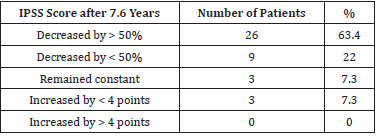
Table 2:IPSS scores after 7.6 years of Alfuzosin treatment.

lupinepublishers-openaccess-journal-urology-nephrology

Mean IPSS scores were 17.9 ± 5.3 before the treatment, and 9.4 ± 5.2 after 7.6 years. A statistically significant (P < 0.05) decrease in IPSS scores by 8.5 ± 6.1 (47.5%) was found. A decrease in IPSS scores by > 50% was noted in 28 patients (68.3%). Overall, a positive dynamic was found in 85.4% of patients. IPSS score dynamics are summarized in Table 2.
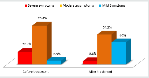
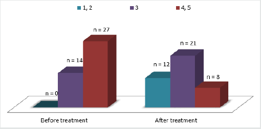
Dynamics of irritative (questions 1, 3, 5 and 6 on the IPSS questionnaire) and obstructive (questions 2, 4 and 7 on the IPSS questionnaire) symptoms were also analyzed. Mean irritative IPSS scores decreased from 6.3 ± 2.9 to 3.7 ± 2.4 (33.7% lower) whilst mean obstructive IPSS scores decreased from 9.6 ± 4 to 5.5 ± 3.7 (35.7% lower). Incidents of nocturia decreased from 2.4 ± 1.1 to 1.5 ± 1.1. Diagram 1 illustrates a major decrease in the percentage of patients with moderate to severe voiding dysfunction symptoms as well as a six-fold increase of patients with mild symptoms. All of the statistics mentioned above were statistically significant (Figure 1).

Figure 1: Severity of LUTS before and after 7.6 years of Alfuzosin treatment.

lupinepublishers-openaccess-journal-urology-nephrology

QoL dynamics are presented in diagram 2. Mean QoL indices decreased from 3.7 ± 1.1 to 2.3 ± 1.1 over the period of observation. The mean decrease in QoL indices was 32.3% (reflecting an increase in QoL). All changes were statistically significant. Overall, 72.7% of patients had an increase in their QoL (Figure 2).

Figure 2: QoL indices before and after 7.6 years of Alfuzosin treatment.

lupinepublishers-openaccess-journal-urology-nephrology

It is important to note that the minority of patients who did not demonstrate clear improvements in symptom severity either underwent surgery or continued to take Alfuzosin. The latter group either demonstrated a minimal but satisfactory improvement or was unable to undergo surgery for various reasons. We believe that it is of utmost importance to understand that the current publication presents a comparative analysis before and after Alfuzosin treatment, and whilst all of the patients had demonstrated an improvement during various periods in the trial, the dynamics of IPSS scores and changes in QoL were not registered at those intervals. It is also important to highlight the role of the detrusor in BPH symptoms, something that was originally demonstrated by Russian authors O.B.Loran and E.L.Vishnevskiy in 1998. The contraction capability, level of energy metabolism, and nature of the biochemical events of the detrusor influence voiding quality no less than the level of intravesical obstruction.[13] This could explain treatment ineffectiveness in certain patients.

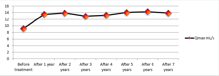
Changes in Qmax over the course of the study were considered as a secondary measurement of treatment effectiveness and were analysed during every follow-up, making this particular variable very intriguing. Mean Qmax values increased from 9.7 ± 0.52 mL/s to 14 ± 0.60 mL/s (an increase of 44.3%). Diagram 3 illustrates Qmax dynamics throughout the entire length of the study. One can conclude that early treatment results remained constant throughout the observation period (Figure 3).

Figure 3: Peak flow rate (Qmax)dynamics during 7.6 years of Alfuzosin treatment.

lupinepublishers-openaccess-journal-urology-nephrology

During the period of observation, 5 patients underwent TURP procedures. The rationale behind TURP in one patient was due to an episode of AUR provoked by alcohol ingestion. After 3 days of catheterisation, the patient did not show any signs of improvement in voiding ability and was subsequently operated. The other 4 patients exhibited worsening LUTS as shown by an increase in their IPSS scores. During ultrasound and transrectal ultrasound testing, a median lobe was detected in all 4 patients. All operations performed did not deviate from a standard TURP procedure.
The median prostate volume of the 16 patients receiving combination therapy had decreased. The value fell from 74.1 cm3 before treatment to 50.1 cm3 after 7.6 years of therapy (a decrease of 32.4%).
Throughout the trial, PSA levels of all patients were measured annually. Median PSA levels were 2.9 ng/mL before the trial and 3.2 ng/mL afterwards, demonstrating no significant variance. PSA levels of patients undergoing combination therapy were considered after the second year of treatment and were doubled. PSA levels of over 4 ng/mL requiring transrectal ultrasound-guided multifocal prostatic biopsies were noted in 3 patients aged 68–74 years. None of the subsequent histological analyses revealed the presence of cancerous cells.
The safety of this treatment was assessed based on the frequency and type of adverse side effects recorded during the trial. In total, 9 patients (22%) exhibited adverse effects. The most common findings were dizziness (2 patients/4.9%) and asthenia (2 patients/4.9%). Another 3 patients experienced dyspepsia, shortness of breath and headaches. Out of the 16-patient subgroup receiving combination therapy, 2 patients complained of decreased libido (possibly attributed to 5-ARIs). One of these patients had their sexual drive normalise after 1 year of therapy, whilst the other retained a low libido throughout the treatment. Considering the relatively small study population involved, a true understanding of the statistical significance of these adverse effects as well as their immediate connection to the treatment in question is not possible. However, the fact that most of the aforementioned adverse effects were present as isolated episodes in patients during the first week of therapy demonstrates a high level of treatment safety.

Discussion

The current study has demonstrated a high level of efficacy with minimal adverse side effects in the treatment of lower urinary tract symptoms (LUTS) arising from benign prostatic hyperplasia (BPH) with α1-adrenergic receptor antagonist (α1-blocker) Alfuzosin. The effectiveness of the treatment was determined using subjective and objective criteria. A 45.7% decrease in LUTS severity as measured by the International Prostate Symptom Score (IPSS) questionnaire was found in 85.4% of the entire study population. Mean Quality of Life (QoL) indices measured using the QoL scale increased by 32.2% and mean peak flow rate (Qmax) values increased by 44.3%. The fact that the positive results achieved during the early months of therapy had stabilised and showed no signs of regression during the entire study period is quite remarkable. Such results verify the notion that a prolonged course of α1-blockers provides consistent long-term results. The high level of patient satisfaction with treatment results and the information that patients themselves have provided us with, mark not only the fact that patients are not bothered by the need to take medication daily, but also (and most importantly) the great sense of confidence patients feel regarding the stability of the results they have achieved, leading to a positive outlook on their future treatment.

Several observational studies indicate results very similar to ours. In the ALFORTI study, 311 patients on Alfuzosin were surveyed over a period of 9 months. A decrease was shown in mean IPSS scores and QoL indices measuring 45.6% and 36.4%, respectively. Adverse effects (most likely due to the medication in question) occurred in 4.4% of the study population.[12] A similar study published by the same authors analyzed the safety and efficacy of Alfuzosin. A decrease in mean IPSS scores and QoL indices was shown and measured 32% and 27.8%, respectively[14].
Combination therapy in the form of α1-blockers and 5-alphareductase inhibitors (5-ARIs) is currently the recommended form of treatment in patients with LUTS and an increased prostate volume according to the European Association of Urology.[12] The safety and efficacy of this treatment has been confirmed by a considerable decrease in LUTS severity and prostate volume (a mean decrease of 32.4%) in our 16 patient subgroup. Similar results have also been demonstrated by such well-known trials as MTOPS, ALTESS and COMBAT[1,2,8,9].
When the information provided by the various authors is analysed, it becomes apparent that Alfuzosin attains a similar degree of effectiveness in every single case. The fact that the results gained from a continuous course of therapy remain consistent throughout prolonged treatment is extremely important, but perhaps the most interesting observation made during our trial is that the considerable results are not only long-lasting, but are achieved and made stable during the first month of treatment.
A study conducted in 2008 by Vallancien G. et al. is one of the few studies (similar to ours in design, but not in duration) that analysed the data collected in a real-life surveillance study format regarding the effectiveness of Alfuzosin during a 3-year course of therapy. The results were as follows: mean IPSS scores decreased by 33.4%, mean QoL indices by 40.7%, and nocturia severity by 25.5%. Adverse side effects (likely related to vascular dilation) were registered in 4.5% of cases, and surgical intervention was required in 5.7% of cases. These results are clearly similar to ours. [10] Similar findings were also recorded earlier during the ALFONE trial published by Elhilali M. et al. in 2006[11].
Whilst several adverse cardiac effects were noted, the analysis of adverse side effects in our trial is far from thorough, which is explained by the lack of the patients enrolled in the trial leading to insufficient statistical integrity. A study involving a multidisciplinary approach could aid the understanding of various guarantees that we may be able to give our patients in the future. These days, patients mostly consist of elderly men whose age and comorbidities make the use of pharmacotherapy ideal for managing the symptoms associated with BPH. Perhaps tomorrow, the necessity of prescribing Alfuzosin for over 10 years will increase considering the continually regressing number of patients who choose surgical intervention for BPH management; those that wish to retain a good quality of life whilst remaining ‘real men’ for many years to come. Today, when patients inquire about the potential length of their treatment, they are told that they may take Alfuzosin for as long as it is required.

Conclusions

This study has demonstrated a high level of safety and efficacy when using Alfuzosin to treat voiding dysfunction in patients suffering from LUTS/BPH. Furthermore, it is important to mention that many researchers have come to the same conclusion. Our study, whilst potentially lacking in statistical magnitude, resolves the issue surrounding a prolonged course of Alfuzosin, which, due to its widespread use, remains the gold standard for medical treatment of lower urinary tract symptoms associated with benign prostatic hyperplasia.

Read More About Lupine Publishers Journal of Urology & Nephrology Please Click on Below Link:
https://lupine-publishers-urology-nephrology.blogspot.com/


Friday, 20 August 2021

Lupine Publishers | Contrast-Induced Nephropathy: Current practice

 Lupine Publishers | Journal of Urology & Nephrology


Abstract

Contrast induced nephropathy (CIN) is a common cause of hospital acquired acute kidney injury (AKI) and associated with adverse clinical outcomes. There is still debate regarding the exact definition, which has greatly influenced the reported incidence of CIN in literature. Recent studies have challenged the universal concern regarding risk of CIN in general population. It is found to occur more commonly after intra-arterial (IA) administration of contrast as in interventional cardiology and vascular procedures especially in patients with multiple comorbidities and underlying renal impairment. Recent studies report negligible risk after intravenous (IV) contrast administration for modern diagnostic radiological examinations. Since it is a potentially preventable clinical condition, it is imperative for health care professional to be well aware of this entity. All patients undergoing iodinated contrast exposure should be risk stratified and preventive measures should be employed in high risk population. This paper will review the epidemiology, controversies regarding definition, pathophysiology, risk stratification, iodinated contrast commonly used in practice and preventive strategies.

Keywords: Contrast Induced Nephropathy,Iodinated Contrast Media,Acute Kidney Injury,Cardiac Angiography,Contrast Enhanced Computed Tomography

Abbrevations: CIN: Contrast Induced Nephropathy,AKI: Acute Kidney Injury,CM: Contrast Media,ICU: Intensive Care Unit,CECT: Contrast Enhanced Computed Tomography,PTCA: Percutaneous Trans-Luminal Coronary Angioplasty,ACR: American College of Radiology,AKIN: Acute Kidney Injury Network,eGFR: Glomerular Filtration Rate,NGAL: Neutrophil Gelatinase-Associated Lipocalin,CES: Cholesterol Embolism Syndrome,ERBP: European Renal Best Practice,KDIGO: Kidney Disease Improving Global Outcomes,NSAIDS: Non-Steroidal Anti-Inflammatory Drugs,HOCM: High Osmolar Contrast Media,LOCM: Low Osmolar Isosmolar CM,HD: hemodialysis,HF: hemofiltration,NAC: N-acetylcysteine,ESUR: European Society of Urogenital Radiology,CAR: Canadian Association of Radiologist,ESRD: End Stage Renal Disease,TOF: Time Of Flight,CKD: Chronic Kidney Disease,NSF: Nephrogenic Systemic Fibrosis,NCCT: Non contrast computed Tomography,CO2: Carbon Dioxide

Introduction

Contrast induced nephropathy (CIN) is defined as acute renal impairment after exposure to iodinated contrast media (CM). In the modern era with the advancement of the diagnostic modalities, increase in number of percutaneous coronary and peripheral artery interventions and frequent use of contrast media, CIN has emerged as a common cause of acute renal failure. Mostly, it results in transient renal impairment; however in patients with multiple comorbidities it can be associated with high morbidity and mortality [1]. It is therefore imperative to identify patients at risk to decrease its occurrence and diagnose it early in course to avoid short and long term clinical adverse effects. It presents a challenging situation that is often encountered by practitioners in varied specialties including emergency medicine, nephrology, cardiology, radiology and intensive care unit (ICU). Since CIN represents a form of acute kidney injury (AKI), which is potentially preventable, it is important that clinicians are well aware of this entity and understands the basic pathogenesis involved. The aim of this review article is to discuss pathogenesis of CIN, associated risk factors, different iodinated CM used in clinical practice and their effect on kidney, alternative diagnostic modalities and preventive measures.

Epidemiology

CIN remains an important cause of in-hospital AKI, accounting for approximately 11% of the total cases of AKI [1]. Literature on CIN mainly reports it to occur following exposure to iodinated contrast during coronary or peripheral angiography [1,2] or diagnostic imaging, however recent data shows contrast exposure associated with modern radiographic procedure is not associated with significant increase in CIN incidence [3]. Exposure following cardiac or peripheral angiography differs from that during diagnostic imaging in that the injection is intra-arterial, requires catheter and dose of CM is concentrated and abrupt [4,5]. In general population without any risk factor, incidence of CIN is very low; however risk increases as comorbidities increases [1,6,7]. Critically ill patients with baseline renal impairment are particularly susceptible and contrast enhanced computed tomography (CECT) might account for 18% of AKI cases in this population [8]. Reported incidence of CIN varies with the procedure and is reported to be 11% after outpatient CECT [9], 4% following intravenous pyelography [10], 9% after peripheral arteriography (4), <3% following percutaneous trans-luminal coronary angioplasty (PTCA) in patient with normal renal function and as high as 40% in patient with underlying renal impairment [2,11,12]. Studies have documented at least 3.1% of patients with CIN required renal replacement therapy. In hospital mortality is reported to be 7.1% in patients with CIN after coronary intervention and as high as 35.7% in patients requiring dialysis, which further increases to 81.2% at 2 year [1]. CIN is reported to be independent predictor of poor outcome in patients with diabetes mellitus with or without renal impairment [6].

Definition

Post- contrast acute kidney injury (PC-AKI) is often defined as acute renal impairment occurring within 48hours of exposure to intravascular iodinated CM. It is a correlational diagnosis and can occur irrespective of CM being cause of renal deterioration. In contrast, CIN comes under subgroup of PC-AKI and is a causative diagnosis. In literature, CIN has been defined as relative change in the baseline serum creatinine (> 25-50%) and absolute elevation from baseline serum creatinine (>0.5-2 mg/dL) [13- 15]. Temporal relationship between exposure to CM and rise in serum creatinine along with exclusion of other causes of AKI are crucial for the diagnosis of CIN. Defining set criteria for diagnosis is important as incidence of CIN in clinical practice can vary greatly with small change in serum creatinine. An absolute increase of serum creatinine by ≥0.5 mg/dL from baseline is still commonly used definition of AKI [16]. Another well-known criterion for CIN proposed by Barrett and Parfrey in early 1990s was an absolute increase in serum creatinine levels by ≥0.5 mg/dL or a relative increase of ≥25% in serum creatinine from baseline within 72 hours after contrast exposure [17]. However, for purpose of standardizing the definitions, recently American College of Radiology (ACR) has recommended using the AKIN (Acute Kidney Injury Network) criteria to define CIN. It employs absolute serum creatinine increase >0.3mg/dl or ≥50% (≥1.5 times) percentage increase in serum creatinine or ≤0.5 mL/kg/hour urine output for at least 6 hours within 48hours of exposure to define AKI [18]. In clinical practice, serum creatinine concentration is commonly used measure of renal function; however it has its own limitations. There is often delay between renal insult and rise of serum creatinine, which delays the diagnosis. It is also affected by other factors including age, gender, nutritional status, volume status, hyper catabolic state, muscle mass and concomitant use of other medications, therefore it is important to interpret results in appropriate clinical settings [19,20]. Changes in serum creatinine concentration are not linearly related to changes in effective glomerular filtration rate (eGFR,) because of which small changes in eGFR can often go unnoticed. Normal serum creatinine is actually maintained till GFR or creatinine clearance is reduced by nearly 50% [20].

Diagnosis

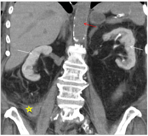
Diagnosing CIN is challenging. It is important to follow systemic approach while evaluating patient with AKI. CIN is a diagnosis of exclusion after other causes of AKI (prerenal/intrinsic/post renal) have been ruled out. There is temporal relationship with contrast exposure, however this should not preclude from searching for other reversible causes of AKI, which may coexist [13-15]. Periprocedural hypotension, bleeding, release of athero-embolic material and use of catheter exchanges may further complicate renal tubular injury after angiography [21]. After exposure to intravascular iodinated contrast, serum creatinine typically rises within the first 24-48 hours, peaks at 3-5 days and returns close to baseline within 1-3 weeks. In most cases, it is asymptomatic and has no significant clinical consequences. However, in rare cases it can lead to oliguria or anuria, irreversible renal damage and need for renal replacement therapy. Basic work up includes clinical assessment, urine output, urinalysis and renal imaging. Volume depleted state can predispose patient to CIN; however CIN can result in intrinsic renal injury with tubular necrosis in extreme cases. Finding on urinalysis may include muddy brown granular cast, tubular epithelial cells and minimal or no proteinuria. Urine examination is neither sensitive nor specific to diagnose CIN, however its usefulness is mainly to exclude underlying glomerular disease or acute interstitial nephritis [22]. Urine studies may show intrinsic renal injury picture with urine sodium < 10 mEq/L, fractional excretion of sodium <1% and urine osmolality <350 mOsm/kg. Persistent nephrogram (Figure 1) may be incidentally seen on follow up imaging because of delayed clearance of contrast [23].

Timing of serum creatinine rise after contrast exposure in literature has been reported to be 24-72hours. However, it is speculated that renal damage actually begins soon after exposure to intravascular iodinated contrast. In study by Ribichini et al. [24], it is reported that measuring change in serum creatinine from baseline within 12hours of contrast exposure significantly predicted occurrence of CIN and was associated with renal damage after 30days [24]. However, relying on serum creatinine for diagnosis of renal injury has its own limitation as discussed above, therefore finding of specific biomarkers that could predict early diagnosis of CIN is desirable. Recently, it has been proposed to classify these biomarkers into two group, namely, (1) reflecting change in renal function (e.g. cystatin C) and (2) representing renal damage {e.g. Plasma or urine neutrophil gelatinase-associated lipocalin (NGAL), interleukin-18} [25]. Briguori et al. [26] reported increase in levels of cystatin C ≥ 10% at 24 h after PTCA had 100% sensitivity and 85.9% specificity for the prediction of CIN [26].

Another condition often encountered after exposure to CM especially catheter related studies is cholesterol embolism syndrome (CES). It can be challenging to distinguish the two conditions clinically; however it is important as both have different treatment approach. CES is usually a rare entity and renal impairment typically develops gradually over 3-8weeks after the procedure. It is a multisystem disease caused by dislodgement of cholesterol crystals from atherosclerotic plaque and occlusion of peripheral arterioles. Other peripheral sign that can help clinch the diagnosis include live do reticularis, petechiae, digital gangrene, splinter hemorrhage and Hollenhorst plaques on ophthalmological examination. Laboratory studies may show peripheral eosinophilia, hypocomplementemia and high erythrocyte sedimentation rate. Spectrum of presentation is varied ranging from mild and asymptomatic to life threatening complications. CES is associated with significantly worse long term renal effects as compared to CIN and is suspected when multiple oral dysfunction occurs following coronary angiography [27].

Pathophysiology

Kidneys are particularly susceptible to ischemic injury in setting of delicate micro vascular circulation, which is particularly vulnerable to systemic and local hypo perfusion. Pathogenesis of CIN is multi factorial and still not completely understood. Interplay of factors including medullary hypoxia, oxidative stress, im balance of renal vaso constrictive and vaso dilatory mediators, changes in renal perfusion and direct tubular toxicity of CM has been suggested [28-30]. Two main mechanisms responsible are direct tubular toxicity of CM and changes in renal micro vascular hemo dynamics. Recently, high end diastolic pulmonary arterial blood pressure and left anterior descending artery lesion has been independently associated with development of CIN, pointing towards role of hemo dynamics and cardiac dynamics in its pathogenesis [31].

Effect of CM on Renal Tubules

CM is freely filtered from renal glomerulus and increases the tubular osmolality. It has direct cytopathological effects on tubular cells, which affects energy metabolism and impairs intracellular transport. Histopathologically, these changes consist of tubular cell vacuolization, necrosis and termed as osmotic nephrosis. There has been conflicting evidence to support precipitation of intratubular proteins triggered by CM. Actually it is proposed that CM induced cellular injury might be the inciting event responsible for CIN which triggers the cascade rather than hypoxia or hypoperfusion. Osmolality, viscosity and ionic properties of CM contribute to its nephrotoxicity. High osmolar CM have been shown to affect erythrocyte deformability, leading of stacking of blood cells and affecting blood flow in animal models. Another mechanism reported is mitochondrial dysfunction and release of catalytic iron, which serve as catalyst in oxidative reactions leading to production of free radicles [28-30].

Effects on Renal Microvascular Hemodynamics

Animal model have suggested that intra-arterial contrast infusion results in biphasic renal perfusion, with initial transient vasodilation followed by sustained prolonged vasoconstriction. Renal medullary perfusion and oxygen tension is lower than outer renal cortex under normal physiological state and thick ascending loop of henle in outer renal medulla has high oxygen requirement exacerbating relative medullary hypoxia. CM further aggravates this hypoxia by causing osmotic diuresis leading to increased sodium transport to thick ascending limb and subsequently increased oxygen extraction. Total medullary hypoxia is actually combination of increased oxygen demand of tubular cells and changes in regional renal microcirculation [32,33]. Subclinical CIN can occur in majority of patients following contrast exposure; however it goes undetected as healthy individuals have intact tubular repair mechanism that prevents clinically significant renal damage. In contrast, in patients with comorbidities like baseline renal impairment and diabetes mellitus even average dose of contrast can have clinical implications in setting of poor reparative mechanism and baseline reduced functional nephrons [34].

Risk Factors

Various risk factors have been described which increases the risk for development of CIN after exposure to iodinated CM. These can be broadly divided into intrinsic or patient related and external or procedural/contrast related. Among the patient related factors, baseline renal impairment (GFR < 60 mL/min per 1.73 m2) and diabetes mellitus are the two important independent risk factors that are commonly coexisting in vascular patient population [7,35]. However, recent analyses by McDonald [16] and Davenport et al. [36] have found increased incidence of CIN in diabetes only if renal function is compromised (GFR < 30 mL/min/1.73 m2) [16,36]. Studies have reported significant risk for CIN if baseline serum creatinine concentration is ≥1.3 mg/dL in men and ≥1.0 mg/dL in women, mostly equivalent to an eGFR <60 mL/min/1.73 m2 [37]. In light of recent studies, European Renal Best Practice (ERBP) along with Kidney Disease Improving Global Outcomes (KDIGO) guidelines suggest that the threshold at which actual risk for CIN increases could be lowered to 45 mL/min/1.73 m2 [38]. Other established risk factors include advanced age (≥70 years), dehydration, anemia, vascular disease, hypertension, coronary artery disease, congestive heart failure, smoking and concomitant use of other medications including metformin, non-steroidal anti-inflammatory drugs (NSAIDS), diuretics or calcium channel blockers.

It was long thought that patients with multiple myeloma have increased risk CIN secondary to precipitation of myeloma proteins in the renal tubules. However, recent studies propose that contrast studies can be safely performed in myeloma patients with normal renal function provided there is no dehydration. Also, studies have reported acute urate nephropathy in patients with hyperuricemia, particularly leukemic patients on chemotherapy, secondary to uricosuric property of CM, however recent studies have failed to show independent association between the two [21,39]. Extrinsic or contrast related factors includes type of CM, route of administration, dose of CM and numbers of contrast exposure. Procedure related factors may include urgent or emergent procedure, use of intra-aortic balloon pump, delayed reperfusion and nature of procedure [12,21,39]. Various risk scoring have been proposed to stratify the risk for CIN. One of them reported by Mehran et al. [6] included eight variables namely age > 75 years, anemia, hypotension, diabetes mellitus, chronic congestive heart failure (CHF), acute pulmonary edema, chronic kidney disease, use of intra-aortic balloon pump and increase volumes of CM. Total risk score ≥ 16 was associated with 57% risk of CIN with 13% of patient requiring dialysis [40]. Another simple risk scoring, which has shown to have clinically significant predictive value for CIN, is ACEF score. It uses three variables, namely age, creatinine level and ejection fraction and has been developed for patients undergoing coronary angiography [41,42].

Iodinated CM

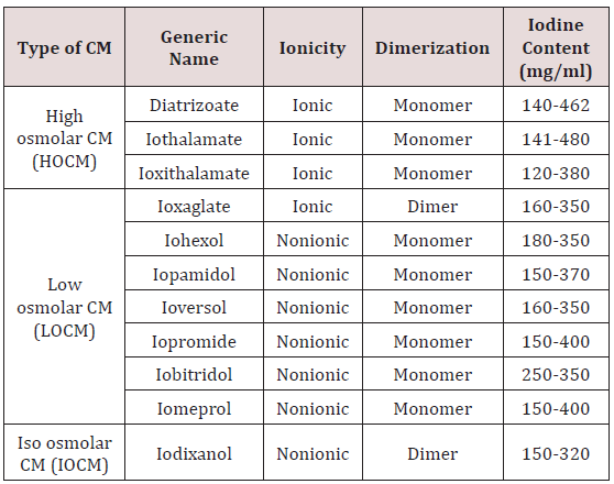
Type of Iodinated CM: Commonly available CMs are benzoic acid derivatives with three iodine atoms. High osmolar CM (HOCM), with osmolality up to eight times that of plasma were frequently used in past and had higher incidence of CIN. To decrease the osmolality, several approaches were considered including production of non-ionic agents, which resist dissociation in solution or by producing double benzoic acid rings. Low osmolar (LOCM) and isosmolar CM (IOCM) are commonly used nowadays and associated with low nephrotoxicity [43]. (Table1) descries the properties of CM. The adverse effect of CM is mainly related to its osmolality, ionicity, viscosity and iodine content. Viscosity instead of osmolality is responsible for vascular resistance [44]. Though IOCM has lower osmolality, because of its dimeric structure it has more viscosity than LOCM. There are conflicting results in studies comparing LOCM with IOCM and in most, no difference was found with respect to renal safety [45]. KDIGO guideline recommends use of LOCM or IOCM instead of HOCM however due to lack of data, there is no recommendation regarding preference of IOCM over LOCM [46]. However, CIN Consensus Working Panel recommends use of nonionic IOCM in patients with high risk for CIN undergoing coronary angiography, whereas IOCM or LOCM can be used for intravenous administration of contrast [47]. The current Canadian Association of Radiologists (CAR) consensus recommends use of CM depending upon route of administration of contrast and renal function; LOCM or IOCM should be used if eGFR < 45 mL/min for intravenous exposure and GFR < 60 mL/min for intra-arterial CM studies [48]. Many radiology department now-a-days uses IOCM in high risk patient population, especially with severe renal impairment with eGFR < 30 mL/min. Local hospital protocols based on guidelines can be used to decide between IOCM and LOCM.

Table 1: Properties of contrast media (information taken from manufacturer manual).
lupinepublishers-openaccess-journal-urology-nephrology

Dose of CM: A number of studies have shown contrast volume to be independent risk factor for subsequent development of CIN after coronary or peripheral angiography [39]. Both volumes of contrast and iodine content needs to be carefully considered taking in account renal safety and optimum imaging. Iodine content is important determinant of image enhancement and degree of attenuation achieved. Iodine content for most of the commonly used contrast ranges from 300 to 370mg/ml. Low dose of CM defined as <30-125ml or <5mg/kg, is less nephrotoxic and associated with lower risk of CIN. However, AKI can even occur with small (30 mL) volumes of iodinated CM, ruling out threshold effect. Some reports suggest performing staged angiography particularly when large volume of contrast use is anticipated, however this is solely dependent on clinical situation [39,43]. Brown et al.[49] proposed formula and use of “maximal allowable contrast (MAC) dose” (contrast volume limit [ml = 5 × body weight {kg}]/ [88.4 × SCr {μmol/l}]), which correlated, with development of CIN [49]. Whenever possible lowest dose of contrast should be used by incorporating MAC as a part of pre-procedure contrast ‘Time-Out’; especially in patients with high risk factors when other alternative modalities cannot be used.

Route of Administration: This is procedure dependent. Intraarterial (IA) injection of iodinated contrast is associated with higher risk of CIN compared to intravenous (IV) administration. Proposed mechanism behind greater risk after IA exposure is amount of contrast directly reaching kidneys if contrast is administered directly in abdominal aorta or renal arteries. Risk is less if contrast is given below origin of renal arteries and minimal risk after IV administration [4,43]. Recent studies by McDonald et al. [16] and Daveport et al. [36] using “propensity matching” as statistical tool in order to compare incidence of CIN in patients who received IV contrast for CECT to control group, found that occurrence of CIN in this population is rare and if it occurs it happens in patients with baseline severe renal impairment (eGFR<30ml/min/1.73 m2) [16,36].

Repeat Contrast Exposure: Multiple dose exposure of contrast within short period of time is well-documented risk factor for CIN [40,43,50]. It is believed that at least 24 hours interval should be considered between two contrast exposures as it takes approximately 20 hours to eliminate contrast from the body, provided patient has normal renal function. Guitterez et al. [51] demonstrated renal impairment may persist for 10 days after contrast exposure and duration of renal impairment is dependent on baseline renal function. Therefore, repeat exposure should be delayed in patients with chronic kidney disease to allow time for renal recovery [51].

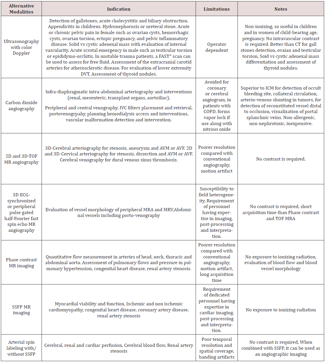
Alternative Imaging Modalities to Prevent CIN: In patients with high risk for CIN, alternative imaging modalities should be chosen after proper risk benefit discussion with the patient. Though contrast enhanced imaging have its own advantages particularly for the diagnosis of inflammatory/ infectious disease and neoplastic conditions as well as diagnosis and treatment of vascular diseases, it is important for the clinicians to be well aware of alternative modalities like ultrasonography, carbon dioxide (CO2) angiography and magnetic resonance imaging (MRI) with or without gadolinium (Table 2). In patients at risk for CIN every effort should be made to avoid iodinated contrast if possible. Non contrast computed tomography (NCCT) is preferred for diagnosis of intracranial hemorrhage, fracture or dislocation and interstitial lung disease [52].

Non contrast MRI [53] including Steady-state free precession MRI (SSFP), Phase contrast MRI, time of flight (TOF) MR angiography should be employed in appropriate setting as described in (Table 2). Gadolinium based contrast agents can be used in patients at risk of CIN, however it is best avoided in patients on dialysis and with severe renal impairment especially chronic kidney disease (CKD) Stage 4 and 5 for concerns of nephrogenic systemic fibrosis (NSF) [54,55]. CO2 angiography is another useful modality often used in various diagnostic and therapeutic interventions when exposure to CM must be avoided to prevent CIN or patient is allergic to iodinated contrast [56]. It is highly soluble, non-allergic, inexpensive and readily available gas with low viscosity relative to blood. For some procedures, CO2 angiography is actually superior to conventional CT including better visualization of collateral circulation, enhanced vascular filing in central venography, detection of AV shunting in tumors and detection of occult GI bleeding. It can be used in patients with chronic lung disease with caution if sufficient time is allowed for elimination of gas from the body. However, it has some limitations as well. It is preferably used for infradiaphragmatic arteriography due to concerns of neurotoxicity. Other complication limiting its use is vapor lock or air trapping which increases with simultaneous use of nitrous oxide. Another limitation for its use is cumbersome delivery system and risk of error of measurement of vessel size, which is dependent on injection technique [56].

Table 2: Alternative non-iodinated contrast and non-contrast diagnostic imaging modalities.
lupinepublishers-openaccess-journal-urology-nephrology

*FAST: Focused Assessment with Sonography for Trauma.

DVT: Deep Venous thrombosis.

TOF: Time of flight.

ICM: Intravascular contrast medium.

SSFP: Steady state Free Precision.

Special Consideration With Use of Iodinated CM:

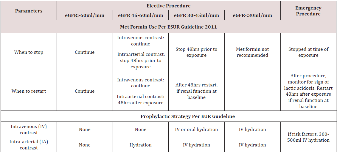
a) Met Formin Use: Metformin has not been recognized as independent risk factor for developing CIN, however serious complications like lactic acidosis can rarely develop in patients on metformin, if they subsequently develop AKI after contrast exposure. Whether it should be stopped in all patients anticipating contrast exposure or only in patients with underlying renal impairment and how earlier it should be stopped is still matter of controversy. The monogram for Glucophage (metformin) recommends that it should be discontinued at the time of or before the procedure in all patients, withheld for at least 48 hours subsequent to the procedure, and restarted only after confirmation of normal renal function [57]. The European Society of Urogenital Radiology (ESUR) (Table 3) and Canadian Association of Radiologist (CAR) consensus recommends holding metformin at the time contrast exposure in patient with normal renal function and 48 hours before in patients with preexisting chronic kidney disease [48,58]. However, ACR recommends withholding it for 48hrs before exposure only in patients with marked renal impairment (eGFR ≤ 30 mL/min/1.73 m2) or undergoing intra-arterial catheter studies and no need to discontinue or withhold if eGFR > 30 mL/min1.73 m2) [59].

Table 3: Preventive strategy to lower the risk of CIN as per EUR guideline 2011.
lupinepublishers-openaccess-journal-urology-nephrology

b) End Stage Renal Disease (ESRD): Patient with ESRD on dialysis usually do not have functional kidneys and can receive intravascular contrast material without further risk of renal damage, however studies have speculated conversion of oliguric patient to anuria. Other consideration is risk of pulmonary edema and fluid overload after osmotic load, though it was reported in past when HOCMs were frequently used. However, whenever possible LOCM should be used as they are not protein bound and have low molecular weight and can be cleared by dialysis. Usually urgent dialysis is never indicated after CM in this population unless there is exposure to large volume of contrast and patient develops life threatening pulmonary edema or cardiac arrhythmia [60].

c) Screening baseline renal function: It is actually general practice to screen baseline renal function prior to contrast exposure; however it can lead to diagnosis or procedural delay, increase financial cost and patient discomfort. ACR recommends to screen renal function (e.g., serum creatinine, eGFR) prior to exposure to iodinated CM in only selected patients with high risk factors including age >60 year, patient with history of renal disease, renal cancer, renal surgery, patient on dialysis, patients with single kidney, hypertension on medical therapy and diabetes mellitus. Patient who do not have above-mentioned risk factors and require routine intravascular contrast do not require routine screening [61,62]. Choyle et al. [62] listed few risk factors (preexisting renal dysfunction, hypertension, gout, proteinuria and prior renal surgery) and found that if none of these factors are present 99% of patients had serum creatinine levels less than 1.7mg/dl and 94% of patients had normal renal function [62]. However, ERBP guidelines recommend measuring baseline serum creatinine in all patients before an intervention and repeat levels 12 and 72 hours after contrast administration in high-risk patients [38]. The decision to repeat serum creatinine after procedure is a matter of debate. According to ESUR, renal function after contrast exposure should be assessed in 3 days, however some studies have documented that in certain patient populations particularly with diabetes mellitus and chronic kidney disease peak rise in creatinine levels is actually delayed, which demands prolonged renal function survey [58,63].

Prevention of CIN

There has been number of preventive measures to reduce incidence of CIN in high- risk population, however there is no clear consensus and current evidence remains inconclusive [64,65]. However, it has been agreed that identification of highrisk population is the foremost. Once this population is identified, best attempt should be done to minimize renal injury by stopping other nephrotoxic medications particularly NSAIDS, loop diuretics and met formin at least 24-48hours prior to contrast exposure particularly if IA exposure is anticipated and in patients with baseline severe insufficiency [47,57,58]. Unfortunately, in emergent procedures it might not be possible to stop medications or delay the procedure, however when possible restarting the nephrotoxic medication should be delayed for at least 48 hours after or as deemed clinically appropriate (Figure 1).

Figure 1: 68-year-old female with drop in hemoglobin status-post cardiac catheterization one day ago. Non-Contrast enhanced CT Abdomen demonstrating persistent nephrogram phase of enhancement from prior cardiac catheterization due to contrast induced nephropathy. Note hyperdense collection suggestive of an acute hematoma extending adjacent to RIGHT psoas muscle (*) lateral to the RIGHT psoas muscle continues with RIGHT iliac vessels, likely cause of drop in hemoglobin. Red arrow depicting no intra-vascular contrast media in the current study.
lupinepublishers-openaccess-journal-urology-nephrology

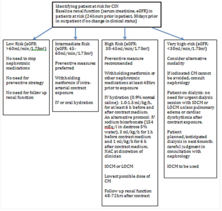
Figure 2: Flowchart for Risk stratification and preventive measures.
lupinepublishers-openaccess-journal-urology-nephrology

The cornerstone of practice to prevent CIN remains volume expansion. It is low-risk practice and carries few complications. Randomized trials have documented effectiveness of IV hydration with normal saline in the prevention of CIN. It should be given in all risk categories, particularly patients with an estimated GFR <60 ml/min/1.73 m2. It is postulated to promote diuresis, increase intravascular volume, induce vasodilation and suppress renin aldosterone axis. However, there is no fixed regimen and in general, starting it well before exposure and continuing even after procedure is the best practice. Oral volume expansion has shown some benefit, however evidence does not support it to be as effective as IV volume expansion. For isotonic saline administration, most studies suggest that 0.9% saline should be started at a rate of 1-1.5 mL/kg/h 3-12 h before and 6-12 h after contrast media exposure [66,67]. IV administration of sodium bicarbonate has also been studied for prevention of CIN; however recent data have shown conflicting and mixed results. The theoretical benefit of sodium bicarbonate is alkalization of tubular fluid and reduced production of free oxygen radicals, along with volume expansion. Most studies have suggested that it should be started at a rate of 3 mL/kg/h 1 h before and 1 mL/ kg/h for 6 h after contrast exposure. Though some studies have documented its beneficial effects, they have been critiqued for being single center, non-blinded and small studies [67,68].

N-acetylcysteine (NAC) also gained popularity for use to prevent CIN. It is believed to be direct free radicals scavenger, which improves blood flow through nitric oxide-mediated pathways. Its antioxidant and vasodilator actions are believed to protect against CIN. As compared to IV saline alone, low-dose NAC along with IV saline has shown significant decrease in CIN in patients receiving either IA or IV contrast. It is commonly employed agent in clinical setting in patients at high risk because of high tolerability, low cost, potential cardio protective properties, even in absence of clear scientific evidence. KDIGO 2012 guideline recommends using oral NAC with IV fluids in patients with high risk of CIN in setting of its low risk profile [38,69]. Algorithm showing preventive measures to be taken is shown in (Figure 2) Various other experimental pharmacologic agents have been studied for CIN prophylaxis including statin, ascorbic acid, tocopherol, dopamine, fenoldopam, theophylline, nebivolol, atrial natriuretic peptide and prostaglandin, however none of them have achieved clinical significant results [70]. Also, no beneficial effect of experimental procedures such as hemo filtration (HF) and prophylactic hemodialysis (HD) was found against CIN [71]. Moreover, prophylactic HD was found to be associated with increased CIN [71]. New investigational study ‘Renal guard system’ which is a fluid management device seems promising except for concern of electrolyte abnormalities and additional studies are needed before renal guard system can be implemented clinically [72,73].

Clinicians should be well aware of CIN, which is potentially serious entity. Since there is no established treatment for CIN, every effort should be taken to prevent it from occurring by recognizing at risk population, weighing risk-benefit ratio in all patients, optimizing volume status prior to contrast exposure, avoiding simultaneous use of other nephrotoxic agents, using newer generation of CM and using lowest possible dose.

Conclusion

Clinicians should be well aware of CIN, which is potentially serious entity. Since there is no established treatment for CIN, every effort should be taken to prevent it from occurring by recognizing at risk population, weighing risk-benefit ratio in all patients, optimizing volume status prior to contrast exposure, avoiding simultaneous use of other nephrotoxic agents, using newer generation of CM and using lowest possible dose.

Read More About Lupine Publishers Journal of Urology & Nephrology Please Click on Below Link:
https://lupine-publishers-urology-nephrology.blogspot.com/