Showing posts with label Open access Journal of Sports Medicine. Show all posts

Tuesday, 21 March 2023

Lupine Publishers| Painfull Posterior Cruciate Ligament Ganglion Cyst. A Case Report

Lupine Publishers| Journal of Orthopedics and Sports Medicine


Introduction

Ganglion cysts (GC) are benign tumor-like lesions usually going out from mucinous degeneration of collagenous structures [1,2]. They could occur in several anatomic areas but GC arising from cruciate ligaments are rare [3,4] with a prevalence of 0.36% or 0.8% respectively when diagnosed by magnetic resonance (MR) or by arthroscopy [3,5-6] However other studies of GC prevalence refer ranges from 0.2% to 1.9% [7-9], with posterior cruciate ligament ganglion cysts (PCLGC) being five times less frequent than those identify in anterior cruciate ligament [5,10]. This lesion is mainly diagnosed in people aged 20-40 years-old and a male predominance has been reported [5,11-13]. The etiology of PCLGC is not clear. They could appear from synovial herniation or congenital translocation of synovial cells. Mesenchymal stem cells proliferation with cysts formation or mucoid degeneration occurring in areas suffering chronic injuries are also reported [7,14-16]. Many of PCLGC are asymptomatic. When symptomatic the main clinical symptoms and signals includes knee pain and / or movement restrictions [17].

The knee joint could present a slight effusion, restriction to extension and particularly in extreme flexion [1,4]. The common classification of cruciate ligament cysts is supported on the position of the cyst, anterior, posterior or between cruciate ligaments [7]. MR is the gold standard for detecting GC1. Recently observation by ultrasonography is considered useful for identifying and locating the lesion, as well as being a conservative approach to treat cystic lesions [1].

Case Report

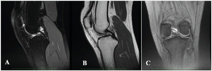
A 17-year-old Caucasian female, a soccer player, presented with a 9 month history of left knee pain, mainly in the posterior and medial aspects of the knee, combined with slightly back swelling. Pain was exacerbated with exercise, especially squatting, and partially alleviated with rest. She had no history of a knee major traumatic event (Figure 1). She complained of knee pain on soccer playing, one of the main reasons to suspend this practice. She alsorefers no confidence on demanding tasks.

Figure 1: Knee MRI of PCL GC; A: T2 sagittal view FS; B: T2 sagittal view FSE; C: T2* coronal view.

Lupinepublishers-openaccess-orthopedics-sports-medicine

In clinical examination we identified a light quadriceps atrophy, compared to the opposite side, and symmetry in active range of motion (ROM). Passive ROM was limited (10 degrees) and painful in extreme flexion and symmetric in hyperextension. Meniscal tests and varus-valgus stress tests were negative. Some tenderness on palpation of the popliteal aspect but no pain or even tenderness was verified in medial or lateral joint line. Plain radiographs were normal. A knee magnetic resonance image (MRI) was then performed regarding the potential diagnosis of a joint cyst so it was acquired weighted T2 sagittal sequences with and without fat suppression and T2* coronal. The MRI revealed a high-signal well-defined, ovoid shaped formation extending along PCL on both T2-weighted images and fat-suppression sequences, measuring 18mmm length compatible with a PCL cyst. Considering that in the last consultation there is no pain or appreciable impairment of mobility, and also the athlete does not intend to continue the practice of soccer, the choice was made for conservative treatment. Proprioceptive closed-chain training and quadriceps and hamstrings muscle strengthening were prescribed for 3 months, 3 x week. Currently she runs 5 x week (40min per day) without pain and functional disability. A new appointment will be made in six months.

Discussion

Etiology and pathogenesis of PCLGC are unclear, however it is proposed that repetitive microtrauma of joint and soft tissue can promote expansion of mucin from ligament fibers and acting as a potential trigger [20]. Recognition of PCLGC as a clinical entity leading knee pain and impairment is increasing due to the sensitivity of MRI to identify intra-articular abnormalities. The typical finding is an ovoid fluid filled cystic lesion which can frequently be multilocular in the intercondylar notch of the knee [22,25]. In our case report MRI shows a cystic multilocular mass with fluid signal intensity within the synovial layer of the PCL. Although most knee cysts are asymptomatic, in some case they could be a relevant source of pain [20,21]. Clinical manifestations of a knee cyst are mostly dependent on the pathologic process involved, along with its location, size, mass effect, and relationship to surrounding structures [26]. The typical presentation of symptomatic PCLGC include posterior knee pain, restriction of ROM, stiffness and mild swelling [20,21].

Limited ROM is a typical finding with an intra-articular ganglion arising from the PCL, mainly with inability and pain to extreme flexion due to the compression of the cyst mass between the PCL and the posterior joint capsule. With this clinical picture in mind, athletes between 20 and 40 years old who present knee pain with restriction on hyperextension or full flexion, with no previous macrotraumatic report or knee instability, should raise a high level of suspicion for intra-articular ganglion cysts. Only symptomatic PCLGC need to undergo treatment. There a broad spectrum of treatments described for these lesions, from a rehabilitation program focused on ROM, strengthening and proprioception to avoid kinetic impairment, to ultrasound or CT-guided aspiration or infiltration, or even arthroscopic excision. Treatment choice must take into account several criteria such as level of activity, time for recovery, risk of joint damage and recurrence of the cyst. Arthroscopic treatment has demonstrated good outcomes with up to 95% of patients reporting good results and associated with the lowest recurrence rate, but it needs an hospitalization, anesthesia and a longer recovery period, which can become a major problem when we are dealing with competitive sports [23,24].

Athletes require quick return to play with minimal side effects, so we need to take into account less invasive treatments like US or CT-guided procedures, or even load management in addition to a rehabilitation program.

Conclusion

CCP is a rare and often asymptomatic condition. Its pathogenesis and prognosis are still unclear. In a young adult with posterior knee pain (popliteal aspect), no history of major event, limited ROM (hyperextension and extreme flexion), meniscus and ligament test negative and no confidence in demanding tasks it is important to think about this condition. The therapeutic option stems from the patient’s characteristics, but US or CT- guided puncture should be considered.

Read More about Lupine Publishers Journal of Orthopedics and Sports Medicine Please Click on Below Link: https://lupinepublishers-orthopedics.blogspot.com/


Monday, 20 March 2023

Lupine Publishers| Outpatient Latarjet Procedure: Early Complications and Feasibility Validation

  Lupine Publishers| Journal of Orthopedics and Sports Medicine


Abstract

Background: The Latarjet procedure may be amenable to outpatient elective surgery as it is often performed on young and healthy patients. Thus, ambulatory surgery for Latarjet procedure is recently rapidly increasing in France with very few validation studies. This feasibility study presents early adverse events following outpatient Latarjet surgery compared to Latarjet surgery performed as an inpatient procedure.

Hypothesis: There is no difference between outpatient or inpatient Latarjet procedure.

Patients and Methods: Thirty patients operated on an outpatient basis and prospectively followed were compared to 30 patients operated on an inpatient basis. All procedures were performed by the same surgeon. Complication rates as well as clinical outcomes at one year were compared between groups.

Results: Post-operative hematomas which did not require surgery occurred more frequently in outpatient group in which no drain was used. No other differences occurred between groups. All outpatients but one was satisfied with the procedure.

Discusion: The latarjet procedure was found to be safe when performed on an outpatient basis. The addition of wax to the base of the coracoid seemed to diminish hematoma formation.

Level of Evidence: level III

Keywords: Shoulder; Latarjet; Instability; Complications; Ambulatory surgery

Introduction

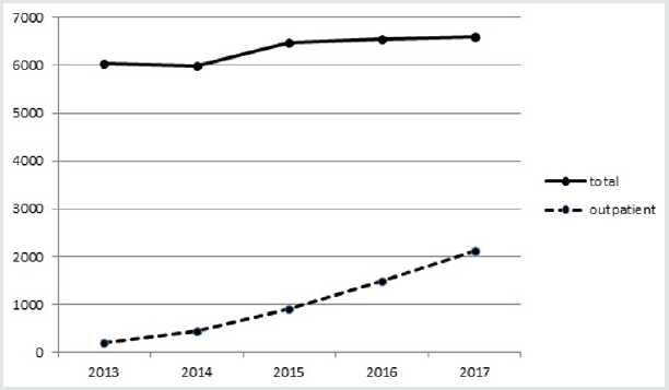
Outpatient surgery provides benefits to patients including a decreased exposure to nosocomial infections, a higher rate of satisfaction [1], and a up to a 68% decrease in direct costs [2]. The Latarjet procedure is now frequently performed on an outpatient basis in up to one third of cases in France in 2017 (Figure 1).

However, the literature regarding the feasibility of outpatient Latarjet surgery is very poor and there is still a need of validation studies [3]. Our aim was to confirm the feasibility of the outpatient Latarjet procedure by comparing the incidence of adverse events and clinical outcomes between patients who underwent inpatient or outpatient Latarjet surgery. Our hypothesis was that the outpatient procedure is both feasible and safe.

Patients and methods

Patients

Study inclusion criteria were as follows: patients were considered if they had a diagnosis of recurrent anterior shoulder instability; were deemed candidates for surgical stabilization; had not undergone prior shoulder surgery, and did not have any significant shoulder co-morbidities. All patients underwent primary surgery for anterior shoulder instability using transfer of the coracoid process (Latarjet procedure). All procedures were performed by a Single Surgeon (SZ). Thirty consecutive patients underwent surgery on an outpatient basis between 2013 and 2017 and were prospectively followed. This group was compared to 30 patients who underwent the Latarjet procedure on an inpatient basis between 2007 and 2012 by the same surgeon.

Surgical protocolA standardized general anaesthesia protocol was followed. An additional interscalene block was administered under ultrasound guidance (single bolus of 20ml of 0.375% ropivacaine) associated with 8mg of a dexamethasone intravenous injection. An open minimally invasive technique was used. The osteotomy of the coracoid process was performed through a deltopectoral approach after the coraco-acromial ligament and pectoralis minor tendon were released and following conjoint tendon exposure and dissection. All harvested coracoid grafts were a minimum of 20mm in length. The subscapularis tendon and muscle was split horizontally. Following glenohumeral capsulotomy, bone on the ventral aspect of the coracoid process and on the anteroinferior aspect of the glenoid rim was decorticated. Any remaining anteroinferior bone bankart fragments were resected. The ventral aspect of the coracoid graft was fixed to the inferior portion of the anterior scapular neck such that the transplant was level with the anterior glenoid rim. The coracoid process was drilled with two 3.5mm holes and fixed with two 3.5 diameter cortical screws; whilst the glenoid neck was drilled with 2.5mm holes to enable compression.

Care was taken to avoid lateral overhang of the graft across the joint line as described by Alain et al. [4]. No additional capsular suture was used. The subscapularis tendon was closed lateral to the graft. Traction on the coracoid graft was avoided all along the procedure to decrease the risk of musculocutaneous nerve injury. The wound was closed in layers with continuous absorbable skin suture. All inpatients had a suction drain inserted. No drain was used in the outpatient group. Sling immobilization was used for one week following surgery. Simple activites of daily living (shower, eating, writing) were immediately permitted. Following one week, self-assisted stretching in all planes was permitted. Running and swimming were allowed after two months, and high-risk sports (rugby, judo…) were allowed after 4 months. All patients were assessed on post-operative day one (by telephone for outpatients); further assessments took place at 1 week, 1 month, 4 months and 12 months post-operative.

Evaluation criteria and statistical analyses

Readmission rates and early complications were recorded. Shoulder range of motion, recurrent instability, persistent subjective apprehension and shoulder pain were compared between groups at one year. Satisfaction rate with the outpatient protocol was assessed. Continuous variables were compared with the independent t-test and categorical variables with the Fisher exact test; statistical significance was set at 0.05.

Results

The 2 groups were comparable at baseline (Table 1). Mean hospital stay in the inpatient group was 2.2±0.4 days. One admission for one night occurred in the outpatient group due to dizziness which resolved without further treatment. No complications occurred related to the interscalene block. There were no reoperations, no nerve injuries and no infections in the series.

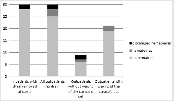
In the inpatient group, drained blood volume prior to drain removal was negligible in 18 patients, less than 100cc in 11 patients and > 100 cc in one patient. All drains were removed on post-operative day one. Seven hematomas occurred within the first 3 weeks following surgery: two in the inpatient group and five in the outpatient group (Figure 2). Four of these hematomas, two in each group, discharged and healed spontaneously. All others healed spontaneously without fistulization. One of these hematomas occurred in a patient with the Factor V Leiden defect (outpatient group). Three of these hematomas, with fistulization twice, occurred in the first 9 patients in the outpatient group and induced a change in the surgical technique. In the following 21 patients, prior to closure, the osteotomy of the coracoid feet was explored, washed and waxed prior to closure during which time a blood clot was typically found. Wax was never used for patients in the inpatient group.

Figure 1: Latarjet procedures statistics in France between 2013 and 2017 (ATIH, technical agency of information on hospitalization, www.atih.sante.fr).

Lupinepublishers-openaccess-orthopedics-sports-medicine

Figure 2: Hematomas formation by group. There was more hematomas in the outpatient group (p<0.01), all hematomas healed spontaneously.

Lupinepublishers-openaccess-orthopedics-sports-medicine

All outpatients but one were satisfied with the procedure. One patient indicated that he would have preferred a one night hospital stay due to postoperative discomfort. At final 12 month followup, no patient had experienced further instability. Six patients answered positively for subjective persistent apprehension in both group (20%). One third of patients of each group have reported occasional shoulder pain. Loss of external rotation was found in half of patients of each group (Table 1).

Read More about Lupine Publishers Journal of Orthopedics and Sports Medicine Please Click on Below Link: https://lupinepublishers-orthopedics.blogspot.com/


Monday, 6 February 2023

Lupine Publishers| Haglund Syndrome – A Case of Bilateral Involvement

 Lupine Publishers| Journal of Orthopedics and Sports Medicine


Abstract

Haglund syndrome is a cause of posterior heel pain and occurs commonly in adolescent girls who wear high heels with restrictive heel counters and may occur in people with rheumatoid arthritis. Is characterized clinically by thickening of the soft tissues at the Achilles tendon insertion or a painful “pump bump” and retrocalcaneal bursitis, Achilles tendinitis, and a prominent posterosuperior calcaneal border or bursal projection. We report a case of a 59 years-old woman with bilateral Haglund syndrome which did not needed surgical treatment, being the conservative treatment enough to improve the patient’s quality of life.

Keywords: Calcaneus; Magnetic resonance imaging; Foot diseases

Introduction

Haglund syndrome is a cause of posterior heel pain and was first described by Patrick Haglund, in 1928, to occur in patients who had a prominence of the posterosuperior surface of the calcaneus [1]. It occurs commonly in adolescent girls who wear high heels with restrictive heel counters and may occur in people with rheumatoid arthritis [1]. Haglund syndrome has also been termed “pump bump,” “winter heel,” “knobby heels,” “calcaneal altus,” “highbrow heels,” and “cucumber heels” [1]. Haglund syndrome is characterized clinically by thickening of the soft tissues at the Achilles tendon insertion or a painful “pump bump” and retrocalcaneal bursitis, Achilles tendinitis, and a prominent posterosuperior calcaneal border or bursal projection [1-4].

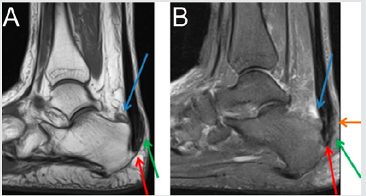
Figure 1: Sagittal section MRI in T1 sequence in A and T2 STIR sequence in B of the right ankle demonstrating posterior calcaneal spur (red arrow), liquid in retrocalcaneal bursa (blue arrow), liquid in subcutaneous calcaneous bursa (orange arrow), and calcaneal tendinopathy (green arrow).

Lupinepublishers-openaccess-orthopedics-sports-medicine

Figure 2: Sagittal section MRI in T1 sequence in A and T2 STIR sequence in B of the left ankle demonstrating posterior calcaneal spur (red arrow), liquid in retrocalcaneal bursa (blue arrow), liquid in subcutaneous calcaneous bursa (orange arrow), and calcaneal tendinopathy (green arrow).

Lupinepublishers-openaccess-orthopedics-sports-medicine

Case Presentation

59 years-old woman with pain and swelling in both feet for four months that get worse when use slippers and when she wake up and put her feet in the floor for the first time in the day. The patient denies other complaints and surgeries. At physical examination refers pain at the palpation of in the posterior and inferior regions of the ankle. The Magnetic Resonance Imaging (MRI) demonstrates posterior and plantar calcaneal spurs, liquid in retrocalcaneal and subcutaneous calcaneous bursa, and calcaneal tendon with heterogeneous signal and thickened (1.4cm at left and 1.2cm at right), markedly in their insertion with adjacent bone edema (Figures 1 & 2). The set of findings are compatible with Haglund syndrome. The patient started conservative treatment with non-steroidal anti-inflammatories, change of footwear and physiotherapy, presenting a good response, not requiring surgery.

Discussion

Haglund syndrome is caused by mechanically induced inflammation of the Achilles tendon and its bursa, from abnormally high pressure between the bursal projection of the calcaneus, the Achilles tendon, and the bursa [1,5]. Patients with bursitis have erythema and swelling over the bursa and tenderness to direct palpation [2] besides pain with dorsiflexion of the foot [6]. Clinically, by physical examination alone, it may be difficult to distinguish Haglund’s syndrome from other causes of hindfoot pain such as Reiter’s disease, rheumatoid arthritis, or isolated local conditions such as superficial tendon Achilles bursitis secondary to poor shoe fit [7]. Plain radiograph in a lateral standing position is useful to assess the presence of a prominent bursal projection of the calcaneum, the Haglund deformity [3]. Loss of a lucent retrocalcaneal recess is an important indication of underlying retrocalcaneal bursitis [3]. The Achilles tendon is swollen, and dystrophic calcifications may also be seen [3].

MRI may be required for ambiguous or clinically equivocal cases [3]. The cardinal soft tissue abnormalities, namely Achilles tendinopathy, and retrocalcaneal and retroachilles bursitis are more easily and directly depicted by MRI [3]. The detection of marrow oedema within the prominent bursal projection is likely to support the repetitive mechanical compression and inflammation as the pathological mechanism in this condition [3]. Treatment of Haglund deformity, with or without bursitis, targets decreasing the pressure and inflammation with openheeled shoes, anti-inflammatory or analgesic medications, and corticosteroid injections [2]. Physical therapy may also help reduce pain. In recalcitrant cases, surgery to remove the Haglund deformity may be necessary [2].

The nonsurgical treatment, combined with modifications in daily shoe wear, is an appropriate initial treatment for pain relief of Haglund’s syndrome [7]. Using ultrasound guidance to inject the retrocalcaneal bursa is a simple, reliable method of ensuring accurate delivery of medication into the bursa and avoiding intratendinous injection [7].

Read More about Lupine Publishers Journal of Orthopedics and Sports Medicine Please Click on Below Link: https://lupinepublishers-orthopedics.blogspot.com/

Wednesday, 21 December 2022

Lupine Publishers| Digit Ratio and Soccer

 Lupine Publishers| Journal of Orthopedics and Sports Medicine


Abstract

The ratio between the lengths of index finger in relation to the length of ring finger of a palm is noticed to as second to fourth digit ratio. Digit ratio tented to shows the quantity of male hormone, to which an individual is exposed in the womb of the mother. Several investigations establish the negative relationship between lower digit ratio and various sports performance as the lower digit ratio settle the high prenatal testosterone hormone. The exposure of prenatal androgenisation masculinizes the human body that impacts the efficiency of sports. Soccer player perceives numerous situations and makes instantaneous decisions. Experienced players are reading the game and anticipating the next move of the opponent. Previous studies have demonstrated the link between cognitive ability and the digit ratio. Similarly, players have numerous cognitive skills to dictate soccer game such as spatial intelligence, awareness, and visual spatial ability. Here, we explore the possible causes of negative associations between lower second to fourth digit ratio and soccer performance. We also think soccer-specific skill performance is likely to be associated with lower digit ratio.

Keywords: Digit ratio; Testosterone; Estrogen; Soccer

Introduction

The peripheral blood cannot be extracted in utero from fetuses [1]. The second to fourth digit ratio (2D:4D) was therefore suggested as a prenatal testosterone marker [2]. Manual second to fourth digit ratio (2D:4D) at the end of the first trimester of pregnancy is believed to be a biomarker of the balance between prenatal testosterone and prenatal estrogen hormone [2-4]. Thereafter, the digit ratio (2D:4D) probably remain unchanged throughout the life [2]. However, Manning (2002) indicated that the digit ratio (2D:4D) was set particularly between week 8 and 12 at the end of the first trimester. The male fetuses mainly produce large quantities of testosterone hormone, primarily from their testis and adrenal glands [5]. This influences brain and other organ systems development [2-6]. In general, the length of index (2D) and ring fingers (4D) in women is about the same (digit ratio=1.00), whereas in men the ring finger is generally slightly longer (digit ratio=0.98) [7]. The length difference between the two digits is higher for men than for women [8-9]. Testosterone influences the growth of the ring finger (4D), whereas estrogen exposure stimulates the growth of the index finger (2D) [7]. The ratio of the index finger to the ring finger (2D:4D) has been shown to be a sexually dimorphic trait [9-10]. Additionally, the ratio of digits (2D:4D) measured by the length of index finger divided by the length of ring finger [7-11]. Therefore, researchers found an index of prenatal testosterone exposure relative to prenatal estrogen exposure [7-12]. The prenatal androgen is likely to be increase, if the digit ratio goes down [7]. Several studies portray the second to fourth digit ratio in which prenatal testosterone hormone was associated. The researches recommend that the lower ratio of digit is a noninvasive feasible indicator for sport success rate Manning and Taylor [13] ; Manning and Hill [14] ;Manning et al. [8]; Hone and McCullough [15] ; Longman et al. [16]; Sudhakar et al. [17]; Sudhakar et al. [18]; Bennett et al. [19]; Kim [20]. As because, adult lower digit ratio (2D:4D) promote the masculine feature [2]. The testosterone (T) is a steroid hormone that develops and maintain masculine feature of human body [21]. The specific aim of this review study was to explore the relationship between digit ratio (2D:4D) and soccer performance.

How does the digit ratio (2d:4d) fixed?

The adult finger length ratio is becoming a widely used research tools to know the tentative trait of prenatal androgens, a diversity of physiological and psychological conditions, athletic ability and sexual orientation [10,22,23]. The differentiation of gonads, fingers, and toes is influenced by HOXA and HOXD genes. HOXA and HOXD genes are also necessary for finger length development and differentiation [24]. Congenital Adrenal Hyperplasia (CAH) is an anomalous hormonal environment that does not function correctly with the adrenal glands [25]. The 21-hydroxylase deficiency, results in the production of surplus quantities of masculine hormones by the adrenal glands [26]. However, researchers Okten and his colleagues studied digit ratio (2D:4D) and 21-hydroxylase deficiency in male (right palm) patients and reported lower digit ratio confirm the 21-hydroxylase deficiency than female and male controls. Women with CAH had a much lower second to fourth digit ratio than women without CAH on the right hand and on the left hand, men with CAH had a much lower digit ratio (2D:4D) than men without CAH [27]. Similarly, researchers [28] reveal the relationship between low digit ratio and CAH. This characteristic also supports a combination of low digit ratio and elevated Fetal Testosterone concentrations [29].

Relationship Between Digit Ratio (2d:4d) With Sports Performance

Researchers [20] widely reviewed the most correlational studies and postulated that low second to fourth digit ratios (high prenatal testosterone and low estrogen hormone) could be a determinant of high sport performance. However, the high performance of rugby depends on low digit ratios [19]. The researchers also discovered differences in the low right-left digit ratios to be a determining factor in elite rugby performance. Keshavarz and his team (2017) studied on three male groups of Wrestlers; they are:

a) World class elite Greco-Roman wrestlers.
b) Collegiate non-elite wrestlers.
c) Sedentary age matched control.

The lower right- and left-hand digit ratios of world class wrestlers were predictors of high wrestling performance compared to other groups [30]. The achievement of the competition phase in team sports was also associated with the ratio of digits (2D:4D). The second to fourth digit ratio was therefore likely to have an impact on the possible athleticism [31]. Similarly, lower digit ratio (high prenatal androgens) has been shown to indicate the sport performance of soccer, surfing, sprinting, endurance, hand grip strength, rowing, kabaddi, swimming, Tennis [8,13-18,32].

Digit Ratio as Soccer Performance Determinant

High prenatal testosterone and low prenatal estrogen hormones are likely to be a strong predictor of soccer performance [7]. Competitive achievement is a major objective of soccer in connection with prenatal androgenization [31]. This prenatal situation influences the judgment of the visual perception [13]. Therefore, according to [7], “Striking a moving opponent or ball requires fine judgment of distance. Determining the exact point of impact demands an accurate perception of the surface of the target as it moves through space” (p.128). However, researchers studied on different types of soccer players and noticed ‘professional’, ‘International’ and ‘1st team players’ had lower digit ratio (2D:4D) than the ‘control group’, ‘youth team’ and the ‘players who had not represent their country’ respectively [13]. Similarly, the International presence of the player in a match is greater for the lower digit ratio individuals [7]. The lower digit ratio could therefore provide an additional discriminator to help estimate soccer capability. Prenatal testosterone exposure also influences professional soccer players’ aggressive behavior. Researchers indicated that exposure to adult and prenatal testosterone detects the number of fouls per match that confirm the aggressiveness of players [33]testified by a low second-to-fourth digit ratio (2D:4D . Furthermore, aggression guarantees the dominant behavior that is essential in competitive sport.

Association among Digit Ratio, Visual-Spatial Ability and Left Handedness

Digit ratio is a putative indicator of sport performance differences [34]. A study concerning several sports related psychological variables (mental toughness, aggression, optimism scale, coping strategies, and goal orientations) with masculine digit ratio reported high scores of optimistic dispositions than those with feminine digit ratio. The study also claims that mental toughness partly determined on gestation period [34] that benefited for gender, age and sporting experience [35]. Mental rotation score test [36] can measure the visual-spatial intelligence [36-38]. Manning and Taylor found negative association between lower digit ratio and high mental rotation scores in males. So, the visual spatial intelligence may partially develop on intrauterine life [13]. High prenatal testosterone exposure is likely to associated with handedness [39,40]. Left-handed people dominated by the right hemisphere and assists visual spatial ability [41]. Interestingly, androgenisation exposure influence the right palm more than the left palm [27,42,43]. Right palm digit ratio is also significantly connected with several psychological and behavioral traits compared to the left palm digit ratio [43].

Cognitive Abilities Influence on Soccer Performance

Most team sports, particularly in soccer players, need to pursue numerous situations that are changing quickly [44]. Elite players perceive the situations and make the appropriate choice at the right moment [45,46]. Therefore, technical and tactical ability influence the outcome of the match [47,48]. Elite players perform the technical and tactical skills better in compared non-elite counterparts [49]. However, researchers postulated that distinguished correlations in male between more masculine digit ratio (lower digit ratio) and in visual-spatial ability [13,22]. On the other side, females have prone to more feminine digit ratios (higher second to fourth digit ratio) should relate to higher scores for depression (Repeat). Therefore, high prenatal testosterone exposure is likely to be predictor of soccer performance as well as cognitive ability [13]. Research also shows that human behavior and the status of cognition can result from prenatal androgenization [50].

Conclusion

Most correlational study reveals the negative relationships between lower digit ratio and sports performance. Low second to fourth digit ratio (2D:4D) can be an indicator in scouting potential athletes especially soccer players. In multifaceted aspects, lower digit ratio is likely to be a potential indicator of soccer specific skill performance. Further studies are required to clarify whether lower second to fourth digit ratio could predict the soccer skill performance in multifaceted aspects including passing, dribbling, control, shooting and decision making within a dynamic situation. In addition, we realized that sporting success might be depended on our hands’ fingers length ratio a long with other variables.

Read More about Lupine Publishers Journal of Orthopedics and Sports Medicine Please Click on Below Link: https://lupinepublishers-orthopedics.blogspot.com/

Wednesday, 9 November 2022

Lupine Publishers| How to Pick a Good Surgeon

Lupine Publishers| Journal of Orthopedics and Sports Medicine


Abstract

A shoulder surgeon has a shoulder injury and requests a specific shoulder surgeon colleague to operate. Instead, the patient is sent to an expert shoulder surgeon with an international reputation who delivers many lectures and writes numerous papers and book chapters. Surgery is performed but the shoulder gets infected and the overall result is unsatisfactory. After a legal challenge, the patient is allowed to go to the surgeon originally requested based on the patient’s own insights into the profession. Revision surgery is performed and iatrogenic damage is discovered from the first surgery. However, the result of this revision surgery is good. This anecdotal case contains many aspects of healthcare decision making, control, and culpability which will be further elucidated here.

Keywords: Picking a Surgeon; Good Surgeon

Introduction

I am sure many of us have either heard or been asked the question, “How do you pick a good surgeon”. Of all the academic questions we deal with on a daily basis as surgeons/physicians, this question perplexes us beyond our educational background due to the multitude of types of information needed to truthfully answer. Now, consider how hard this decision is to make for the lay person/patient who does not have the same insights into the inner workings of the profession. Before being able to pick a good surgeon, we need to define what makes a good surgeon. A surgeon is a medically trained doctor who has chosen to specialize in pathologies that can be treated with a surgical option but also has the knowledge and ethics to know when to treat non-surgically. However, most pathologies have a spectrum ranging from near normal to end-stage and management options that often depend on several factors: stage of the pathology, chronological versus physiological age, activity level, co-morbidities, availability of resources, etc. Therefore, anyone dealing with a specific pathology (e.g. osteoarthritis or shoulder instability) should not only understand the whole spectrum of the pathological process and its variants, but also be adept at managing these different stages with “stage specific solutions” instead of employing a “one operation fits all” philosophy. Picking a surgeon for oneself or one’s family has several pathways, some controlled by the patient, and some by the agency responsible for the financial settlement.

There are no standard pathways for this choice, but when trying to understand what factors are of influence, we are able to identify many.

Commonly Used Factors When A Patient Seeks A Surgeon

a) A primary caretaker (family member/family doctor/ friend) recommends a surgeon due to personal experience or word of mouth recommendation [1,2]. While this sort of recommendation can be based on limited interactions with the surgeon, and his/ her outcomes, someone who has had a prolonged interaction would be able to gain a better insight into the outcomes from that surgeon. However, when limited interactions are the basis of a recommendation, this may prove to be detrimental to the patient since, for example, a surgeon who once performed a successful carpal tunnel surgery may not be the best option for performing a shoulder replacement.

b) An insurance company/workers compensation board will limit choice from surgeons within their approved network based upon whom they have an agreed terms and conditions contract [3]. This network is not based purely on quality of surgical skill, but is more based on a financial agreement with the insurer [3]. The financial entity does not have the responsibility of auditing the surgeon’s outcomes, and hence can rarely make the choice easier for the patient except to limit the choice to a specific group of surgeons.

c) A surgeon with a higher rating on social media platforms is viewed as a better option than one with a lower rating [4]. Very few people understand how social media ratings come about. Individual ratings tend to be either strongly positive or strongly negative. Unhappy patients are more likely to complain, and thus there is a bias toward negative spontaneous social media ratings. Social media provides a platform from which the angry patient can voice their discontent. Happy patients are generally more interested in getting on with their lives following a successful surgery than devoting a lot of time to grandstanding on social media. Conversely, many well-run private practices are very efficient at collecting ratings from happy patients while avoiding collecting ratings from unhappy patients in their clinics. The latter practice may balance the equation by counteracting the negative bias or perhaps tilt responses to excessively positive. Social media ratings by patients are also more determined by their overall customer experience than their surgical outcome. Factors such as wait time, parking, office atmosphere, and interpersonal interactions with the surgeon and staff have been shown to have a stronger impact on social media ratings than quality of the outcome. This is logical since humans have daily interpersonal interactions which provide a standard for comparison, unlike a surgical procedure which is generally limited to a small number of times in an individual’s life. Hence, social media may focus on extraneous factors and can be manipulated. Thus, online patient ratings are not the best indicator of a good surgeon. If every patient had to input a rating, usefulness might be improved and this may represent a direction for the future.

d) Speed of Surgeon [5]. Is a slow surgeon better than fast surgeon, or vice versa? Some surgeons have a reputation for being very quick while others have a reputation for being very slow, or sometimes described as meticulous. Inorder to fully appreciate this issue, one needs to understand what a particular pathology requires in order to resolve the symptoms. Taking a very simple example, carpal tunnel release, some surgeons take 4 minutes to complete this through a 1 cm incision, versus others take 90 minutes or more with a 5 cm incision or an endoscopic technique. While a fast surgery may miss some steps, although all being achievable for a carpal tunnel surgery within the 4 minutes, the same surgery with a larger incision and a long operative time may have a greater risk of infection and scarring, due to many more surgically performed strokes of the knife. The latter refers to the surgeon performing more surgery than is necessary to achieve the result, under the label of being “meticulous” and “precise”. Hence, performing more surgery than is necessary to treat the pathology is detrimental to the outcome. Therefore the speed of the surgeon is not always easy to understand without the context of the surgical results achieved by that surgeon.

e) Communication skills [6]. A surgeon who can communicate well is better than a surgeon who is not a good communicator [6]. Communication is a central and under-valued aspect of any surgeon’s skill set. The most important factor in healthcare is the needs of the patient. Hence, it is the patient who needs to understand the problem, and how to fix it, to a level compatible with his/her educational background. Communication between the surgeon and the patient allows the necessary information to be patient-centric, in order that confidence is built within that relationship [6]. A surgeon who is able to communicate well with all the patient demographics and educational levels in his/her practice should be viewed in a positive light, while the abrupt, minimal communicator cannot justifiably impart the necessary information. If a patient is not made to understand the whole process, he/she cannot be an effective part of the treating team, and patients should be considered as part of the team.

f) Academic impact [7]. “A surgeon who writes a lot of papers is better than one who doesn’t write papers”. Surgery, like any athletic pursuit or manual trade (carpenter, motor mechanic, etc.) is a dexterous skill, combined with knowledge and the translation of that knowledge to the physical activity. As any athlete knows, it takes a very long time to train one’s body to perform a physical task at a certain level, more than 90% of their time. So if an athlete spends a significant amount of time reading about a skill, and less time translating the skill into his/her own physical performance, the results are failure in competition. Surgeons are no different. Those who spend significant amounts of their time in research, giving talks, writing papers, advocacy and committee work, etc. have less time to see and diagnose patients, operate and treat patients, follow up and assess patients’ outcomes etc. Although there is not a welldefined parameter to judge the optimal times for cerebral versus dexterous activities, the principle of the athlete is relevant to the surgeon.

g) Education at high profile institutions [7]. Those with educational backgrounds in high profile institutions are considered with greater regard than those with lesser academic profile institutions [7]. It should be constantly remembered that surgery is an intricately intermingled cerebral and dexterous skill. Being overweight in either may not compensate for achieving the best results. High profile institutions achieve their profile by many different means, but none achieve it by the quality of the training they provide to the next generation of surgeons. The institutions select their surgeons based on qualities important to the mission of the institution, and it can be assumed the trainees who are trained there will both actively and passively achieve some of those qualities. However, great institutions can produce poor quality surgeons and lesser institutions can produce outstanding surgeons so it is not a guaranteed assurance to rely on the surgeon’s educational institution’s reputation.

h) Specialty Board Certification [8]. The American Board of Medical Specialties (ABMS) and its 24 member boards are organizations established to ensure consistency of training and practice within established guidelines for surgical specialists. These organizations seek to ensure that surgeons have a specific knowledge base and have completed requisite numbers of surgical procedures during their training. In certain competitive areas such as cosmetic surgery, physicians lacking ABMS certification seek to deceive prospective patients by creation of non-ABMS boards which do not require any specific training and allow surgeons to join simply by paying a fee. In orthopaedic surgery the ABOS (American Board of Orthopaedic Surgery) seeks to ensure this quality with didactic exams and a review of cases performed by the surgeon. While these boards try to ensure quality, they do not have the capacity to be stringent enough and would require considerable investment to improve quality of candidates. So a “Board Certified” surgeon may not always be a better choice than a non-board certified surgeon, but the certification ensures a minimum quality.

i) Availability of the surgeon [9]. Some consider a surgeon who is easily available for new appointments to be better than those who are really difficult, or have a very long waiting list. Similar to a restaurant, would you prefer to eat at an empty restaurant or one where it is always busy? The three ‘A’s of medical practice priority used to be (and should be) “Ability, Affability, Availability”. Unfortunately, with the societal shift in attitudes the order has reprioritized to “Availability, Affability, Ability” [9].

j) Intellectual honesty. One of the most important qualities for a medical professional is the ability to honestly assess one’s own level of knowledge and skill. Not all surgeons are equal, just like not all tennis players or restaurants are equal. Hence, when a surgeon is unable to perform a certain operation that may be the optimal treatment for a particular patient, it takes intellectual honesty for that surgeon to communicate this discrepancy and refer the patient to another surgeon who is able to perform that optimal procedure. It is dishonest to perform a procedure that is not in the best interest of the patient just because it is a procedure that the surgeon is able to perform, regardless of the suitability of the performed procedure to the presenting pathology. An example of this is when surgeons perform reverse shoulder replacements instead of anatomical total shoulder replacements despite the presence of all the factors that can make an anatomical replacement successful. Although a reverse replacement is an easier procedure to perform, it sacrifices an intact rotator cuff. The price of an inappropriate operation is paid at a later date by the patient, with very fewer good options if the reverse shoulder replacement fails.

k) Volume and institutional protocols [10]. Frequent performance of a procedure by a surgeon naturally leads to technique refinements if the surgeon is thoughtful about the process. Likewise, the operating room staff and other associated individuals such as anesthesiologists become familiar with the surgeon’s routine and optimize their functions accordingly. Further, some surgical procedures benefit from ancillary services such as specialized nursing care, physical therapy or occupational therapy working in a coordinated manner immediately following the procedure. In these instances, the good surgeon’s skills are only one component of a successful surgical outcome, albeit an important one. Surgeons who perform low volumes of surgery are likely to be less dexterous than those who perform high volumes of surgery, with a required debate regarding the definition of high, low, and adequate volumes to maintain dexterous skills.

l) Word of mouth reputation [1,2]. The natural course of surgical practice is that the surgeon reaps what he sows. That is, if a surgeon performs an operation successfully on one patient, that patient is more likely to tell their friends and their referring physician about their experience [1,2]. Those patients and providers are more likely to send other patients, and thus forms the basis of “word of mouth” referenced in the first paragraph. This leads to a snowball effect on the surgeon’s practice which grows over the course of a career. Name recognition for surgeon grows over time and hence the effect of word of mouth becomes more powerful for them individually. Name recognition would seem to imply a larger pool of satisfied patients, but this is not always true as other factors can lead to name recognition such as advertising. Further, it is likely this effect is more powerful in smaller communities with fewer providers compared to larger communities.

m) Access to specialized equipment [10]. Specialized equipment may allow an appropriately-skilled surgeon the ability to perform a procedure in a less invasive, safer, or more efficient manner. The recent innovations in robotic surgery provide an example for certain procedures. However, it is important to note that just because a technology is new or “hi-tech”, it is not necessarily better or safer than established methods. For instance, endoscopic carpal tunnel release has no superior data compared to minimal incision release. Furthermore, technology presents a constantly changing environment and yet it can take years for a particular innovation to mature to its fullest potential or fade out.

Read More About our Lupine Publishers Journal of Orthopedics and Sports Medicine Please Click on Below Link: https://lupinepublishers-orthopedics.blogspot.com/ 

Tuesday, 9 August 2022

Lupine Publishers| Rehabilitation Following UCL Repair with Internal Brace

 Lupine Publishers| Journal of Orthopedics and Sports Medicine


Introduction

Elbow injuries in the overhead athlete, particularly baseball pitchers, continue to increase in frequency because of extreme repetitive valgus stress [1-4]. This repetitive stress results in ulnar collateral ligament (UCL) insufficiency which produces elbow pain, medial joint laxity, and an inability to throw.16 Pitchers are the most injured players in Major League Baseball and elbow injuries account for 22-26% of pitching injuries [1,5,6]. The risk factors related to sustaining a pitching related UCL injury are pitching when fatigued, a high pitch volume, improper mechanics, and repetitive throwing at maximal effort in the youth player and throwing a high number of pitches at peak velocity in the professional athlete [7]. The current preferred surgical treatment for most UCL tears that fail conservative management is a reconstruction using one of several autogenous grafts. [8] Extensive follow-up data on UCL reconstructions with a minimum 2-year follow-up shows that just 83% of the athletes undergoing reconstruction were able to return to the same level of play or higher and that on average return to competition took 11.6 months [9].

However, recent technological advances have sparked renewed interest in repair of the UCL augmented by an internal brace (Internal Brace; Arthrex Inc) in a search for a surgical option that would allow a faster recovery than what is typical following UCL reconstruction. Repair of the UCL with internal brace is a direct repair of the native ligament with a spanning tape dipped in collagen (Internal Brace) anchored on each end of the UCL [10]. (Figure 1) Two 3.5mm Swive Locks spanned with a 2mm piece of Fiber Tape (Arthrex, Inc. 1370 Creekside Blvd., Naples, FL, 34108) and size 0 nonabsorbable sutures are used to repair the native ligament back to its anatomic origin and insertion ensuring that tension of the Fiber Tape matches that of the native UCL during range of motion (ROM). A UCL repair with internal brace is reserved for use in cases of partial or complete tears at the origin or insertion of the UCL with good ligament tissue and low-grade mid-substance partial UCL tears [10]. In patients with chronic, attritional damage to the UCL and associated loss of elbow joint stability, reconstruction remains the most appropriate surgical intervention [8,9,11,12]. The decision to perform a surgical repair of the UCL, rather than a reconstruction, can only be made intra-operatively from direct visual assessment of the UCL.


Rehabilitation Guidelines

Rehabilitation after UCL repair with internal brace surgery is accomplished via a sequential and progressive 5 phased approach, designed to return the athlete to their previous level or higher as quickly and safely as possible [13-15] (Table 1). Initially rehabilitation interventions are designed to minimize the effects of immobilization, facilitate early healing of the UCL, re-establishing pain-free ROM, reduce pain and inflammation, and retard muscular atrophy. Early limited passive elbow/forearm ROM exercises and grade I/II joint mobilizations are incorporated in conjunction to neuromodulate pain, promote articular cartilage nutrition and aide in the synthesis, alignment, and organization of collagen tissue [16-19], [20-25]. Local modalites, including Cryotherapy, electrical stimulation and Class IV deep tissue laser are used to control pain, inflammation, speed healing of the incision and increase nitrous oxide in the healing tissue [26]. Pain free, submaximal isometrics are used to initiate muscle activation and retard atrophy for all planes of elbow, forearm, wrist and shoulder movements. Shoulder external rotation (ER) and internal rotation (IR) isometrics are performed with caution and must be completely pain free. Rhythmic stabilization and neuromuscular control drills for shoulder, elbow and wrist along with seated scapular and postural exercises are also introduced early in the rehabilitation process.

The controlled mobility phase runs for a total of 3 weeks starting at the second week after surgery and focuses on a stepped restoration in elbow ROM (outlined in Table 1), improved muscular strength/endurance, and normalizing joint arthrokinematics. Active-assisted, active, and passive ROM exercises, as well as more aggressive joint mobilizations, are all incorporated for the elbow, forearm and wrist with the primary goal to achieve full elbow extension and minimize the risk of developing an elbow flexion contracture [26-29]. Elbow flexion contractures are the most common postoperative complication following elbow surgery and must be diligently avoided. At any sign of flexion contracture, we find using a low load–long duration (LLLD) stretch in conjunction with joint mobilization and stretching to be extremely beneficial for regaining full elbow extension. A light resistance exercise band (Theraband CLX Performance Health, 1245 Home Ave, Akron, OH 44310) is applied to the wrist and used to place a LLLD stretch on the anterior elbow structures for 12-15 minutes, for a total of 60 minutes a day. (Figure 2) Strengthening exercises at this point are performed beginning with concentric and progressing to eccentric muscle contractions with the focus placed on a comprehensive strengthening program for the throwing athlete, such as the Thrower’s Ten Program [30,31].

The intermediate phase is from postoperative week 6 to 8 and emphasizes the maintenance of joint mobility, improving muscular strength, endurance, neuromuscular control of the elbow complex, and continuing with a functional progression of activity. Stretching, flexibility and mobilizations are used to maintain full motion with a particular focus on elbow extension and forearm pronation flexibility. At 4 weeks the athlete is progressed to the advanced thrower’s 10 program to place greater demands on the posterior shoulder and scapular muscles [32]. Neuromuscular control manual resistance exercises are incorporated for both the shoulder and elbow, proprioceptive neuromuscular facilitation, rhythmic stabilizations, and slow reversal hold techniques. 2-handed plyometrics are introduced 6 weeks following surgery progressing to 1-hand exercises 2 weeks later. The fourth phase of UCL repair rehabilitation is the advanced phase which runs from weeks 9 to 14 and is specifically designed to increase strength, power, endurance, and neuromuscular control to prepare for a return to sports using strengthening activities that emphasize high speed, eccentric contractions, and plyometrics. Elbow flexion exercises here emphasize high speed eccentric control training elbow deceleration. Weight machine exercises are begun 10 weeks after surgery and include, seated chest press, seated rowing, and front latissimus dorsi pull-downs. A hitting program is permitted at week 10 and an interval throwing program 11 weeks after surgery if the athlete meets the objective criteria for throwing. Reinold et al. [33] provides the best description of sports specific interval programs. Pitchers generally are able to advance to throwing off of a mound 8 weeks after they begin a throwing program.

The return to activity phase is the last part of the process and emphasizes a proper dynamic warm-up, continued exercise loads and managing the progression back to unrestricted activity and competitive throwing [34,35]. The general time frame to return to play following a UCL repair with internal brace is approximately 5 months. Functional testing can aide the return to play decision process. We use the prone ball drop test, developed by the senior author (KEW) which utilizes a 1kg (2 pound) plyoball with the patient prone, shoulder abducted to 90˚, and elbow extended. The patient is instructed to perform as many ball drops and catches as possible in a 30 second timeframe, comparing successful cathese bilaterally seeking a goal of 110% for the throwing side (Figure 3). At our center, 350 UCL repairs with internal brace have been performed. Of these, 1-year follow-up data is available for 79 throwers, showing 98% of the 1-year follow ups returned to their pre-injury level of activity.

Summary

The UCL is a frequently injured in overhead athletes and these injuries continue to climb in number in youth athletes. Surgical repair of the UCL with internal brace is a viable option in athletes who meet specific findings at the time of surgery. The rehabilitation of this unique surgical procedure has been presented based on our experience treating in excess of 350 athletes over the past 3 years. The average time required for an athlete to return to participation in our cohort is 7 months which is approximately 5 months less than average return to play times after UCL reconstruction surgery. Long-term results of this surgery and rehabilitation program are still needed but our initial experience is extremely promising.

Read More about Lupine Publishers Journal of Orthopedics and Sports Medicine Please Click on below Link: https://diabetes-obesity-lupine-publishers.blogspot.com/

Tuesday, 8 March 2022

Lupine Publishers| Midshaft Clavicle Malunion with an Atypical Posterior Apex Deformity

 Lupine Publishers| Journal of Orthopedics and Sports Medicine


Abstract

Purpose: We are presenting this pattern of a rare variant of a clavicle malunion with an apex posterior-inferior deformity. This occurred in an elite major junior hockey player during his draft season. This illustrates that such a deformity will most likely result in shoulder weakness, altered shoulder mechanics and may cause brachial plexus neurological findings. In addition, this can cause associated sterno-clavicular deformity which can lead to sternoclavicular joint subluxation secondary to the increased strain placed on the sternoclavicular joint from an apex posterior inferior malunited clavicle. Deformity of > 20 degrees in any direction interferes with normal motion and normal cortical strength even in a young patient.

Introduction: Symptomatic malunion is fortunately less frequently observed (4) since the significant shift to operative treatment for displaced shortened mid shaft clavicle fractures. Symptomatic patients are typically those with marked displacement and significant shortening at the fracture site. Patient’s report weakness of the involved shoulder with rapid fatigability plus an increased deformity comes with an increased risk of recurrent fractures. Although not commonly described in the literature, clavicle malunion usually has a very consistent deformity pattern. As illustrated by McKee et al, the patient usually presents with a complex three dimensional deformity with shortening, an anterior apex at the fracture site and associated joint pain around the shoulder or sternum (6). The influence of the coraco-clavicular and a cromio-clavicular ligaments on the fracture fragments is hypothesized to cause an effect on the displacement of these fractures which involves the lateral segment of the clavicle being carried forward by virtue of its retained a cromio-clavicular and residual coraco-clavicular attachments. Angulations are more acute the closer the break is to these pivot points. This has had associated significant alteration in normal clavico-scapular motion.

Method: Case report and literature review.

Conclusion: Symptomatic clavicular malunion is rare but definitely higher with non-operative management and can cause discomfort and shoulder weakness. Neurological symptoms and signs are more likely to occur in inferior malunited clavicle, particularly with an inferior-posterior deformity. We illustrated the steps necessary to correct all deformities and lengthen the clavicle using a long working length precountored plate construct. This has improved the clinical symptoms of the patient and illuminated the risk of repeat fracture due to deformity. Plate removal is planned but is still an unanswered question.

Keywords: Mid Shaft; Clavicle Symptomatic; Malunion; Nonunion; Deformity

Case

An 18 year old elite Canadian Hockey player presented with a new fracture to his left clavicle and associated pain at the sternoclavicular joint with an obvious deformity. He had sustained a previous injury to his left mid shaft clavicle two years ago playing hockey. This was treated on operatively and went on to heal with a 25 degree posterior-inferior deformity. A review of his initial injury films, from two years ago, illustrated a moderately displaced mid shaft clavicle with a significant amount of shortening (2 cm)due to inferior apex deformity( 25 degrees).However, it was decided to treat him on operatively as it was a closed injury in a relatively young male and he was neurovascularlyintact. His fracture healed with 2.5 cm of shortening, slight scapular inward rotation and a 25-30 degree posterior-inferior deformity. The sternoclavicular joint deformity on the left side stopped him from playing hockey at an elite level for about two months but a steroid injection seemed to remove most of his symptoms and allowed him to compete. He also complained of an ongoing occasional shoulder weakness and an occasional fleeting numbness in his arm and hand. This was significant enough to warrant a CT of the chest to rule out thoracic outlet syndrome.n 2010, the Czech Republic participated in the World Health OrgAn 18 year old elite Canadian Hockey player presented with a new fracture to his left clavicle and associated pain at the sternoclavicular joint with an obvious deformity. He had sustained a previous injury to his left mid shaft clavicle two years ago playing hockey. This was treated on operatively and went on to heal with a 25 degree posterior-inferior deformity. A review of his initial injury films, from two years ago, illustrated a moderately displaced mid shaft clavicle with a significant amount of shortening (2 cm)due to inferior apex deformity( 25 degrees).However, it was decided to treat him on operatively as it was a closed injury in a relatively young male and he was neurovascularlyintact. His fracture healed with 2.5 cm of shortening, slight scapular inward rotation and a 25-30 degree posterior-inferior deformity. The sternoclavicular joint deformity on the left side stopped him from playing hockey at an elite level for about two months but a steroid injection seemed to remove most of his symptoms and allowed him to compete. He also complained of an ongoing occasional shoulder weakness and an occasional fleeting numbness in his arm and hand. This was significant enough to warrant a CT of the chest to rule out thoracic outlet syndrome.nization WHO Research to determine the quality of medical decAn 18 year old elite Canadian Hockey player presented with a new fracture to his left clavicle and associated pain at the sternoclavicular joint with an obvious deformity. He had sustained a previous injury to his left mid shaft clavicle two years ago playing hockey. This was treated on operatively and went on to heal with a 25 degree posterior-inferior deformity. A review of his initial injury films, from two years ago, illustrated a moderately displaced mid shaft clavicle with a significant amount of shortening (2 cm)due to inferior apex deformity( 25 degrees).However, it was decided to treat him on operatively as it was a closed injury in a relatively young male and he was neurovascularlyintact. His fracture healed with 2.5 cm of shortening, slight scapular inward rotation and a 25-30 degree posterior-inferior deformity. The sternoclavicular joint deformity on the left side stopped him from playing hockey at an elite level for about two months but a steroid injection seemed to remove most of his symptoms and allowed him to compete. He also complained of an ongoing occasional shoulder weakness and an occasional fleeting numbness in his arm and hand. This was significant enough to warrant a CT of the chest to rule out thoracic outlet syndrome.

This 18 year old male continued to play elite major junior hockey (prime pathway to the NHL in Canada) then unfortunately sustained another injury where he was checked into the boards during an elite hockey game. He felt immediate pain and tenderness along his clavicle and therefore presented to the hospital emergency. Interestingly, since his initial incident, he had never been free of symptoms and he subsequently fractured his clavicle with relatively low trauma within 18 months of his last fracture. Plus he had significant sterno-clavicular associated symptoms with pain and anterior subluxation of the ipsilateral sterno-clavicular joint

In the Emergency Department he was evaluated by the ER physician and the orthopaedic on call team. He had normal vital signs and good air entry bilateral chest, his neurological exam of both motor and sensory nerves of his left upper extremity showed no deficit, no signs of thoracic outlet syndrome and he illustrated a normal vascular exam. His investigation included x ray of his left clavicle with a contra lateral clavicle x ray for comparison. Both clavicles had an AP and orthogonal clavicular views (see images below). His clavicle demonstrated a more pronounced posterior-inferiorapex deformity (30-35 degrees), shortening and malrotation plus a significantly deformed (anterior subluxation) sternoclavicular joint as noted over the last year.

A detailed discussion with the patient about the findings was complete along with the possible operative and non operative treatment modalities available. Given the latest research and paper by McKee et al on the increased fracture rate in significantly deformed clavicles, an operative approach was chosen. This choice was also enhanced by the history of increased discomfort generally around the shoulder girdle discomfort plus the significant shoulder weakness, sterno-clavicular pain, neurological symptoms and reduced maximal function. We, therefore, elected to book him for a corrective osteotomy to restore length, alignment, rotation and angulations to augment the mechanics of his shoulder and the biomechanical ability of this clavicle to absorb an impact without re-fracturing.

Operative Procedure

The patient underwent general anaesthesia and was placed in a beach chair position in a 45 degree semi sitting position with a small pad behind the left shoulder blade and the involved upper extremity was draped freely with the distal arm placed in a sterile extremity drape. An oblique incision was made along the superior surface of the clavicle to expose the nonunion site. The skin and subcutaneous tissue was raised as a flap, and the underlying myofascial planes identified. This layer was raised as contiguous flaps and was preserved so that a two-layered closure could subsequently be achieved. Next, the malunion site was identified, and a long oblique, superior to inferior, osteotomy was performed. This provided a long osteotomy surface to correct the inferior apex deformity while allowing for the three dimensional correction with excellent bone to bone contact.

The osteotomy was performed with a, well irrigated, cooled, micro sagital saw. After careful dissection a small blunt Haworth elevator was placed underneath the clavicle to protect the neurovascular structures during the osteotomy and elevation of the deformity. Very importantly, the medullar canal was re-established, on both sides of the osteotomy, with a 3.5-mm drill-bit plus very aggressive curettage of the sclerotic bone in order to obtain an excellent opening in the medullar canal in the proximal and distal segments.

However, we have a very novel solution in the Czech Republic - whetSmall reduction clamps were then utilized to perform a reduction that would allow lengthening of the clavicle along with rotational and ambulatory correction utilizing the precountored plate as a reduction tool. First, shortening was corrected and held by translating the medial lateral fragment over the large surface osteotomy area to gain the planned length of 2.5 cm based on our preoperative planning. This was accomplished almost entirely by deformity correction. Secondly, rotation was corrected by rotating the lateral fragment about forty degrees clockwise until the flat surface of the lateral fragment was facing superior as desired. We then placed a long 10 whole precountored clavicle plate on the superior surface of the clavicle using the construct, with its long working length, to help gradually realign the bone back to the plate. This was and should be done very slowly and carefully as the underlying neurovascular structures can be tethered to the deformed bone. This was then held using absolute stability fixation with non locking screws on each side of the osteotomy. The screws were then gradually tightening of screws on either side of the deformity.r you are an individual patient crippled and dying for legal or iSmall reduction clamps were then utilized to perform a reduction that would allow lengthening of the clavicle along with rotational and ambulatory correction utilizing the precountored plate as a reduction tool. First, shortening was corrected and held by translating the medial lateral fragment over the large surface osteotomy area to gain the planned length of 2.5 cm based on our preoperative planning. This was accomplished almost entirely by deformity correction. Secondly, rotation was corrected by rotating the lateral fragment about forty degrees clockwise until the flat surface of the lateral fragment was facing superior as desired. We then placed a long 10 whole precountored clavicle plate on the superior surface of the clavicle using the construct, with its long working length, to help gradually realign the bone back to the plate. This was and should be done very slowly and carefully as the underlying neurovascular structures can be tethered to the deformed bone. This was then held using absolute stability fixation with non locking screws on each side of the osteotomy. The screws were then gradually tightening of screws on either side of the deformity.

Intra operatively, significant improvement in the shoulder contour was obvious as well as a noticeable reduction in the anterior subluxation of the sternoclavicular joint. Screw length was checked with an image at the end of the procedure. Deformity correction usually necessitates some screw changes as the initial screws can be long once the deformity is reduced. Wound closure was done in layers closing the myofascial flap over the plate and subsequently the subcutaneous tissue and the skin was re approximated with narrow skin staples.

Post operatively the patient was placed in a shoulder sling for comfort and scheduled for early physio to initiate shoulder and elbow function. His post op exam confirmed intact neurovascular status of his left upper extremity. Chest x ray taken in recovery room confirmed we had not created a pneumothorax. The operative procedure was performed as an outpatient. The patient went home on the same day and returned at 10 days for wound examination and staple removal. Aggressive physio was initiated that day following the initial gentle ROM and pendulum exercises which were initiated immediately post op (Figures 1-9).

Figure 1: Axial CAT scan of the chest delineating the sternoclavicular deformity related to the clavicle malunion.

Figure 2: Coronal CT showing the direction of malunited clavicle.
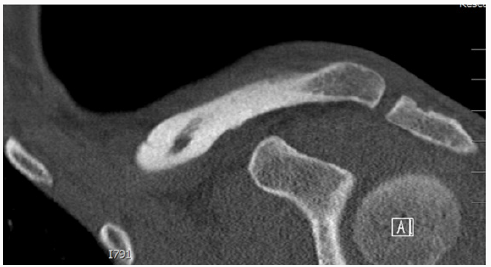
Figure 3:
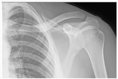
Figure 4: (a) Comparison right (normal)(b) Left (Malunited) clavicle
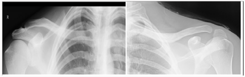
Figure 5:
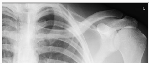
Figure 6:
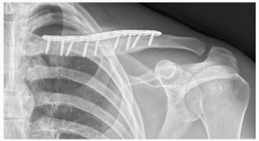
Figure 7: Early post operative.
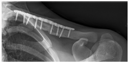
Figure 8:
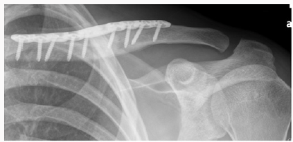
Figure 9: Three months post-operative (signs of radiographic healing).

Discussion

Clavicles fractures are common injuries and are reported to represent 2% to 5% of all adult fractures [1]. More recent evidence suggests that specific subsets of patients may be at higher risk for nonunion, symptomatic malunion, or suboptimal functional outcomes [2]. A recent meta-analysis suggests that the incidence of clavicle nonunion after nonsurgical treatment is approximately 5.9%, but can be as high as 15%for some fracture subtypes [3]. Nonsurgical treatment universally results in some degree of malunion; however, symptomatic malunion is fortunately quite low and is usually used particularly in very young patients [4]. Symptomatic patients are typically those with marked displacement at the fracture site, with shortening of >2 cm. Patients that are symptomatic may report weakness of the involved shoulder, rapid fatigability, numbness and paresthesia of the hand and forearm with elevation of the limb, and an asymmetric, “droopy,” “ptotic,” or “driven in”shoulder [5].

McKee et al performed a review of a cohort of patients to analyze the functional results of corrective osteotomy of a mal united clavicular fracture in patients with chronic pain, weakness, neurologic symptoms, and dissatisfaction with the appearance of the shoulder. Fifteen patients (nine men and six women with a mean age of thirty-seven years) who had amalunion following non operative treatment of a displaced mid shaft fracture of the clavicle were reviewed both preoperatively and postoperatively. The mean time from the injury to presentation was three years (range, one to fifteen years).Follow-up, at a mean of twenty months (range, twelve to forty-two months) postoperatively, illustrated that the osteotomy site had united in fourteen of the fifteen patients. All fourteen patients expressed satisfaction with the result. There was one nonunion, and two patients had elective removal of their plates. With regards to the patho anatomy of the deformed clavicle, McKee et al. noted that the deformity of the clavicle was a complex three-dimensional problem with all their patients illustrating a superior-anterior apex deformity. In his series there were certain consistent features seen in patients who presented with symptoms following non operative treatment and a healed clavicle. The hall mark characteristic is shortening in the medial-lateral dimension, with inferior displacement of the distal fragment and superior displacement of the proximal fragment. They, therefore, concluded that the shortening in the medial-lateral plane had a negative effect on muscle-tendon tension, and muscle balance. The anatomic boundaries of neurovascular structures were of paramount importance in the development of symptoms [6].

In a study by Edelson et al, he studied the bony anatomic details in 73 cadaver specimens which had clavicle malunions in different regions of the clavicle. According to the Allman classification. Edelson found that in the middle-third fractures, similar anterior angulations to the lateral third fracture malunion was indeed present. The most consistent finding at the middle-third level was that the lateral shaft fragment was almost invariably displaced posterior to the medial shaft fragment. The author also commented that initial anterior-posterior radiographs of clavicle fractures are often dominated by inferior displacement or ptosis of the lateral fragment. However, in the cadaveric specimens, anterior angulations rather than drooping of the lateral fragment were the predominant deformity. Although often initially displaced in a down ward direction, the lateral fragment does not usually heal in this position, unless it is a greenstick fracture as occurred in our patient.

Read More About Lupine Publishers Journal of Orthopedics and Sports Medicine Please Click on Below Link: https://lupinepublishers-orthopedics.blogspot.com/

Wednesday, 9 February 2022

Lupine Publishers| On A Criminal Trace of Crippling and Often Even Lethal False Diagnosis: Lege Artis Cz

 Lupine Publishers| Journal of Orthopedics and Sports Medicine


Case Report

In 2010, the Czech Republic participated in the World Health Organization WHO Research to determine the quality of medical decision making in determining diagnosis and to determine an adequate individual choice of patient treatment. The conclusions were really frightening: the relative frequency of fatal medical decision-making errors is 10%, with a statistical accuracy of the tolerance estimate of +/- 2%, with a statistical reliability of conclusions of about 95%. Approximately rewarding results are found in all OECD countries - in all types of outpatient or inpatient health services. In real terms, this means that approximately every 10th accidental visit of the patient to the doctor will end up with an additional health problem. For example, a late-diagnosis of B-type jaundice, a medical prescription of an inappropriate medication that contravenes the health of a particular patient, or contraindicates the contraindication effects of other concurrent medications, or a radiologist misinterprets an X-ray image-with overlooked by emerging stomach cancer, overlooked severely fatal osteoporosis-disturbed bone, a preventive standard examination of the colonoscopy will occur with careless medical manipulation of the colon perforation probe in a hitherto healthy patient. In general, they are seriously threatened patients, sometimes even seriously endangered patient lives.

If the above mentioned documented WHO research results refer to the whole Czech pupil - that is, for the total population of the Czech Republic, 10.6 million living people and an average of at least one annual patient visit to any doctor in the Czech Republic. approximately 1 million citizens are probable expected to be expected to receive approximately one million people instead of the actual assistance needed in the Diagnosis and appropriate individual treatment, likely to have an unexpected, addictive - crippling health risky next complication - and some patients even due to a deteriorated health condition, probably some of them die sooner - many years before the bio statistic mean survival estimate for the remaining years would be the same-if the medical error did not become the correct diagnosis or treatment. The Providers Health care and medical staff said: It is fatal legal yearly irrevocable Facts - but I have said NO!

On a Criminal Trace of Repeated Mass Medical Mistakes With False Coded Working with Regards Lege Artis CZ

The First Criminal suspicious for Mass Illegal Medical Workflow CZ

If approximately one million Patients CZ are seriously threatened annually with incorrect medical decision-making contrary medical Knowledge’s WHO, logically at least Sum 50,000 Patients CZ are unnecessarily mutilated yearly. They are mostly casual preliminary dying - a long time ago from the adequate Cluster with similar Patients CZ with the same Diagnoses, Age, similar variant of Treatments they are growing differenced Health consequences and ever-increasing causal next health risks to die after medical mistakes - All these cases should end annually with ignoring of the Czech Justice Courts, supervising Criminal Police CZ, supervising from State Penalty Offices CZ - and survivors or heavily crippled patients of the Czech Republic should receive from the responsibly Health instance CZ of the Providers nets of Health Services of the Czech Republic - approximately there are ignored 3 million compensation for financial satisfaction per 1 case per annum.

The total should therefore be a probable compensation in the astronomical annual amount yearly - 50 thousand persons CZ x 3 million crowns - the result is 15 x 10x10x10x10x10x10x10x10x10x10 = 150 milliard CZK / year!!! Thus, in the Czech Republic’s national income about Sum CZK 330 Milliard, almou nearly half of the annual income is a waste of medical work. This is for the Healthcare of the Czech Republic, for the Provider of Health Services and their Employees, for the Judiciary CZ, for the State Budget of the Czech Republic, for the Criminal Police of the Czech Republic, for the Public Prosecutor’s Office of the Czech Republic unprivileged by national, political.

However, we have a very novel solution in the Czech Republic - whether you are an individual patient crippled and dying for legal or illegal medical mistakes very Cheap: All fatal cases will be placed in the category of “Unwanted inevitable medical and legal results of medical processes” with the unique cover false slogan LEGE ARTIS CZ. It is similar as a Italy Mafia with more strong laws, more servos as a OMERTA, who has taken a keen interest in all the injured patients in the medical, security, criminal and custody organizations: You are either a healthy, unnecessarily frozen or unnecessarily dying patient in the Czech Republic - but absolutely under the monotonic motto of observing Medical cases with false wisdom declination the principles of LEGE ARTIS CZ. So that any private truth criminalist evidence of Patients CZ, when that you are the Victim of a criminal way of providing medical health care will come out monotonously: You will almost never receive adequate financial justice satisfaction, because it is only derived of your preliminary “informed Patient consent” to planned risky and probably often including very technically illegal medical practice with Medical Devices, often with explicitly wrongly misused The Medical Devices- you have only one direction right Cementary route: sooner or later, without any patient comments to Court dealing CZ: to die without delay as a false result of the absolutely indifference responsible medical work “LEGE ARTIS CZ” with content many fatal explicit illegal technical partial medical mistakes [1-3].

Annual unnecessary severe mutilation of at least about 40 to maybe perhaps 50,000 patients in the Czech Republic with a much earlier causal death - this is a pre-agreed probable loss of life and health of the Czech Patients, which is the number of deaths and astronomically large national economic losses similar to the lives of Victims of the secret national continual war Physicians CZ - against their own patients in the Czech Republic, often absolute out of legal technical usage Medical Devices in hospital net CZ in regards to Technician Requirements of Laws EU/CZ.

The Perspective for Solutions to Limit and to Prevency the Mass Repeated Medical Mistakes in OECD

I know namely reliable coherent scientific processing to limit the Mass repeated similar medical mistakes with more efficiency sharing the best medical experiences only as perfect redefined scientific principles “LEGE ARTIS CZ“ - with more effective managing medical workflow with more effective usage samplings, clustering, validated medical processing, continual testing elementary partial medical activities of partial medical processing step by steps“ with more efficiency implanting Artificial intelligence, with more regards to Technician requirements of Laws EU/CZ for usage Medical Devices - with acquired my decision making method S_T_A_R_S in daily medical workflow - see the Literature Antonín Cuc: The Utility model 21532 CZ 2010, Czech Office for Industrial Laws. The Equipment for Retrieval and Search of sufficient statistic information to repeated similar strategic decision making with risk and computer support “the opened Sources to usage for Medicine US since April 17, 2017!

As a scientist, I am well educated in the field of Cybernetics and Statistics and in the field of H+S for Medical Devices, Technician requirements of Laws EU/CZ for legal processing usage Medical Devices in Providers Health care nets!

The précised and Redefined Logical Criminalist Definition Lege Artis Medical Processing in Health Care OECD

For example, violation of the Technical Requirements of the Laws on the Legal Safe Use of Medical Devices, for example in contravention of the Technical Requirements of EU / the use of Medical Devices, contrary to the requirements of related harmonized European standards, contrary to the instructions of the manufacturers of medical devices and contrary to the certified use of their installation and measuring instruments, contrary to the obligation to prevent known hazards declared in the Protocols on Medical Devices as products with use management conformity assessment rules “CE”.

The Logical Substantiation in Medical Processing OECD

In the Binary Logics so as in the Criminalist Logics for the conjunctions partial mixed procedural phenomenon’s with evaluating True, False - there are sum resulting evaluation the coherent sequential processing always resulting in evaluation F A L S E, when there are occurrence at least the One partial unit with evaluation with partial well criminal evidenced FALSE, definitely!

I am just needless dying Patient CZ because the implanting processing set Total Hip Arthroplasty since November 13, 2007 contained many illegal processing medical mistakes contrary functional assembling product processing for set THA Bicontact S, no cemented, nominal dimension 13 mm N, B.Braun Germany

- with false coaxial ties between the metallic Stem and my right femoral bone in the Surgery hall without preliminary Clinic Plan, with illegal declination coaxial ties 13,68 grads in sagittal Direction, out of acceptable assembling tolerance +/- 1 grad - The post operational first RTG images from date November 16, it was the sufficient criminal evidences about false illegal placement the spice the Stem in coordinate Xi, Yi, Zi - out of the firm installing radiologic Masks - but it should be controlled by the Laws and by the product ovoid on the Orthopaedic screen with the same scaling.

The wisdom false Court Medical Message from the Knowing Medical Institute - The Central Army Hospital Prague, CZ No. 36 C 181/2009-123 since the March 27, 2012 defined all Medical Orthopaedic Processing in Limits Lege Artis CZ, The fatal unhappy was defined not as Diagnose Fausse route stem on the Orthopaedic Surgery Hall - but as the results of the false medical Hypotheses in Complot of the Radiologic, Orthopads Doctors from the Orthopady Clinic and from the Knowing Medical Institute as Post operational tragic Event by Patient fallen“, despite such substitutions in truth Criminal traceing, Radiology, Geometry 3 D, Health Patient Documentation EHRs.

I laid down my life so that you and your physicians and technicians can work together in a responsible manner and in the real truth of God and in the same truth of the criminalist evidences.

I became an unnecessary Merthyr of human Stupidity and irresponsibility that threatens humanity and the patients of the Czech Republic who are denied the Constitutional Human Rights of the Czech Republic: for life, for a fair Court trials, for legal health care CZ. I believe that Medicine.com will never accept the falsity of the current approaches of „LEGE ARTIS CZ“, as now submitted to the world public by the Czech courts, the Criminal Police of the Czech Republic, the network of the State Prosecutor’s Offices of the Czech Republic.

I am just placing the principal private RTG images for Forensic reconstruction Crash of set THA Bicontact S, no cemented in Geometry 3 D since date November 13, 2007 on the Orthopaedic Surgery Hall of the Regional Hospital Mladá Boleslav. Most of Patients with similar Crash are dying in duration since 3 till 7 days after overlooked FAUSSE ROUTE STEM, I was reoperation till 17 days after Poly trauma on the Surgery Hall, when the preoperational THA processing was realised in the same profile of Surgeon catting as in first THA, but the Spice of the Stem was created met he blooding and full plegic dysfunction Nervus is achidici with destructions my structure muscles in right calf, I am just frequential happened fallen with risk the repeated breaking the proximal femoral bone with just 8x worse probabilities and I am dying earlier about many years as other Patients CZ in Cluster with right legal first THA surgery.

Figure 1:

My Orthopads, my Judge, the forensic doctors from the Knowing Medical Institute CZ, Departments of Criminal Police CZ, State Penalty Offices CZ are smiling to my nearly Death within forced me many growing casual orthopaedic and leukemic heavy injuring LEGE ARTIS CZ (Figure 1). The virtual „Shorten of the Length the Axes of metallic Stem “It is caused the false placement with fatal declination in sagittal Direction in angle 13, 68 grads, there are false installing position of the spice Stem on coordinates Xo, Yo, Zi - it is illegal „to observing the RTG image in hands and by intuitive views“, when the Orthopads should taken the Orthopaedic screen and comparison the placement the Spice contrary firm radiologic Mask for the stem!

The Doctors said by illegal daily habits: We are working Lege artis, only a few orthopaedic patient are sometimes legal dying! The Criminal police CZ gave legal agreements so as the Knowing Medical institute CZ too - The mortal injured Patient CZ on the Prosector Hall haven´t commentaries! We are dying needless and very Cheap and frequently in CZ, always Lege Artis CZ (Figure 2). This is the Image from date November 28, 2007 too, this is forensic nonsense to prented this Criminal traceing could be realised as Patient fatal post operational happened! The last moving of the Stem was realised in Surgery hall November 13, 2007 in Regional Hospital Mladá Boleslav CZ.

Figure 2:

In Court trials CZ the Judge could believe for medical illegal Stupidities each of workaday. No Protests from Patients CZ are accepted in legal care for Constitutional Human Rights CZ by Ombudsman CZ- despite me is the Sate investigator for occupational mortalities in Branche Medical Devices. My Death is very awaiting CZ Events, like a public Execution of a disagreeable world Scientist in 21st Century.

Read More About Lupine Publishers Journal of Orthopedics and Sports Medicine Please Click on Below Link: https://lupinepublishers-orthopedics.blogspot.com