Lupine Publishers | Journal of Surgery & Case Studies

Abstract
Background:Tooth extraction is usually followed by partial resorption of the residual alveolar ridge. Different techniques such as ridge preservation procedure have been proposed to maintain the ridge dimension. However, applying these methods to extraction sockets could not completely preserve the coronal part of facial bone walls, which were comprised almost entirely of bundle bone.
Aim: To assess the effectiveness of partially tooth extraction (SST) for completely preservation of alveolar ridge.
Materials and Method: Case series study, which includes 6 participants medically fit, undergoes extraction of Non-restorable teeth, which doesn’t have any periapical or periodontal pathology. After clinical and radiographical assessment, computed tomography (CBCT), indicated insufficient width of buccal bone plate therefore socket shield technique was planned for simultaneous immediate implant placement with immediate provisionalization in area between the maxillary first premolars. Initial follow up after two weeks then after 2 months final restoration by screw retained crowns inserted, after 6 and 12 months of loading follow up by using CBTC, for evaluation bone remodelling and clinical evaluation of soft tissue changes around implants.
Result: Two weeks follow up revealed the healing was uneventful, and after 6 and 12 months the clinical and (CBCT) revealed, that retaining root fragment adjacent to the buccal crestal bone and placing an implant engaged to the palatal socket wall immediately are able to maintain the contour of the ridge. And the implant can achieve osseointegration without any inflammation at Periimplant tissue and also soft tissue contour preserved.
Conclusion: within limitation of this study we can conclude, after one year follow up, SST can prevent soft and hard tissue changes can be happens during healing of alveolar socket after tooth extraction. However the using SST as routine clinical practice stile need to higher level of evidence.
Abbreviations: SST: Socket Shield Technique, RST: Root Submergence Technique
Introduction
Literature has reported after tooth extraction, the alveolar bone undergoes a remodeling process, which leads to approximately 3.87 mm of horizontal ridge reduction and 1.67mm of vertical ridge reduction in the 6-12 months following extraction, primarily in the initial 3 months [1]. Conversely, the loss of a tooth triggers a remodeling reaction as part of the healing process, involving various degrees of alveolar bone resorption, especially affecting the buccal lamella: The bundle bone is primarily vascularized by the periodontal membrane of the tooth. Therefore, this part of the alveolar bone is compromised by the extraction, to such an extent that the buccal lamella is insufficiently nourished, leading to its total or partial resorption [2]. A higher degree of resorption is to be expected in the presence of flap formation, thin soft tissue biotype and prominent roots, especially with frontal teeth in buccal position [1,3]. These resorption processes complicate dental rehabilitation, particularly in connection with implants [4]. Immediate implant placement does not necessarily prevent resorption of the alveolar ridge [5]. A variety of ridge preservation techniques using tissue and augmentative materials have been proposed in the literature [6]. It has also been suggested that resorption of the buccal bundle bone can be avoided by leaving a buccal root segment (socket shield technique) in place, because the biological integrity of the buccal periodontium (bundle bone) remains untouched. This method has also been described in connection with immediate implant placement [7]. Clinical Concept, Procedures and Indications With the root submergence technique (RST), submucosal root retention can virtually eliminate bone resorption [8]. Based on this concept, the retention and stabilization of the coronal and buccal bundle bone and the retention of the periodontal membrane by retaining a coronal tooth fragment (so-called “socket shield”) [6]. Therefore, potential indications for such techniques include their use as part of the (delayed) late implantation approach or the optimization of pontic support in crown-bridge reconstructions or to improve the prosthesis base for removable dentures [7].
Figure 1: Socket Shield Technique by immediate implant placement.
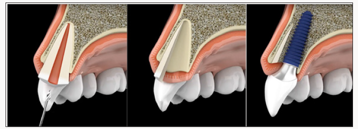
A step-by-step illustration of the proposed procedure: Firstly, the hopeless tooth is split supragingivally, the crown fragment is carefully dislocated and removed using a suitable instrument. Then, the root is separated vertically in a ratio between 1:3 and 2:3. The smaller, buccal root fragment is retained and the larger lingual root fragment is removed in a manner that spares bone and soft tissue to the greatest possible extent. The height of the buccal socket shield is reduced to the level of the bone. Simultaneous immediate implant placement Figure 1. After the procedure, patients rinse with 0.2% Chlorhexidine mouthwash two to three times daily for one minute over a period of at least ten days. During this time, mechanical oral hygiene is avoided in the affected area and only restarted after the follow-up examination. Anti-inflammatory drugs (e.g., Amoxicillin 500mg tid) are prescribed as needed. Each patient was informed verbally and in writing about the treatment and the materials used as well as the associated pre-and post-operative risks and gave their written consent to the use of the collected data and photos.
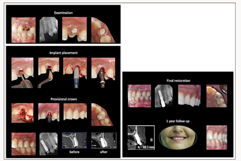
Figure 2: Treatment procedure for case 1.
Case Presentations
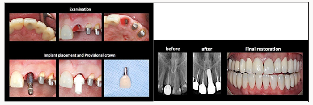
Case 1
Medical history: A 45-year-old woman, non-smoker with noncontributory medical history. Extra-oral findings: The patient’s facial features were symmetrical with average smile lines. On inspection and palpation, no abnormalities detected. Intra-oral findings: Non-restorable tooth #23, which doesn’t has any periapical or periodontal pathology. Clinical and radiographical assessment, computed tomography (CBCT), indicated insufficient width of buccal bone plate. Treatment plan: partial root extraction of tooth #23, prevention of alveolar ridge resorption using the socket shield technique, simultaneous immediate implant placement (Straumann 4.1x12 RN) with immediate provisionalization crown. Initial follow up after two weeks, then after 2 months final restoration by screwretained crown inserted. After 6 and 12 months of loading follow up by using CBTC, for evaluation bone remodeling and clinical evaluation of soft tissue changes around implants. The treatment is depicted in Figure 2.
Case 2
Medical history: A 35-year-old man, smoker with noncontributory medical history. Extra-oral findings: The patient’s facial features were symmetrical with average smile lines. On inspection and palpation, no abnormalities detected. Intra-oral findings: Non-restorable tooth #24, which doesn’t has any periapical or periodontal pathology. Clinical and radiographical assessment, computed tomography (CBCT), indicated insufficient width of buccal bone plate. Treatment plan: partial root extraction of tooth #24, prevention of alveolar ridge resorption using the socket shield technique, simultaneous immediate implant placement (Straumann 4.1x10 RN) with immediate provisionalization crown. Initial follow up after two weeks, then after 2 months final restoration by screwretained crown inserted. After 6 and 12 months of loading follow up by using CBTC, for evaluation bone remodelling and clinical evaluation of soft tissue changes around implants. The treatment is depicted in Figure 3.
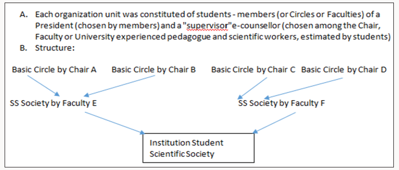
Figure 3: Schema of a Student Scientific Society structure.
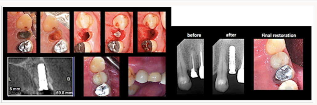
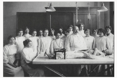
Figure 4: Professor SHOR GV (1812-1944) - well known Pathologist - with students at a meeting of the student scientific circle of Pathologic Anatomy in the 30-ties.
Case 3
Medical history: A 37-year-old woman, non-smoker with noncontributory medical history. Extra-oral findings: The patient’s facial features were symmetrical with average smile lines. On inspection and palpation, no abnormalities detected. Intra-oral findings: Non-restorable tooth #21, which doesn’t has any periapical or periodontal pathology. Clinical and radiographical assessment, computed tomography (CBCT), indicated insufficient width of buccal bone plate. Treatment plan: partial root extraction of tooth #21, prevention of alveolar ridge resorption using the socket shield technique, simultaneous immediate implant placement (Straumann 4.1x10 RN) with immediate provisionalization crown. Initial follow up after two weeks, then after 2 months final restoration by screwretained crown inserted. After 6 and 12 months of loading follow up by using CBTC, for evaluation bone remodelling and clinical evaluation of soft tissue changes around implants. The treatment is depicted in Figure 4.
Discussion
Post extraction ridge collapse with degrees of alveolar resorption has been extensively documented in the literature. These hard and soft tissue defects can negatively affect ideal planned implant placement with a potential for esthetic failure [9]. Bundle bone arises from a functionally loaded PDL and is lost following extraction, resulting in an almost certain collapse of the buccofacial tissues [10]. Ideally, a method for the prevention of alveolar ridge resorption should be a cost-effective and minimally invasive, with only minimal material requirements. However, these criteria are not entirely met by any of the methods available today. Moreover, the goal of complete preservation of the alveolar ridge after tooth extraction has not even been reached with sophisticated techniques [11]. First reported in 2010 the SS technique had progressed from concepts introduced in the 1950s that the retention of a tooth limits tissue alterations following extraction. The submergence of tooth roots was introduced originally to preserve alveolar ridge volume beneath removable full prostheses [12,13]. The retention of tooth roots in the alveolar process can preserve the ridge tissues Histologically this was demonstrated by Hürzeler and coworkers. The buccal plate crest showed an absence of osteoclastic activity an absence of active remodelling [14]. The histological analysis suggests that the buccal bone plate was preserved. Therefore, it may be speculated that this technique may have the potential to avoid the marked resorption of the buccal bone plate after tooth extraction [15]. The clinical outcome of Hürzeler and coworkers’ report presented the successful osseointegration of an implant placed simultaneous to the SS technique and a restoration with aesthetics indistinguishable from the adjacent maxillary central incisor. Whilst the authors reported preservation of the buccofacial tissues, it should be noted that absolute preservation has not yet been shown.
The authors later reported a mean of 1 mm horizontal loss after final restoration, Chen and coworkers reported 0.72 mm of buccal resorption [16,17]. Retaining the buccal aspect of the root in conjunction with immediate implant placement is a viable technique to achieve osseointegration without any inflammatory or resorptive response [15]. It needs to be highlighted that the presented partial extraction is technique-sensitive and associated with the risk of displacement of the buccal root fragment, or even the buccal lamellar bone. Already at the time of extraction, the root fragment should be reduced along its vertical axis to the level of the height of the alveolar ridge to prevent perforation of the buccal mucosa during the healing period. Should the root fragment become exposed despite this preventative measure, height reduction and tissue freshening can be performed during follow-up visits [7]. The SS technique offers a promising solution to the difficulties encountered when managing the post-extraction tissues. This case series of immediate placement simultaneous to the SS technique is among the first to demonstrate with a 1 year follow up successful preservation of post-extraction tissues coinciding with successful restorative implant treatment.
Conclusion
Within limitation of this study we can conclude, after one year follow up, SST can prevent soft and hard tissue changes can be happens during healing of alveolar socket after tooth extraction. However the using SST as routine clinical practice stile need to higher level of evidence.
Read More About Lupine Publishers Surgery Journal Please Click on Below Link:
https://surgery-casestudies-lupine-publishers.blogspot.com/