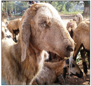
A cross-sectional study was conducted from October 2016 to July 2017
on ovine slaughtered at Jimma municipal abattoir with the aim of
identifying main pathological lesions causing organs and carcass
condemnation, and associated direct financial losses. In this, 384 sheep
were recruited to the study using systematic random sampling and
standard antemortem (AM), and postmortem inspection (PMI) procedures
were employed. Nasal discharge, tick infestation, coughing, lameness,
emaciation, depression and salivation are recorded as the major AM
findings of the current study. Accordingly, 47 (12.2%) sheep showed
signs of diseases and abnormalities; of which 23(6%) were conditionally
approved whereas 11 (2.9%) sheep were unfit and judged to be detained
and rejected. In the present study age, body conditions and geographic
origin of the animals were considered as study variables, and the
results showed BCS and age groups had statistically higher (p ≤ 0.05)
rejection probabilities. PM examination findings indicated a total of
192 lesions were encountered, of which 48.9%, 29.2%, 6.8%, and 5.7%
lesions were recorded from livers, lungs, GIT, and hearts and kidneys,
respectively. During the PM inspection, both total and partial
condemnation judgments were passed on organs and carcass appeared with a
sign of abnormality. C. teniculosis 58 (30.2%), calcification
36(18.7%), abscess 31(16.2%), hydatid cyst 23 (12%), hepatitis 10
(5.2%), pericarditis 4(2%), bruise 3(1.6%) and nephritis 2(1%) were
found to be the major pathological lesions recorded. In two sheep all
organs and carcass were totally condemned as their entire bodies
appeared yellowish. The direct loss due to the condemnation of organs
and carcasses at Jimma municipal abattoir was also investigated in
slaughtered sheep, and there were about 56,576 USD losses per year. In
conclusion, this study has identified the pathological lesions affecting
edible organs and meat, and then rendering them unfit for human
consumption. The study also estimated pathological lesions associated
direct financial losses at Jimma abattoir. Therefore, further studies
focusing on the primary causes of the abnormalities were recommended in
the study area.
Keywords: Abattoir; Financial loss; Lesion; Organ condemnation; PMI; Sheep; Jimma
Introduction
The livestock sector globally is highly dynamic, contributes 40% of
the global value of agricultural output and support the livelihoods and
food security of almost a billion people (Thornton [1]). Within African
society, small ruminant comprises a greater proportion of the total
wealth of the rural families, because of the low input requirements such
as low initial capital, fewer resources and maintenance cost. They are
also able to produce milk and meat in readily usable quantities using
marginal lands and poor pasture and crop residues. Furthermore, their
production cycle makes them need only short periods to reconstitute
flocks after a disaster and respond quickly to the demand (Getenby,
[2]). Ethiopia is the leading African country in livestock population,
having around 34-40 million TLU (Tropical livestock unit) out of which
17% and 12% cattle and small ruminants, respectively, are found in
Ethiopia (Ministry of information (MOI) [3]. According to (Development,
pp Statis, [4]), the population of sheep and goats in Ethiopia is
estimated to be 26.1 and 21.7 million respectively. It was the third
largest number of sheep and goat among African nations and rank eighth
in the world (Alemu and Merkel, [5]).
They generate cash income from export of meat, edible organs, skins
and live animals (Ibrahim, 1998). There is also a high domestic meat
demand from these animals, particularly during religious festivals. Even
though this sub-sector contributes much to the national economy, its
development is hampered by various constraints. These include endemic
animal diseases, insufficient nutrition, poor husbandry, and lack of
sufficient infrastructure, trained labor and government policies (PACE,
[6]). Each year a large loss results from the death of animals and
weight loss during transportation; and condemnation of edible organs and
carcasses at slaughter.
Abattoir meat inspection is essential to remove gross abnormalities
from meat and its products, to prevent the distribution of contaminated
meat and to assist detecting and eradication of certain livestock
diseases. More specifically, antemortem inspection attempts to avoid
introduction of clinically diseased animals into slaughter house and
also serves to obtain information that will be useful in making sound
post mortem inspection. Likewise, postmortem inspection is the center
around which meat hygiene revolves since it provides information
essential for evaluation of clinical signs and pathological process that
affect the wholesomeness of meat (Herenda, et al. [7]).
As the meat is the sources of protein to a human being, it should be
clean and free from diseases of particular importance to the public such
as tuberculosis and cysticercosis. Meat is also condemned at
slaughterhouse to break the chain of some zoonoses which are not
transmitted to man directly via meat like hydatidosis and other
important diseases of animals such as fasciolosis (Arbabi and Hooshyr
[8]; Fufa et al. [9]).
Each year a significant economic loss results from mortality, poor
weight gain, condemnation of edible organs and carcasses at slaughter.
This production loss in the livestock industry is estimated at more than
900 million USD annually (Jacob, [10]; Abebe, [11]; Jobre et al. [12]).
The major causes of pathological lesion during PMI of slaughtered ovine
at abattoir are the disease caused by parasites, bacterial and other
abnormalities. The final judgment as to action to be taken with an
organ, the carcass or part of a carcass is based on the total evidence
produced by the visual observation, palpation and incision (Teka, [13]).
Abattoir data is an important option for observing the diseases of both
economic and public health importance (Arbabi and Hooshyr, [8]; Fufa,
et al. [9]). Nowadays, several modern abattoirs like: HELMEX, ELFORA,
Metehara, Modjo and Luna are established in Ethiopia. This increase in a
number of slaughterhouse shows that increase in demand for meat supply,
but the provisions have been challenging due to diseases, production
problems and other factors. Given this, proper evaluation of financial
losses due to organ condemnation resulting from various diseases at
abattoirs is needed (Ezana, [14]). It is necessary to have enough
information on a pathological lesion that causes organs and carcass
condemnation at the abattoir. Hence, having information on where and how
to reduce the losses that may be caused by the various abnormalities
(lesions/pathology). Various studies (Jembere, [15]; Yimam, [16];
Aseffa, [17]; Getachew, [18]; Regessa et al. [19]) were carried out in
the country in this regard to know the causes and losses associated.
However, in Jimma there are no recorded studies conducted on major
causes and financial losses associated with organs and carcass
condemnation along with survey on pathological lesions. Therefore, the
objectives of this study were to:
a) Identify major pathological lesions causing organs and carcass condemnation in slaughtered sheep at Jimma municipal abattoir
b) Estimate the direct financial losses attributed to condemned organs and carcass in sheep
Materials and Methods
Study Area
The study was conducted from November 2016 to July 2017 at Jimma
municipal abattoir in Jimma zone. Jimma two is found in Oromia region
south-western part of Ethiopia at a distance of 346km away from Addis
Ababa and lies between 36°50'E longitude and 7°40'N latitude atan
average elevation of 1750 meter above sea level. Jimma is the largest
city in south-western Ethiopia. It is special zone of the Oromia
Regional state and is surrounded by different Jimma woreda. The climate
of the area is characterized by humid tropical with bimodal heavy
rainfall which is uniform in amount and distribution, ranging from 1200
to 2800mm per year, with short and main seasons occurring from
mid-February to May and June to September, respectively. The rainy
season extends from mid-February to early October. Temperatures at Jimma
are in a comfortable range, with the daily mean staying between 20 °C
and 25 °C year-round. The total human populations of Jimma town was
about 174, 446(88, 766 males and 85, 680 females). The livestock
population of the area was reported to be about 2, 016, 823cattle, 942,
908 sheep, 288, 411 goats, 74, 574 horses, 49, 489 donkey, 28, 371
mules, 1, 139, 735 poultry and 418, 831bee hives (GOR, [20]).
Study Population
The study animals were sheep brought Jimma municipal abattoir and
destined for slaughter. All animals were male and belonged to indigenous
breeds kept under extensive management system. Sheep destined for
slaughter had come from different parts of the weredas in the Jimma zone
such as Dedo, Serbo, Saqa and Bilida inspected by standard AM, and PMI.
Study Design
A cross-sectional study using systemic random sampling technique was
conducted from December 2016 to April 2017 to determine the pathological
lesion that causes organs and carcass condemnation and to estimate the
magnitude of direct financial loss attributed in sheep slaughtered at
Jimma abattoir.
Sampling Method
Sample size determination
In this study, systematic random sampling method was applied to
include study animals, and study animals were grouped into young (under
1year and three months) and adult above this based on the eruption of
one or more incisor teeth according to Vatta et al. [21]. Since there
was no published work on lesion survey from Jimma abattoir, 50% expected
prevalence is considered to calculate the total sample size with 95%
CI, 5% level of precision (Thrushfield, [22]). The sample size was 384
and determined using the formula given by

Where N= required sample size, Pexp= expected prevalence and d is desired absolute precision. Accordingly, the total sheep included in the study were 384.
Abattoir Survey
In the cross-sectional study of active abattoir survey, both AM and
PMI were carried out in accordance by the procedures of Ethiopian Meat
Inspection Regulation (Belina and Melese, [23]).
Ante-mortem Inspection (AMI)
The AMI (pre-slaughter examinations) of ovine was conducted at
lairage both in motion and at rest and information related to study
variables such as the behavior of an animal, age, BCS and origin and
were recorded. At the same time, various signs of diseases and
abnormalities were inspected with physical animal examination and its
judgment were approved, conditionally approved, detained and rejected.
Study animals were grouped into young and adult age groups according to
standard dentation method (Vatta et al.[21]).
Post Mortem inspection (PMI)
During PMI all internal organs (liver, lungs, heart, kidney,
gastro-intestinal tract), and carcasses were thoroughly inspected by
visualization, palpation and making systematic incisions for the
presence of cysts, parasites and other abnormalities. Pathological
lesions were differentiated and judged according to guidelines on meat
inspection for developing countries as totally fit for human
consumption, and conditionally approved, and totally or partially
condemned when unfit for human consumption (FAO, [24]).
Assessment of direct Financial Loss
In the current study, the total financial loss due to organs and
carcass condemnation was computed by considering the condemnation rate
or percentage of each edible organ and carcass, average number of
animals slaughtered in the abattoir per year from retrospective data of
the abattoir. The average weight of each organ and carcasses in kg,
average current local market price of major organs and carcass, and each
condemned organ was counted to estimate the financial loss. The average
current local market price of each organ and mutton was collected by
questionnaire from the butcheries in Jimma town for ease of computing
the loss.
The retail average market prices obtained from butcher shops found in
Jimma town in ETB were: Liver=30, lung=20, kidney=15, heart=18,
GIT=90,whole carcass=4000 and mutton =150ETB/kg. In the case when there
was whole carcass plus organs (whole body) rejection at PM, the average
price of sheep came for slaughter was considered (4,000ETB). The direct
loss is calculated according to the procedures described by Ogurinade
and Ogunrinade [25], and the formula:
LOS is direct annual financial loss due to organs and carcass loss,
MAK is annual average number of sheep slaughtered at Jimma abattoir, PL
is overall prevalence of lesion, Pi is prevalence of each organ and carcass condemned, Ci
is average market price of each organ and 1kg mutton at butcher shops
of the Jimma town. The direct financial loss was expressed in.com Dollar
($) based on the current currency exchange rate of 1 USD = 22.5
Ethiopian Birr (ETB).
Data Analysis
The active abattoir data, and questionnaire survey were entered into
Microsoft Excel- 2016 spread sheet and the process of coding, cleaning
and validating was done on this sheet and analyzed using SPSS version
20. For the data from PMI, descriptive statistics were used to determine
organ and carcass condemnation rates, defined as the proportion of
organs and carcasses condemned to the total number of organs and
carcasses examined. Each financial loss was also calculated. Possible
variation between rejection rates of specific organs, age groups and
origin, were taken into consideration.
Results
Abattoir Survey
Antemortem Inspection (AMI)
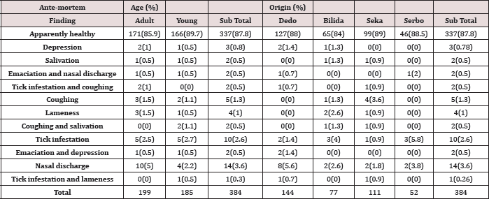
Detail AMI was conducted on a total of 384 sheep destined for
slaughter at Jimma municipal abattoir and 47 (12.2%) of ovine were found
to have different abnormalities. Nasal discharge, coughing, tick
infestation, depression, emaciation, and lameness were those frequently
observed among signs of diseases encountered in both age groups. The
result also showed 6% (23/384) animals were conditionally passed for
slaughter because of abnormalities such as lameness, respiratory
problem, and their collection with tick infestation. On the other hand2%
(8/384) sheep were unfit for human consumption and rejected during AMI.
Since, they showed two and more signs of diseases such as emaciation
with nasal discharge and depression 1% (4/384), salivation and
salivation with coughing 1%(4/384) were the major cause of rejection
(Table 1).
Table 1: Abnormalities encountered during AM inspection within Age groups and Origin of the animals.
The AMI result also depicted of 28 sheep with poor body conditions
46.43% were found to have one or more sign/s of illness whereas in those
with good BCS only 7% of the sheep were showed sign/s of diseases
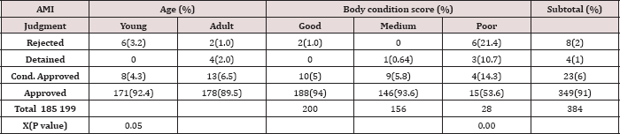
(Graph 1). In the current study, four different decisions were passed as
AM judgments where apparently healthy sheep were passed for slaughter
(91%), and others showed mild sign of illness and conditionally approved
(6%) whereas 2.9% of sheep were detained and rejected as unfit for
slaughter. Rejection rate was significantly higher (p ≤ 0.05) in young
animals with poor BCS than in adult animals with good and medium BCS
(Table 2).
Figure 1: proportion of ante-mortem finding by animals'boay condition score.
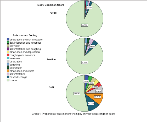
Table 2: Proportion of animals rejected and passed the AMI judgments by age group and BCS at the abattoirs.
Postmortem Inspection (PMI)
Among animals that had been examined during AMI 373 were slaughtered
and subjected to through PMI following standard postmortem procedure and
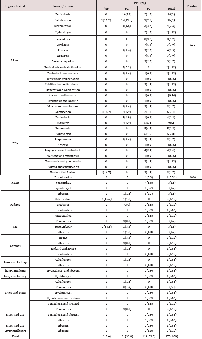
a total of192gross pathological lesion leading to partial and total
condemnation of organs and carcasses were recorded. Among these
abnormalities, lesions were frequently encountered from liver; and of
which Cyst cercus teniculosis (34%) and calcification accounted 25.5%.
These followed by abscess (13.8%), hepatitis (10.6%), cirrhosis (7.4%)
and hydatid cyst (7.4%), fasciolosis (5.3%) and stelezia hepatica
(3.2%).
Table 3: Relative percentages of pathological lesions resulted in condemnations of organs and or carcasses at the abattoir.
*AP=organs conditionally passed for human consumption
A total of 56 lungs were also condemned as they were affected abscess
(3.6%) and 5% with unidentified lesions. In this study by teniculosis
(35.7%), Hydatid cyst (21.4%), marbling lesion abscessation was also
inspected in other organs like heart, kidney (17.8%), Emphysema and
calcification (16%), pneumonia (12.5%), and GIT, and carcass (Table 3).
Table 4: Summary of direct financial losses and organs and carcass condemned at abattoir.

There was a condition of examining, a single to multiple lesions per
organ e.g., we examined teniculosis and Calcification from a single
liver; and all are recorded as different lesions.
*=Whole carcass plus organs totally rejected at PM; except in carcass, PC indicates 50% loss.
Out of a total of 11 hearts condemned (Table 4), hydatid cyst Renal
problems were observed in 11 kidney examined and 54.5% and pericarditis
recorded as major causes contributing 45.5% and found to be caused by
abscess whereas 27.3% and 18.2% were 36.4% which followed by abscess
(18.2%) and discoloration (0.9%). due to calcification and nephritis and
other unidentified causes,
respectively.13 GITs were also encounter as with abnormalities likecyst
cercus teniculosis and abscess and foreign bodies, and they were
subjected to total and partial condemnation accordingly.
The major pathological conditions for carcass rejection from local
market were bruising accounting for 42.9%. Out of 7 rejections
judgments2 were total and the rest 5 were partial (Table 3&4). There
was a condition of examining, a single to multiple lesions per organ
e.g., we examined teniculosis and Calcification from a single liver; and
all are recorded as different lesions.
*=Whole carcass plus organs totally rejected at PM; except in carcass, PC indicates 50% loss.
Assessment of Direct Financial Losses
The direct financial loss was computed based on average cost/ price
of individual condemned organs and carcasses during the study period,
applying the formula given by Ogurinade and Ogunrinade [25]. The study
indicated that there had been a total loss of 12,729,960 ETB which is
56,576 USD due to a partial and total condemnation of organs and carcass
at slaughterhouse annually. The study result also indicated there was a
total condemnation of 2(1.3%) whole carcasses (carcass plus organs)
(Table 4). For the calculation, alive price of one sheep is considered
as 4000ETB for total carcass condemnation.
Discussion
The AM and PM inspections were conducted in the abattoir for the
purpose of identifying an abnormality and removing animals' products
with pathological lesions which were unsafe for human consumption and
having poor aesthetic values (Van L logtestijn, [26]; Gracey et al.
[27]). In this research, out of 384 study animals 11(2.9%) were rejected
and detained as unfit for human consumption suspecting different
zoonotic diseases such as rabies in the case of salivation with high
fever and tuberculosis in animals with a sign of emaciation with high
coughing, depression and nasal discharge. Similarly, suspecting rabies
in the case of salivation and bovine TB in animals with sign of
emaciation with high depression and coughing was reported by Belina and
Melese [23]. Radostits et al. [28] stated that in domestic ruminants,
cobalt deficiency results in appetence and loss of body weight,
emaciation, weakness, decreased growth, unthrifty appearance, diarrhea,
and anemia. Again during the AMI, 23(6%) of sheep were found to be
showing signs of abnormalities such as lameness, tick infestation along
with coughing and nasal discharge frequently encountered and passed with
judgments of conditional approval where due attention was given for
whole body part and specific organ at postmortem examinations.
One of the causes of lameness was trauma caused by hitting with a
thick stick during driving to abattoir on foot and inappropriate
vehicles and loading and off-loading negligence during transportation to
marketplaces and to the abattoir. During the AM examinations, it was
found that respiratory disorders were higher than other abnormalities
encountered during the AMI 14(3.7%) nasal discharge and 5(1.3%)
coughing. The respiratory signs such as the presence of nasal discharge
and coughing were most probably related to stress due to lack of feed
and water that may lead to immune suppression enhancing opportunistic
pathogens. On the other hand, overcrowding during transportation is also
a source of stress (Getachew, [18]). In agreement to the current study,
coughing, depression and lameness are frequently observed abnormalities
encountered during AMI (Mandefro et al.
[29] ) at Elfora Export Abattoir, Ethiopia.
The rejection rate was significantly higher (p<0.05) for those
poor body conditions than good and medium body conditions (Table 2).
Because of poor BC by itself may be due to unidentified abnormalities
that increase rejection probability. Jibat et al.
[30] studied and determined the rate of organs and carcasses condemned
and the associated annual financial loss at HELMEX abattoir in Ethiopia
and they reported out of 2688 sheep and goats examined 188 (7%)
carcasses were condemned due to poor body condition cases.
On the other hand, there was a significant difference (p=0.051)
within the age groups of animals in rejection at AM more young than the
adult which were 3.2% (6/185) and 1% (2/199) respectively. It may due to
difficulty in protection from stress, shortage of feed and water, not
getting enough rest. Herenda et al. [31] stated that leanness (Poorness)
is often observed in case of poor quality pasture and young growing
animals which have had protein-deficient diet.
In the present study, organ condemnation rate showed that, liver and
lung were the most frequently affected organs with the highest
condemnation rate followed by GIT, kidney and heart and carcass
significantly (p=00), which is 94(48.9%), 56(29.2%), 13(6.8%), 11(5.7%),
11(5.7%) and 7(3.6%) respectively. This finding is in agreement with
reports of Cadmus and Adesokan [32] who recorded that lungs (45.7%) and
the liver (32.9%) were the most affected organs with the kidney (0.02%)
and the heart (0.01%) being the least. The current study introduced that
parasites are the major causes of organ condemnations. Parasitic causes
like, Cyst cercus teniculosis, hydatidosis, fasciolosis and Stelezia
hepatica were found to be the major parasitic conditions responsible for
organ condemnation. There was no statistical difference in the rate of
organ and carcass condemnation from parasitic infestation considering
the age and origin of animals. This shows that parasitic diseases of
sheep are widely spread in all age groups and everywhere in the country.
The presence of small ruminant hydatidosis at slaughterhouse has been
documented in Ethiopia. (Bekele et al., [33]) reported a prevalence
rate of 16.4% in sheep which is higher than the finding in this study
(7.4%). Similarly (Jobre et al. [12]) reported prevalence rate of 11%
and 6% from South Omo and Debrezeit slaughterhouses, respectively in
sheep and goats. In present finding, hydatid cysts were more frequently
observed in lungs than liver of sheep (6.3%) and (3.6%) respectively.
Additionally, similar findings were also reported by different authors
(Khan et al. [34], Dalimai et al. [35] and Daryani et al. [36]).
However, the most common site for hydatid cyst was the liver followed by
the lungs in the Middle East (Kamhawi et al. [37]). Lungs are most
commonly affected by hydatidosis because at old age the liver
capillaries are dilated, and most cysts passed directly to the lung.
Secondly, the cyst passes to the lung via the thoracic duct without
involving the liver (Gracey, [38]). And also, many researchers reported
that liver and lung are the most commonly affected organs by hydatid
cyst (Abunna and Hordofa, [39]; Denbarga, [40]; Jobre, [12]). The reason
being that lung and liver contain highest capillary bed in the body and
therefore, the majority of the oncospheres were filtered out and
trapped in the fine blood capillaries and only small number of
oncospheres reaches the remaining organs (Gracey, [38]). In present
study also lungs and liver 12(6.3%) and 7(3.6%) respectively, were
affected by hydatidosis.
Out of 94 condemned liver teniculosis is the most frequent cause of
organ lesion (34%) followed by calcification (25.5%), abscess 13(13.8%),
hepatitis 10(10.6%), cirrhosis 7(7.4%), discoloration 4(4.3%), more
than 3 lesion on liver 3(3.2%) and parasites like teniculosis, Stelezia
hepatica, Fasciola species and hydatid cyst were found to be the major
causes that rendered liver rejection from the local market (Table 3).
Fascioliasis constitutes both economic and public health constrains to
ruminant production. It is caused by two trematode species, Fasciola
hepatica and F.gigantica, which develop in different livestock species
mainly sheep and cattle; but, also in many other domestic herbivores
(Gracey, and Collins, [41]). The reported prevalence of Fasciolaspp.
(5.3%) was lower than other studies in bovine, like Belina and Melese
[23] study result showed fasciolosis and hydatidosis alone contributed
690(35.1%) gross pathological lesions.
Previous studies have indicated a higher economic loss resulting from
a condemnation of edible organs and carcasses due to parasitic causes
(Negategize et al. [42]; Jembere, [15]; Jibat, [43]). In the current
study, these parasitic causes of liver lesion might be due to improper
wasting of condemned organ and the stray dog feed it at abattoir and
selling of infected offal for dog which is final host for teniculosis
and hydatidosis and stay them. Sissay et al. [44] studied the prevalence
and seasonal incidence of cestodeparasite infections of sheep in
Eastern Ethiopia for two years (2003-2005). During this period, viscera
including liver, lungs, heart, kidneys and the gastro-intestinal tract
were collected from 655 sheep slaughtered at four abattoirs. One of the
most prevalent metacestodes was C. teniculosis. In sheep, the overall
prevalence was 79% for C.teniculosis.
The causes for calcification abscess, hepatitis, cirrhosis and
discoloration were difficult to identify grossly and it may be due to
systemic infectious diseases. Calcification is also another lesion that
we encountered; it can be caused by injury, infection, and autoimmune
disorders. Large-scale tissue damage is associated with extensive loss
of cells, a situation referred to as tissue necrosis. The death of
tissue in a specific area of the body leads to the release of signaling
factors that attracts cells to clean up and heals the dead tissue. This
process, known as an inflammatory response, attracts calcium into the
damaged area as it heals (Carne, [45]). This study indicates 24(12.5%),
9(4.7%) and 3(1.6%) of Liver, lung, and kidney, respectively were
affected by calcification.
Abscess was also apathological condition; which is a collection of
.com circumscribed by fibrous tissues. It occurs with great frequency
throughout many organs and the carcasses of the meat animals and may be
associated with a general condition or be found as isolated lesions
(Libby, [46]). In present study 13(6.8%), 6(3.1%), 6(3.1%), 2(1%) and
2(1%) of liver, GIT, kidney, heart and carcass were affected by abscess.
In agreement with (FSIS, [47]) stated that caseous lymphadenitis is a
disease of sheep and goats caused by the C.Pseudotuberculosis.
Postmortem findings may include, enlarged abscessed lymph nodes with
greenish white- yellow caseous exudate, which tends to become dry and
granular, cross-sections of lesions contain remnants of connective
tissue capsules (resembles the concentric rings seen on the cut surface
of an onion). Lesions found in many lymph nodes, especially the
subiliac, superficial cervical, deep popliteal, tracheobronchial, and
mediastinal lymph nodes, as well as lungs, heart, liver, spleen, and
kidneys. (Asrat, [48]) stated that occasionally the worms penetrate the
bile duct wall into the liver parenchyma causing liver abscesses.
The study conducted in Gondar abattoir (Mesele et al. [49]) and
Nekemte (Moje et al. [50]) also revealed that livers and lungs are the
most rejected organs by PM inspection and fasciolosis and hydatidosis
are the major causes of rejections. However, in the current study
different calcifications, cirrhosis, hepatitis, abscessations,
emphysema, pneumonic lesions, marbling (contagious caprine
pleuropneumonia (CCPP)) lesion, nephritis, foreign body, traumatic
lesions and others non parasitic abnormalities and unidentified lesion
contributed to a condemnation of organs and carcasses were investigated.
Lungs were condemned because of C.teniculosis, hydatid cysts, marbling,
emphysema, calcification, pneumonia, other unknown caused lesion and
abscess which were (35.7%), (21.4%), (17.8%),(16%), (16%), (12.5%), (5%)
and (3.6%) respectively. C.teniculosis accounts for 35.7% as a
principal cause of lung condemnation in sheep This might because of
increased number of a stray dog in the area, the principal cause of lung
condemnation was parasitic. However, the report observed during a
retrospective study (Regassa et al., [19]) reported pneumonia as a
principal cause of lung condemnation in central Ethiopia accounting for
42.1% (Getachew, [18]). In current study, from the total lungs inspected
higher 56 (29.2%) lungs were condemned. It may because of the animals
unable to resist stress within a short period of time during
transportation along way on foot, shortage of feed and water, stress due
to hitting of animal by personnel who driving animal to market from the
farmer and to abattoir and does not getting sufficient amount of rest
at lairage may causes this respiratory problem.
FSIS [47] reported that pneumonia is an inflammatory condition of the
lungs that maybe caused by infectious agents, parasites, physical
trauma, or foreign material inhalation. In similar reports pneumonia
might also be as a result of endemic diseases of sheep and goats such as
pasteurellosis, which is triggered by stress, contagious caprine
pleuropneumonia (Radiostitis et al. [51]). The other cause was marbled
appearance (CCPP) lesions: CCPP is a disease peculiar to shoat and takes
the form of a chronic inflammation of the lungs and pleura. It is not
communicable to man and the carcass and the lungs found to be positive
for CCPP can be passed for human consumption after a partial
condemnation of the diseased part Gracey and Collins [41]. On the other
hand, Emphysema is an abnormal and permanent enlargement of air spaces
distal to terminal bronchioles with destruction of their alveolar walls,
whereas oedema is a nonspecific lesion in which interstitium and
alveoli are accumulated with fluid (Carne, [45]).
Also, different lesions of infectious and noninfectious causes like
abscess, pericarditis, nephritis, and discoloration were found to be
important causes for the condemnation of edible organs like liver,
heart, and kidney. Similarly the same causes were found at central
Ethiopia (Getachew, [18]; Regassa et al. [19]) and in goats slaughtered
at Nigeria (Ojo, [52]).
In the present study, out of the 11 (5.7%) kidneys condemned abscess 6
(54.5%) account, whereas calcification accounts 3(27.2%), nephritis
2(18.2%) and other unidentified causes 2(18.2%) (Table 3). In this study
abscess was a principal cause of kidney condemnation, however, the
result in (Dejene et al. [53]) study revealed out of the 57 (6.71%)
kidneys condemned Nephritis 20 (2.35%) accounting for 11 (2.59%) and 9
(2.12%) kidneys in Ovine and Caprine respectively, was the principal
cause of condemnation. Radostitis et al., [28] stated that embolic
nephritis occurs after septicemia or bacteremia when bacteria lodge in
renal tissue.
The major causes of heart condemnation were found to be pericarditis,
hydatid cyst and abscess. Out of the total of 11 (5.7%) hearts
condemned due to gross abnormalities, pericarditis contributes about 4
(36.4%) and hydatid cyst also contribute 4(36.4%) and abscess 3(27.3%)
out of condemned organs (Table 3). The main cause of lesion in GIT
condemnation primarily parasitic C.teniculosis and abscess 6(46.2%) and
foreign body 4(30.8%). As a septic lesion, whenever localized abscess is
found, partial condemnation is recommended Gracey and Collins [41].
The main management practices that rendered organs and carcasses
unfit for human consumption were bruising of the carcass mainly brought
about by not proper handling of animals during transportation to the
slaughterhouses by hitting the animal with thick stick and mechanical
damage to organs due to faulty evisceration especially liver. Apart from
affecting carcass value, bruising has also animal welfare implications
as excessive use of sticks while driving to the abattoir, mishandling of
animals during loading and unloading, improper transport vehicle and at
slaughter could be responsible causes (Mungube et al. [54]). It is
stated that bruising of animals during transport is the major source of
economic loss in Africa and Asia (Mitchell and slough, [25]). In the
present study out of 7 carcass condemnations, 2 (28.6%) whole carcass
was also totally condemned due to the yellowish discoloration,
suspecting liver disease which may toxicity, systemic disease causing
prehepatic and hepatic jaundice. Herenda et al. (2000) stated that
icterus is the result of an abnormal accumulation of bile pigment,
bilirubin, or of hemoglobin in the blood. Jaundice is divided into three
main categories. Prehepatic jaundice occurs following an excessive
destruction of red blood cells. Tick-borne diseases such as Babesiaovis
and Anaplasmosis cause this type of icterus. Hepatic jaundice occurs due
to direct damage to liver cells as seen in liver cirrhosis, systemic
infections, and in chemical and plant poisoning. In sheep, jaundice may
have been caused by phytogenic chronic copper poisoning. Obstructive
jaundice occurs when the drainage of the bile pigment bilirubin is
blocked from entry into the intestine.
However, parasitic C.teniculosis and Stelezia hepatic have no public
health importance; they are considered as the important cause of
economic loss in the meat industry since viscera harboring them are
rejected for aesthetic reasons. The threat these parasites pose to small
ruminants' meat industry in Ethiopia is evident due to the present
situation of improper disposal of offal at abattoirs and backyard
slaughter. The presence of freely roaming stray dogs on grazing land
together with livestock and the deeply-rooted habit of feeding dogs with
offal, including sheep heads, are important risk factors. This may lead
to the perpetuation of the life cycle between intermediate hosts
(sheep) and the final hosts (dogs) for C.teniculosis and hydatidosis.
The financial loss in the abattoir was high, in this study analyzed
those losses through condemnation of organs and carcass from local
market. A total loss of (56,576 USD USD) was incurred in the abattoir.
Carcass condemnation accounts highest part of the losses of the total
direct losses whereas liver, lung, GIT, heart and kidney takes,
respectively. The indirect losses from body weight gain, mortality at
the farms, public health implications (cause of treatment for a human
when diseased upon eating of the affected edible organ which is
zoonotic) were not included in the analysis in this study. Thus, the
total financial loss attributable to diseases of ovine and, hence,
abattoir wastage could be much higher. The economic analysis of
livestock diseases in Ethiopia is scarce and inadequate because of lack
of information on the prevalence and partly by the complexity of the
analysis. Negategize et al. [42] have reported a financial loss
associated with a liver condemnation due to ovine fasciolosis alone in
the central highlands of Ethiopia amounting to be 2.3 million Ethiopian
Birr (460,000 USD). Similarly Jobre et al. [12] have estimated a total
annual loss of 1.3 million Ethiopian Birr (260000 USD) resulting from
offal condemnation and carcass weight loss [55,56].
Conclusion And Recommendations
The current study revealed that, during the study period different
signs of diseases, and abnormalities leading to conditional approval,
rejection and detain of animals were encountered at AM inspection. In
lesion survey, a total of 192 gross pathological lesions resulting in
partial and total condemnations of liver, lung, kidney, heart, GIT and
carcass were investigated. Different calcifications, parasitic
teniculosis, hydatidosis, fasciolosis, Stelezia hepatica, pneumonic
lesions, abscess, cirrhosis, marbling (CCPP), emphysema, hepatitis,
nephritis, pericarditis, bruising (mechanical damage), discoloration and
foreign body were the main abnormalities recorded as causes of (56,576
USD) losses. There is perpetuation of the life cycle between
intermediate hosts (sheep) and the final hosts (dogs) for C. teniculosis
and hydatidosis by wasting the condemned organ near abattoir; and
sometimes selling of affected organ. The results of the study showed
that teniculosis and calcification were the two most frequently examined
conditions, contributing 58(30.2%) of gross pathological lesions
[57,58].
Based on this conclusion, the following recommendations are recommended:
a) Awareness should be created for the animal attendants, farmers,
customers, abattoir workers and butchers regarding the public health
significance of diseases of animal origin and the related losses.
b) The government must empower veterinarians and other meat inspector
more in passing professional judgments and, avoid complains of
investors working in meat industry sector on inspectors judgments.
c) Immediate, safe and controlled elimination of all condemned abattoir materials and the sale of contaminated.