Showing posts with label Journal of Biomedical and Biosciences. Show all posts

Tuesday, 14 June 2022

Lupine Publishers| Employing Blockchain Technology to Understand Personalized Medicine

 Lupine Publishers| Journal of Biomedical Engineering and Biosciences


Abstract

Blockchain technology is yet another method in helping in the understanding and employment of personalized medicine. This technology can complement the use of mathematical models for the amalgamation of complex data from genetics, sequencing, mutations, cellular reactions, interactomes, medications, medication dosing and scheduling, and drug reactions. Additional uses of blockchain technology are described.

Keywords: Blockchain Technology; Personalized Medicine; Computational Mathematics; Genetics; Drug Delivery Systems; Interactomes

Introduction

In order to interpret the vast amount of information available on sequencing the genetic composition of patients, the data on analyzing diseases, genetic mutations, cellular reactions with genes, interactomes, medications, drug reactions and toxicities, doses of drugs and therapeutic schedules, and epidemiology problems, it is necessary to have secure methods with which to work. Reference is made to an earlier discussion on the use of computational mathematics and mathematical models for understanding personalized medicine. Kushner [1]. This article seeks to explain the benefits of yet another method available to decipher the vast amount of data that is being generated. Recently there has been a tremendous amount of discussion and publicity about crypto currency and blockchain technology among public and financial institutions. Most of this has been centered on financial transfers using Bit coin and other crypto currency avenues.

And while this has been in the limelight, there has been a quieter and more subtle revolution with the application of blockchain technology in the medical world. Blockchain creates a chain of transaction blocks to store information. One reason why there is such an attraction to blockchain technology is that this technique enables data exchange to occur in a secure and irrevocable world. Blockchain technology can deal with medical claim adjudication and billing management, as well as controlling the supply of drugs and aiding in the avoidance of counterfeit medicines. Perhaps one of the most obvious applications of this technology would be in the field of management of health care paper records and the electronic health care record. This technology permits the sharing of patient information by increasing the speed and availability of research data.

In order to make good decisions in medical research and personalized medicine, the employment of blockchain technology can aid in determining treatment protocols and medical choices. In fact, clinical trials with various medications will be suspect if there is missing data, endpoint switching, or data dredging. Polacheck [2]. Now health care professionals are interested in blockchain technology because the extended functionality of the blockchain and the emerging application program interface (API) services that enable several collaborative service processes. A transaction block is replicated across a collection of computers connected as a peer-to-peer network that constitutes a blockchain. Each of these computers is referred to as a node. Advanced cryptography allows for the nodes to interact anonymously and securely on the network. Culver [3]. Boderson C et al. [4] notes that another definition would be: 'A distributed tamperproof database that secures all records that are added to it, wherever they exist. Each record contains a timestamp and secure links to the previous record" Boderson C et al. [4].

A block of one or more new transactions is collected into the transaction data. A series of hashes connect via the header, and with the header referring to the previous hashed copy thus forming the ability to chain together transactions. Copies of each transaction are hashed, and the hashes are paired, hashed, paired again, and hashed again until a single hash remains Stagnaro [5]. Broder et al. [6] writes "Blockchains are cryptographic protocols that allow a network of computers (nodes) collectively to maintain shared ledger of information without the need for complete trust between the nodes. Each blockchain database is a time-sequenced chain of events that have been authenticated using a consensus mechanism specified by the protocol. The mechanism guarantees that, as long as the majority of the network validates the blocks posted to the ledger as per the stated governance rules, information stored on the blockchain can be trusted as reliable. The effect of the distributed consensus mechanisms often means that all of the nodes of the network hold all the information stored on the blockchain" Broder et al. [6].

Blockchain technology can be used in five data-driven areas:

    a. Longitudinal health care records

    b. Automated health care claims adjudication

    c. Interoperability

    d. Online patient access

    e. Supply chain management.

This technology has an extended functionality, which enables collaborative and operational services that deal with data-sharing. Das [5,7]. Longitudinal health care records use blockchain to link various healthcare provider organizations. For example, Weiss gives an example in which a patient has an acute episode and is attended by an emergency medical technician (EMT), who swipes the patient's interactive wristband containing his healthcare blockchain ID number. This information is broadcast to the patient's primary care doctor and to the hospital. Using a blockchain encryption security key to obtain messaging, they can access the patient’s s updated blockchain health care account Weiss M [8].

A second use of blockchain in healthcare concentrates on automated health claim adjudication which ensures correct completion of the claims and supports compliance audits using business rules. This process allows payers to send remittances to providers and allows patients to process payments using their bank or health savings accounts Miliard M [9]. The third use of blockchain for this discussion involves the gathering of massive amounts of patient data. This technique supports HIPAA’S Protected Health Information federal laws and regulations. This use of blockchain for personalized medicine complements the earlier article on the employment of computational mathematics and mathematical models to deal with large amounts of important clinical and research medical data Kushner et al. [1].

The fourth use of blockchain technology in healthcare allows for a patient's access to his healthcare records. The patient is provided with a security key that matches his provider’s key. Eckblaw has proposed a variation of the technique, which is used by a company named "MedRec". This company uses Ethereum, a clinical and research blockchain, for their medical records, and believes that this provides improved data quality for medical research. Eckblaw A et al. [10]. And finally, blockchain technology can be used for supply chain management by providing real time contract tracking, execution, and the ability to determine if a satisfactory completion of the contracts has been obtained. Thus information will be available concerning the ingredients, quality, and source of drugs being purchased. This could aid in the fight against counterfeit drugs being sold worldwide.

The quest for managing big data is not limited to health care, but is a problem for many organizations and corporations. Jennifer Bresnick has proposed the creation of an ecosystem that stresses accuracy, timeliness, and shared-decision making Bresnick J [11]. Personalized medicine would benefit by the use of blockchain technology in integrating all of the patient's health data by using tools such as the American College of Surgeons’ National Surgical Quality Improvement Program (ACS NSQUIP) Surgical Risk Calculator. If all of the patient’s data were already on his electronic health care record, then the cost of manually including this information on databases such as the National Trauma Data Bank and the National Cancer Database could be reduced.

In addition, all genetic data and phenotypic data from online sources and from wearable data could be integrated into the electronic health care record Peters AW et al. [12]. Blockchain technology can help solve some of the problems experienced with the present health IT systems since interoperability is critical to the Precision Medicine Initiative (PMI). Linn and Koo have suggested that all medical data could be stored in a data lake which is scalable Linn L [13]. These data lakes could be used for determining the best treatment based on the genetic information obtained from text mining, text analytics, and machine learning. All of this information would be encrypted and digitally signed to ensure privacy. Blockchain works with standard algorithms and protocols for cryptography and data encryption. In summary, blockchain technology may be used in conjunction with computational mathematics and mathematical models to determine whether or not the disease originates from a known specific genetic defect. This technology can tract whether or not the data supports the diagnosis and the treatment recommended. And finally, the massive amount of data should reveal the effectiveness of any medications proposed.

Read More About Lupine Publishers Journal of Biomedical Engineering and Biosciences  Please Click on Below Link: https://biomedical-sciences-lupine-publishers.blogspot.com/

Lupine Publishers| Binding Model of Antidiabetic Constituents from Capsosiphon fulvescens with Human Aldose Reductase

 Lupine Publishers| Journal of Biomedical Engineering and Biosciences


Abstract

The use of seaweeds as functional foods and drugs is well known. Capsosiphon fulvescens (C. fulvescens) is a green sea alga for which beneficial health impacts have been reported even in diabetes. In the present study, three C. fulvescens constituents (chalinasterol, capsofulvesin A and capsofulvesin B) were evaluated for their molecular binding signature and interaction patterns against human aldose reductase as an antidiabetic target. The enzyme has been robustly implicated in the development of secondary complications of diabetes. The results generated here with the aid of in silico tools revealed a favourable binding affinity for the seawed constituents on the active site of aldose reductase. However, detailed observation shows no interference with the NADP binding pocket. The binding configuration of capsofulvesin A and capsofulvesin B were comparable to fidarestatas the compounds inserted one of their elongated aliphatic long chains with double bonds into the binding pocket. Nonetheless, subtle differences were sighted with the binding format of the aliphatic chain of capsofulvesin B resulting from a possible hydrophobic hinderance or influence within the binding pocket. Only one of the aliphatic chains of capsofulvesin A enjoyed hydrophobic interaction with residues Val47, Trp219 and Phe122 compared to the capsofulvesin B having hydrophobic interaction with Phe122 at the second chain while the other aliphatic chain formed hydrophobic bond with Trp20, Val47, Trp219 and Leu300. Meanwhile, the galactose unit of the two compounds displayed similar binding orientation and interaction with amino acid residues Ser22 and Trp20 at the active site. Chalinasterol, on the other hand, established hydrogen bond with the imidazole ring of His110 and indole ring of Trp111 respectively. The current study represents the first attempt to model the interaction of these ligands with aldose reductase and the unique binding patterns might explain the mechanism and potency of C. fulvescens in the management of diabetes and its complications.

Keywords: Capsosiphon fulvescens; Aldose reductase; Molecular interaction; Capsofulvesin A; Capsofulvesin B; Chalinasterol

Abbrevations: NADP: Nicotinamide Adenosine Diphosphate; 3D: Three Dimensional

Introduction

The human aldose reductase is the protein that catalyzes the rate-limiting step in polyol pathway which generates sorbitol. Due to elevated blood glucose level (hyperglycemia) in diabetic patients, metabolism of glucose by this enzyme in the pathway may lead to biochemical imbalances causing increased intraocular pressure [13]. Moreover, accumulation of sorbitol within the mesangial and proximal tubule cells can lead to alterations in cellular myoinositol level [2]. It can also lower Na+/K+-ATPase activity [3,4]. Each ofthese effects has inherent detrimental effect in diabetes. In addition, the conversion of glucose to sorbitol requires NADPH while oxidation of sorbitol enhances NADH concentration with a concomitant rapid imbalance in the cytoplasmic redox state [4]. Therefore, a reduction in the concentration of NADPH in the cytosolic will upset the NADPH+/NADP+ ratio. This may compromise the process which generates a reduced glutathione in oxidatively stressed cells [4,5]. Interestingly, aldose reductase inhibition can prevent or delay such numerous early alterations in hyperglycemic patient. Little wonder the enzyme has been validated as a therapeutic target in the management of diabetes and its complications.

Seaweed extracts have been reported for their diverse pharmacological and biological activities such as immunomodulating, anticoagulating, anticancer effects, etc [6- 8]. Capsosiphon fulvescens (C. fulvescens) commonly known as 'Maesaengi' is a green alga widely found in the coastal area of Koreaas well as along the coasts of North America and Europe [9,10]. The alga is generally eaten as food among the Koreans because of its unique flavor and soft texture as well as its acclaimed health-promoting properties. Studies have shown that it is rich in macronutrients like iron, potassium, micronutrient like iodine and vitamins (A and C) [11]. The seaweed is reportedly cultivated on a large-scale in both the laboratory and the field which enhances its economic value [12]. However, research efforts still continue till date to investigate its nutritional profile and potential therapeutic benefits [12-14]. A lot of studies carried out on C. fulvescens using in vitro and in vivo assays have unraveled the pharmacological potentials of the alga. When eaten, the seawee dis said to treat stomach disorders as well as in hangovers [11]. Extracts obtained from C. fulvescens was reported to inhibit platelet aggregation and reduce serum lipid level in ovariectomized animals [15]. The radical scavenging activities and ferric reducing ability of the seaweed extracts was reported by Cho et al. [16]. The presence of phenolic compounds and flavonoids in the sea plant was also reported and correlated with the antioxidant effectiveness of the plant. These compounds contribute to the capacity of C. fulvescens to improve lipid peroxidation rats orally fed high-carbohydrate and high-fat diet [17]. Son and coworkers observed the potent capacity of the seaweed to inhibit colonic aberrant Crypt Foci in experimental animals treated with azoxymethane [18]. The seaweed also reduces serum cholesterol levels in hypercholesterolemic animal model [17]. Furthermore, extracts of this alga is useful in developing natural herbicidal compounds.

Components of C. fulvescens have also been proved to be biologically active. For instance, a water-soluble polysaccharide obtained from this seaweed was reported to possess anticancer activity against various human cancer cell lines including prostate (PC-3), colon (HT-29), lung (A-549), and gastric cancer cell line (MK-N-45) [19,20]. Another report claimed that polysaccharides isolated from the alga can stimulate the growth of gastrointestinal cells [21]. A group of sulfated polysaccharides from C. fulvescens also shows immunomodulatory, anti-coagulant and hepatoprotective activities in vivo [10,22].

Kim and coworkers reported that C. fulvescens-derived glycoprotein had pro-apoptotic potential on human gastric carcinoma cells [19]. Similarly, a protein purified from the seaweed demonstrated excellent antioxidant property against 2,2-diphenyl- 1-picrylhydrazyl radical, hydroxyl radical, superoxide anion, and hydrogen peroxide (H2O2) in vitro [23]. The protein, which was obtained from the hydrophilic compartments of the seaweed, also alleviate impaired spatial memory induced by chronic ethanol exposure in vivo [24]. Another compound, pheophorbide A, from the seaweed inhibited endothelial dysfunction mediated by glycation end product formation [25].

Few reports are available in literature on the inhibitory effect of C. fulvescens and its components on enzymatic activity of proteins. Cho and colleague showed that extract from the alga can inhibit the enzymatic activity of alcohol dehydrogenase, glucosidase, elastase and angiotensin converting enzyme in vivo in a dose-dependent manner [9] while Yoo et al. [26] reveals the ability of the C. fulvescens to inhibit melanogenesis via downregulation of tyrosinase in B16 cells. These compounds also exert inhibitory effect on acetylcholinesterase as the target for their anti-neurodegenerative activity [27,28]. Although the antidiabetic potential of C. fulvescens and its constituents have been reported, there is dearth of evidence to show the precise molecular interaction with the target protein. Herein, an attempt was made to build the interaction model of antidiabetic components isolated from C. fulvescens with aldose reductase in silico towards unravelling the possible mechanisms involved in their inhibitory potential against the protein.

Material and Methods

In silico methods used in this study has been described previously [29].

Preparation of Ligands

Briefly, a total of five ligands were selected from the literature for docking study. Out of these compounds, three are natural compounds isolated from C. fulvescens while the other two are known aldose reductase inhibitors which were adopted as reference ligands. The chemical structure of these compounds was obtained from NCBI PubChem compound database (http://www. ncbi.nlm.nih.gov/pccompound) and prepared using Marvinsketch. Three-dimensional optimization of the ligand structures was done using Merck molecular force field (MMFF94) before use in docking studies. The ligands were saved as MOL SD (3D) format after optimization.

Selection and Preparation of Protein Structure

The starting three dimensional (3D) structure of the proteins used in this study was downloaded from the RCSB protein data bank (http://www.rcsb.org) with PDB ID: 1EF3. The macromolecule was found in complex with fidarestat and Nicotinamide Adenosine Diphosphate (NADP). All the co-crystallized water molecules found with the protein structure and fidarestat were deleted before molecular docking procedures.

Validation of Molecular Docking Procedure

One of the major ways of validating docking procedure is to accurately regenerate both the pose and the molecular interaction of the co-crystallized ligand on the crystallographically-determined protein structure. The ligand found at the binding site of the experimentally determined aldose reductase was deleted. The structure of the ligand (sdf format) was separately prepared using Marvin sketch as described above and re-docked into the enzyme active site. The molecular interaction, majorly hydrogen bond in this case, was compared to that of the x-ray diffraction crystal structure.

Molecular Docking

Autodock Vina 4.2 was used for molecular docking analysis and scoring [30,31]. The optimized ligand molecules were docked into the active site of refined model of aldose reductase. The rotatable bonds of the ligands were set to be free while the protein macromolecule was treated as a rigid structure. Grid box size was set at 100x100x100 whereas grid points were adjusted to -8.62, 40.82 and -7.18 A° (x, y, and z) to include all the amino acid residues at the binding site and permit. This gives enough space to enhance adequate ligand rotation and translation. However, the spacing between grid points was kept at 0.375 angstroms. After molecular docking experiment, the best results in terms of binding energy and pose were selected for analysis.

Data Analysis

All protein snapshots were taken using PYMOL while receptor- ligand molecular interaction was analysed on proteinsplus server [32].

Results and Discussion

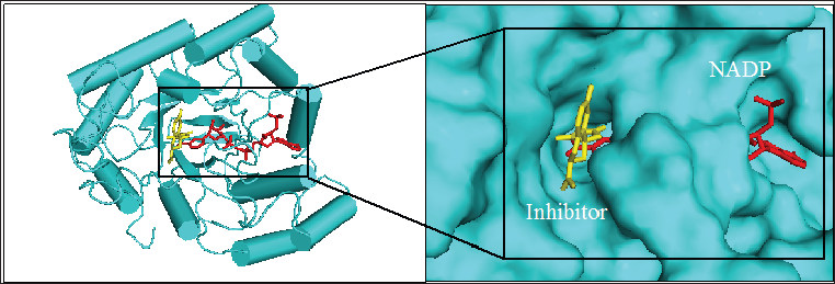
Figure 1: 3D structure of aldose reductase in cartoon and surface representation respectively showing the binding site for inhibitor fidarestat (yellow stick) and NADPH (red stick) at the active site

Lupinepublishers-openaccess-biomedicalengineering-biosciences

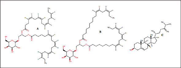
Figure 2: Chemical structure of the seaweed-derived compounds used as ligands in this study. (A)capsofulvesin A, (B) capsofulvesin B and (C) chalinasterol

Lupinepublishers-openaccess-biomedicalengineering-biosciences

Figure 1 shows the structure of the protein, aldose reductase used as a target in the current study. Aldose reductase is an NADP- dependent enzyme which catalyzes the conversion of glucose to sorbitol in the polyol pathway. This pathway is essential because hyperglycemia, an occasion where blood glucose level becomes excessively high, can trigger an increased activity of aldose reductase which can lead to the accumulation of intracellular sorbitol. This effect has been implicated in the pathogenesis of diabetic complication such as retinopathy, neuropathy and nephropathy. Therefore, inhibition of the catalytic function of aldose reductase has been proposed as a promising therapeutic target in the prevention and treatment of type II diabetes and its associated it's severe degenerative complications [33]. As shown in Figure 1, the enzyme active site is connected to the NADP binding pocket. This provides a chance for a ligand to potentially be a competitor of either the substrate of the coenzyme. Some natural and synthetic inhibitors of this protein have been sought over the years. Most of the reported inhibitors of aldose reductase preferably bind to the substrate binding location rather than competing with NADPH [34]. For more accurate understanding of the aldose reductase- ligand compex in the present study, two known inhibitors of the protein (fidarestat [35] and inhibitor-IDD384 [34]) were employed to compare the interaction pattern. The 2D chemical structure of the alga-derived ligands are given in Figure 2. Chalinasterol is a cholesterol nucleus-containing compound and it is structurally different from capsofulvesin A and capsofulvesin B which are galactolipids comprising of double aliphatic long chains with double bonds. These ligands are naturally-occurring compounds isolated from pharmacologically-important seaweed (C. fulvescens) [28]. It is essential to emphasize that these bioactive compounds have earlier been reported to inhibit aldose reductase in vitro [36]. However, the mechanism of the inhibitory effect still remains uncler.

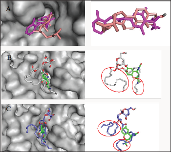
Figure 3: Analysis of the binding configuration of (A) chalinasterol, (B) capsofulvesin A and (C) capsofulvesin B. The binding pose for the compounds were compared against inhibitors of aldose reductase,fidarestat (green) and inhibitor- IDD384 (magenta). Areas with subtle differences in binding pose are identified in red spheres.

Lupinepublishers-openaccess-biomedicalengineering-biosciences

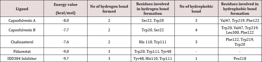
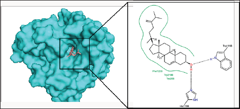
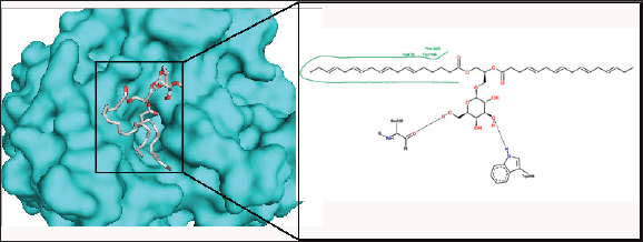
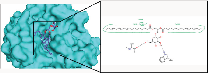
Chalinasterol shared a comparable binding pose with inhibitor- IDD384 (Figure 3). The molecular interaction as analyzed on proteinsplus server shows the contribution of both hydrophilic and hydrophobic bonds to the stability of the complex formed by the enzyme and C. fulvescens compounds (Figure 4-6). The bulky steroidal rings of chalinasterol was seen deeply buried within the fidarestat-binding pocket on aldose reductase where it is participated in hydrophobic bonds while the alkyl portion was exposed to the solvent-accessible region. Meanwhile, the inability of chalinasterol to completely block the active site may favor enzyme-substrate-chalinasterol complex formation as a mixed type of inhibition. As presented in Figure 4, two hydrogen bonds were established with amino acid residues His 110 and Trp111 respectively while Phe122, Trp219 and Trp20 participated in hydrophobic interaction with the ligand. The significance of hydrophobic bonds in the inhibition of aldose reductase cannot be overemphasized as it has been reported with many inhibitors of this protein including inhibitor IDD384.Inhibitory potential of chalinasterol against the carbohydrate-metabolizing enzyme was further proved by its binding affinity which is consistent with previous in vitro study that the compound possessed antidiabetic property [36,37]. The binding energy was relatively higher than that of the control ligands indicating a lower inhibitory activity for chalinasterol (Table 1). For capsofulvesin A and capsofulvesin B, the aromatic ring was placed at the solvent exposed region of the active site where it established hydrophilic interactions between its methoxyl moieties and the indole ring of Trp20 as well as the hydroxyl side chain of Ser22 (Figure 5). Possibly, the determining factor in the potency of these compounds as depicted in their IC50 is the binding configuration of the two long aliphatic chain. Capsofulvesin A has both of its side chain obstructing the entrance to the binding cavity while one of the aliphatic chains in capsofulvesin B was found moving away from the active site (Figure 6). This elongated side chain established a unique hydrophobic bond with residue Phe122. An efficient blockage of aldose reductase active site by capsofulvesin A may alter substrate access to the protein binding site, hence interfering with the enzymatic activity. Furthermore, the extra hydrophobic bond found with Phe122 in capsofulvesin B might have hindered the possible "swinging" of the aliphatic side chain from occupying the active site. It is also not impossible that these ligands interact with the enzyme-substrate complex in a mixed type of inhibition [36]. These mechanisms possibly underline the variations in the IC50 values previously obtained for these compounds. The binding energy of -7.9 kcal/mol estimated for capsofulvesin A compared to capsofulvesin B (-7.7 kcal/mol) suggests a higher affinity of the former to the protein. This result is in agreement with the previous in vitro report showing IC50 values of 52.53 |iM and 101.92 |iM for capsofulvesin A and capsofulvesin B respectively [36]. Although seaweeds have been investigated in diabetes and its complications, only scanty reports are available for C. fulvescens. Among the few studies, effect of extracts from the seaweed were observed for antidiabetic activity in vivo using streptozotocin-induced diabetic rats [37,38]. Therefore, this study provides insights into at least a part of the mechanisms responsible for the activity.

Table 1: Docking results showing binding energy and molecular interaction.

Lupinepublishers-openaccess-Biomedicalengineering-Biosciences

Figure 4: The binding pose and molecular the molecular interactions of chalinasterol with aldose reductase. Only key residues involved in hydrogen and hydrophobic interactions are shown as prepared on proteinsplus server.

Lupinepublishers-openaccess-biomedicalengineering-biosciences

Figure 5: The binding pose and molecular the molecular interactions of capsofulvesin A with aldose reductase. Only key residues involved in hydrogen and hydrophobic interactions are shown as prepared on proteinsplus server.

Lupinepublishers-openaccess-biomedicalengineering-biosciences

Figure 6: The binding pose and molecular the molecular interactions of capsofulvesin B with aldose reductase. Only key residues involved in hydrogen and hydrophobic interactions are shown as prepared on proteinsplus server.

Lupinepublishers-openaccess-biomedicalengineering-biosciences

Conclusion

This study investigated three antidiabetic compounds from C. fulvescens (a well-known green sea algae) against a diabetes- related target (aldose reductase) for their possible binding and molecular interaction pattern to provide insights into their mechanism of inhibition. With binding configurations similar to that of known inhibitors of aldose reductase, the results of the current study divulge the alga-derived compounds as suitable inhibitors of the protein. They bind at the active site and established both hydrophilic and hydrophobic bonds with some amino acid residues around site. This indicates their potential to interfere with the catalytic function of the protein. However, it was clear that the compounds did not interact with the NADP binding pocket. Hence, they are not competitive with NADPH for their inhibitory activity against aldose reductase. The subtle contrasts in the binding mode of these compounds might be responsible for the differences in their IC50 values as previously reported in in vitro experiments. Taken together, the results of this study verify aldose reductase inhibitory activity of the C. fulvescens constituents and validate the antidiabetic property of the seaweed.

Read More About Lupine Publishers Journal of Biomedical Engineering and Biosciences  Please Click on Below Link: https://biomedical-sciences-lupine-publishers.blogspot.com/

Monday, 13 June 2022

Lupine Publishers| Study About the Despeckling Methods for Retinal Optical Coherence Tomography Images

 Lupine Publishers| Journal of Biomedical Engineering and Biosciences


Abstract

This paper presents study about the different despeckling methods used for quality enhancement of retinal optical coherence tomography (OCT) images. Speckle noise is an inherent property of an OCT images which affects the visual quality of the images, hence difficult to diagnosis the patients. Therefore speckle noise reduction from the OCT images is an important prerequisite, whenever OCT imaging is used for diagnosis. The speckle noise intensity depends on the various imaging system parameters of its systems and the different structure representations used for the image tissues. A despeckling technique is to be designed in such a way that it should be able to reduce the speckle noise from the OCT images while preserving the tissues and fine details of the images.

Keywords: Despeckling; Optical coherence tomography; Speckle noise

Introduction

Optical Coherence Tomography (OCT) is a most powerful biomedical imaging technique used to extract the required information from an object. The working approach of this method is almost similar to ultrasound imaging except the medium used in obtaining its. OCT imaging uses light beams instead of sound [1]. OCT has proved its significant importance in the field of ophthalmology, especially in the detection of diseases related to retina and glaucoma [2-4]. Currently OCT is a best suitable method for imaging the internal structure of biological systems and also an essential part of a procedure to obtain high-resolution images of the retina [5-7]. It is also observe that retinal layer thickness of the OCT images improve the clinical finding in the field of ophthalmology Retinal layer segmentation improves the clinical finding in the case of glaucoma progression and macular degeneration. Therefore, the pre-processing of ophthalmic OCT images is a most importance step to improve the clinical diagnosis. Ophthalmic OCT imaging technique is based on detection of coherent waves; therefore these images are accompanied with a significant amount of speckle noise, which degrade the image quality and limit the contrast to noise and signal to noise ratio of the image.

To reduce the effect of speckle noise and to preserve the fine image features. A good speckle noise reduction algorithm is required that reduce the effect of speckle noise while preserving fine details of image. A lot of efforts have been made to overcome the effect of speckle noise. All methods proposed for speckle noise reduction from the OCT images are mainly divided into two categories namely transform domain techniques and spatial domain techniques. Organization of manuscript is as follows: section-2: describes brief about speckle noise; section-3: presents example of speckle noise reduction from the OCT image; section-4: discusses about different despeckling methods for OCT images and its summary is also presented in tabular form; section-5: presents summary and conclusion of the manuscript.

Speckle Noise

When a signal or an image is acquired by narrowband detection systems like SAR, ultrasound and OCT, then a pattern known as speckle effect the quality of these objects. The main reasons that can affect the speckle are optical properties of the system, status of the subject, size and coherence nature of source, multiple scattering, phase difference of the beam and aperture of the detector [8,9]. Further we can classify the speckle present in OCT images into two categories first one signal- carrying speckle and the second one is signal- degrading speckle .The mathematical distribution of the speckle can be model with a Rayleigh distribution. In this modelling, speckle noise assumed as a multiplicative behaviour, in contrast to additive nature of the noise. The mathematical model of Speckle noise can be represented as:

Y (m,n)= X (m,n)S (m, n) (1)

Where Y, X and S represent the noisy image data, original image data and speckle noise respectively. Normally, the multiplicative nature of speckle noise is converting into additive nature by logarithmic transformation of equation (1).

Example of Noise Reduction in OCT image

Figure 1: Speckle Noise Reduction from the Noisy Image.

Lupinepublishers-openaccess-biomedicalengineering-biosciences

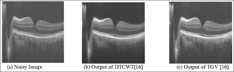
A noise reduction technique is required to improve the visual quality of the OCT image. All noise reduction techniques can be implemented into two ways, first one implemented during the acquisition time and second one implemented after acquisition of images. Normally, the noise reduction technique during the acquisition time is not good solution because these techniques require repetition of the same data which increase the acquisition time. Therefore, the use of post-processing techniques is more favourable solution to reduce the speckle noise from the OCT images. The example of speckle noise reduction from the noisy image is shown in the Figure 1. In this Figure 1(a) is the noisy image, 1(b) is the output image of DTCWT method [9] and 1(c) is the output image of second order total generalized variation (TGV) [9] method. From the Figure 1, it is clear that the effect of speckle noise reduction is very clear.

Despeckling methods

In the literature, a number of researchers proposed different post-processing methods to overcome the effect of for speckle noise from the OCT images. Few classic despeckling techniques that are used for SAR images, successfully used for OCT images also which are Lee [10], kuan [11] and frost filters [12]. An image restoration method is introduced that overcome the limitations of medical tomographic imaging system, by recovering the noiseless high frequency information corrupted by imaging system [13]. Especially for the multi-dimensional image data a better technique, based on compressive sensing principles is developed. This technique has capability to reduce the speckle noise while interpolate the missing data. This technique termed sparsity based simultaneous denoising and interpolation (SBSDI) [14]. A more appropriate approach based on dual-tree complex wavelet transform (DT-CWT) has been developed to overcome the effect of speckle noise from the OCT images. In this process adaptive-weighted bilateral filter (AWBF) is use to further enhance the OCT imaging by use of smoothing process [15].

For better image representation a new multiscale geometric analysis tool knows as wave atom transform was also proposed. The wave atom transform give better visual results in comparison to other transform such as wavelet and curvelet transform [16]. Image decomposition may also help in image denoising if the speckle noise in the OCT image is consider as texture or oscillatory patterns. A second order total generalized variation (TGV) decomposition model is helpful to remove the texture from the OCT image [16]. TGV is also removing the staircase side effect from the resulted images. Adaptive bilateral filter is also help to enhance the multi-frame OCT data [17]. The processed frames are averaged to convert into denoised output OCT image [17]. Variational image decomposition is also used to reduce the speckle noise from the ophthalmic OCT images [18].

The method converts the original image into cartoon part, texture part and speckle noise part to reduce the speckle noise from the image [18]. This method has the capability to suppress the speckle noise, while preserve image features such as edges and texture [18]. It may be useful for edge detection, segmentation and thickness calculation [18]. Independent Component analysis (ICA) techniques are also used in noise reduction of retinal OCT images [19]. The ICA technique can be beneficial when fewer number of B-scans images are available [19]. In, Raheleh kafieh et al. [20] proposed method for speckle denoising of OCT images based on dictionary learning approach with dual tree complex wavelet transform. In Zahra Amini [21], and Hossein Rabbani proposed method for speckle denoising of OCT images based Nonlinear transform based approach. To design the structural dictionaries, the information related to retinal layer is extracted automatically in the SSR method [22].

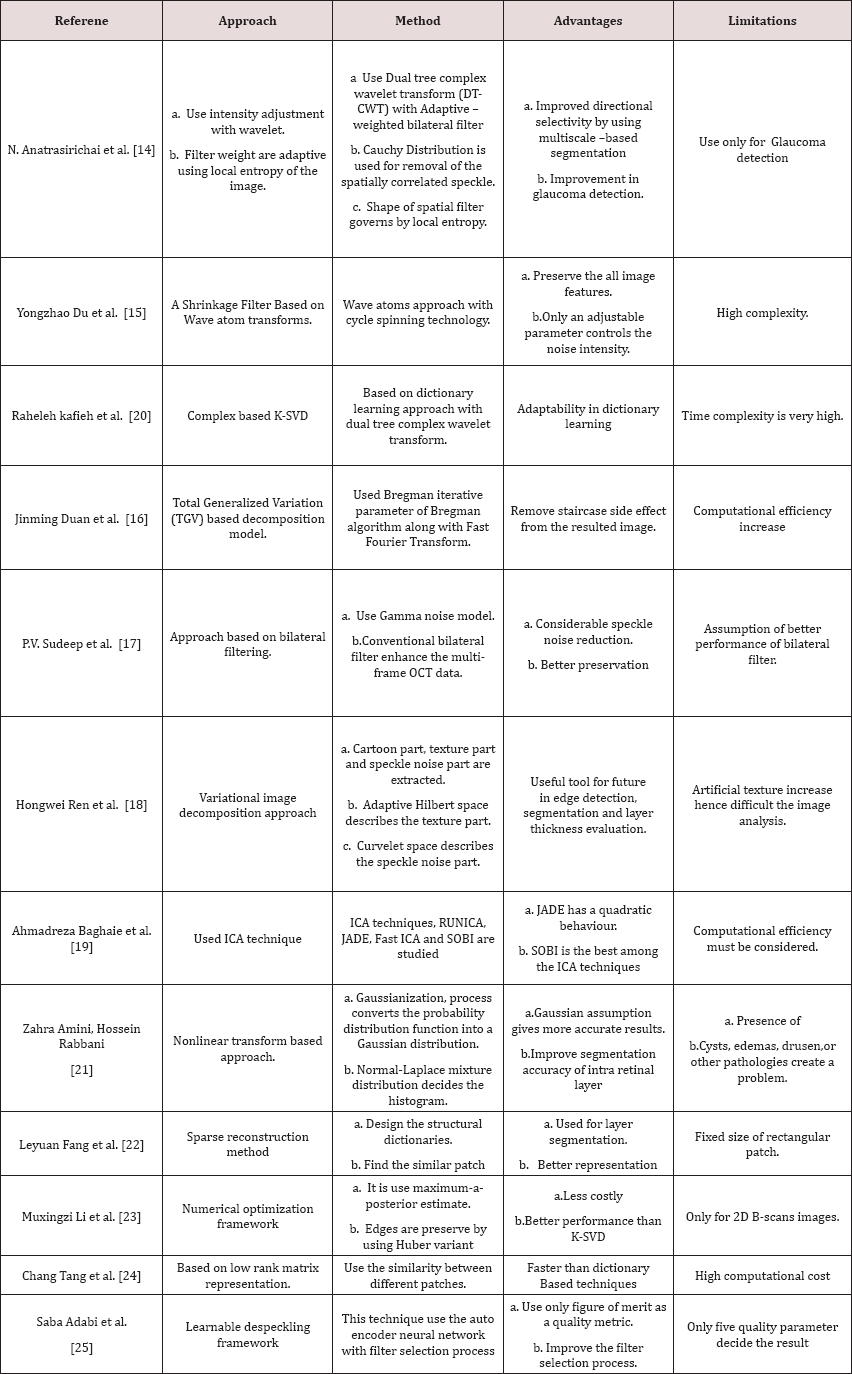
This method also uses the patch similarity index to enhance the performance [22]. Recently, optimization approach based on MAP estimate is proposed [23]. It use a suitable noise reduction approach along with Huber variant of TVR that overcome the effect of speckle noise, while preserve the edges as well [23]. To reduce the speckle noise, selection of a digital filter is a very difficult task. To overcome this limitation a framework based on learning approach known as learnable despeckling framework (LDF) has been proposed [24]. This approach only uses a single quality metric for selecting the appropriate filter for the task [25]. The different despeckling techniques are also studied and summarize into a Table 1.

Table 1: Summary of different despeckling methods for OCT Images.

Lupinepublishers-openaccess-Biomedicalengineering-Biosciences

Conclusion

This paper highlighted study about the different despeckling methods used for quality enhancement of retinal optical coherence tomography (OCT) images. The above study shows that among the different type of despeckling methods are used for OCT images. Study shows that an approach based on bilateral filter removes the significant amount of speckle noise while preserves the edges of the images. The Wavelet transform based approach is also a better solution for speckle noise reduction because of its multiscale nature. Out of different ICA techniques, SOBI is the one of the best techniques used for speckle noise reduction. Further work can be carried out in the variational image decomposition approach, because this approach is separate the speckle noise from the cartoon part and the texture part of the OCT images. Finally, learnable despeckling framework is also helpful for the selection of digital filter as per the requirement of the images features.

Read More About Lupine Publishers Journal of Biomedical Engineering and Biosciences  Please Click on Below Link: https://biomedical-sciences-lupine-publishers.blogspot.com/


Wednesday, 8 June 2022

Lupine Publishers| The Most Devastating of Life’s Disasters

 Lupine Publishers| Journal of Biomedical Engineering and Biosciences


Abstract

Spinal Cord Injuries are the most devastating of life's disasters. Three different case histories are presented. The effect these injuries have on our society from both an emotional and economic point of view is discussed. Examples are given of just how some patients and their families have adapted to their circumstances.

Keywords: Spinal Cord Injuries

Introduction

Robin was 18 years old at the time of his injury. He was working at the Rainbow Cleaners located in Annapolis, Maryland when one late afternoon, a customer walked into the store. He demanded all of the money in the store and Robin complied. Then the thief told Robin to turn around which he did, but then the thief shot Robin in the back of the neck causing an immediate paralysis below the neck. In just a few fateful minutes, Robin's life was turned upside down. After the thief escaped another customer entered the store and found Robin on the floor. The customer called 911 to obtain help from the Emergency Medical service and the police. The Emergency Medical service took Robin to the Anne Arundel General Hospital and I was called. Robin was examined and found to be tetraplegic with a gunshot wound to the back of the neck. X-rays obtained showed a bullet lodged right next to the C6-C7 vertebrate. Arrangements were made to transfer Robin to the operating room where he was placed on the operating table after being anesthetized. A posterior cervical surgical procedure was performed and the bullet removed from its adjacent position next to the vertebrate. The spinal cord was examined and the dura was opened more so as to examine the spinal cord itself. The spinal cord was bruised and was slightly hemorrhagic, but it was not transected. The dura was repaired and a good closure was obtained. The remainder of the wound was debrided and closed. Robin was treated with steroids, intravenous fluids, and antibiotics. Although initially tetraplegic, as each day progressed Robin seemed to acquire some partial movement in his extremities. Physical therapy was started and day-by-day Robin improved. He remained paralyzed below the C6-C7 level. As time went on, Robin was able to make adjustments with hand brakes in his automobile so that he could safely drive. He then went on to George Washington University where he commuted to class for four years and graduated. For many years he was able to work in the insurance and real estate fields and to support his family until eventually he was appointed as Director of the State of Maryland’s Criminal Victim’s Compensation Fund.

Although there is some evidence that the ancient Egyptians thought about treating patients with spinal cord injuries, there is more evidence that they felt that this condition was hopeless and should not be treated at all. It was probably not until the time of a Greek physician, Hippocrates, that a form of traction and immobilization was attempted [1]. No significant advances were made in the treatment of spinal cord injuries until 1846, when Ignaz Philipp Semmelweis began working at the General Hospital in Vienna, Austria and brought the infection issue to the forefront of medicine. Semmelweis observed that the mortality rate for women who were cared for by medical students was lower than the mortality rate for women who delivered babies, and who were cared for by midwifery pupils, who did not wash their hands and carried infectious material from patient to patient. In 1847, Semmelweiss made it mandatory that medical students and doctors washed their hands in between performing autopsies and delivering babies and prior to going to different patients. A few years before Semmelweiss wrote a book on this subject, Oliver Wendell Holmes (1809-94) gave a lecture in Boston suggesting that childhood fever was an infection caused by "germs". Many doctors doubted these ideas, but were finally convinced that pathogens did cause infection in 1879, by none other than Louis Pasteur [2]. Even so, in WWI, 90 per cent of the patients who had a spinal cord injury died within one year of the injury and only 1 per cent of these patients survived for 20 years [3]. Since that time, patients have been living longer with these injuries, but still severely incapacitated. There have been improvements in the general medical care of all patients with the development of antibiotics, respirators, and in physical therapy techniques.

Although there were many such injuries in WWII, one of the more publicized was that involving General George S. Patton, General Patton was riding as a passenger in the back seat of a Cadillac limousine. After waiting at a railroad junction for a long train to pass, suddenly an Army truck made a left turn right into the Cadillac not going even twenty miles an hour. But this accident threw General Patton into the steel frame of the partition causing a severe laceration of the scalp which resulted in a great deal of blood loss. More importantly, Patton told Major General Hobart R. Gay who was riding with him that he was having trouble breathing and wanted Gay to work his fingers for him. Shortly afterward, an army ambulance arrived with Captain Ned Snyder, a medical officer Dr. Snyder found General Patton motionless in the back seat of the car. Patton told the doctor that he thought he was paralyzed. Dr Snyder diagnosed that Patton had a broken neck and ordered him to be bundled up and transferred to the outskirts of Heidelberg to the new station hospital of the Seventh Army. Patton was conscious during the whole ordeal and was very quiet during the twenty five minute trip to the hospital operated by the 130th Station Hospital with Colonel Lawrence C. Ball as the commanding officer [4]. Lieutenant Colonel Paul S. Hill, Jr., chief of the surgical service, took over the management of this case immediately. Upon examining General Patton, Dr. Hill found him to have cyanotic lips and cold feet with his neck being flexed forward. Because Patton was in shock, Dr. Hill had him transfused with 1500 cc. of plasma and 300 cc .of whole blood type A from four GI donors. Color returned to Patton's face and his blood pressure returned to 110 over 76, whereas it had been only 86 over 80 upon arrival. Dr. Hill then proceeded to deal with the huge scalp wound which was a Y shaped laceration.

Then Dr. Hill went on to do a more detailed physical examination. There was no evidence of any long bone fractures. There was a flaccid paralysis of all the muscles of the thorax, abdomen, and the extremities. There was no sensation below the clavicles and he did not have any deep tendon reflexes. Patton was not able to move any of his extremities. A neurosurgeon, Captain William R. Duane, Jr, examined the patient and the x-rays. He diagnosed that the third and fourth cervical vertebrate were fractured and dislocated and that the spinal cord had been transected. Crutchfield tongs were placed in the skull which would hold pounds of traction in order to attempt to reduce the fracture dislocation. The urinary bladder needed to be emptied and an indwelling catheter was inserted. Penicillin was administered as well. Dr. Hill told the others that General Patton would not recover and would not live very long. Spinal cord injuries accounted for approximately twelve per cent of neurosurgical casualties in WWII, a number higher than anticipated. Initially, thee was no inclination to explore these injuries surgically and many neurosurgeons questioned the value of any surgical exploration. But there were others who felt that surgery might just help and that it would be beneficial to remove bone fragments from the spinal cord itself and to debride any dead tissue in order to decrease the chances of an infection. Eventually those who favored surgical exploration prevailed. During WWII, the Army Medical Corp adopted the policy of exploring wounds to the brain and to the spinal cord when there was reasonable doubt concerning the extent of the damage to the central nervous system. The author of this policy was Dr. R. Glen Spurling from Louisville, Kentucky who had given up his practice after Pearl Harbor was attacked and served as a neurosurgical consultant in Europe during the war. He worked with Dr. Barnes Woodhall who became Chief of Neurosurgery at Duke University, but by December, 1945, neither of them was in Europe.

The Surgeon General in Washington was able to contact the War Office in London to request that Brigadier Hugh Cairns, professor of Neurosurgery at Oxford University School of Medicine consult on this case. An Army plane was sent to London and on December 10, Brigadier Cairns flew to Heidelberg. Cairns then examined Patton and his findings confirmed earlier exams. Professor Cairns felt that the patient had an incomplete lesion at the fifth cervical vertebrate and that the third cervical vertebrate had also been fractured. Professor Cairns felt that there was reason to be hopeful when he detected that Patton had felt the tapping of the right patellar tendon and the pinching of the right thoracic skin. Since they were having difficulty keeping the Crutchfield tongs in place, Professor Cairns recommended that they use zygomatic hooks instead which are ordinary fish hooks fixed into the cheekbones which were uncomfortable since they now had ten pounds of weight on them. General Eisenhower arranged for Spurling to fly to Germany with Mrs. Patton. No one believed that Spurling could add anything else but they felt Mrs. Patton just might improve Patton's spirits. On December 12, Spurling examined Patton and found that he was paralyzed, but there was some voluntary motion of the right biceps muscle. He detected some deep sensation in his arms and legs. The deep tendon reflexes had returned and were hyperactive in both legs. He detected a definite sweating at the C 5 dermatome and the most recent x-rays showed a complete realignment of the cervical spine. Patton's general condition had stabilized as the temperature and the blood pressure were normal. Patton was able to breathe on his own. Fourteen hours later, Patton's condition deteriorated at which point Spurling became less optimistic.

Captain Duane found that Patton had improved. He detected some movements in the left quadriceps muscle and in the right abductor group of muscles. Patton was positioned in a 45 degree angle and was able to take some liquids by mouth such as soup. On December 19, he was able to eat an egg and drink a cup of coffee. On December 18, his vital signs appeared to be normal with a blood pressure of 108 over 70. Colonel Spurling cabled Dr. Michael DeBakey who was a 37 year old assistant professor of surgery at Tulane University serving as a colonel in the office of the Surgeon General about arrangements for Patton’s care in the United States. Later that night, Patton started coughing and bringing up bloody sputum. Colonel Spurling diagnosed an infarction of the lungs due to a pulmonary embolus. This is a piece of clotted blood that can lodge in the heart or in the brain and bring on instant death. By the morning of December 20, Patton was given Atropine and oxygen as well as saline and blood, but nothing helped him. General Patton died December 21, 1945 at 17:45. Dean was a teenager of 16 years when he dove into unknown waters near his home on the Magothy river near Annapolis, Maryland. He was rendered tetraplegic immediately when his neck was acutely flexed as he hit the bottom of the river. He was flown to the Shock-Trauma Center at the University of Maryland and received immediate treatment. In addition, he had an iliac bone graft to his posterior cervical spine area in order to fuse the vertebrate at C5-C6 where there was a fracture dislocation. Afterwards he received rehabilitation and physical therapy. He later married a nurse at the Anne Arundel Medical Center. I met this couple at the Naval Academy golf driving range where she was practicing golf drives and he was observing in a wheelchair which she managed to remove from their SUV.

It is estimated that 11,000 spinal cord injuries occur each year in the United States, and that approximately there are a quarter of a million Americans living with this catastrophic injury [5]. The records indicate that the average age at the time of injury has increased from 28 years to 38 years. More people over the age of 60 are having these injuries mainly due to auto accidents and falls. Only a small percentage of the victims are children. Based on reports from the NSCISC, the major causes of these injuries are due to automobile crashes, falls, violent acts, gunshot wounds, and recreational sports. Probably because of the preventive attention called to these problems by insurance companies and others, there has been a decline in the number of work related injuries resulting in spinal cord injuries and there has been an increase in the number of spinal cord injuries resulting from recreational sports such as football. The higher the level of injury to the spinal cord, the more severe will be the disability. For example, a C4-5 injury will result in the loss of movement in the arms and perhaps difficulty in breathing in some patients. In addition, the patients with lesions at this level will have no sensation below C4-5, no deep tendon reflexes, no superficial abdominal reflexes, no bowel or bladder control, and no movement in the lower extremities. A patient with a lower lesion at T12-L1 will retain strength and movement in his arms and will retain deep tendon reflexes in his arms. He will not have difficulty breathing and may very well have some superficial abdominal reflexes. He will not be able to move his legs and will not have bowel nor bladder control. Estimating the costs of these injuries to society is more difficult due to the decrease in the value of the American dollar and the increase in inflation. According to Berkowitz [6], the costs for the first year of care after such a devastating injury can be close to $2,500,000 and then the annual costs for the care of such a patient can be close to $4.500,000 with an additional indirect costs amounting close to another $2,500,000 annually.

How does the incidence of spinal cord injuries compare with the incidence of other tragic diseases? According to some reports, if there are a quarter of a million spinal cord cases in the United States requiring chronic care, there are about 400,000 cases of multiple sclerosis, 2.3 million cases of epilepsy, half a million cases of Parkinson's disease, 4.5 million cases of Alzheimer’s disease, and 5.4 million stroke patients [6-11]. Unfortunately, there is no way to reverse damage to the spinal cord. Intravenous methylprednisolone has been used as a treatment option for a spinal cord injury, but recent research has demonstrated the potential side effects such as blood clots and pneumonia. Hence steroids are no longer recommended after a spinal cord injury. Some experimental treatments are still under investigation such as hypothermia and stem cell therapy. In addition, rehabilitation involves the use of electrical stimulation devices to control arm and leg muscles. Robotic gait training and computer adaptations are also used in some medical centers. Finally, spinal cord injury treatment should also focus on preventing such an injury.

Read More About Lupine Publishers Journal of Biomedical Engineering and Biosciences  Please Click on Below Link: https://biomedical-sciences-lupine-publishers.blogspot.com/
Tuesday, 7 June 2022

Lupine Publishers| Novel Meta-Structures for Am Bioengineering Applications

 Lupine Publishers| Journal of Biomedical Engineering and Biosciences


Abstract

Additive Manufacturing (AM), also termed 3D-printing, has recently exploded-with rapidly evolving processes, systems, and new bio-compatible materials. At ABËMIS labs, we have been working on a novel AM enabling meta-structure that, to our knowledge, has not been previously described. Meshagons™, as we call them, are the first programmable, fractal-like meta-mesh structures that are fully manifold and multi-scale. Description and examples are presented.

Introduction

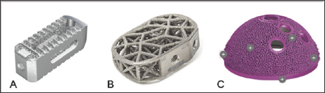
Additive Manufacturing (AM, 3Dprinting) technologies are recently enabling extraordinary new implant and prosthetic designs. Materials for AM have recently reached medical-grade (e.g. titanium, PEEK, hydroxyapatite) and are progressing [1]. Several companies and research/university organizations have demonstrated exciting AM implant products. A selected few are shown in Figure 1 [2-4]. We note that these, and to our knowledge, all others are lattice- based truss structures, generally having some kind of shell with a lattice or truss type infill to reduce weight, and encourage bonein growth. Although improved over a simple surface treated solid, lattices/trusses have topological and material limitations. Lattices, for example, cannot easily conform to complex bio-topological boundaries (complex shapes, holes, etc.) or multi-materials, or related boundary conditions and currently require significant manual intervention/design. To address these limitations, we present here Meshagons™*, a new type of procedurally-generated finite element (FE) based mesh-meta-structure.

Figure 1: 3D-printed spine and hip implants from (A) Stryker Inc., Tritanium series spine [2] (B) 4WEB Inc., LG series disc replacement [3], (C) NEXTSTEP Arthropedix Inc., Acetabular cup systems [4]. Images are credit to their respective websites (see refs.).

Lupinepublishers-openaccess-biomedicalengineering-biosciences

Methods

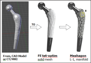
A Meshagon™ is a hyper-mesh. There are three main steps for generating a Meshagon (Figure 2). Briefly:

    a) Take any solid model, such as femoral implant (e.g. from CAD, CT/MRI, or any STL file).

    b) Convert using our FE meshing into a tetrahedral/hex/ hybrid mesh, optimized (proprietary).

    c) Transform the FE edge-node mesh into a manifold metamesh structure: Level-1 Meshagon.

We can repeat this process as many times as needed. For example, we can take one of the complex joints of the Level-1 Meshagon and re-apply the procedure (Figure 3). We call this a Level-2 Meshagon, and so on... A Meshagon of the size in Figure 1 (appx. 10K elements, 5M triangles) currently takes about 30 min to process/generate on an Intel i7 with dual Nvidia 1070 GPU processors. Improvements in computational speed are ongoing†.

Figure 2: Meshagon generation; 3 main steps (from left). Start with a CAD or CT/MRI model of astructure such as this prototype femoral implant ==> Generate FE solid tetrahedral or hybrid mesh, optimized for structure/loads/BCs and Meshagon generation ==> L-1 Meshagon is fully manifold†.

Lupinepublishers-openaccess-biomedicalengineering-biosciences

Figure 3: Close-up of internal joint (left, as indicated in Fig. 2) and L-2 Meshagon (right). Parameters for each Level can be independent and/or dependent, depending on the desired simple or complex functional model(s). For example, L-1 can be made more dense and L-2 less dense, etc. Any parameter can be added to the topology and the chosen underlying model(s)†.

Lupinepublishers-openaccess-biomedicalengineering-biosciences

Results/Examples

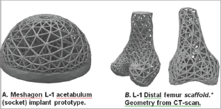
Figure 4: Two examples of Meshagon implant prototypes: (left) An acetabular cup Meshagon, note that this is just one density- all parameters can be changed and Meshagon updated accordingly. (right) Two views of a prototype distal knee scaffold; (credit: CT and segmentation from the SimTK Open Knee project [5]). This demonstrates capability for complex structured Meshagon scaffold generation (not as an implant, to be precise). This Meshagon meta-structure could be printed in combination of titanium and hydroxyapatite or other bio-compatible AM materials and related models [6-7].

Lupinepublishers-openaccess-biomedicalengineering-biosciences

The hip femoral component Level-1 and Level-2 prototypes (Figures 2 & 3) have good strength characteristics and ultralight weight (54+% reduced mass, assuming fused titanium or stainless steel material). Strut thickness was held here to 2.5 mm but of course can be resized, changed, or adapted to suit physical 3D-printer capabilities. The hydroxyapatite coating was simulated and not part of the FE optimization. The acetabular component prototype example (Figure 4a) was designed to be adjustable to topologies/factors important for bone in growth and mesh density for bone-implant integration [5-7]. There is only one central hole in this model (for fixation) but this can be easily modified, holes added, to suit. This early prototype supports 30x+ BW in simulation. The distal femur and chondyle model (Figure 4b) is manifold and fully printable, successfully demonstrating unique potential for Meshagon scaffolds that can be parametrically (programmed) as well as conforming to complex topologies.

Discussion

Meshagons and the related concepts presented here, along with rapidly advancing AM/3D printing technologies, have promise for generating unprecedented, multi-scale, ultra-light meta-structures. We have produced and printed a number of medical and implant related Meshagon structures as shown. A significant advantage of Meshagons is the ability to automatically (procedurally) compute multi- scale Meshagons that conform to complex biological shapes/topologies that until now were not possible to achieve (to our knowledge).

Limitations

We have not performed precision experimental testing on the printed implant structures (e.g. tension-compression, fatigue). The results and discussions here are based upon FE model simulations, and basic tests of actual printed structures. We have done simple compression tests of standard 'brick' Meshagons with positive confirmation of simulation results, but further work and collaboration is needed in this area. Another aspect that needs improvement is defining interpolation between L-levels. We are currently using a simple modified Boolean method, but is now working on improved fully implicit method(s) that will enable better control of (any) combining parameters.

Our recent work enables large scale meta-mesh parallel- calculations, now limited only by computer RAM and SSD storage (multi-scene). We can now generate Meshagons with appx 1M elements (est. 50M triangles) in appx 80 minutes for Level-1*. This can improve (work needed). Also, we can now control the thicknesses of individual struts and joints, the chamfer (or transitions), local and global density-porosity, optimize local stresses and vibrations using modal and explicit analyses. An interesting parallel between Meshagons and actual connective tissues is that they are both heirarchical structures. Tissues have multi-scale topologies, for example, Tendon → Fascicle→ Fiber bundle → Fibril → Tropocollagen. Meshagon meta-structures are also multi-scale, L1 → L2 → L3 → etc. Thus, Meshagons have a unique potential to improve description of actual multi-scale tissue geometries and related topologies/structure. Another concept we introduce here is the capability of Meshagon "multilevel porosity". The different levels L-1 and L-2 (Figure 3) each have different, porosities and other properties that can be programmed/ controlled. That means with Meshagons we can generate multiporosity structures that may better encourage bone-in growth and/or restorability properties, possibly leading to improved tissue engineered constructs and regeneration. Development is also ongoing for multi-scale, multi-material structures with enhanced optimized physical, electrical, and biochemical properties. New AM materials are now capable of directing and controlling electrical charge, which will open whole new frontiers for active 'smart' Meshagons and related structures. We are also extending to multimesh combinations, and further innovations in AM hyper metamaterials for medical implants and other areas-such as Aerospace, where Meshagon ultra- light, ultra-strength capabilities are also critical factors. Ultimately we believe that AM and Meshagon-type structures (and even the simpler lattice-type), will lead to dramatic improvements in medical implant, prosthetic, and system/device designs in the near future.

Read More About Lupine Publishers Journal of Biomedical Engineering and Biosciences  Please Click on Below Link: https://biomedical-sciences-lupine-publishers.blogspot.com/
Monday, 6 June 2022

Lupine Publishers| Cervical Cancer, Types of Diagnosis and Recent Treatment Methodologies

 Lupine Publishers| Journal of Biomedical Engineering and Biosciences


Abstract

Cervical cancer is one of the most common cancers among women in developing countries. Human papilloma virus (HPV) with sexually transmitted infection is the major causative agent and risk factor for developing cervical cancer. Women aged between "21-65 years” are the major risk group for cervical cancer. Increase in incidence and mortality rate due to cervical cancer, led to interventions for preventing and screening of this disease. Pap smear and HPV test are the two major screening techniques. According to National Cancer Institute, HPV vaccines can prevent cancerous and pre-cancerous cases infected by high risk HPV Recent advancement by targeting specific genes provides new scope in the treatment of cervical cancer

Keywords: Cervical cancer; HPV (human papilloma virus); Pap smear; HPV vaccines

Abbrevations: CDC: Center for Disease Control and Prevention, FIGO: International Federation of Gynecology and Obstetrics, AJCC: American Joint Committee on Cancer, HPV: Human papilloma Virus, CIN: Cervical Intraepithelial Neoplasia, DES: Diethylstilbestrol, LEEP: Loop Electrosurgical Excision Procedure.

Introduction

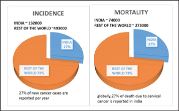
In case of women cervical cancer is the second most detected type of cancer [1]. Squamous cell cancer is the major type which accounts about 90% and 10 to 20% for Adenocarcinoma [2] According to CDC, there is a high rate of occurrence of cervical cancer in Hispanic women [3]. Based on global cancer statistics in 2012 it was found that 14.1 million new cases of cervical cancer are diagnosed and 8.2 million deaths per year occur in the world [4]. A study done by Dr. Surendra Srinivas Shastri reported that 30% of global mortality rate of cervical cancer is accounted by Indian Women [5](Figure 1).

Figure 1: ACCF/AHA Stages of HF.

Lupinepublishers-openaccess-biomedicalengineering-biosciences

Cervical Cancer Disease Rate

Women in between 35 to 55 years show more incidence to this type of cancer [6,7]. The main cause is human papilloma virus (HPV) which is accounted for 90% and other causes involves immune system deficiency, smoking , use of oral contraceptives ,exposure to diethylstilbestrol etc., [8]. HPV 16 and HPV 18 are the high risk HPV types which causes 70% of cervical cancer [1]. The major signs and symptoms includes: Abnormal bleeding, Pelvic pain, unusual vaginal discharge, and abnormal menstrual cycle [9]. In case of cervical cancer the main screening procedure is Pap smear [10]. This Pap test helps to diagnose the presence of precancerous and cancerous cells in the cervix [11]. For staging of cervical cancer mainly 2 systems are used, named as FIGO and AJCC. According to FIGO the cervical cancer is staged from 1 to 4 based on clinical examination. The AJCC classifies cervical cancer based on 3 factors:

    a. Extend of tumor (T)

    b. Lymph node spread (N)

    c. Distant spread (M)[12]

Discussion

Screening of Cervical Cancer

    i. Cervical screening detects cervical cancer and pre-cancerous changes in the cervix.

    ii. Cellular abnormalities are tested in laboratories by collecting cells from the surface of cervix.

    iii. This screening cannot be done for cancer of reproductive system and sexually transmitted diseases [13].

    iv. At the age of 70 two normal results within the past five years is recommended to stop screening.

    v. After first exposure of HPV, it develops within 3 to 5 years and leads to cervical cancer [14].

Screening tests are of two types

    a) Pap smear (cytological based screening): Pap smear detects cancerous cells, infection and inflammation like noncancerous conditions. Screening of sample shows the presence of abnormal cells.

    b) HPV testing: Detects high-risk HPV types in cervical cells and cell abnormalities. 13- 14 high risk HPV types can be identified through sample screening [15].

Types of Screening Results

Sometimes, the screening results of cervical cancer shows:

    A. False-positive result: normal cells are present, but the result may show abnormal cells.

    B. False-negative result: abnormal cells may be present but detects as normal.

    C. Douching, sexual intercourse, and using vaginal medications or hygiene products before 2 days of test should be avoided to prevent such results. During menstrual period cervical cancer screening should be avoided [15].

In.com, at the age of 21 screening can be done, but in some other countries screening begins at the age of 25. Screening below 21 year old girls are not recommended because of high risk of false positive result. [16].

Pap smear and HPV Tests

Named after Dr.Georgios Papanikolaou, who determined PAP test [17]. Pap test was first introduced in 1940's. In 1999, FDA approved screening methods to detect cancerous cells and first vaccine was developed in 2006 for the prevention of HPV [18]. Cervical cancer can be screened by Pap test for the detection of cancerous and precancerous cells in cervix by scrapping the cells and examines the abnormal cell growth. Collected cells from the cervix will be looked under the microscope to find cancer and precancer cell growth [19]. Pap smear can be done for all the women aged between 21- 65 years, whereas HPV is not advisable for women under the age of 29 years. If PAP and HPV are co tested, I can be done once in 5 years [15].

High risk types of HPV was found by HPV tests. Untreating the HPV types can lead to abnormal cell synthesis. In HPV test, cells from cervix are collected using small soft brush and send to the lab for further investigation or directly from pap sample HPV test sample can be taken. Worldwide about 70% of cervical cancer was caused by HPV16 and HPV18, high risk HPV types (genotype). HPV test can be used to detect presence of HPV, when Pap test is not clear [20].

Preparation for Pap and HPV Test:

    I. Schedule the date for test, by avoiding the menstruation days. If menstruation begins unexpectedly reschedule the date.

    II. 2 days before the test, sexual intercourse should be avoided.

    III. Avoid douche 2 days before test.

    IV. Avoid vaginal creams and medication inserted in vagina for 48 hrs before test [21].

Procedure

A speculum, which is a metal or plastic instrument used to keep the vagina open, will be placed inside the vagina to see the cervix properly. Samples of cells and mucus will be scraped from the exocervix using a small spatula. Through the opening of the cervix a small cotton swab or brush will be inserted to take a sample. For hysterectomy patients samples are taken from vaginal cuff (upper part of the vagina).

Prepared cell samples are examined under a microscope in the laboratory by:

    A) The sample are smeared on the slide, sprayed with preservative and send to the laboratory, known as conventional cytology

    B) Instead of directly putting the samples on the slide, they will add preservative liquids and send to the lab. This will be spread on to the slide known as liquid based Pap smear.

    C) Conventional cytology finds more abnormal cancer cells than liquid based Pap smear. Advantages of liquid based pap smear includes less chance of repeating pap test, and same sample can be for used HPV testing. The disadvantages of this test are, it also detects the non-cancerous cell changes along with cancerous cells and it is more expensive than conventional cytology [19]. The Pap test results show cell changes, usually known as cervical dysplasia [20].

Reporting of Results for Cervical Cancer:

i. In case of Pap test, the result interprets as normal or abnormal. Similarly HPV test can either be positive (infection with high risk HPV) or negative (no presence of high risk HPV type).

ii. In United States, Bethesda system is the type of standard term used for reporting the results.

iii. Based on Bethesda system, negative is reported for intraepithelial malignancies even if there are no abnormal cells.

iv. Pap test negative report may find non-cancerous cells, like inflammation and common infection [22] (Table 1).

Table 1: Difference between the Pap Smear, A Biopsy and an Hpv Test.

Lupinepublishers-openaccess-biomedicalengineering-biosciences

Who have to Screen for Cervical Cancer?

Recommendations say that all women between the ages of 21-65 should screen for Pap smear [23,24]. But guidelines recommend women with age 30 or above for starting of cervical cancer screening. It is mainly because of higher risk of cervical cancer in them. 30-49 years aged women should be screened prior to reduce the screening test in their lifetime. Screening risk of CIN2 determines the extent of disease to younger and older women [25].

The latest recommendations suggested by American Cancer Society for screening of cervical cancer are:

    A. All women should begin screening at the age of 21 regardless of their sexual activeness [26].

    B. Women between the ages of 21-29should have a Pap test every 3 years. They should not be tested for HPV until it is needed after an abnormal Pap test result.

    C. Women between the age of 30 and 65 should undergo Pap test and an HPV test every 5 years. This is the preferred approach, but it is also good to have a Pap test alone every 3 years.

    D. Women elder than 65, who have had regular screenings with normal results need not be screened again for cervical cancer But women who have been diagnosed with cervical pre-cancer should continue with the screening.

    E. Women who have taken the HPV vaccine should still follow the screening recommendations for their specific age group.

    F. Women who are at high risk are needed to be screened for cervical cancer more often. This high risk conditions may include those with exposure to the drug DES, HIV infection, or organ transplant [25,27].

Cervical cancer and pap smear: who is not screened and why?

Pap smear is the screening test for cervical cancer. Precancerous or cancerous tissues can be detected by this. With a proper diagnosis and treatment cervical cancer could be cured. Regular screening and early detection of this disease can improve the survival rates and reduce the mortality.

WHO guidelines for screening and treatment of preneoplasmic lesions for cervical cancer recommend a screening and treatment program for patients to achieve the goal [28]. But there are some reasons for which women are not screening for cervical cancer

    i. ACCS (advisory committee of cervical screening) in England recommends no screening test for women aged below 25 as it results in real harm to patient like miscarriage and pre term delivery and over treatment [29].

    ii. Harlan et al. compared Hispanic women with black and whites; most of the Hispanic women were unknown about the screening of cervical cancer than blacks or whites. It shows the compliance for screening which is related to race. In the same study they compared Hispanics speaking Spanish and those who are speaking English. It is evident that Spanish speaking women has less compliance than English speaking women because of their low knowledge about this.

    iii. Lucina et al. [29] studied the effect of acculturation (or foreign born) on cervical cancer screening in Mexican American women. It is described that the positive influence of Hispanics familism helps in increasing the effectiveness of cancer control interventions.

    iv. In the comparison study of women below and above poverty index in the past 3 years, the former are less likely to have a Pap smear screening. Low educated women (includes minorities and low socio economic status people) also showed a good rate of noncompliance to Pap smear. That means education as well as income influence the compliance to screen.

    v. Studies showing that, individuals with no usual problems in their health will not screen for cervical cancer.

    vi. Menopausal state of women also showed effect on screening compliance. Non-surgical menopause had less compliance than pre menopause or surgical menopause.

    vii. Women who have had their uterus and cervix removed by a hysterectomy and have no history of cervical cancer or precancer need not be screened [30-32].

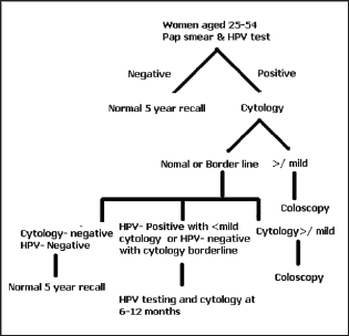
New Screening Algorithm for Cervical Cancer (Figure 2)

Figure 2: Treatment algorithm for cervical cancer.

Lupinepublishers-openaccess-biomedicalengineering-biosciences

Benefits of Pap smear

    a. Pap smear is a rapid test that can be done in few minutes [33].

    b. It can detect cervical dysplasia or cervical precancerous changes.

    c. Pap test along with HPV test can improve the detection of abnormalities in glandular cells such as cervical adenocarcinoma [34-39].

Limitations of Pap Smear:

    a. Pap test is not efficient to detect Squamous cell abnormalities and cancers.

    b. Chance to occur false negative results due to clustering of blood cells on the glass slide.

    c. There may be a chance to contagion the sample with other content of cervical specimen like blood, yeast and bacteria which leads to prevent the detection of abnormal cells.

    d. Long air contact of the sample may falcificate the cervical cells.

    e. By examining the sample, human error may come which affects the detection.

    f. Sometimes inadequate collection of sample will be there [34-39].

Complications of Pap smear

After doing Pap test, it's having some complications which are following

Pelvic Discomfort: Little sensation of pelvic discomfort or pain may experience when the doctor takes cervical cells. But, it will recover fast.

Cramping: Abdominal cramping may occur in a mild form. It's a temporary one. If it prolong, analgesics can be taken under the supervision of physician.

Vaginal Bleeding: Scraping cells from the cervix may cause discomfort to the reproductive part which leads to vaginal bleeding. It is in mild form and subside within one day [36].

Recent Advancement In Cervical Cancer

Targeted Therapy: Targeted Therapy inhibits the spreading and further growth of cancerous cells. These works by targeting specific genes or proteins which is found in cancerous cells [40]. It is used along with chemotherapy or radiation therapy [41]. Mainly there are 2 types of targeted therapy: Monoclonal antibodies and small molecule drugs.

Monoclonal antibodies: It blocks the signal activation which is necessary for cell growth, antibody dependent cellular cytotoxicity

Small molecule drugs: It inhibits cancerous cell multiplication and spreading. One of the examples include, Bevacizumab, it is an angiogenesis blocker and it also belongs to class of monoclonal antibodies which block VEGF-A to prevent angiogenesis. Pazopanib is a targeted drug therapy it act as VEGF and tyrosine kinase inhibitor [42].

Hyperthermia: It includes raising the temperature in the particular area where the tumor is present. It is also indicated along with radiation therapy which avoids the reoccurrence and increase in life span of patients [43].

HPV Vaccine: These are developed in order to prevent infections associated with HPV types [41,44]. Mainly three types of HPV vaccines which are named as Gardasil, Gardasil 9 and Cervarix. The major mechanism by them is to prevent infection due to HPV types 16 and 18. Among these, Gardasil and Gardasil 9 inhibit infection due to other high risk HPV types [45].

LEEP Surgery: In this, through a thin wire hook electrical current is passed, which remove the abnormal cervical tissue found during colposcopy.

Exenteration: This is done when cervical cancer spread to colon, vagina, uterus, rectum along with radiation therapy [44].

Drugs for pre-cancers: It includes conization and cryotherapy which are the standard treatment for CIN. In one study it is found that after administration of an anti-viral drug called Cidofovir, half of the women with CIN treated with this drug got resolved [41].

Adoptive cell Therapy: In this T cells from the patients are removed and in order to enhance the activity either by genetic modification or treated with chemicals. And this is reintroduced in order to improve the anti-cancer response.

Therapeutic vaccines: TVGV-1 vaccine: A phase 2 clinical trial is done for patient with pre cancer which is induced by HPV. VGX- 3100: A phase 2 clinical trial of this vaccine which mainly target HPV types 16 And 18 [46].

Conclusion

Incidence and mortality rate of cervical cancer can be reduced by screening techniques like Pap smear and HPV test. Cervical cancer can be prevented by early detection and by treating precancerous conditions. Review on articles based on cervical cancer and screening techniques gives more information about prevention screening of cervical cancer. New Advancement in the field of cervical cancer led a tremendous improvement in prevention.