Showing posts with label Biological and Biomedical materials open access Journals. Show all posts

Wednesday, 3 May 2023

Lupine Publishers| A Model for Metastasis for Hybrid Cancer Cells

Lupine Publishers| Journal of Biomedical Engineering and Biosciences


Abstract

Hybrid cancer cells have been recently discovered. They have greater ability to form metastasis. Here a simple mathematical model is given for this phenomenon. Some comments about the possibility of their reaching brain are given.

Hybrid Tumor Cells

Recently [1,2,3,4] hybrid tumor cells have been discovered. They have the following properties:

i. They circulate more than ordinary tumor cells.

ii. They have greater ability to migrate and invade other tumors.

iii. They have greater ability to form metastasis.

The Metastasis Model

Metastasis comprises a sequence of linked steps leading to the dissemination of cancer cells from a primary tumor to other distant tissues the overwhelming majority of cancer-related deaths still result from the progressive growth of metastasis that are resistant to conventional therapies [1,2].

Motivated by this the following model is presented for the metastasis of hybrid cancer cells:

Let T1, H1 be the ordinary and hybrid tumor cells respectively of the first tumor. Let N=T1+H1. The second tumor is assumed to contain ordinary tumor cells T2. Hence the model can be represented by

dH1/dt=a1H1-N-c2H1, dT1/dt=b1T1^(2/3)-N, dT2/dt= (b2-1)T2+c2H1    (1)

where a1,b1,b2,c2 are positive constants. The metastasis term is c2H1.

The reason for the power 2/3 is that ordinary tumor cells grow due to surface cells [3,4].

The equilibrium solution for the coexistence of both tumors is:

H1eq=T1eq/(a1-c2-1)

T1eq= [b1(a1-1-c2)/(a1-c2)]^3    (2)

T2eq=c2H1/(1-b2)

It is locally asymptotically stable if:

b2<1,

[1-(2/3)(a1-c2)/(a1-c2-1)][1+c2-a1]-1>0    (3)

[1-(2/3)(a1-c2)/(a1-c2-1)]+[1+c2-a1]>0

Since hybrid cells have a greater ability to invade other cells, it is expected that they will invade brain cells. Hence brain tumors can be a good source for identifying them. Moreover trying to attract them to less important sites can be a feasible strategy to deal with them. It may be difficult to test this idea experimentally, because the hybrid state, in general, is unstable [5].

Read More Lupine Publishers Journal of Biomedical Engineering and Biosciences Please Click on Below Link:
https://biomedical-sciences-lupine-publishers.blogspot.com/

Wednesday, 22 February 2023

Lupine Publishers| Electro Elastic Actuator for Micro and Nano Surgical Repairs

 Lupine Publishers| Journal of Biomedical Engineering and Biosciences


abstract

The structural scheme and the transfer functions, the characteristics of the electro elastic actuator for micro and nano surgical repairs are obtained. The transfer functions of the electro elastic actuator are described the characteristics of the actuator with regard to its physical parameters and external load.

Keywords: Electro elastic actuator; Piezo actuator; Structural scheme; Transfer function

Introduction

The electro elastic actuator on the piezoelectric, electrostriction effects is used in the mechatronics systems for the micro and nano surgical repairs, for the micro and nano robotics, for the micro and nano manipulators and injectors [1-6]. The mathematical model, the structural scheme and transfer functions of the electro elastic actuator are calculated for designing the control system for the micro and nano surgical repairs [4-11]. The structural scheme and transfer functions the electro elastic actuator based on the electro elasticity make it possible to describe the dynamic and static properties of the electro elastic actuator for the micro and nano surgical repairs with regard to its physical parameters and external load [12-23].

Structural Scheme Electro Elastic Actuator

The method of mathematical physics with Laplace transform is applied for the solution the wave equation. The structural scheme of the electro elastic actuator for the micro and nano surgical repairs is changed from Cady and Mason electrical equivalent circuits [7- 8]. The equation of the electro elasticity [6,8,12] has the following form

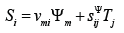
where Si is the relative displacement along axis i of the cross section of the piezo actuator, is the control parameter, Em, is the electric field strength for the voltage control along axis m, Dm is the electric induction for the current control along axis m, Tj is the mechanical stress along axis j, νmi is the electro elastic module, for example, the piezo module, ij sΨ is the elastic compliance for the control parameter Ψ = const , and the indexes i= 1, 2, … , 6; j = 1, 2, … , 6; m = 1, 2, 3. The main size along axis i for the electro elastic actuator is determined us the working length l = {δ , h,b} in form the thickness, the height or the width for the longitudinal, transverse or shift piezo effect.

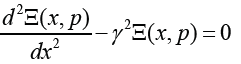
For the construction the structural scheme of the electro elastic actuator is used the wave equation [8,10,14] for the wave propagation in the long line with damping but without distortions. With using Laplace transform is obtained the linear ordinary second-order differential equation. The problem for the partial differential equation of hyperbolic type using the Laplace transform is reduced to the simpler problem [8,14] for the linear ordinary differential equation

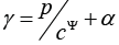
where Ξ(x, p) is the Laplace transform of the displacement of the section of the electro elastic actuator α Ψ = + is the propagation coefficient, cΨ is the sound speed for the control parameterΨ = const ,α is the damping coefficient.

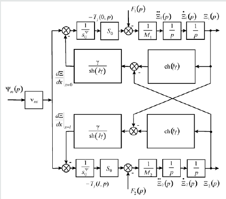
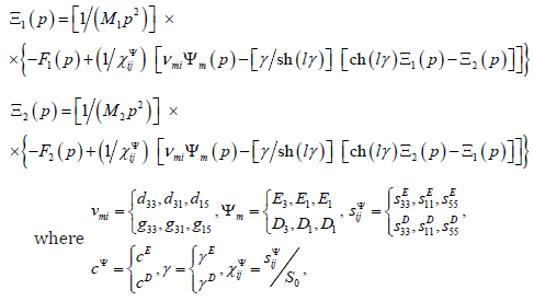
The mathematical model [6, 23] and the structural scheme of the electro elastic actuator for the micro and nano surgical repairs on Figure 1 are determined, using method of the mathematical physics for the solution of the wave equation, the boundary conditions and the equation of the electro elasticity, in the following form

Figure 1: Structural scheme of electro elastic actuator for micro and nano surgical repairs.

lupinepublishers-openaccess-journal-environmental-soil-sciences

vmi is the electro elastic module, is the control parameter, m E is the electric field strength for the voltage control along axis m, m D is the electric induction for the current control along axis m, sΨij is the elastic compliance, dmi is the piezo module at the voltage-controlled piezo actuator, gmi is the piezo module at the current-controlled piezo actuator, S0 is the cross section area, M1 , M2 are the mass of the load, are the Laplace transforms of the appropriate displacements and the forces on the faces 1, 2. For the micro and nano surgical repairs the structural schemes of the voltage-controlled or current-controlled piezo actuator are obtained from its mathematical model.

Transfer Function Electro Elastic Actuator

The matrix transfer function [6,18,21] of the electro elastic actuator for the micro and nano surgical repairs is derived from its mathematical model in the following form

where (Ξ( p)) is the column-matrix of the Laplace transforms of the displacements for the faces 1, 2 of the electro elastic actuator, (W( p)) is the matrix transfer function, (P( p)) the column-matrix of the Laplace transforms of the control parameter and the forces for the faces 1, 2.

Conclusions

The structural scheme, the transfer functions of the electro elastic actuator for the micro and nano surgical repairs, for the micro and nano robotics, for the micro and nano manipulators and injectors are described the characteristics of the electro elastic actuator with regard to its physical parameters, external load.

Read More Lupine Publishers Journal of Biomedical Engineering and Biosciences Please Click on Below Link:
https://biomedical-sciences-lupine-publishers.blogspot.com/

Wednesday, 11 January 2023

Lupine Publishers| Electro Elastic Actuator for Micro and Nano Surgical Repairs

 Lupine Publishers| Journal of  Biomedical Engineering and Biosciences


abstract

The structural scheme and the transfer functions, the characteristics of the electro elastic actuator for micro and nano surgical repairs are obtained. The transfer functions of the electro elastic actuator are described the characteristics of the actuator with regard to its physical parameters and external load.

Keywords: Electro elastic actuator; Piezo actuator; Structural scheme; Transfer function

Introduction

The electro elastic actuator on the piezoelectric, electrostriction effects is used in the mechatronics systems for the micro and nano surgical repairs, for the micro and nano robotics, for the micro and nano manipulators and injectors [1-6]. The mathematical model, the structural scheme and transfer functions of the electro elastic actuator are calculated for designing the control system for the micro and nano surgical repairs [4-11]. The structural scheme and transfer functions the electro elastic actuator based on the electro elasticity make it possible to describe the dynamic and static properties of the electro elastic actuator for the micro and nano surgical repairs with regard to its physical parameters and external load [12-23].

Structural Scheme Electro Elastic Actuator

The method of mathematical physics with Laplace transform is applied for the solution the wave equation. The structural scheme of the electro elastic actuator for the micro and nano surgical repairs is changed from Cady and Mason electrical equivalent circuits [7- 8]. The equation of the electro elasticity [6,8,12] has the following form

where Si is the relative displacement along axis i of the cross section of the piezo actuator, is the control parameter, Em, is the electric field strength for the voltage control along axis m, Dm is the electric induction for the current control along axis m, Tj is the mechanical stress along axis j, νmi is the electro elastic module, for example, the piezo module, ij sΨ is the elastic compliance for the control parameter Ψ = const , and the indexes i= 1, 2, … , 6; j = 1, 2, … , 6; m = 1, 2, 3. The main size along axis i for the electro elastic actuator is determined us the working length l = {δ , h,b} in form the thickness, the height or the width for the longitudinal, transverse or shift piezo effect.

For the construction the structural scheme of the electro elastic actuator is used the wave equation [8,10,14] for the wave propagation in the long line with damping but without distortions. With using Laplace transform is obtained the linear ordinary second-order differential equation. The problem for the partial differential equation of hyperbolic type using the Laplace transform is reduced to the simpler problem [8,14] for the linear ordinary differential equation

where Ξ(x, p) is the Laplace transform of the displacement of the section of the electro elastic actuator α Ψ = + is the propagation coefficient, cΨ is the sound speed for the control parameterΨ = const ,α is the damping coefficient.

The mathematical model [6, 23] and the structural scheme of the electro elastic actuator for the micro and nano surgical repairs on Figure 1 are determined, using method of the mathematical physics for the solution of the wave equation, the boundary conditions and the equation of the electro elasticity, in the following form

Figure 1: Structural scheme of electro elastic actuator for micro and nano surgical repairs.

lupinepublishers-openaccess-journal-environmental-soil-sciences

vmi is the electro elastic module, is the control parameter, m E is the electric field strength for the voltage control along axis m, m D is the electric induction for the current control along axis m, sΨij is the elastic compliance, dmi is the piezo module at the voltage-controlled piezo actuator, gmi is the piezo module at the current-controlled piezo actuator, S0 is the cross section area, M1 , M2 are the mass of the load, are the Laplace transforms of the appropriate displacements and the forces on the faces 1, 2. For the micro and nano surgical repairs the structural schemes of the voltage-controlled or current-controlled piezo actuator are obtained from its mathematical model.

Transfer Function Electro Elastic Actuator

The matrix transfer function [6,18,21] of the electro elastic actuator for the micro and nano surgical repairs is derived from its mathematical model in the following form

where (Ξ( p)) is the column-matrix of the Laplace transforms of the displacements for the faces 1, 2 of the electro elastic actuator, (W( p)) is the matrix transfer function, (P( p)) the column-matrix of the Laplace transforms of the control parameter and the forces for the faces 1, 2.

Conclusions

The structural scheme, the transfer functions of the electro elastic actuator for the micro and nano surgical repairs, for the micro and nano robotics, for the micro and nano manipulators and injectors are described the characteristics of the electro elastic actuator with regard to its physical parameters, external load.

Read More About  Lupine Publishers Journal of  Biomedical Engineering and Biosciences Please Click on Below Link: https://biomedical-sciences-lupine-publishers.blogspot.com/

Monday, 5 December 2022

Lupine Publishers| The Role of Biofield Energy Treated DMEM in Erectile Dysfunction using Detection of Cgmp Levels in Human Endothelial Cell Line

 Lupine Publishers| Journal of Biomedical Engineering and Biosciences


abstract

The study was performed to assess the impact of Biofield Treated test item (DMEM medium) on Human Endothelial Hybrid Cell Line (EA. hy926) for the expression of cyclic guanosine monophosphate (cGMP). The test item was divided into three parts. The first part was received one-time Consciousness Energy Treatment by a renowned Biofield Energy Healer, Mahendra Kumar Trivedi and labeled as BT-I, while second part received two-times Biofield Treatment and is denoted as BT-II. The third part did not receive any types of treatment and denoted as untreated DMEM. The level of intracellular cGMP in the BT-I and BT-II groups showed a significantly (p≤0.001) increased by 296.06% and 339.37%, respectively in Ea. hy926 cells compared to the untreated DMEM group. These results suggest that BT-II group showed a significant improved the level of cGMP with respect to the BT-I group. Therefore, the Biofield Energy Healing Treatment can be used to treat the erectile dysfunction patients along with other associated disorders such as orgasmic disorders, frotteuristic disorder, female sexual arousal disorder, vaginismus, fetishistic disorder, sex addiction, hypoactive sexual desire disorder, premature or delayed ejaculation.

Keywords: Biofield Energy; cGMP; Endothelial Hybrid Cell; PDE-5; DMEM; Erectile Dysfunction

Abbrevations:BT-I: One-time Biofield Energy Treated DMEM; BT-II: Two-times Biofield Energy Treated DMEM; CAM: Complementary and Alternative Medicine; NCCAM: National Center for Complementary and Alternative Medicine; DMEM: Dulbecco’s Modified Eagle’s Medium; cGMP: Cyclic guanosine 3′,5′-monophosphate; FBS: Fetal Bovine Serum

Introduction

Erectile dysfunction (ED) or impotence is the inability to get and keep an erection firm enough for sex. This is the most common sexual disorder in men across the globe [1]. Erectile dysfunction symptoms include persistent trouble in getting and keeping an erection and less sexual desire. Male sexual arousal is a complex process, which includes the coordination of the brain, hormones, emotions, nerves, muscles, and blood vessels [2]. ED might occur due to stress and severe mental health conditions. Besides, the literature data reported that physical cause of ED are heart disease, Parkinson’s disease, multiple sclerosis, atherosclerosis, high cholesterol, peronei’s disease high blood pressure, diabetes, obesity, sleep disorders, use of tobacco, alcoholism, injury of spinal cord and various metabolic syndromes such as high insulin levels, body fat around the waist, etc. [3]. Along with physical and psychological factors, impaired function of arteries and corpora cavernosa within the penis are the primary condition for impotence. While the lack of smooth muscle tone and imperfections in neuronal stimuli can lead to unsuccessful penile erection [4]. ED eventually leads to neuronal and cardiovascular disorders [5,6].

Nitric oxide synthase (NOS) enzymes are the major mechanism involved in ED, and it enhanced the production of cyclic guanosine monophosphate (cGMP), which results in smooth muscle relaxation and vasodilation via NO/cGMP pathway [7,8]. Inadequate level of NO/cGMP leads to ED. Thus, cGMP is the major therapeutically important target to overcome ED by inhibiting the cGMP-specific phosphodiesterase (PDE-5) enzyme [9]. However, sildenafil a PDE5 inhibitor has been used to treat ED, but it has life-threatening sideeffects viz. cardiac arrhythmia and hypotension [10] and vascular or neuronal deficiency like diabetes [11]. Thus, some alternative or complementary therapeutic approach is the best method to treat impotence without any side effects. In recent years, a remarkable outstanding alternative Complementary and Alternative Medicine (CAM) therapies approach known as Biofield Energy Healing Treatment (The Trivedi Effect®) have been scientifically reported in various fields. The human Biofield Energy is a weak electromagnetic field around of the human body. The Trivedi Effect® can be able to transform all the living organisms and non-living materials through a unique energy transmission process [12]

The effects of the CAM therapies have great potential, which include Johrei, Qi Gong, external qigong, Tai Chi, Reiki, therapeutic touch, deep breathing, yoga, polarity therapy, pranic healing, chiropractic/osteopathic manipulation, guided imagery, meditation, massage, homeopathy, hypnotherapy, special diets, progressive relaxation, acupressure, acupuncture, relaxation techniques, mindfulness, Rolfing structural integration, healing touch, movement therapy, pilates, Ayurvedic medicine, traditional Chinese herbs and medicines in biological systems both in vitro and in vivo [12]. Biofield Energy Healing as a CAM showed significant results in biological studies [13]. Also, the National Center for Complementary and Alternative Medicine (NCCAM), well-defined Biofield therapies in the subcategory of Energy Therapies [14]. The Trivedi Effect® has been reported to have created significant changes in the materials science [15-17], agricultural science [18,19], microbiology [20-22], biotechnology [23,24], improved bioavailability [25-27], skin health [27-29], nutraceuticals [30,31], cancer research [32,33], bone health [34-36], human health and wellness. Based on the outstanding benefits of The Trivedi Effect®, the present study was aimed to investigate the effect of Biofield Treated DMEM on the level of cGMP, in order to eradicate the ED using standard in vitro assay in Human Endothelial Hybrid Cell Line (EA. hy926).

Material and Methods

Requirement of Chemicals

Dulbecco’s Modified Eagle’s Medium (DMEM) and fetal bovine serum (FBS) were obtained from Life Technology, USA. Antibiotics solution (penicillin-streptomycin) was purchased from HiMedia, India, while ethylenediaminetetraacetic acid (EDTA) was purchased from Sigma, USA. Sildenafil citrate was purchased from Clearsynth, India. All the other chemicals used in this experiment were analytical grade procured from India.

Cell Culture

Human Endothelial Hybrid Cell Line (EA. hy926) was used as a test system in this experiment. The cells were maintained in DMEM growth medium for routine culture supplemented with 10% FBS. Growth conditions were maintained at 37°C, 5%CO2, and 95% humidity and subcultured by trypsinization followed by splitting the cell suspension into new flasks and supplementing with fresh cell growth medium. Three days before the start of the experiment, the growth medium of near-confluent cells was replaced with fresh phenol-free DMEM, supplemented with 10% charcoal-dextran stripped FBS (CD-FBS) and 1% penicillin-streptomycin [37].

Study Design

The experimental groups consisted of group 1 (G-I) with serumfree DMEM defined as the untreated DMEM. Group 2 (G-II) consisted of positive control (sildenafil citrate) at different concentrations. Further, group 3 (G-III) included DMEM medium (test item group) with the one-time Biofield Energy Treatment and denoted as BT-I, while the group 4 (G-IV) included the test item with the two-times Biofield Energy Treatment and indicated as the BT-II.

Biofield Energy Healing Treatment Strategies

The test item, DMEM was divided into three parts. One part of the test item was treated with the one-time Biofield Energy Healing Treatment by a renowned Biofield Energy Healer (The Trivedi Effect®) and coded as the Biofield Energy Treated DMEM (BT-I), while the second part was received the two-times Biofield Energy Healing Treatment and denoted as the BT-II. Further, the third part did not receive any treatment and defined as the untreated DMEM group. This Biofield Energy Healing Treatment was provided by a renowned Biofield Energy Healer, Mahendra Kumar Trivedi, remotely for~3 minutes. The Biofield Energy Healer was located in the USA, while the test item was located in the research laboratory of Dabur Research Foundation, New Delhi, India. This Biofield Energy Treatment was administered for ~3 minutes through the Healer’s unique Energy Transmission process remotely to the test items under the standard laboratory conditions. Mahendra Kumar Trivedi never visited the laboratory in person, nor had any contact with the test item (DMEM medium). Further, the untreated DMEM group was treated with a “sham” healer for comparative purposes. The “sham” healer did not have any knowledge about the Biofield Energy Treatment. After that, the Biofield Energy Treated and untreated samples were kept in similar sealed conditions for experimental study.

Assessment of PDE-5 Enzyme Inhibition

The cells were counted using an hemocytometer and were seeded at a density of 0.4 X 10⁶ cells/well in DMEM with 10 % FBS in 6-well plates. The details test procedure was followed as per Branton et al. 2018 [38-40]. Increase in cGMP level was determined as the following equation (1)

% Increase in intracellular cGMP level = {(B-A)/A} x 100----- -- (1)

Where, B = OD of cells treated with test item and A is the OD of untreated wells (media treated).

Statistical Analysis

Values were expressed as Mean ± SEM of three independent experiments. For multiple group comparison, one-way analysis of variance (ANOVA) was used followed by post-hoc analysis by Dunnett’s test. Statistically significant values were set at the level of p≤0.05.

Result and Discussion

Detection of PDE-5 Enzyme Inhibition

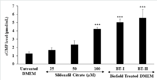
The result of the intracellular cGMP level in Ea. hy926 cells is shown in Figure 1. Sildenafil citrate, used as positive control at 25 μM, 50 μM, and 100 μM exhibited a significant increase in the intracellular cGMP in Ea. hy926 cells by 34%, 84%, and 234%, respectively compared to the untreated DMEM group. The onetime Biofield Energy Treated DMEM (BT-I) group showed 5.03 pmol/mL, while two-times Biofield Energy Treated DMEM (BTII) group showed 5.58 pmol/mL level of cGMP. Thus, BT-I group showed a significant (p≤0.001) increase in intracellular cGMP level by 296.06% in Ea. hy926 cells with respect to untreated DMEM. Similarly, the BT-II group showed a significant increase in the intracellular cGMP levels by 339.37% in Ea.hy926 cells than untreated DMEM. Thus, the data suggest that the two-times Biofield Energy Healing Treatment (BT-II) showed better results with respect to the increased cGMP level as compared with the one-time Biofield Treated DMEM group, which meant that the Biofield Energy Healing Treatment inhibited PDE-5 enzyme; resulting to the higher level of cGMP which can help to treat the erectile dysfunction (ED).

Figure 1: Effect of the test items (untreated and Biofield Treated DMEM) on the expression of intracellular cyclic guanosine monophosphate (cGMP) in human endothelial hybrid (Ea. hy916) cells. BT-I: One-time Biofield Energy Treated DMEM; BT-II: Two-times Biofield Energy Treated DMEM. ***p≤0.001 vs. untreated DMEM group.
lupinepublishers-openaccess-journal-environmental-soil-sciences

The drugs, which are available in the market for the treatment of ED, are having serious side-effects along with short time treatment of ED [41]. Thus, ED can be treated with some alternative mode of treatment without having any type of adverse effects. Biofield Energy Healing Treatment is one of the best CAM approach worldwide to treat various clinical disorders along with a significant change in different scientific fields. The results are outstanding and can be comparable with the marketed synthetic drug, sildenafil citrate. Scientific literature results showed that relaxation in smooth muscles results in improved level of cGMP production leading to penile erection [42]. Increase in cGMP results in decreased level of intracellular calcium, which supports penile erection [43]. However, cGMP activation is regulated by the PDE-5 enzyme, which is abundant in the corpus cavernosum and results in improved blood circulation that leads to penile erection [44]. Biofield Energy Healing based DMEM and Biofield Energy Healing Treatment might work by the relaxation of penile smooth muscles, which could lead to penile erection.

Conclusion

Erectile dysfunction results an unsatisfactory sex life, mental stress or anxiety, embarrassment or low self-esteem, relationship problems, inability to get your partner pregnant, which can produce lot of pathological implications like hypertension, hypercholesterolemia, diabetes mellitus, cardiovascular disease, and depression. Thus, the Biofield Energy Healing Therapy is one of the best approach to treat various sexual disorders and its related diseases. The present study results showed that the Biofield Energy Treated DMEM significantly increased the level of intracellular cGMP in Ea. hy926 cells compared with the untreated DMEM group. PDE- 5 is the predominant phosphodiesterase, while the intracellular cGMP level was significantly (p≤0.001) increased by 296.06% in the one-time Biofield Energy Treated DMEM group (BT-I) in Ea. hy926 cells compared to the untreated DMEM group. Additionally, the BT-II group i.e., the two-times blessed in DMEM also showed a significantly (p≤0.001) increased the level of cGMP by 339.37% compared with the untreated DMEM group. Henceforth, it can be concluded that the Biofield Energy Treated (The Trivedi Effect®) DMEM were found to have a significant impact on cGMP level, which might significantly inhibit the PDE-5 enzyme that leads to the penile erection. Thus, the Biofield Therapy can be used for the treatment of numerous sexual disorders viz. hypoactive sexual desire disorder, fetishistic disorder, dyspareunia, frotteuristic disorder, vaginismus, exhibitionistic disorder, voyeuristic disorder, sex addiction, premature or delayed ejaculation (or sexual malfunction or sexual disorder) improve normal sexual activity, desire, including physical pleasure, arousal or orgasm, preference, and neurological disorders, hormonal imbalances, sexual performance, desire disorders (lack of sexual desire or interest in sex), marital or relationship problems, effects of a past sexual trauma, feelings of guilt, depression, and pain disorders (pain during intercourse).

Read More Lupine Publishers Journal of Biomedical Engineering and Biosciences Please Click on Below Link:
https://biomedical-sciences-lupine-publishers.blogspot.com/

Monday, 17 October 2022

Lupine Publishers| Thermodynamic, HOMO-LUMO, MEP and ADMET Studies of Metronidazole and its Modified Derivatives Based on DFT

 Lupine Publishers| Journal of Biomedical Engineering and Biosciences


Abstract

In this study, Metronidazole (Met) and it’s modified derivatives are optimized by employing density functional theory with B3LYP/6-31g (d,p) level theory to explore their structural and thermodynamical properties. Molecular electrostatic potential (MEP) calculation has performed to calculate their possible electrophilic and nucleophilic attack. ADMET prediction was performed to search the absorption, metabolism and toxic level. Finally, this study can be helpful to design a potent candidate.

Keywords: Metronidazole; Density functional theory; HOMO-LUMO; MEP; ADMET

Abbrevations:Met: Metronidazole; DFT: Density functional theory; HOMO: Highest occupied molecular orbital; LUMO: Lowest unoccupied molecular orbital; MEP: Molecular electrostatic potential; ADMET: Absorption, distribution, metabolism, excretion, and toxicity

Introduction

Metronidazole (Met) is an antibiotic [1] and antiprotozoal drug [2]. It is widely used in the treatment of amoebiasis, trichomoniasis and giardiasis [3,4]. It has some demerits depending on the type and nature of unusual physical condition and on the limit of dose. High and long term dose can cause of leucopenia, neutropenia and peripheral neuropathy diseases [5]. Adverse effect and resistance of drugs indicate the importance of the discovery of new potential candidate. In computer aided drug design system, physicochemical, molecular docking, nonbonding interactions, and ADMET predictions are important criteria to evaluate newly designed molecules [6]. Drug modification is another alternative way to search better agent, which can increase the selective action of drug and reduce the side effect. Recently, it has been seen the trait of modifying drugs using halogens and alkyl group play important role in improving drug performance [7].

Herein, I report the optimization of Metronidazole (Met) and its modified derivatives to investigate their biochemical behavior on the basis of quantum mechanical approach. The free energy, electronic energy, enthalpy, dipole moment, HOMO-LUMO gap, hardness, softness, chemical potential and electrostatic potential have been calculated. All the newly designed derivatives show better thermodynamic properties, and some of them exhibit better chemical reactivity than parent drug. From the regarding quantum chemical studies, it’s assuming that, some of the designed compounds may have profound effect as drug.

Methods and Materials

Computational Details

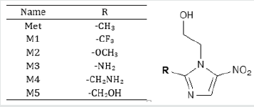
In computer aided drug design, quantum mechanical methods are widely used to predict thermal, molecular orbital, and molecular electrostatic potential properties [8]. Initial geometry of Metronidazole (Met) was taken from the online structure database named ChemSpider [9]. Geometry optimization and further modification of all structures carried out using Gaussian 09 program [10]. Density functional theory (DFT) with Becke’s (B) [11] threeparameter hybrid model, Lee, Yang and Parr’s (LYP) correlation functional [12] under Pople’s 6-31g (d,p) basis set has been employed to optimize and elucidate their thermal and molecular orbital properties [13]. Initial optimization of all compounds was performed in the gas phase. Dipole moment, electronic energy, enthalpy, free energy and electrostatic potential are calculated for all the compounds (Figure 1).

Figure 1: Chemical structure of Metronidazole (Met) and its modified analogues.

lupinepublishers-openaccess-journal-environmental-soil-sciences

Frontier molecular orbital features HOMO (highest occupied molecular orbital), LUMO (lowest unoccupied molecular orbital) were calculated at the same level of theory. For each of the drugs, HOMO-LUMO energy gap, hardness (η), softness (S) and chemical potential were calculated from the energies of frontier HOMO and LUMO as reported considering Parr and Pearson interpretation [14,15] of DFT and Koopmans theorem [16] on the correlation of ionization potential and electron affinities with HOMO and LUMO energy (𝜀). The following equations are used to calculate hardness (η), softness (S) and chemical potential (μ);

In computer aided drug discovery system, computational predictions are using to explore absorption, distribution, metabolism, excretion, and toxicity (ADMET) which saves on time and investment. AdmetSAR online database was utilized to predict ADMET properties of Metronidazole and its analogues [17].

Result and Discussion

Thermodynamic Analysis

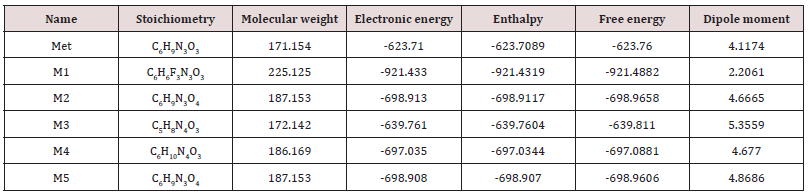
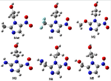
Simple modifications of molecular structure significantly influence the structural properties including thermal and molecular orbital parameters. From the free energy, and enthalpy values, spontaneity of a reaction and stability of a product can be predicted [18]. In drug design, hydrogen bond formation and nonbonded interactions also influenced by dipole moment. Increased dipole moment can improve the binding property [19]. From thermodynamic data (Table 1), the free energy of Metronidazole is -623.7600 Hartree, where M1 shows the highest negative value (-921.4882 Hartree). The –F substitution (M1) influence the free energy significantly. Highly negative free energy is favourable for stable configuration. Again, the dipole moment of Metronidazole is 4.1174 Debye where M3 shows the maximum dipole moment (5.3559 Debye) due to substitution of –NH2 group (Figure 2).

Table 1: The stoichiometry, molecular weight, electronic energy, enthalpy, free energy in Hartree and dipole moment (Debye) of Metronidazole (Met) and its analogues.

lupinepublishers-openaccess-journal-environmental-soil-sciences

Figure 2: Most stable optimized structures of Metronidazole (Met) and newly designed analogues. Optimized with B3LYP/6- 31g (d, p) level theory.

lupinepublishers-openaccess-journal-environmental-soil-sciences

Molecular Orbital Properties

Table 2: Energy (eV) of HOMO, LUMO, gap, hardness, softness and chemical potential of the designed drugs.

lupinepublishers-openaccess-journal-environmental-soil-sciences

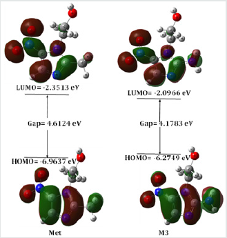
The HOMO-LUMO energies, hardness, softness, chemical potential of all compounds is presented in Table 2. The electronic absorption relates to the transition from the ground state to the first excited state and mainly described by one electron excitation from HOMO to LUMO [20]. The chemical hardness, softness, and potential values depend on the energy gap of HOMO-LUMO [21,22]. Kinetic stability decreases with the decrease of HOMO-LUMO gap. As a result, removal of electrons from ground state HOMO to excited state LUMO requires less energy. In our studies, Metronidazole shows the HOMO-LUMO gap 4.6124 eV, where M3 have the lowest energy gap (4.1783 eV) with highest softness (0.4787 eV) which may contribute higher chemical reactivity (Figure 3).

Figure 3: Frontier molecular orbital (HOMO-LUMO) and related energy of Metronidazole (Met) and M3.

lupinepublishers-openaccess-journal-environmental-soil-sciences

Molecular Electrostatic Potential Analysis

Molecular electrostatic potential (MEP) was calculated at B3LYP/6-31G (d,p) level of theory to forecast the reactive sites for electrophilic and nucleophilic attack of all optimized structures [23]. Red color represents maximum negative area which favorable site for electrophilic attack, blue color indicates the maximum positive area which favorable site for nucleophilic attack and green color represent zero potential area. MEP displays molecular size, shape as well as positive, negative and neutral electrostatic potential regions simultaneously in terms of color grading. It is seen from MEP map, region having the negative potential are over electronegative atom (oxygen atoms) and having positive potential are over hydrogen atoms. Here, the maximum negative potentiality is found for M3 is -0.3497 a.u (deepest red) for oxygen atoms and the highest positive potentiality of M1 is +0.3903 a.u (deepest blue) of hydrogen atoms.

Read More About Lupine Publishers Journal of Biomedical Engineering and Biosciences Please Click on Below Link: https://biomedical-sciences-lupine-publishers.blogspot.com/

Tuesday, 14 June 2022

Lupine Publishers| Employing Blockchain Technology to Understand Personalized Medicine

 Lupine Publishers| Journal of Biomedical Engineering and Biosciences


Abstract

Blockchain technology is yet another method in helping in the understanding and employment of personalized medicine. This technology can complement the use of mathematical models for the amalgamation of complex data from genetics, sequencing, mutations, cellular reactions, interactomes, medications, medication dosing and scheduling, and drug reactions. Additional uses of blockchain technology are described.

Keywords: Blockchain Technology; Personalized Medicine; Computational Mathematics; Genetics; Drug Delivery Systems; Interactomes

Introduction

In order to interpret the vast amount of information available on sequencing the genetic composition of patients, the data on analyzing diseases, genetic mutations, cellular reactions with genes, interactomes, medications, drug reactions and toxicities, doses of drugs and therapeutic schedules, and epidemiology problems, it is necessary to have secure methods with which to work. Reference is made to an earlier discussion on the use of computational mathematics and mathematical models for understanding personalized medicine. Kushner [1]. This article seeks to explain the benefits of yet another method available to decipher the vast amount of data that is being generated. Recently there has been a tremendous amount of discussion and publicity about crypto currency and blockchain technology among public and financial institutions. Most of this has been centered on financial transfers using Bit coin and other crypto currency avenues.

And while this has been in the limelight, there has been a quieter and more subtle revolution with the application of blockchain technology in the medical world. Blockchain creates a chain of transaction blocks to store information. One reason why there is such an attraction to blockchain technology is that this technique enables data exchange to occur in a secure and irrevocable world. Blockchain technology can deal with medical claim adjudication and billing management, as well as controlling the supply of drugs and aiding in the avoidance of counterfeit medicines. Perhaps one of the most obvious applications of this technology would be in the field of management of health care paper records and the electronic health care record. This technology permits the sharing of patient information by increasing the speed and availability of research data.

In order to make good decisions in medical research and personalized medicine, the employment of blockchain technology can aid in determining treatment protocols and medical choices. In fact, clinical trials with various medications will be suspect if there is missing data, endpoint switching, or data dredging. Polacheck [2]. Now health care professionals are interested in blockchain technology because the extended functionality of the blockchain and the emerging application program interface (API) services that enable several collaborative service processes. A transaction block is replicated across a collection of computers connected as a peer-to-peer network that constitutes a blockchain. Each of these computers is referred to as a node. Advanced cryptography allows for the nodes to interact anonymously and securely on the network. Culver [3]. Boderson C et al. [4] notes that another definition would be: 'A distributed tamperproof database that secures all records that are added to it, wherever they exist. Each record contains a timestamp and secure links to the previous record" Boderson C et al. [4].

A block of one or more new transactions is collected into the transaction data. A series of hashes connect via the header, and with the header referring to the previous hashed copy thus forming the ability to chain together transactions. Copies of each transaction are hashed, and the hashes are paired, hashed, paired again, and hashed again until a single hash remains Stagnaro [5]. Broder et al. [6] writes "Blockchains are cryptographic protocols that allow a network of computers (nodes) collectively to maintain shared ledger of information without the need for complete trust between the nodes. Each blockchain database is a time-sequenced chain of events that have been authenticated using a consensus mechanism specified by the protocol. The mechanism guarantees that, as long as the majority of the network validates the blocks posted to the ledger as per the stated governance rules, information stored on the blockchain can be trusted as reliable. The effect of the distributed consensus mechanisms often means that all of the nodes of the network hold all the information stored on the blockchain" Broder et al. [6].

Blockchain technology can be used in five data-driven areas:

    a. Longitudinal health care records

    b. Automated health care claims adjudication

    c. Interoperability

    d. Online patient access

    e. Supply chain management.

This technology has an extended functionality, which enables collaborative and operational services that deal with data-sharing. Das [5,7]. Longitudinal health care records use blockchain to link various healthcare provider organizations. For example, Weiss gives an example in which a patient has an acute episode and is attended by an emergency medical technician (EMT), who swipes the patient's interactive wristband containing his healthcare blockchain ID number. This information is broadcast to the patient's primary care doctor and to the hospital. Using a blockchain encryption security key to obtain messaging, they can access the patient’s s updated blockchain health care account Weiss M [8].

A second use of blockchain in healthcare concentrates on automated health claim adjudication which ensures correct completion of the claims and supports compliance audits using business rules. This process allows payers to send remittances to providers and allows patients to process payments using their bank or health savings accounts Miliard M [9]. The third use of blockchain for this discussion involves the gathering of massive amounts of patient data. This technique supports HIPAA’S Protected Health Information federal laws and regulations. This use of blockchain for personalized medicine complements the earlier article on the employment of computational mathematics and mathematical models to deal with large amounts of important clinical and research medical data Kushner et al. [1].

The fourth use of blockchain technology in healthcare allows for a patient's access to his healthcare records. The patient is provided with a security key that matches his provider’s key. Eckblaw has proposed a variation of the technique, which is used by a company named "MedRec". This company uses Ethereum, a clinical and research blockchain, for their medical records, and believes that this provides improved data quality for medical research. Eckblaw A et al. [10]. And finally, blockchain technology can be used for supply chain management by providing real time contract tracking, execution, and the ability to determine if a satisfactory completion of the contracts has been obtained. Thus information will be available concerning the ingredients, quality, and source of drugs being purchased. This could aid in the fight against counterfeit drugs being sold worldwide.

The quest for managing big data is not limited to health care, but is a problem for many organizations and corporations. Jennifer Bresnick has proposed the creation of an ecosystem that stresses accuracy, timeliness, and shared-decision making Bresnick J [11]. Personalized medicine would benefit by the use of blockchain technology in integrating all of the patient's health data by using tools such as the American College of Surgeons’ National Surgical Quality Improvement Program (ACS NSQUIP) Surgical Risk Calculator. If all of the patient’s data were already on his electronic health care record, then the cost of manually including this information on databases such as the National Trauma Data Bank and the National Cancer Database could be reduced.

In addition, all genetic data and phenotypic data from online sources and from wearable data could be integrated into the electronic health care record Peters AW et al. [12]. Blockchain technology can help solve some of the problems experienced with the present health IT systems since interoperability is critical to the Precision Medicine Initiative (PMI). Linn and Koo have suggested that all medical data could be stored in a data lake which is scalable Linn L [13]. These data lakes could be used for determining the best treatment based on the genetic information obtained from text mining, text analytics, and machine learning. All of this information would be encrypted and digitally signed to ensure privacy. Blockchain works with standard algorithms and protocols for cryptography and data encryption. In summary, blockchain technology may be used in conjunction with computational mathematics and mathematical models to determine whether or not the disease originates from a known specific genetic defect. This technology can tract whether or not the data supports the diagnosis and the treatment recommended. And finally, the massive amount of data should reveal the effectiveness of any medications proposed.

Read More About Lupine Publishers Journal of Biomedical Engineering and Biosciences  Please Click on Below Link: https://biomedical-sciences-lupine-publishers.blogspot.com/
Lupine Publishers| Binding Model of Antidiabetic Constituents from Capsosiphon fulvescens with Human Aldose Reductase

 Lupine Publishers| Journal of Biomedical Engineering and Biosciences


Abstract

The use of seaweeds as functional foods and drugs is well known. Capsosiphon fulvescens (C. fulvescens) is a green sea alga for which beneficial health impacts have been reported even in diabetes. In the present study, three C. fulvescens constituents (chalinasterol, capsofulvesin A and capsofulvesin B) were evaluated for their molecular binding signature and interaction patterns against human aldose reductase as an antidiabetic target. The enzyme has been robustly implicated in the development of secondary complications of diabetes. The results generated here with the aid of in silico tools revealed a favourable binding affinity for the seawed constituents on the active site of aldose reductase. However, detailed observation shows no interference with the NADP binding pocket. The binding configuration of capsofulvesin A and capsofulvesin B were comparable to fidarestatas the compounds inserted one of their elongated aliphatic long chains with double bonds into the binding pocket. Nonetheless, subtle differences were sighted with the binding format of the aliphatic chain of capsofulvesin B resulting from a possible hydrophobic hinderance or influence within the binding pocket. Only one of the aliphatic chains of capsofulvesin A enjoyed hydrophobic interaction with residues Val47, Trp219 and Phe122 compared to the capsofulvesin B having hydrophobic interaction with Phe122 at the second chain while the other aliphatic chain formed hydrophobic bond with Trp20, Val47, Trp219 and Leu300. Meanwhile, the galactose unit of the two compounds displayed similar binding orientation and interaction with amino acid residues Ser22 and Trp20 at the active site. Chalinasterol, on the other hand, established hydrogen bond with the imidazole ring of His110 and indole ring of Trp111 respectively. The current study represents the first attempt to model the interaction of these ligands with aldose reductase and the unique binding patterns might explain the mechanism and potency of C. fulvescens in the management of diabetes and its complications.

Keywords: Capsosiphon fulvescens; Aldose reductase; Molecular interaction; Capsofulvesin A; Capsofulvesin B; Chalinasterol

Abbrevations: NADP: Nicotinamide Adenosine Diphosphate; 3D: Three Dimensional

Introduction

The human aldose reductase is the protein that catalyzes the rate-limiting step in polyol pathway which generates sorbitol. Due to elevated blood glucose level (hyperglycemia) in diabetic patients, metabolism of glucose by this enzyme in the pathway may lead to biochemical imbalances causing increased intraocular pressure [13]. Moreover, accumulation of sorbitol within the mesangial and proximal tubule cells can lead to alterations in cellular myoinositol level [2]. It can also lower Na+/K+-ATPase activity [3,4]. Each ofthese effects has inherent detrimental effect in diabetes. In addition, the conversion of glucose to sorbitol requires NADPH while oxidation of sorbitol enhances NADH concentration with a concomitant rapid imbalance in the cytoplasmic redox state [4]. Therefore, a reduction in the concentration of NADPH in the cytosolic will upset the NADPH+/NADP+ ratio. This may compromise the process which generates a reduced glutathione in oxidatively stressed cells [4,5]. Interestingly, aldose reductase inhibition can prevent or delay such numerous early alterations in hyperglycemic patient. Little wonder the enzyme has been validated as a therapeutic target in the management of diabetes and its complications.

Seaweed extracts have been reported for their diverse pharmacological and biological activities such as immunomodulating, anticoagulating, anticancer effects, etc [6- 8]. Capsosiphon fulvescens (C. fulvescens) commonly known as 'Maesaengi' is a green alga widely found in the coastal area of Koreaas well as along the coasts of North America and Europe [9,10]. The alga is generally eaten as food among the Koreans because of its unique flavor and soft texture as well as its acclaimed health-promoting properties. Studies have shown that it is rich in macronutrients like iron, potassium, micronutrient like iodine and vitamins (A and C) [11]. The seaweed is reportedly cultivated on a large-scale in both the laboratory and the field which enhances its economic value [12]. However, research efforts still continue till date to investigate its nutritional profile and potential therapeutic benefits [12-14]. A lot of studies carried out on C. fulvescens using in vitro and in vivo assays have unraveled the pharmacological potentials of the alga. When eaten, the seawee dis said to treat stomach disorders as well as in hangovers [11]. Extracts obtained from C. fulvescens was reported to inhibit platelet aggregation and reduce serum lipid level in ovariectomized animals [15]. The radical scavenging activities and ferric reducing ability of the seaweed extracts was reported by Cho et al. [16]. The presence of phenolic compounds and flavonoids in the sea plant was also reported and correlated with the antioxidant effectiveness of the plant. These compounds contribute to the capacity of C. fulvescens to improve lipid peroxidation rats orally fed high-carbohydrate and high-fat diet [17]. Son and coworkers observed the potent capacity of the seaweed to inhibit colonic aberrant Crypt Foci in experimental animals treated with azoxymethane [18]. The seaweed also reduces serum cholesterol levels in hypercholesterolemic animal model [17]. Furthermore, extracts of this alga is useful in developing natural herbicidal compounds.

Components of C. fulvescens have also been proved to be biologically active. For instance, a water-soluble polysaccharide obtained from this seaweed was reported to possess anticancer activity against various human cancer cell lines including prostate (PC-3), colon (HT-29), lung (A-549), and gastric cancer cell line (MK-N-45) [19,20]. Another report claimed that polysaccharides isolated from the alga can stimulate the growth of gastrointestinal cells [21]. A group of sulfated polysaccharides from C. fulvescens also shows immunomodulatory, anti-coagulant and hepatoprotective activities in vivo [10,22].

Kim and coworkers reported that C. fulvescens-derived glycoprotein had pro-apoptotic potential on human gastric carcinoma cells [19]. Similarly, a protein purified from the seaweed demonstrated excellent antioxidant property against 2,2-diphenyl- 1-picrylhydrazyl radical, hydroxyl radical, superoxide anion, and hydrogen peroxide (H2O2) in vitro [23]. The protein, which was obtained from the hydrophilic compartments of the seaweed, also alleviate impaired spatial memory induced by chronic ethanol exposure in vivo [24]. Another compound, pheophorbide A, from the seaweed inhibited endothelial dysfunction mediated by glycation end product formation [25].

Few reports are available in literature on the inhibitory effect of C. fulvescens and its components on enzymatic activity of proteins. Cho and colleague showed that extract from the alga can inhibit the enzymatic activity of alcohol dehydrogenase, glucosidase, elastase and angiotensin converting enzyme in vivo in a dose-dependent manner [9] while Yoo et al. [26] reveals the ability of the C. fulvescens to inhibit melanogenesis via downregulation of tyrosinase in B16 cells. These compounds also exert inhibitory effect on acetylcholinesterase as the target for their anti-neurodegenerative activity [27,28]. Although the antidiabetic potential of C. fulvescens and its constituents have been reported, there is dearth of evidence to show the precise molecular interaction with the target protein. Herein, an attempt was made to build the interaction model of antidiabetic components isolated from C. fulvescens with aldose reductase in silico towards unravelling the possible mechanisms involved in their inhibitory potential against the protein.

Material and Methods

In silico methods used in this study has been described previously [29].

Preparation of Ligands

Briefly, a total of five ligands were selected from the literature for docking study. Out of these compounds, three are natural compounds isolated from C. fulvescens while the other two are known aldose reductase inhibitors which were adopted as reference ligands. The chemical structure of these compounds was obtained from NCBI PubChem compound database (http://www. ncbi.nlm.nih.gov/pccompound) and prepared using Marvinsketch. Three-dimensional optimization of the ligand structures was done using Merck molecular force field (MMFF94) before use in docking studies. The ligands were saved as MOL SD (3D) format after optimization.

Selection and Preparation of Protein Structure

The starting three dimensional (3D) structure of the proteins used in this study was downloaded from the RCSB protein data bank (http://www.rcsb.org) with PDB ID: 1EF3. The macromolecule was found in complex with fidarestat and Nicotinamide Adenosine Diphosphate (NADP). All the co-crystallized water molecules found with the protein structure and fidarestat were deleted before molecular docking procedures.

Validation of Molecular Docking Procedure

One of the major ways of validating docking procedure is to accurately regenerate both the pose and the molecular interaction of the co-crystallized ligand on the crystallographically-determined protein structure. The ligand found at the binding site of the experimentally determined aldose reductase was deleted. The structure of the ligand (sdf format) was separately prepared using Marvin sketch as described above and re-docked into the enzyme active site. The molecular interaction, majorly hydrogen bond in this case, was compared to that of the x-ray diffraction crystal structure.

Molecular Docking

Autodock Vina 4.2 was used for molecular docking analysis and scoring [30,31]. The optimized ligand molecules were docked into the active site of refined model of aldose reductase. The rotatable bonds of the ligands were set to be free while the protein macromolecule was treated as a rigid structure. Grid box size was set at 100x100x100 whereas grid points were adjusted to -8.62, 40.82 and -7.18 A° (x, y, and z) to include all the amino acid residues at the binding site and permit. This gives enough space to enhance adequate ligand rotation and translation. However, the spacing between grid points was kept at 0.375 angstroms. After molecular docking experiment, the best results in terms of binding energy and pose were selected for analysis.

Data Analysis

All protein snapshots were taken using PYMOL while receptor- ligand molecular interaction was analysed on proteinsplus server [32].

Results and Discussion

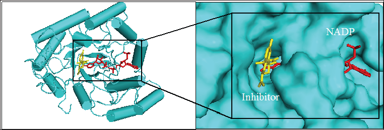
Figure 1: 3D structure of aldose reductase in cartoon and surface representation respectively showing the binding site for inhibitor fidarestat (yellow stick) and NADPH (red stick) at the active site

Lupinepublishers-openaccess-biomedicalengineering-biosciences

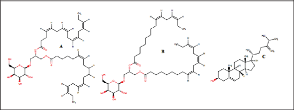
Figure 2: Chemical structure of the seaweed-derived compounds used as ligands in this study. (A)capsofulvesin A, (B) capsofulvesin B and (C) chalinasterol

Lupinepublishers-openaccess-biomedicalengineering-biosciences

Figure 1 shows the structure of the protein, aldose reductase used as a target in the current study. Aldose reductase is an NADP- dependent enzyme which catalyzes the conversion of glucose to sorbitol in the polyol pathway. This pathway is essential because hyperglycemia, an occasion where blood glucose level becomes excessively high, can trigger an increased activity of aldose reductase which can lead to the accumulation of intracellular sorbitol. This effect has been implicated in the pathogenesis of diabetic complication such as retinopathy, neuropathy and nephropathy. Therefore, inhibition of the catalytic function of aldose reductase has been proposed as a promising therapeutic target in the prevention and treatment of type II diabetes and its associated it's severe degenerative complications [33]. As shown in Figure 1, the enzyme active site is connected to the NADP binding pocket. This provides a chance for a ligand to potentially be a competitor of either the substrate of the coenzyme. Some natural and synthetic inhibitors of this protein have been sought over the years. Most of the reported inhibitors of aldose reductase preferably bind to the substrate binding location rather than competing with NADPH [34]. For more accurate understanding of the aldose reductase- ligand compex in the present study, two known inhibitors of the protein (fidarestat [35] and inhibitor-IDD384 [34]) were employed to compare the interaction pattern. The 2D chemical structure of the alga-derived ligands are given in Figure 2. Chalinasterol is a cholesterol nucleus-containing compound and it is structurally different from capsofulvesin A and capsofulvesin B which are galactolipids comprising of double aliphatic long chains with double bonds. These ligands are naturally-occurring compounds isolated from pharmacologically-important seaweed (C. fulvescens) [28]. It is essential to emphasize that these bioactive compounds have earlier been reported to inhibit aldose reductase in vitro [36]. However, the mechanism of the inhibitory effect still remains uncler.

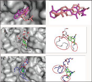
Figure 3: Analysis of the binding configuration of (A) chalinasterol, (B) capsofulvesin A and (C) capsofulvesin B. The binding pose for the compounds were compared against inhibitors of aldose reductase,fidarestat (green) and inhibitor- IDD384 (magenta). Areas with subtle differences in binding pose are identified in red spheres.

Lupinepublishers-openaccess-biomedicalengineering-biosciences

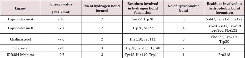
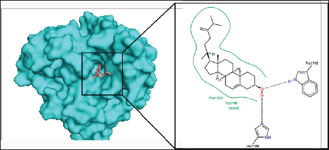
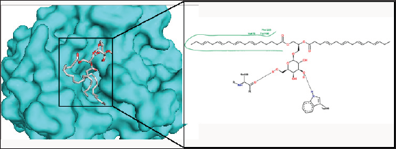
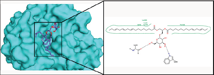
Chalinasterol shared a comparable binding pose with inhibitor- IDD384 (Figure 3). The molecular interaction as analyzed on proteinsplus server shows the contribution of both hydrophilic and hydrophobic bonds to the stability of the complex formed by the enzyme and C. fulvescens compounds (Figure 4-6). The bulky steroidal rings of chalinasterol was seen deeply buried within the fidarestat-binding pocket on aldose reductase where it is participated in hydrophobic bonds while the alkyl portion was exposed to the solvent-accessible region. Meanwhile, the inability of chalinasterol to completely block the active site may favor enzyme-substrate-chalinasterol complex formation as a mixed type of inhibition. As presented in Figure 4, two hydrogen bonds were established with amino acid residues His 110 and Trp111 respectively while Phe122, Trp219 and Trp20 participated in hydrophobic interaction with the ligand. The significance of hydrophobic bonds in the inhibition of aldose reductase cannot be overemphasized as it has been reported with many inhibitors of this protein including inhibitor IDD384.Inhibitory potential of chalinasterol against the carbohydrate-metabolizing enzyme was further proved by its binding affinity which is consistent with previous in vitro study that the compound possessed antidiabetic property [36,37]. The binding energy was relatively higher than that of the control ligands indicating a lower inhibitory activity for chalinasterol (Table 1). For capsofulvesin A and capsofulvesin B, the aromatic ring was placed at the solvent exposed region of the active site where it established hydrophilic interactions between its methoxyl moieties and the indole ring of Trp20 as well as the hydroxyl side chain of Ser22 (Figure 5). Possibly, the determining factor in the potency of these compounds as depicted in their IC50 is the binding configuration of the two long aliphatic chain. Capsofulvesin A has both of its side chain obstructing the entrance to the binding cavity while one of the aliphatic chains in capsofulvesin B was found moving away from the active site (Figure 6). This elongated side chain established a unique hydrophobic bond with residue Phe122. An efficient blockage of aldose reductase active site by capsofulvesin A may alter substrate access to the protein binding site, hence interfering with the enzymatic activity. Furthermore, the extra hydrophobic bond found with Phe122 in capsofulvesin B might have hindered the possible "swinging" of the aliphatic side chain from occupying the active site. It is also not impossible that these ligands interact with the enzyme-substrate complex in a mixed type of inhibition [36]. These mechanisms possibly underline the variations in the IC50 values previously obtained for these compounds. The binding energy of -7.9 kcal/mol estimated for capsofulvesin A compared to capsofulvesin B (-7.7 kcal/mol) suggests a higher affinity of the former to the protein. This result is in agreement with the previous in vitro report showing IC50 values of 52.53 |iM and 101.92 |iM for capsofulvesin A and capsofulvesin B respectively [36]. Although seaweeds have been investigated in diabetes and its complications, only scanty reports are available for C. fulvescens. Among the few studies, effect of extracts from the seaweed were observed for antidiabetic activity in vivo using streptozotocin-induced diabetic rats [37,38]. Therefore, this study provides insights into at least a part of the mechanisms responsible for the activity.

Table 1: Docking results showing binding energy and molecular interaction.

Lupinepublishers-openaccess-Biomedicalengineering-Biosciences

Figure 4: The binding pose and molecular the molecular interactions of chalinasterol with aldose reductase. Only key residues involved in hydrogen and hydrophobic interactions are shown as prepared on proteinsplus server.

Lupinepublishers-openaccess-biomedicalengineering-biosciences

Figure 5: The binding pose and molecular the molecular interactions of capsofulvesin A with aldose reductase. Only key residues involved in hydrogen and hydrophobic interactions are shown as prepared on proteinsplus server.

Lupinepublishers-openaccess-biomedicalengineering-biosciences

Figure 6: The binding pose and molecular the molecular interactions of capsofulvesin B with aldose reductase. Only key residues involved in hydrogen and hydrophobic interactions are shown as prepared on proteinsplus server.

Lupinepublishers-openaccess-biomedicalengineering-biosciences

Conclusion

This study investigated three antidiabetic compounds from C. fulvescens (a well-known green sea algae) against a diabetes- related target (aldose reductase) for their possible binding and molecular interaction pattern to provide insights into their mechanism of inhibition. With binding configurations similar to that of known inhibitors of aldose reductase, the results of the current study divulge the alga-derived compounds as suitable inhibitors of the protein. They bind at the active site and established both hydrophilic and hydrophobic bonds with some amino acid residues around site. This indicates their potential to interfere with the catalytic function of the protein. However, it was clear that the compounds did not interact with the NADP binding pocket. Hence, they are not competitive with NADPH for their inhibitory activity against aldose reductase. The subtle contrasts in the binding mode of these compounds might be responsible for the differences in their IC50 values as previously reported in in vitro experiments. Taken together, the results of this study verify aldose reductase inhibitory activity of the C. fulvescens constituents and validate the antidiabetic property of the seaweed.

Read More About Lupine Publishers Journal of Biomedical Engineering and Biosciences  Please Click on Below Link: https://biomedical-sciences-lupine-publishers.blogspot.com/

Monday, 13 June 2022

Lupine Publishers| Study About the Despeckling Methods for Retinal Optical Coherence Tomography Images

 Lupine Publishers| Journal of Biomedical Engineering and Biosciences


Abstract

This paper presents study about the different despeckling methods used for quality enhancement of retinal optical coherence tomography (OCT) images. Speckle noise is an inherent property of an OCT images which affects the visual quality of the images, hence difficult to diagnosis the patients. Therefore speckle noise reduction from the OCT images is an important prerequisite, whenever OCT imaging is used for diagnosis. The speckle noise intensity depends on the various imaging system parameters of its systems and the different structure representations used for the image tissues. A despeckling technique is to be designed in such a way that it should be able to reduce the speckle noise from the OCT images while preserving the tissues and fine details of the images.

Keywords: Despeckling; Optical coherence tomography; Speckle noise

Introduction

Optical Coherence Tomography (OCT) is a most powerful biomedical imaging technique used to extract the required information from an object. The working approach of this method is almost similar to ultrasound imaging except the medium used in obtaining its. OCT imaging uses light beams instead of sound [1]. OCT has proved its significant importance in the field of ophthalmology, especially in the detection of diseases related to retina and glaucoma [2-4]. Currently OCT is a best suitable method for imaging the internal structure of biological systems and also an essential part of a procedure to obtain high-resolution images of the retina [5-7]. It is also observe that retinal layer thickness of the OCT images improve the clinical finding in the field of ophthalmology Retinal layer segmentation improves the clinical finding in the case of glaucoma progression and macular degeneration. Therefore, the pre-processing of ophthalmic OCT images is a most importance step to improve the clinical diagnosis. Ophthalmic OCT imaging technique is based on detection of coherent waves; therefore these images are accompanied with a significant amount of speckle noise, which degrade the image quality and limit the contrast to noise and signal to noise ratio of the image.

To reduce the effect of speckle noise and to preserve the fine image features. A good speckle noise reduction algorithm is required that reduce the effect of speckle noise while preserving fine details of image. A lot of efforts have been made to overcome the effect of speckle noise. All methods proposed for speckle noise reduction from the OCT images are mainly divided into two categories namely transform domain techniques and spatial domain techniques. Organization of manuscript is as follows: section-2: describes brief about speckle noise; section-3: presents example of speckle noise reduction from the OCT image; section-4: discusses about different despeckling methods for OCT images and its summary is also presented in tabular form; section-5: presents summary and conclusion of the manuscript.

Speckle Noise

When a signal or an image is acquired by narrowband detection systems like SAR, ultrasound and OCT, then a pattern known as speckle effect the quality of these objects. The main reasons that can affect the speckle are optical properties of the system, status of the subject, size and coherence nature of source, multiple scattering, phase difference of the beam and aperture of the detector [8,9]. Further we can classify the speckle present in OCT images into two categories first one signal- carrying speckle and the second one is signal- degrading speckle .The mathematical distribution of the speckle can be model with a Rayleigh distribution. In this modelling, speckle noise assumed as a multiplicative behaviour, in contrast to additive nature of the noise. The mathematical model of Speckle noise can be represented as:

Y (m,n)= X (m,n)S (m, n) (1)

Where Y, X and S represent the noisy image data, original image data and speckle noise respectively. Normally, the multiplicative nature of speckle noise is converting into additive nature by logarithmic transformation of equation (1).

Example of Noise Reduction in OCT image

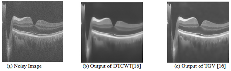
Figure 1: Speckle Noise Reduction from the Noisy Image.

Lupinepublishers-openaccess-biomedicalengineering-biosciences

A noise reduction technique is required to improve the visual quality of the OCT image. All noise reduction techniques can be implemented into two ways, first one implemented during the acquisition time and second one implemented after acquisition of images. Normally, the noise reduction technique during the acquisition time is not good solution because these techniques require repetition of the same data which increase the acquisition time. Therefore, the use of post-processing techniques is more favourable solution to reduce the speckle noise from the OCT images. The example of speckle noise reduction from the noisy image is shown in the Figure 1. In this Figure 1(a) is the noisy image, 1(b) is the output image of DTCWT method [9] and 1(c) is the output image of second order total generalized variation (TGV) [9] method. From the Figure 1, it is clear that the effect of speckle noise reduction is very clear.

Despeckling methods

In the literature, a number of researchers proposed different post-processing methods to overcome the effect of for speckle noise from the OCT images. Few classic despeckling techniques that are used for SAR images, successfully used for OCT images also which are Lee [10], kuan [11] and frost filters [12]. An image restoration method is introduced that overcome the limitations of medical tomographic imaging system, by recovering the noiseless high frequency information corrupted by imaging system [13]. Especially for the multi-dimensional image data a better technique, based on compressive sensing principles is developed. This technique has capability to reduce the speckle noise while interpolate the missing data. This technique termed sparsity based simultaneous denoising and interpolation (SBSDI) [14]. A more appropriate approach based on dual-tree complex wavelet transform (DT-CWT) has been developed to overcome the effect of speckle noise from the OCT images. In this process adaptive-weighted bilateral filter (AWBF) is use to further enhance the OCT imaging by use of smoothing process [15].

For better image representation a new multiscale geometric analysis tool knows as wave atom transform was also proposed. The wave atom transform give better visual results in comparison to other transform such as wavelet and curvelet transform [16]. Image decomposition may also help in image denoising if the speckle noise in the OCT image is consider as texture or oscillatory patterns. A second order total generalized variation (TGV) decomposition model is helpful to remove the texture from the OCT image [16]. TGV is also removing the staircase side effect from the resulted images. Adaptive bilateral filter is also help to enhance the multi-frame OCT data [17]. The processed frames are averaged to convert into denoised output OCT image [17]. Variational image decomposition is also used to reduce the speckle noise from the ophthalmic OCT images [18].

The method converts the original image into cartoon part, texture part and speckle noise part to reduce the speckle noise from the image [18]. This method has the capability to suppress the speckle noise, while preserve image features such as edges and texture [18]. It may be useful for edge detection, segmentation and thickness calculation [18]. Independent Component analysis (ICA) techniques are also used in noise reduction of retinal OCT images [19]. The ICA technique can be beneficial when fewer number of B-scans images are available [19]. In, Raheleh kafieh et al. [20] proposed method for speckle denoising of OCT images based on dictionary learning approach with dual tree complex wavelet transform. In Zahra Amini [21], and Hossein Rabbani proposed method for speckle denoising of OCT images based Nonlinear transform based approach. To design the structural dictionaries, the information related to retinal layer is extracted automatically in the SSR method [22].

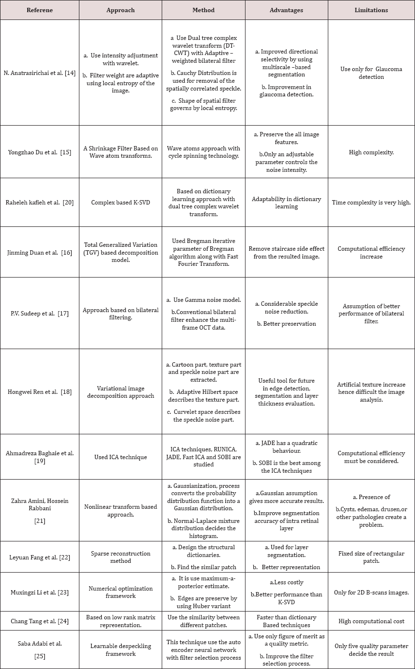
This method also uses the patch similarity index to enhance the performance [22]. Recently, optimization approach based on MAP estimate is proposed [23]. It use a suitable noise reduction approach along with Huber variant of TVR that overcome the effect of speckle noise, while preserve the edges as well [23]. To reduce the speckle noise, selection of a digital filter is a very difficult task. To overcome this limitation a framework based on learning approach known as learnable despeckling framework (LDF) has been proposed [24]. This approach only uses a single quality metric for selecting the appropriate filter for the task [25]. The different despeckling techniques are also studied and summarize into a Table 1.

Table 1: Summary of different despeckling methods for OCT Images.

Lupinepublishers-openaccess-Biomedicalengineering-Biosciences

Conclusion

This paper highlighted study about the different despeckling methods used for quality enhancement of retinal optical coherence tomography (OCT) images. The above study shows that among the different type of despeckling methods are used for OCT images. Study shows that an approach based on bilateral filter removes the significant amount of speckle noise while preserves the edges of the images. The Wavelet transform based approach is also a better solution for speckle noise reduction because of its multiscale nature. Out of different ICA techniques, SOBI is the one of the best techniques used for speckle noise reduction. Further work can be carried out in the variational image decomposition approach, because this approach is separate the speckle noise from the cartoon part and the texture part of the OCT images. Finally, learnable despeckling framework is also helpful for the selection of digital filter as per the requirement of the images features.

Read More About Lupine Publishers Journal of Biomedical Engineering and Biosciences  Please Click on Below Link: https://biomedical-sciences-lupine-publishers.blogspot.com/