Showing posts with label Journal of Clinical Gastroenterology. Show all posts

Tuesday, 15 March 2022

Lupine Publishers| Successful Endoscopic Treatment of Refractory Postoperative Duodenocutaneous Fistula using Fibrin Sealant

 Lupine Publishers| Journal of Gastroenterology and Hepatology


Case Report

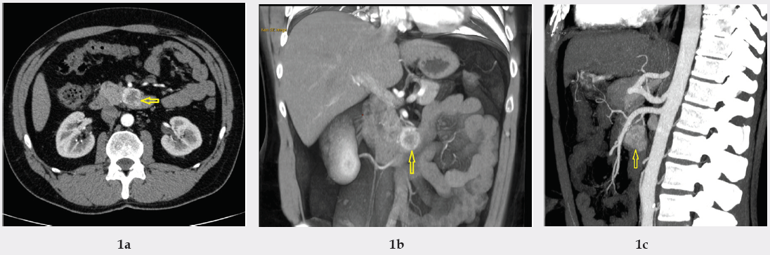
Iatrogenic, postoperative enteroatmospheric fistulas remain difficult to manage but common problem after gastrointestinal surgery. Surgical revision, after failure of conservative treatment is not only partially efficient but also carries a high mortality risk, mainly due to patients’ general condition and usually multiples comorbidities. Because of that alternative minimally invasive techniques are extensively studied, but unfortunately the data are still confounding [1,2]. We present a case of 38 – y.o. man with past history of nephrolithiasis, accidentally diagnosed with 30 mm gastrointestinal stromal tumour of the duodenum (Figures 1a-1c).

Figure 1a: Contrast enhanced multidetector multiphase computed tomography of the abdomen and pelvis showing solid tumor (Ø 30 mm) between duodenum and pancreas (cross-section) (arrow).

Figure 1b: Contrast enhanced multidetector multiphase computed tomography of the abdomen and pelvis showing solid tumor (Ø 30 mm) between duodenum and pancreas (anterior-posterior section) (arrow).

Figure 1c: Contrast enhanced multidetector multiphase computed tomography of the abdomen and pelvis showing solid tumor (Ø 30 mm) between duodenum and pancreas (latteral-section) (arrow).

lupinepublishers-openaccess-journal-gastroenterology-hepatology

Tumour resection with end-to-end anastomosis of transversal duodenum was performed, but the procedure was complicated by massive intraabdominal bleeding leading to acute pancreatitis and finally to enterocutaneous fistula forming. In course of treatment, the patient required five reoperations with Roux-en-Y resection and sewing patch on the fistula’s enteral orifice, but no one was efficient (Figures 2a& 2b). To avoid another reoperation we decided to perform endoscopic fistula closure by injection fibrin glue (Tisseel, Baxter) into the internal opening (Figures 3a-3c). Procedure was performed via catheter using standard Olympus videoendoscope. We observed immediate fistula closing, and there were no early nor late complications related to the procedure. Patient was discharged after 67 days of hospital stay. After 11 months of follow-up patient remains cured (Figure 4). In conclusion, the fibrin sealant treatment is safer and much less invasive viable option, and it should be taken into account in selected cases [3].It appears that fistula closure is more efficient if performed from the internal opening.

Read More About Lupine Publishers Journal of Gastroenterology and Hepatology Click on Below Link: https://currenttrendsingastroenterology.blogspot.com/

Wednesday, 14 July 2021

Lupine Publishers| Utilization of Traditional Anticoagulation for Venous Thromboembolism in Patients with Liver Cirrhosis: Is It Safe?

 Lupine Publishers| Current Trends in Gastroenterology and Hepatology (CTGH)


Abstract

Patients with liver cirrhosis are known to have increased risk bleeding particularly from gastrointestinal tract. However, recent literature has shown that patients with liver cirrhosis are also at increased risk of developing deep venous thrombosis and pulmonary embolism. Therefore, it is important to consider prophylactic and therapeutic anticoagulation in cirrhotic patients. In this article, we have reviewed the available literature on the safety and efficacy of the utilization of prophylactic and therapeutic anticoagulation in cirrhotic patients.

Abbreviations: INR: International Normalized Ratio, VTE: Venous Thromboembolism, DVT: Deep Venous Thrombosis, PE: Pulmonary Embolism, PTT: Partial Thromboplastin Time, LMWH: Low-Molecular-Weight Heparin, UFH: Unfractionated Heparin, CLD: Chronic Liver Disease

Introduction

Liver plays central physiologic role in hemostasis as it synthesizes the majority of the procoagulant and anticoagulant factors. The levels of these factors are markedly affected by decrease function of liver associated with cirrhosis resulting in abnormal hemostatic mechanism. Generally, the impression in the clinical world is that liver cirrhosis is associated with decrease synthesis of procoagulant factors resulting in increased risk of bleeding. This phenomenon is known as auto-anticoagulation and is supported by elevated international normalized ratio (INR) and low platelet count usually observed in cirrhotic patients. In this regard, gastrointestinal bleeding and more specifically variceal bleed are of major concern since they contribute significantly to the mortality of patients with liver cirrhosis. At least, 30% mortality has been reported at the first episode with a 70% recurrence rate in this patient population and a 1year survival estimate ranging from 32% to 80% [1]. However, decrease function of cirrhotic liver also results in reduce level of anticoagulant factors including antithrombin III, protein S, and C which may result in increased tendency to form clots. Interestingly, recent data has also noted cases of venous thromboembolism (VTE) including both deep venous thrombosis (DVT) and pulmonary embolism (PE) in cirrhotic patients ranging between 0.5% to 6.3% [2-10]. Dabbagh et al. [4] found that even an elevated INR > 2.2 was not protective against VTE in this patient population. Gulley D et al. [10] noted that hospitalized cirrhotic patients without predisposing co-morbidities (e.g. neoplasm, congestive heart disease and chronic renal failure) had similar risks for VTE as compared to noncirrhotic patients [10]. Thus, the myth of auto-anticoagulation seems to be only partially true. Therefore, the abnormal routine blood tests (like elevated INR, Partial Thromboplastin Time [PTT], high MELD score and low platelet count) may indicate increased hemorrhage risk in this patient population but may not be necessarily completely true as these tests do not accurately reflect the activity of aforementioned anticoagulant factors in the serum. The purpose of this article is to review the epidemiological data available on the utilization of traditional VTE prophylaxis and treatment on the increased bleeding risk in patients with liver cirrhosis.

Methods

An electronic Medline search was conducted using the key terms anticoagulation, low-molecular-weight heparin (LMWH), unfractionated heparin (UFH), oral anticoagulant, deep venous thrombosis, pulmonary embolism, venous thromboembolism, liver cirrhosis, chronic liver disease, and decompensated liver disease. Studies written in the English from January 2000 to March 2018 were considered for this review article. All search results were reviewed.

Results

Intagliata N et al. [11] studied the complication rates associated with administration of thromboprophylaxis in 235 hospitalized cirrhosis patients admitted to the University of Virginia between 2007 and 2010 accounting for 355 discrete hospitalizations. They noticed only nine gastrointestinal bleeding events (2.5% of admissions) and concluded that VTE prophylaxis was not associated with increased risk of bleeding or death in their patient population [11]. Vivarelli M et al. [12] conducted a retrospective study in Italy on 229 consecutive cirrhotic patients with HCC who underwent hepatic resection. They assess the safety and effectiveness of prophylaxis for VTE in terms of thrombotic or hemorrhagic complications between those who received and those who did not receive prophylaxis with low-molecular weight heparin. They divided the patients into two groups i.e. Group A included 68.5% of patients and Group B included 31.5 % patients. Group B patients had higher Child-Pugh and MELD scores, lower platelet counts, a higher prevalence of esophageal varices and higher requirements for intraoperative transfusion of FFP. Group A received VTE prophylaxis with LMWH while Group B did not receive VTE prophylaxis. The cases of VTE were higher in group B (1.38%) as compared to the group A (0.63%) but the difference was not statistically significant (P = 0.530). On the other hand, the prevalence of hemorrhagic complications was higher in group A as compared to group B but again the difference was not statistically significant (P = 0.380). Moreover, only the presence of varices was associated with increased risk of bleeding (P=0.05) among other studied risk factors including age, CTP class and Model for End-stage Liver disease scores, platelet count and intraoperative transfusion requirements [12].

Bechmann L et al. [13] conducted a retrospective study in Germany to evaluate the safety of LMWH utilization for either prophylactic or therapeutic indications anticoagulation among 84 patients with cirrhosis. Only seven patients developed variceal bleeding at a rate comparable with the baseline rate in patients with advanced cirrhosis. The hemorrhagic complications were either secondary to bleeding esophageal varices or 2whypertensive gastropathy with no mortality. They concluded that a prophylactic use of LMWH in patients with cirrhosis appears to be safe [13].

Smith C et al. [14] also conducted a retrospective chart review of 410 chronic liver disease (CLD) patients admitted to a tertiary care academic medical center in US from August 2009 through July 2011. 225 (55%) patients received thromboprophylaxis including mechanical (154), pharmacologic (49), and combined (22). They noticed a significant decrease in overall thromboprophylaxis use and pharmacologic prophylaxis use for patients with INR >2.0 as compared to those with INR 1.4 to 2.0 (P = 0.013 and P < 0.001, respectively). 0.7% of patients developed VTE (DVT) and fifteen (3.7 %) patients had bleeding event. Out of fifteen patients who developed bleeding, nine were on mechanical prophylaxis, 1 on pharmacologic, 3 on combination, and 2 with no prophylaxis. Majority of patients experiencing a bleeding event had an INR >2.0 (P = 0.001). They concluded that use of VTE pharmacologic prophylaxis does not appear to increase bleeding risk in CLD patients with INR ≤2.0 [14].

Barclay S et al. [15] conducted another retrospective study to determine whether pharmacologic prophylaxis for VTE was associated with a decrease in the incidence of VTE or an increased incidence of bleeding in patients with CLD. The study sample consisted of a total of 1581 CLD patients hospitalized over a 3-year period and divided into two groups based on receipt of pharmacologic VTE prophylaxis. 392 (24.7%) patients received pharmacologic VTE prophylaxis. Decrease risk of VTE (0.5% vs 1.8%, p=0.05) and bleeding (2.0% vs 10.3%, p<0.001) were noted in the prophylaxis group as compared to the nonprophylaxis group [15].

In a multicenter retrospective study conducted in 5 hospitals, Reichert J et al, studied the pharmacologic VTE prophylaxis on risk of hemorrhage while hospitalized in patients with CLD and concurrent coagulopathy. They utilized the ICD-9 codes to identify subjects with CLD admitted from January 1, 2012, until December 31, 2012. 256 patients met criteria for analysis, with 80 received pharmacologic VTE prophylaxis and 176 did not receive pharmacologic VTE prophylaxis. They found that pharmacologic thromboprophylaxis was associated with increased number of hemorrhagic events among cirrhotic patients who received it as compared to those who did not (17.5% vs 7.4%, p=0.02). However, it was noted that difference in rate of overall hemorrhage was driven primarily by a difference in minor hemorrhage [16].

Conclusion

The use of anticoagulation for VTE prophylaxis and treatment in cirrhosis remains underutilized and it is still not a universal practice to provide hospitalized cirrhotic patients with such prophylaxis and treatment due to the concerns of increased bleeding risk. Also, the literature on the safety and efficacy of anticoagulation in liver cirrhosis is in initial stages and is based mainly on retrospective studies. Six studies have investigated the use of VTE prophylaxis. Two studies have shown that DVT prophylaxis does decrease the risk VTE in cirrhotic patients [12,15]. However, the results of one of the study did not reached the statistical significance which could be due to low power of the study because of the small sample size [12]. On the other hand, only one study have shown that pharmacologic thromboprophylaxis was associated with increased number of hemorrhagic events among cirrhotic patients which they attributed to increased incidence of minor hemorrhage [16]. Another study noticed higher prevalence of hemorrhagic complication in VTE prophylaxis group as compared to cirrhotic patients who did not received VTE prophylaxis, but their result did not achieve statistical significance [12]. Moreover, only the presence of varices was associated with increased risk of bleeding.

It is possible that risk of hemorrhage is underestimated in these studies because of retrospective nature of studies with possibility of underreporting of bleeding events and underutilization of thromboprophylaxis of VTE in patients at higher risk of bleeding such as those with high INR, low platelets and presence of esophageal varices. Only one study investigated the use of VTE treatment in cirrhotic patients and they found no increased bleeding risk with it [13]. However, it was a small sampled study and there was no control group utilized to compare [13]. In general, recent data suggest that utilization of VTE prophylaxis in cirrhotic patients is safe and should be considered. Underutilization of both chemical and mechanical VTE prophylaxis in hospitalized cirrhotic patients has been noted ranging to as high as 75% [8,17]. In another study, only 9% of hospitalized patients with CLD were treated with pharmacological VTE prophylaxis, while only 16% received mechanical VTE prophylaxis [2]. The major concern regarding the use of anticoagulation in this population is the gastrointestinal bleed and specifically esophageal variceal bleed. However, VTE prophylaxis appears relatively safe in cirrhotic patients and should be considered in all hospitalized cirrhotic patients with minimal varices, no evidence of clinical bleeding and with an appropriate clinical indication.

Infact, the safety of prophylactic or therapeutic anticoagulation in carefully selected patients with cirrhosis without the presence of high-risk esophageal varices appears to be comparable to general medical patients. Also, in patients with esophageal varices, primary or secondary prophylaxis with either endoscopic variceal ligation or use of non-selective beta-blockers is suggested prior to initiation of anticoagulation and after discussing about the risks and benefits of anticoagulation with the patients. Periodical screening endoscopy should be considered to assess varices and the risk of bleeding in these patients. Various classes of heparin are currently available for prophylactic or therapeutic purposes including UFH, LMWH and fondaparinux. LMWH seems to be the treatment of choice for both the prevention and treatment of VTE in cirrhotic patients. UFH is an alternative in cirrhotic patients for shorter-term use and in cases of severe renal dysfunction and/or hemodynamic instability. The possible role for the new antithrombotic drugs with direct action on factor Xa or thrombin has yet to be evaluated.

Clinical tools to evaluate the risk of VTE in cirrhotic patient population also need to be developed to target the cirrhotic patient who will benefit most from the anticoagulation. Bogari H et al. [18] conducted a retrospective cohort study to evaluate the utilization of the Padua Predictor Score (PPS) as a risk-stratification tool for the development of VTE in 163 patients with liver cirrhosis [18,19]. They categorized the patients into two groups based on whether they developed a VTE (11%) or not (89%). Patients were further risk stratified into high-risk (score ≥4) and low-risk (score <4) using the PPS. Patients in the VTE group had significantly greater mean PPS than in the non-VTE group (5.8 ± 2.0 versus 3.0 ± 2.1, respectively; p<0.001). Also, High-risk patients were more likely to have VTE (OR 12.7, 95% CI 2.8 to 57.4, p=0.001) as compared to low risk. Based on their findings, Bogari H et al. [18] concluded that PPS is an effective risk assessment tool for development of VTE in patients hospitalized with chronic liver disease. Various other factors like low serum albumin and decompensated liver cirrhosis have also shown to be associated with increased risk of VTE in patients with liver cirrhosis. In two retrospective studies conducted by Northup et al. [2], Garcia Fuster et al. [3] low serum albumin was found to be associated with increased risk of developing VTE independent from elevated INR or low platelet count [2,3]. Certain other factors like INR > 2 and low platelet count may lower the risk of developing VTE. It will be prudent to develop tools including such factors to help accurately characterizing cirrhotic patients at higher risk of developing VTE. This will help provide clinicians with more confident to start anticoagulation in cirrhotic patients at high risk of bleeding secondary to esophageal varices.

Further studies are needed to determine the utility of novel methods to monitor the anticoagulation therapy in cirrhotic patients. LMWH is conventionally monitored by anti-Xa assay, while UFH can be monitored by activated PTT (aPTT) assay in the general population. However, monitoring of LMWH anticoagulation with anti-Xa levels for dose adjustment is challenging in cirrhotic patients since the levels of anti-Xa factor have been found to be lower in cirrhotic patients compared to normal controls after administration of prophylactic or therapeutic dose of LMWH [13]. For example, Bechmann et al. [13] studied the pharmacokinetics of LMWH in patients with cirrhosis and confirmed that standard doses of enoxaparin failed to achieve target anti Xa levels recommended for prophylactic or therapeutic use against VTE [13]. However, in vitro studies evaluating the effect of LMWH on thrombin generation found that despite reduced antithrombin and anti-Xa activity levels, cirrhotic patients had an increased response to LMWH [20]. They concluded that the likely explanation of low anti-Xa levels could be a laboratory artifact while the efficacy of LMWH is preserved [20]. Thus, targeting its levels for the prophylaxis and treatment of VTE in noncirrhotic patients might underestimate the true degree of anticoagulation which could lead to incorrect dosing and subsequent increase morbidity and mortality secondary to bleeding. The role of thrombin generation in monitoring the response to LMWH in cirrhotic patients has been investigated by Lisman T et al. [21] and they found it to be a useful tool and an alternative to anti-Xa levels [21]. On the other hand, UFH is monitored with aPTT in general population. But again, aPTT levels are also known to be prolonged in patients with liver cirrhosis thus making it difficult to predict the dose accuracy of UFH in this patient population.

In the end, the treatment and prophylaxis for VTE should be utilized in patients with liver cirrhosis who are at low risk of bleeding i.e. does not have esophageal varices. Moreover, randomized prospective studies needs to be performed to confirm the efficacy of VTE prophylaxis in cirrhotic patients, to determine the safety of therapeutic anticoagulation against VTE in cirrhotic patients, to figure out accurate markers for monitoring the use anticoagulation in cirrhotic patients, and to better understand the role of coumadin and newer oral anticoagulants in VTE prophylaxis and treatment in cirrhotic patients.

Read More About Lupine Publishers Current Trends in Gastroenterology and Hepatology (CTGH) Please Click on Below Link: https://currenttrendsingastroenterology.blogspot.com/


Tuesday, 8 June 2021

Lupine Publishers| Non-Surgical Pneumoperitoneum Complicating Mechanical Ventilation

 Lupine Publishers| Current Trends in Gastroenterology and Hepatology


Abstract

The occurrence of a pneumoperitoneum is usually linked to intra-abdominal hollow-organ perforation and generally requires emergency abdominal surgery. In 5 to 15% of cases, the pneumoperitoneum is not related to organ perforation and could be caused by various aetiologies. Barotrauma secondary to either invasive or non invasive mechanical ventilation can cause a pneumomediastinum with air diffusion to retroperitoneum and peritoneal cavity with extensive subcutaneous emphysema. We present a case of a fifty-six-year-old patient with a permanent tracheostomy for oropharyngeal neoplasm, admitted to Intensive Care Unit (ICU) for severe pneumonia requiring mechanical ventilation leading to pneumoperitoneum and retropneumoperitoneum with extensive subcutaneous emphysema. We also discuss Investigations and management challenges for this patient.

Keywords: Mechanicalventilation; Barotraumas; Pneumoperitoneum; Retropneumo-peritoneum; Conservative Management

Abbreviations: ICU: Intensive Care Unit, CT: Commuted Tomography

Introduction

Pneumoperitoneum is the presence of air in peritoneal cavity. Chest x-ray commonly reveals a subdiaphragmatic crescent of air. It is due to hollow-organ perforation in 85 to 95% of cases. Other aetiologies of pneumoperitoneum do not require emergency surgical exploration [1,2]. Presence of gas in peritoneal cavity without bowel perforation is called spontaneous pneumoperitoneum that results from thoracic, abdominal or pelvic causes [3]. Mechanical ventilation can cause a pneumoperitoneum during ventilator induced barotrauma which is usually revealed by subcutaneous emphysema. We report the case of a non-surgical pneumoperitoneum and retroperitoneum, in a patient with permanent tracheostomy for pharyngeal cancer, that occured after mechanical ventilation and favorable clinical outcome with conservative management.

Case report

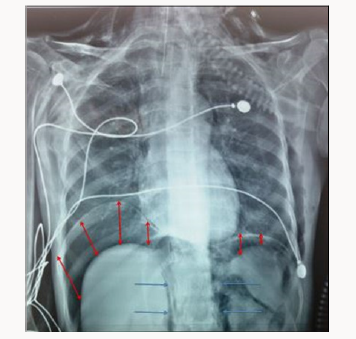
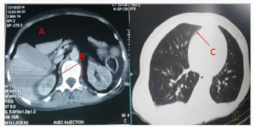
A 56-year-old with permanent tracheostomy for hypopharyngeal cancer on radiotherapy was admitted to ICU for community acquired pneumonia with septic shock. She required mechanical ventilation via tracheostomy, broad spectrum antibi otics and she was started on vasopressors (Noradrenalin) through righ internal jugular central venous catheter. Mechanical ventilation was undertaken with Assist Control Ventilation, a tidal volume of 6ml/kg ideal body weight, a respiratory rate of 12 cycles/min, a positive end expiratory pressure of 6 cm H2O and FiO2 of 60%, the patient was sedated with propofol and remifentanyl. 24 hours later, examination revealed extensive subcutaeous emphysema to chest wall and abdomen, peak pressures were normal and breath sounds were equally transmitted on auscultation. The chest x-ray did not show any pneumothorax but revealed obvious bilateral crescent of air beneath diaphragm compatible with pneumoperitoneum (Figure 1). Haemodynamically, the patient improved and vasopressors were stopped 30 hours later despite the pneumoperitoneum. Thoraco-abdominal Commuted Tomography (CT) scan confirmed the pneumoperitoneum, revealed a retropneumoperitoneum, pneumomediastinum and ruled out a pneumothorax (Figure 2). Investigation was completed by a gastrograffin enema that was injected through the gastrostomy tube inserted before the start of radiotherapy, and did not reveal leakage from the digestive tract.

Conservative management was adopted as the patient was clinically improving, she was weaned of vasopressors and mechanical ventilation 5 days later, subcutaneous emphysema and abdominal distension steadily resolved and the patient was discharged to medical ward after 10 days stag in ICU.

Figure 1: Standard epidemic of Sheep bluetongue in Tamilnadu.

lupinepublishers-openaccess-journal-gastroenterology-hepatology

Figure 2: Standard epidemic of Sheep bluetongue in Tamilnadu.

lupinepublishers-openaccess-journal-gastroenterology-hepatology

Discussion

Pneumoperitoneum is an uncommon complication of mechanical ventilation and its causes need to be correctly diagnosed to avoid unnecessary laparotomy. Non-surgical pneumoperitoneum is the presence of air in the peritoneal cavity and is usually detected on chest x-ray, but its management is controversial based on observation or laparotomy [4]. According to Mularskei, there are five main causes of non-surgical pneumoperitoneum: False or pseudo- pneumoperitoneum describes a similar radiological aspect of a pneumoperitoneum but can be caused by a pulmonary atelectasis simulating the aspect of air beneath the diaphragm, subphrenic extraperitoneal fat or interposition of the right hepatic lobe to the diaphragm [5,6]. Abdominal causes: abdominal surgery [7], peritoneal dialysis [8,9], gastro-intestinal explorations [10,11], colonoscopy [12,13] Thoracic causes: mechanical ventilation, cardio-pulmonary resuscitation, pneumothorax. Gynaecologic causes: Hysterosalpingography, pelvic inflammatory diseases by air diffusion into the peritoneal cavity via the uterus and the Fallopian tubes Other causes: Scleroderma, dental extraction, cocaine and idiopathic [3].

In this case, investigations including thoraco-abdominal CT, oeso-gastro-duodenal endoscopy and digestive opacification with gastrografin enema ruled out a hollow-organ perforation and indication for emergency laparotomy. The CT has revealed a pneumomediastinum without pneumothorax, likely caused by a barotrauma with subsequent retroperitoneum and pneumoperitoneum. This can be explained by the passage of air by a number of routes. Mediastinal air my pass directly through micro-perforations of the pleura and diaphragm or via thoracoabdominal communicating pores to the retro-peritoneum and then the peritoneal cavity [3]. Pneumoperitoneum can also occur as a complication of non-invasive ventilation [14] or following multiple laryngoscopies in difficult intubation [15]. Predictive factors of pneumoperitoneum during mechanical ventilation in adults include high airway pressures, ventilation with high tidal volumes, low lung compliance and underlying lung diseases such as chronic obstructive pulmonary disease or Acute Respiratory Distress Syndrom [3]. The association between high airway pressures and barotrauma is demonstrated in animals with both subcutaneous emphysema and pneumoperitoneum are induced by presures over 60 cm H2O [16]. Management of spontaneous pneumoperitoneum is controversial. No consensus exists on the optimal management of non-surgical pneumoperitoneum complicating mechanical ventilation but some authors advocate conservative management when abdominal distension is minimal, absence of signs of peritonitis, absence of fever or leucocytosis and most importantly absence of surgical cause of the pneumoperitoneum [3]. In this case, pneumoperitoneum was cleared with conservative management, decision was made in consultation with the surgical team in view of the absence of abdominal cause, absence of signs of abdominal compartment syndrom and the clinical improvement of the patient [17].

Conclusion

The occurrence of subcutaneous emphysema in a patient under mechanical ventilation should raise suspicion of a pneumoperitoneum, either isolated or associated to pneumothorax or retropneumoperitoneum and a surgical cause should be ruled out. In the absence of signs of peritoneal irritation and obvious surgical cause, a non surgical cause of pneumoperitoneum in patients under mechanical ventilation should be recognised and a conservative management adopted to avoid unnecessary laparotomies.

Read More About Lupine Publishers Current Trends in Gastroenterology and Hepatology Please Click on Below Link: https://currenttrendsingastroenterology.blogspot.com/


Monday, 5 April 2021

Lupine Publishers| Pregnancy after Liver Transplantation

 Lupine Publishers|  Current Trends in Gastroenterology and Hepatology


Introduction

In 1978, Walcott and coworkers documented the first pregnancy in a liver transplant recipient [1]. The recipient’s immunosuppressive regimen included prednisone and azathioprine. Her pregnancy was uncomplicated, and she delivered a healthy male, weighing 2400 g, at term. Since Walcott’s report, there have been hundreds of pregnancies reported worldwide in liver transplant recipients. Whether the transplant could take in an adverse effect of the pregnancy or whether the pregnancy could potentiate liver transplant it is not settled [2].

Liver Transplant and Timing of Pregnancy

a) Women should wait at least 1 year after receiving a solidorgan transplant before attempting to get significant, and then should do so but when earned by the transplant team and obstetrician, with close monitoring [3].

b) The best contraceptive method, at least early after liver transplantation, is a barrier method. Later, when the patient is stabilized, hormonal methods may be considered. Sterilization is an option for patients who have completed their family [4].

c) Liver transplant recipients considering pregnancy should have preconception counseling with a maternal–fetal medicine specialist and their transplant physician. There are no specific guidelines for liver transplant recipients to decide whether an allograft is functioning sufficiently well for pregnancy as there is for kidney transplant recipients. Nevertheless, a general assessment of graft function and the status of comorbidities, such as high blood pressure, should be done before conception. The preconceptional visit also provides an opportunity to look back with the patient outcomes of pregnancies with liver transplants, potential effects of immunosuppressive agents on the pregnancy and fetus, and the plan of management for the gestation.

Pregnancy outcome after liver transplantation

Despite advances in immunosuppressive therapy, pregnancies in liver transplant recipients are still more risky than in the general population for both the mother and the fetus. Pregnancies in liver transplant recipients should be followed up more cautiously by a multidisciplinary team because [5,6]:

a) Overall, 78% of pregnancies were successful.

b) Many recipients of solid-organ transplants, including liver transplants, develop high blood pressure and renal dysfunction. It is easily grounded in the obstetrical literature that these conditions are independently linked with adverse pregnancy events. The most common obstetric complications were high blood pressure (28%), preeclampsia (12%), and reversible liver dysfunction (27%) [7].

c) The rate of acute rejection was 10% [8].

d) The incidence of perinatal and neonatal death was about 4%.

e) Thither was a 3% incidence of congenital malformations, after exclusion of an infant with the presumed fetal alcohol syndrome and an infant with an autosomal dominant genetic disorder inherited from the female parent.

f) The maternal death rate was 5.5%, over a 2-year period following delivery [9].

g) Additionally, it seems that the danger of infection during gestation is also significantly increased after liver transplant; this finding may be a consequence of chronic immunosuppressive therapy [1].

h) Liver transplantation does not significantly modify pretransplant diabetes mellitus. Diabetes mellitus frequently develops de novo after liver transplantation, although this complication is usually short-lived and likely linked to immunosuppressive drug administration. The prognosis of patients with post-transplant diabetes mellitus is worse than that of those without this complication [10].

i) The incidence of preterm delivery is 24% [8].

j) The rate of cesarean delivery for liver transplant recipients is higher (20% -100%) than the national average [11].

k) The rate of newborn complications was also high, at 29%.

l) Subjective hearing impairment is frequent in patients after OLT and contributes to post-OLT morbidity. Calcineurin inhibitor-related neurotoxicity appears as a potential mechanism. Further prospective investigations with objective hearing tests are necessary to substantiate these results and to assess the use of immunosuppression [12].

Management Recommendations

a) Patients with liver transplants considering pregnancy should receive preconception counseling. The preconceptional evaluation will require an appraisal of the current graft status and episodes of rejection.

b) Preconceptional counseling should be provided to both the patient and her spouse. For patients who bear a genetic disorder underlying their liver disease, genetic counseling should also be provided. These will include patients with alpha-1antitrypsin deficiency and hemochromatosis, as well as less common disorders such as Alagille syndrome and Caroli syndrome. Prenatal testing may be usable for certain disorders.

c) Ideally, immunosuppressive medications are at stable doses, and no episodes of rejection have occurred in the old yr. An assessment of renal, cardiovascular, and pulmonary status should be performed.

d) Comorbidities such as diabetes and hypertension should be addressed and ideally should be easily-controlled before conception.

e) The status of hepatitis B and C infections as easily as the CMV status should be known and the possible impact on pregnancy discussed. Patients should have up-to-date vaccinations for influenza, pneumococcus, tetanus, and hepatitis B if indicated. In summation, a discussion of pregnancy results in transplant recipients should take place, concentrating especially on the potential for and long-term outcomes of preterm parturition.

f) Pregnancies in patients with liver transplants should be done in an interdisciplinary fashion between a maternal–fetal medicine specialist, a transplant physician, and other specialists as indicated.

g) Transplant recipients will require frequent prenatal visits throughout the class of their pregnancies.

h) Close monitoring of line pressure, proteinuria, and laboratory studies throughout the pregnancy are all-important.

i) Patients on chronic prednisone should have early screening for gestational diabetes. Although the frequency of congenital birth defects does not appear to be increased with immunosuppressive agents in studies thus far, a genetic sonogram is indicated for transplant patients to boot out the presence of congenital abnormalities.

j) Serial growth scans should be done throughout pregnancy to monitor for fetal growth restriction.

k) In the third trimester, antenatal testing may be done, although the benefit of testing in this population has not been established.

l) Cesarean delivery should be performed for standard obstetrical indications.

m) Stress-dose steroids are shown at the time of delivery for patients who have been taking corticosteroids during pregnancy.

n) An increase in baseline liver function needs an aggressive evaluation in these pregnancies. On that point is no contraindication to liver biopsy in these patients to evaluate for graft rejection.

o) Several other complications of pregnancy, such as preeclampsia, HELLP syndrome, and cholestasis of pregnancy, may result in elevations of liver enzymes. Some small reports have demonstrated an increase in the rate of cholestasis of pregnancy in liver transplant recipients, but this has not been measured in larger works. It may be rather challenging to identify these complications of pregnancy from graft rejection or exacerbation of the underlying liver disease, such as hepatitis C.

p) The goal of managing these pregnancies is to optimize maternal health, including graft function and co morbidities, to find and manage complications of pregnancy such as preeclampsia and infection, and to downplay the complications of preterm birth whenever possible.

q) From the literature, it appears that many liver transplant recipients can look at safe gestation and neonatal outcomes. However, at that place is a big deal of research needed to clarify the optimal management of these pregnancies.

Read More  Lupine Publishers Current Trends in Gastroenterology and Hepatology Please Click on Below Link https://currenttrendsingastroenterology.blogspot.com/

Friday, 5 October 2018

Rifaximin for Treatment of Small Bowel Bacterial Overgrowth: (CTGH)- Lupine publishers


Rifaximin for Treatment of Small Bowel BacterialOvergrowth by Chung Owyang in Current Trends in Gastroenterologyand Hepatology in Lupine publishers

Small intestinal bacterial overgrowth (SIBO) is often secondary to intestinal dysmotility and several other predisposing conditions. Treatment of SIBO typically includes antibiotics and treatment of underlying etiology. Identification and treatment of the underlying modifiable causes is critical for management of refractory and recurrent SIBO. We report here a case of scleroderma with small bowel dysmotility and bacterial overgrowth, on different occasions the patient failed to respond to Rifaximin. We showed that restoring small bowel motility is critical in maintaining the effectiveness of Rifaximin in the treatment of SIBO. Rifaximin resistance may also develop following prolonged use of antibiotics. Rotation of antibiotic regimen is important to prevent drug resistance.

https://www.lupinepublishers.com/gastroenterology-hepatology-journal/pdf/CTGH.MS.ID.000115.pdf
https://www.lupinepublishers.com/gastroenterology-hepatology-journal/abstracts/CTGH.MS.ID.000115.php

Thursday, 4 October 2018

Fungal Infections in Gastroenterology: (CTGH)- Lupine publishers


Fungal infections are increasing in gastroenterology. Cirrhotic patients, liver transplantation recipients, and patients with inflammatory bowel disease (IBD) are vulnerable to these infections. The origin of these fungal infections is frequently the GI tract. The advent of sequencing technology can identify now previously unculturable fungi. Dysbiosis of the mycobiome and microbiome can uncheck normal commensal fungi and turn pathogenic by largely unknown mechanisms Resistance to antifungals is becoming a global problem. New antifungals are in development and they are badly needed. In this mini-review the enteric mycobiota and fungal infections in GI disease are discussed.


Tuesday, 28 August 2018

Successful Endoscopic Treatment of Refractory Postoperative Duodenocutaneous Fistula using Fibrin Sealant: (CTGH)- Lupine Publishers


Iatrogenic, postoperative enteroatmospheric fistulas remain difficult to manage but common problem after gastrointestinal surgery. Surgical revision, after failure of conservative treatment is not only partially efficient but also carries a high mortality risk, mainly due to patients’ general condition and usually multiples comorbidities. Because of that alternative minimally invasive techniques are extensively studied, but unfortunately the data are still confounding [1,2]. We present a case of 38 – y.o. man with past history of nephrolithiasis, accidentally diagnosed with 30 mm gastrointestinal stromal tumour of the duodenum (Figures 1a-1c).

Chemically-Induced Peptic Ulcer: Gastroprotective Effects of Peach Fruit: CTGH-Lupine Publishers


Peptic ulcer, an inflammatory breach in the gastric and duodenal mucosa, is caused by hyper secretion of gastric acid and defective mucosal protective elements, affecting approximately 4 million people each year. The currently available antiulcer drugs are reported to have severe side effects. Hence, there is a need to develop more effective and less toxic alternative therapeutic agents from natural sources. The current study was conducted to investigate the therapeutic potential of Prunus persica Linn. Batsch fruit (Peach) against chemically-induced gastric and duodenal ulcers.

Monday, 20 August 2018

Utilization of Traditional Anticoagulation for Venous Thromboembolism in Patients with Liver Cirrhosis: Is It Safe?: (CTGH)- Lupine Publishers


Patients with liver cirrhosis are known to have increased risk bleeding particularly from gastrointestinal tract. However, recent literature has shown that patients with liver cirrhosis are also at increased risk of developing deep venous thrombosis and pulmonary embolism. Therefore, it is important to consider prophylactic and therapeutic anticoagulation in cirrhotic patients. In this article, we have reviewed the available literature on the safety and efficacy of the utilization of prophylactic and therapeutic anticoagulation in cirrhotic patients.

Friday, 17 August 2018

Hygiene and Dietary Measures in the Treatment of Type 2 Diabetes Population of Chief Region (Algeria): (CTGH)- Lupine Publishers

Hygiene and Dietary Measures in the  Treatment of Type 2 Diabetes Population of  Chief Region (Algeria) by Ghouini A and Boukoufa H in Current Trends in Gastroenterology and Hepatology (CTGH) in Lupine Publishers

Diabetes mellitus is a chronic, sometimes silent disease that exposes cardio-vascular morbidities by its micro and macroangiopathic complications with heavy socio-economic repercussions. Strategies of various kinds are put in place to contain this metabolic and nutritional pathology; hygiene and dietary measures participate in this program to fight against the outbreak and worsening of the diabetic disease.We wanted in this study to make an inventory of places concerning the therapeutic education, the knowledge of the patients on the disease and the hygienic-dietary rules which should accompany any therapeutic scheme for an optimal control of the evolution of the diabetes. The socio-economic and school level, the food beliefs and the non-access to a specialized consultation, translate the non-observance of the hygienic-dietary measures which cause difficulties optimal control of diabetes.