Showing posts with label Gastroenterology open access journal. Show all posts

Wednesday, 29 March 2023

Lupine Publishers| Acute Upper Gastrointestinal Bleeding (UGIB) In A Resource Limited Setting Highly Endemic for Viral Hepatitis B: Which Etiologies for Which Real Clinical Practices?

 Lupine Publishers| Journal of Gastroenterology and Hepatology


Abstract

Aim : To determine the aetiologies of acute upper gastrointestinal bleeding (UGIB) in a setting highly endemic for hepatitis B and to describe actual clinical practices in a resource-limited setting.

Patients and methods: This study was conducted in two parts. The first part was retrospective from January 1st 2010, to December 31st 2019 on the epidemiological profile of UGIB and the second was a prospective study from December 1st 2017 to May 31st 2018 to evaluate, in a blinded experiment, the actual clinical practices in front of an acute UGIB at the emergency units in Yaounde (Cameroon), and included: recognizing UGIB, assessing for severity, taking emergency measures and prescribing emergency Eosogastroduodenal endoscopy (EGDE).

Results : During the retrospective period, 506 patients (prevalence of acute UGIB in the services 5.6%) were included of which 71.3% were men (sex ratio 2.5). The mean age was 49.9 +/- 8 years. Haematemesis was inaugural in 350 patients (69.1%), nonsteroidal anti-inflammatory drugs were the main risk factor in 297 (43.6%), in 78 (15.4%), this was a second episode. Clinical parameters showed initial instability in 435 patients (85.9%) and haemoglobin (Hb) was <7g/dl in 359 (83.4%). EGDE was performed in 203 patients (40.2%), the main causes of UGIB were lesions of portal hypertension in 111 (44.7%), followed by peptic ulcers in 108 (43.5%). Treatment was mainly medical. However, 94 patients (84.7%) with portal hypertension lesions received endoscopic treatment, mainly by injection of sclerosing agent (69.1%), as well as 13 (1.2%) with peptic ulcers, mainly by isolated injection of dilute adrenaline (1: 10,000) in 11 (84.6%). A total of 75 patients (14.8%) died. The second part concerned 74 patients admitted for acute UGIB at the emergency services of five hospitals in Yaounde. To recognize UGIB, a digital rectal examination was done in 43 patients (58.1%), no patient received a nasogastric tube. For assessment of severity, blood pressure was taken in 73 patients (98.6%), pulse rate in 61 (82.4%), respiratory rate in 17 (23%), saturation in 17 (23%), no patient had prognostic scores in their record. For resuscitation measures, 10 patients (13.5%) received a double peripheral venous line, 20 (27%) were filled with crystalloids, restrictive blood transfusion (Hb < 7 g /dl) was carried out in 24 out of 27 patients (88.9%), 9 (12.2%) received nasal oxygen therapy. EGDE was carried out in 43 patients (60.6%), all beyond 24 hours and none had a prognostic score (Forrest or Rockall).

Conclusion: Rupture of oesogastric varices plays a significant role in the occurrence of UGIB in areas with high hepatitis B endemicity, with exceptional severity and high mortality among young people. The lack of qualified human resources and insufficient technical facilities constitute a serious problem. Locally applicable protocols are needed. In the long term, eliminating viral hepatitis B and C should reduce the prevalence of UGIB

Keywords:Gastrointestinal Bleeding; Hepatitis B Virus; Portal Hypertension; Limited Resources; Endoscopy; Clinical Practice.

Background

Acute gastrointestinal bleeding is one of the major medical and surgical emergencies whose severity should never be underestimated [1]. In approximately 80% of cases, acute gastrointestinal bleeding is of high origin, i.e. the aetiology of the bleeding is located upstream of the duodenojejunal angle or Treitz angle [2]. Acute upper gastrointestinal bleeding (UGIB) is the most frequent emergency in hepato-gastroenterology and remains a major cause of mortality, despite improvements in technical facilities, the mortality rate remains stable at about 10-15% [1-4]. The UGIB is exteriorized in 66% of cases in the form of hematemesis and the aetiologies involved are varied [2,5-7]. In the West, the proportion of peptic ulcers is significantly high. Indeed, the most common causes of acute UGIB are non-varicose (80-90%) and include gastric and duodenal ulcers in 20-50% [2,3,5-7]. Contrarily, in sub-Saharan Africa, the proportion of portal hypertension lesions is significant [8,9]. The impact of chronic hepatitis B virus (HBV) infection in this highly endemic area is significant. In highly endemic countries, ≥8% HBsAg positivity, the HBV-related disease burden is due to liver cancer and cirrhosis in adulthood, responsible for portal hypertension. The majority (80%) of the world population lives in high- or intermediate-endemic areas [10]. The way to handle acute UGIB is well codified. Gastrointestinal bleeding must be recognised, its severity assessed, and blood loss compensated. Finally, the cause of the bleeding must be found and treated [1,2,7,11]. The diagnostic approach, non-specific measures to prevent or treat haemorrhagic shock and specific haemostasis measures according to the aetiology of UGIB are often not all implemented in resources limited countries and this has an impact on evolution and prognosis. Based on data collected in the files of patients admitted in emergency and those obtained following the daily clinical practice of emergency staff, the study aimed to highlight the epidemiology and actual management of acute UGIB in our context dominated by HBV infection and limited resources.

Methods

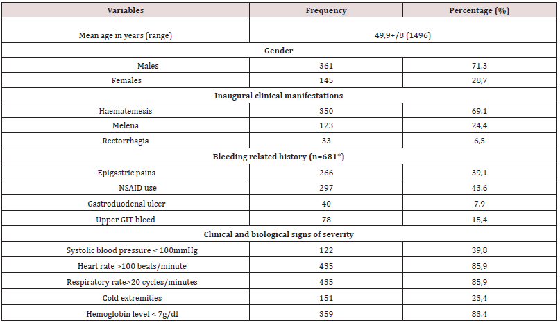
A retrospective collection of data contained in the files of patients admitted for acute UGIB at the Yaounde Central Hospital (Cameroon) between January 1st2010 and December 31st2019, was carried out. Located at the heart of the city of Yaounde, the Yaounde Central Hospital (YCH) was created in 1930. It is the largest public referral hospital in Cameroon with capacity of …. Beds that also acts as a teaching hospital. It houses several services including the Hepato-gastroenterology service with a capacity of 34 beds, three university specialists, four general practitioners and several permanent workers and residents. This service has an upper and lower gastrointestinal endoscopy room and endoscopy equipment. The variables recorded included: demographics (age, sex); clinical and biological characteristics (onset of UGIB, history of bleeding, physical parameters of bleeding severity, and haemoglobin levels on admission); Esogastroduodenal endoscopy (EGDE) findings; specific management of the cause of the bleeding and outcomes (Table1).

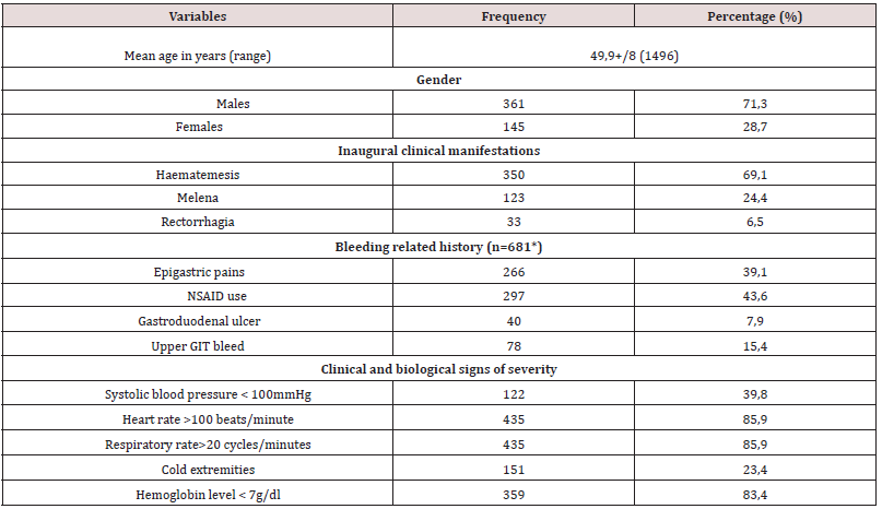
Table 1: Clinical and biological parameters of 506 patients admitted at the Yaounde Central hospital for a recognized acute UGIB.

Lupinepublishers-openaccess-gastroenterology-hepatology-journal

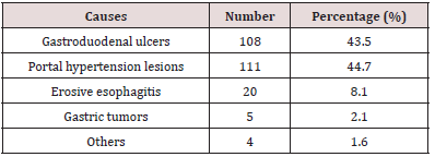
Table 2: Main causes of acute UGIB at the Yaounde Central Hospital amongst 203 patients who had an EGD.

Lupinepublishers-openaccess-gastroenterology-hepatology-journal

The second component, cross-sectional and observational blinded experiments, conducted from December 1st 2017 to May 31st 2018, assessed the diagnostic approach and actual management of patients admitted for acute UGIB in five hospitals of different categories (1st to 3rd) of the Cameroonian health pyramid, in the city of Yaounde. Twenty (20) doctors and 39 nurses in the emergency service who managed 74 patients admitted for acute UGIB were followed. The elements to take care of acute UGIB included recognition of acute UGIB by placing a nasogastric tube or performing a digital rectal exam; assessment of severity by clinical criteria (taking blood pressure (BP), pulse rate (PR), respiratory rate (RR) and room oxygen saturation (SPO2), Glasgow- Blatchford score; fluid resuscitation and transfusions (double venous line, crystalloids filling, proton pump inhibitors (PPI) or vasoactive treatment, oxygenation, restrictive blood transfusion (haemoglobin (Hb) <7 g/dl), hourly monitoring of vital signs and neurological status) and finally, the performance of EGDE and the Rockall and Forrest scores.

Statistical Analysis

Data was analysed using Statistical Package for Social Sciences (SSPS Inc, Chicago, Illinois, USA) version 23.0. Means ± standard deviation was used for quantitative variables; Categorical data was expressed as numbers and proportions. A p value of less than 0.05 was considered statistically significant.

Results

During the retrospective period, 506 patients (prevalence of acute UGIB in the services 5.6%) were included of which 361 men (71.3% and 145 women (28.7 %), given a 2.5 sex ratio. The mean age was 49.9+/- 8 years (maximum 14-96 years) and the peak of bleeding was in the 55-65-year age group. Haematemesis was the initial complaint in 350 patients (69.1%), non-steroidal antiinflammatory drugs (NSAIDs) constituted the most important risk factor in 297 patients (43.6%), in 78 (15.4%). This was a second episode. On admission, clinical parameters relevant to severity were systolic BP <100 mmHg in 122 patients (39.8%); HR >100 beats/ minutes in 435 (85.9%) and RR >20 cycles/minute in 435 (85.9%). Hb was <7g/dl in 359 patients (83.4%). EGDE was performed in 203 patients (40.2%), the major causes of bleeding were: portal hypertension lesions in 111 patients (44.7%) followed by peptic ulcers in 108 (43.5%). Treatment was mainly medical. However, 94 patients (84.7%) with portal hypertension lesions received endoscopic treatment, mainly by injection of sclerosing agent (69.1%), as well as 13 patients (1.2%) with peptic ulcers, mainly isolated injection of dilute adrenaline (1: 10,000) in 11 (84.6%). A total of 75 patients (14.8%) died during hospitalization (Table 2).

The second part concerned the assessment of care offered to 74 patients admitted for acute UGIB (mean age 55 years; sex ratio 2.1). For the recognition of bleeding, digital rectal examination was performed in 43 patients (58.1%) and no patient received nasogastric tube. Regarding the assessment of severity by clinical criteria, BP was taken in 73 patients (98.6%), HR in 61 (82.4%), and RR in 17 (23%), no patient had prognostic scores in the record, including the Glasgow-Blatchford score. Only 17 patients (23%) had SPO2 measurements. Regarding intensive care measures, only 10 patients (13.5%) received a double peripheral venous line, the majority of which was a small-bore venous line. Twenty patients (27%) were filled with crystalloids; restrictive blood transfusion was performed in 24 out of 27 patients (88.9%) with Hb < 7 g / dl. Only 9 patients (12.2%) received nasal oxygen therapy. EGDE was performed in 43 patients (60.6%), all beyond 24 hours after admission and none had a prognostic score after endoscopy (Forrest or Rockall).

Discussion

The study showed that acute UGIB is an emergency with exceptional severity in our environment, as mortality is very high at around 15%. In this study, it was found that acute UGIB affected two and a half times more men than women with a mean age of about 50 years. This is the case in studies conducted in Mali (sex ratio 2.78; mean age 47.45 years), and in Côte d’Ivoire (sex ratio 3.38; mean age 47 years) [8,12]. Indeed, in the sub-Saharan African region, the occurrence of acute UGIB often involves relatively young patients. This can be explained by the fact that the causes of bleeding are often dominated by portal hypertension lesions, especially in young patients, as reported in Mali in a rural area [8]. As opposed to sub-Saharan Africa, in the West, the age of onset of acute UGIB is higher than 70 years due to the use of NSAIDs in the elderly population. Of chronic NSAID users, 25% develop an ulcer, of which 2-4% are complicated by bleeding [2,7,13,14]. The male predominance is universal, and reverses in the West after the age of 80 years due to the higher life expectancy in the female population [2,4,6-9, 11,13,14].

As in several studies reported in literature, hematemesis was the most common initial clinical presentation [2,6,7,15,16]. This initial clinical presentation can be explained by the different lesions found on endoscopy. In fact, portal hypertension lesions, led by oesophageal varices, were the most frequent, alongside peptic ulcers. The frequency of the various aetiologies varies from one region to another [5,6,8]. Thus, in the sub-Saharan African region, several studies report very high frequencies of portal hypertension lesions. This is the case of the study by Diarra et al. in Mali in 2007 [8]. The authors reported a frequency of 55.2% in favour of ruptured oesophageal varices, far ahead of peptic ulcers which represented 16%. The frequency of portal hypertension lesions is also high in Burundi (28.2%) [16] and Gabon (29.5%) [17]. These various countries have liver diseases related to chronic HBV infection in common. Indeed, sub-Saharan African countries are located in a zone of high endemicity according to the World Health Organisation (WHO), i.e. the prevalence of hepatitis B is 8-20% of the general population [10]. Cirrhosis, which causes portal hypertension, is often the result of chronic HBV infection acquired at birth or in early childhood [18]. The annual incidence of varicose veins is approximately 5% [19]. UGIB from ruptured esophageal varices accounts for 70% of gastrointestinal bleeding in cirrhosis, with an estimated overall 2-year bleeding risk of 20% [19]. The aetiological approach to acute UGIB in high- and intermediate-endemic areas where the majority (80%) of the world population lives should therefore consider this high frequency of portal hypertension lesions and bleeding complications.

Regarding the severity of the bleeding, more than 85% of patients were initially unstable with clinical and laboratory signs of severity, despite the absence of Glasgow-Blatchford and Rockall scores in their records. This initial haemodynamic instability can be explained, on the one hand, by the causes of bleeding, in particular the rupture of oesogastric varices, which are exceptionally serious, but also, on the other hand, by the late arrival in hospital structures due to distance, cultural considerations or difficulties of mobility in our country.

Treatment was essentially medical. PPI treatment was most often initiated on admission, even if this treatment did not always fully comply with current recommendations in Europe and Asia [2,7,20,21]. Contrarily, in cases of suspected acute UGIB related to rupture oesogastric varices, vasoactive therapy to reduce portal blood flow was never initiated on admission, in line with international recommendations, including those adapted by the European Society of Gastrointestinal Endoscopy to be applicable to resource-limited settings, including some African countries [2,7,22-24]. Patients with bleeding peptic ulcers have rarely benefited from endoscopic haemostasis, unlike those with portal hypertension lesions. Late arrival at hospital would explain why in sub-Saharan Africa Forrest scores IIc and III, i.e. pigmented stain or clean base of the ulcer, are most frequently found [25]. For the Forrest classification guiding the choice of endoscopic treatment modality for ulcers, there is no endoscopic treatment for these scores [2,3,7,26]. The practice of injecting adrenaline alone instead of combined adrenaline and bipolar coagulation or endoclips was inappropriate. This could be explained by factors such as excessive procedure costs, insufficient training of practitioners and limited logistic equipment.

Read More About Lupine Publishers Journal of Gastroenterology and Hepatology Please Click on Below Link: https://currenttrendsingastroenterology.blogspot.com/

Tuesday, 28 March 2023

Lupine Publishers| Improvement of Liver Function by a Short-term Administration of Luseogliflozin in Patients with Type 2 Diabetes: A Single-arm Study and the Mini-literature Review

Lupine Publishers| Journal of Gastroenterology and Hepatology


Abstract

Background: Non-alcoholic fatty liver disease is not simply the hepatic manifestation of obesity and diabetes but also linked to hepatocellular carcinoma. Yet, its effective treatment has not been established. In this study, we evaluated the effect of a short-term administration of luseogliflozin to patients with type 2 diabetes having non-alcohol fatty liver disease. Luseogliflozin is a unique sodium-glucose cotransporter 2 inhibitor which is metabolized in and excreted by the liver in addition to the kidney. Therefore, the drug might possess an additional effect on other agents of the same class which are exclusively metabolized in the kidney.

Methods: Using alanine aminotransferase >20 IU/L as a diagnostic basis for non-alcoholic fatty liver disease, 19 patients, not taking alcohol, with type 2 diabetes (male/female 15/4, the median age 57 years) was treated with 2.5 mg luseogliflozin for 12 weeks.

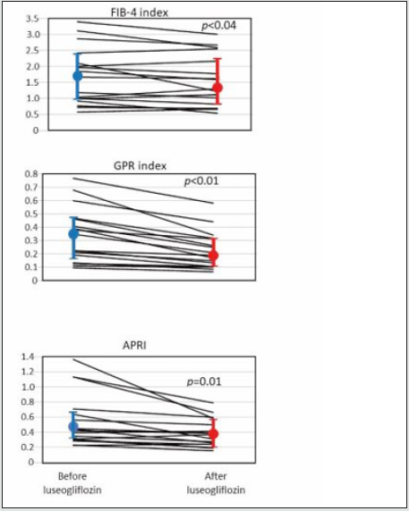
Results: Pre- and post-treatment median values for alanine aminotransferase were 51 IU/L and 33 IU/L (p = 0.001), and the corresponding values for the fibrosis index based on the four factors [age (years) ∙ alanine aminotransferase (IU/L)] / [platelets (109/L) ∙ alanine aminotransferase (IU/L)1/2)] were 1.669 and 1.314 (p = 0.043). There was no adverse effect of the drug. Our findings were essentially compatible with the results of the previous studies reviewed.

Conclusion: We conclude that sodium-glucose cotransporter 2 inhibitor could be the choice for the pharmacological treatment of non-alcoholic fatty liver disease. Especially, a short-term administration of it is consistently effective in mild cases.

Keywords: SGLT2 inhibitor; NAFLD; Fibrosis-4 index; GPR-index; APRI.

Abbreviations: SGLT2i: sodium-glucose cotransporter 2 inhibitor; NAFLD: non-alcoholic fatty liver disease; NASH: non-alcoholic steatohepatitis; T2DM: type 2 diabetes mellitus; AST: aspartate aminotransferase; ALT: alanine aminotransferase; GGT: gammaglutamyl transpeptidase; Fib-4 index: Fibrosis-4 index; GPR-index: gamma-glutamyl transpeptidase to platelet ratio; APRI: aspartate aminotransferase to platelet ratio

Introduction

Obesity or overweight is the motherland of non-alcoholic fatty liver disease (NAFLD) and non-alcoholic steatohepatitis (NASH) [1]. Accordingly, with increasing trend of body weight worldwide, the number of patients with the liver problem is relentlessly increasing [2]. Recently, NASH has also been attracting the attention as a cause of hepatoma [3,4]. Under such yet, treatment of NAFLD (NASH and NAFLD are collectively called NAFLD hereafter in this communication), irrespective of presence or absence of diabetes, has not been established. Results of treatment of patients with type 2 diabetes (T2DM) having NAFLD with sodium-glucose cotransporter- 2 inhibitor (SGLT2i) appears promising [3-7], the effectiveness of a short-term treatment, such as 12 week-treatment, has not been established.
Here, we evaluated effectiveness of a short-term administration of SGLT2i, luseogliflozin, that is metabolized not only in the kidney but in the liver [8]. A mini literature review on this issue was also performed to resolve the current inconsistency. This study was approved by the Clinical Research Ethics Committee of Aizawa Hospital (No. 2019-094).

Subjects and Methods

Subjects

Consecutive 29 patients with T2DM who took luseogliflozin for 3 months or longer between January 1, 2019 to May 31, 2020 were initially registered. Because the purpose of this study was to investigate the effect of luseogliflozin on NAFLD/NASH, 8 with alanine aminotransferase (ALT) less than 20 IU/L [9], and other 2 with a habitual alcohol drinking of 20 g/day or more were excluded, and the remaining 19 were analyzed.
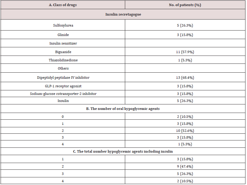
The study was a single-arm, add-on study. Namely, 2.5 mg luseogliflozin was additively prescribed on top of the hypoglycemic agents already taken by the patients, which are shown in Supplemental Table 1. The data before and 12 weeks after luseogliflozin administration was critically compared.

Laboratory measurements

In addition to the routine clinical chemistries, indices of the hepatic fibrosis including Fibrosis-4 index (Fib-4 index), gammaglutamyl trans peptidase (GGT) to platelet ratio (GPR-index), and aspartate aminotransferase (AST) to platelet ratio (APRI) were calculated10: the unit for AST, ALT, GGT as IU/L, platelet counts as 109/L and age as a year. Equations for each index were as follows [10].

Fib-4 = [(AST) ∙ (age)] / [(Platelet counts) ∙ (ALT)1/2]
GPR index = 100 ∙ (GGT) / (Platelet counts)
APRI = 100 ∙ (AST) / (Platelet counts)

Statistical analysis

The data were collected retrospectively and analyzed cross-sectionally and longitudinally. We evaluated the effect of luseogliflozin on the liver function tests and the indices of liver fibrosis. In addition, delta, i.e., the basal value minus 12 week-value for the indices of liver fibrosis was calculated for each study subject, and correlation between the delta values of fibrosis indices and the basal plasma glucose (PG), glycosylated hemoglobin (HbA1c), AST, ALT, GGT, body weight (BW) and body mass index (BMI) were examined. Furthermore, the delta value of the liver fibrosis indices and the delta value of PG, HbA1c, AST, ALT, GGT, BW and BMI were also examined. The statistical analysis was performed using JMP ver.15. Wilcoxon rank-sum test, Wilcoxon signed rank test and Spearman rank correlation were used as needed.

Literaturereview

Representative 10 original reports published in the English language on the treatment of patients with T2DM having NAFLD by SGLT2 inhibitors [6,7,11-18] were summarized to provide a current overview of the issue. In these literature, five kinds of SGLT2i agents were prescribed, with the number of the patients ranging from 9 to 32 and the treatment duration from 12 to 48 weeks.

Results

Baseline characteristics of the patients

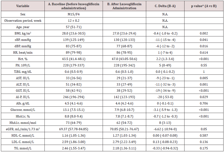
Table 1. Baseline data (A), the data 3 months after the luseogliflozin treatment (B) and the difference (C) between the two (luseogliflozin- basal).

Lupinepublishers-openaccess-gastroenterology-hepatology-journal

Values are median and interquartile ranges except for sex and the observation period: the latter was shown as mean and SD. sBP, systolic blood pressure; dBP, diastolic blood pressure; HR, heart rate; Hct, hematocrit; Plt, platelet count; T.Bil, total bilirubin; AST, aspartate aminotransferase; ALT, alanine aminotransferase; GGT, gamma-glutamyl transpeptidase; Al-P, alkaline phosphatase; Alb, albumin; eGFR, estimated glomerular filtration rate; HDL-C, high density-lipoprotein cholesterol; LDL-C, low density-lipoprotein cholesterol; TG, triglycerides. N.A., not applicable. *Wilcoxon signed-rank test.

The study patients were male dominant (the proportion of males, 79%), middle-aged Japanese adults with the median BMI of 28.0 kg/m2 which was larger than the representative value in the Japanese patients with T2DM in general11(Table 1A). The median HbA1c value of the entire group before luseogliflozin was 8.8% (77 mmol/mol) so that the level of glycemic control was unsatisfactory (Table 1A). Regarding the hypoglycemic agents used before subscribing luseogliflozin, Dipeptidyl Peptidase-4 (DPP-4) inhibitors and biguanide were most frequently employed (Supplemental Table 1), which was typical for the Japanese patients [19].

Supplemental Table 1: Medications before administration of luseogliflozin. 2.5 mg luseogliflozin was added in each patient on top of the medication described above.

Lupinepublishers-openaccess-gastroenterology-hepatology-journal

Values are median and interquartile ranges except for sex and the observation period: the latter was shown as mean and SD. sBP, systolic blood pressure; dBP, diastolic blood pressure; HR, heart rate; Hct, hematocrit; Plt, platelet count; T.Bil, total bilirubin; AST, aspartate aminotransferase; ALT, alanine aminotransferase; GGT, gamma-glutamyl transpeptidase; Al-P, alkaline phosphatase; Alb, albumin; eGFR, estimated glomerular filtration rate; HDL-C, high density-lipoprotein cholesterol; LDL-C, low density-lipoprotein cholesterol; TG, triglycerides. N.A., not applicable. *Wilcoxon signed-rank test.

Liver function and indices of hepatic fibrosis following luseogliflozin administration

Elevated serum level of ALT was an inclusion criterion, so that the ALT was clearly elevated as a group with the median value, 51 IU/L. As well expected, administration of luseogliflozin significantly decreased PG and HbA1c (Table 1, A and B, before and after luseogliflozin, respectively). In addition, it significantly lowered the serum level of AST, ALT, GGT, and alkaline phosphatase (ALP). The degree of lowering was 12%, 34%, 35 and 42% for the respective enzyme levels. Importantly, the luseogliflozin treatment also significantly lowered the values for Fib-4 index, GPR index, and APRI (Figure 1).

Correlation between delta Fib-4, GPR index, and APRI and baseline and delta values of liver function and the body weight and BMI

Figure 1: Change of indices of liver fibrosis produced by luseogliflozin. Individual lines represent a change of the value in each person and the circles, and the vertical lines indicate the median and interquartile ranges: blue ones for before and red ones for after luseogliflozin. Wilcoxon signed-rank test was used for the statistical analysis.

Lupinepublishers-openaccess-gastroenterology-hepatology-journal

The delta Fib-4 index was significantly and positively correlated associated with higher baseline AST and ALT levels; the delta GPR index was correlated with baseline AST and GGT; the greater delta APRI was associated with higher baseline AST and ALT (Table 2A). Correlation between the delta values and the baseline of liver function was absent for BW and BMI (Table 2A).
On the other hand, there was an inverse correlation between ‘delta Fib-4 and delta AST and delta ALT’, ‘delta GPR index and delta GGT’ and ‘delta APRI with delta AST and delta ALT’ (Table 2B). There was no significant correlation between delta values of the fibrosis indices and the delta of BW and BMI (Table 2B).

Table 2. Correlation between delta Fib-4, GPR index and APRI and baseline value (A) and delta (B) of liver function. Delta means the difference between the value of each test performed on the day of starting luseoglifrozin and 12 weeks later.

Lupinepublishers-openaccess-gastroenterology-hepatology-journal

Values are median and interquartile ranges except for sex and the observation period: the latter was shown as mean and SD. sBP, systolic blood pressure; dBP, diastolic blood pressure; HR, heart rate; Hct, hematocrit; Plt, platelet count; T.Bil, total bilirubin; AST, aspartate aminotransferase; ALT, alanine aminotransferase; GGT, gamma-glutamyl transpeptidase; Al-P, alkaline phosphatase; Alb, albumin; eGFR, estimated glomerular filtration rate; HDL-C, high density-lipoprotein cholesterol; LDL-C, low density-lipoprotein cholesterol; TG, triglycerides. N.A., not applicable. *Wilcoxon signed-rank test.

Read More About Lupine Publishers Journal of Gastroenterology and Hepatology Please Click on Below Link: https://currenttrendsingastroenterology.blogspot.com/

Wednesday, 8 February 2023

Lupine Publishers| Is there a utility of Nordin’s Index in the evaluation of osteoporosis in patients with post viral cirrhosis? Results of a pilot study in Cameroonians

 Lupine Publishers| Journal of Gastroenterology and Hepatology


Abstract

Background: Metabolic bone disorders are frequent in patients with cirrhosis. These two conditions are usually misdiagnosed in Sub-Saharan Africa before the onset of complications. A full evaluation of metabolic bone disorders in patients with liver cirrhosis is difficult in our milieu and osteodensitometry is rarely available. In this preliminary study, we sought to determine the necessity of a cost-effective method, the Nordin’s Index, in the evaluation of osteoporosis in patients with liver cirrhosis.

Methods: This was a prospective cross-sectional study from January to May 2017 in Yaoundé. We compared the data of 19 patients (15 men, 4 women) with post viral liver cirrhosis and 17 controls (13 men, 4 women) paired with age, gender and body mass index (BMI). Data collected included vitamin D levels, serum and urine concentrations of calcium and phosphorus, Nordin’s Index and results of the bone mineral density using an x-ray absorptiometry scan. Statistical analysis was performed using the software SPSS 21. A p value of less than 0.05 was considered to be statistically significant.

Results: The mean age of patients was 38 ±15 years, with a mean BMI of 25 ±8 kg/m². Three of the four women were on menopause. Etiologies of cirrhosis were viral hepatitis B (8 patients), viral hepatitis B and D coinfection (7 patients), and viral hepatitis C (4 patients). The median duration of cirrhosis was 19 [8; 48] months, and 14 patients were classified grade A in Child Pugh classification. There was no statistical difference in the serum and urine concentrations of calcium and phosphorus. Osteoporosis was more frequent in cirrhosis (31.6% versus 11.8%, p<0.05). Nordin’s Index was significantly elevated in patients with cirrhosis compared to controls (0.12 [0.06; 0.13] mg/mg, versus 0.03 [0.01; 0.08] mg/mg, p<0.05), and in patients with cirrhosis associated to osteoporosis compared to those without (0.13 [0.09; 0.13] mg/mg versus 0.07 [0.03; 0.08] mg/mg, p<0.05). Vitamin D deficiency was more observed in controls (13/17 versus 7/19, p<0.05). Factors associated with osteoporosis were disease duration, elevated Nordin’s Index and elevated serum level of transaminases.

Conclusion: The Nordin’s Index, a simple and inexpensive tool for exploration of the phosphocalcic metabolism, could be useful for the evaluation of osteoporosis during viral cirrhosis. However, its performance has to be evaluated in a larger sample.

Keywords: Cirrhosis, chronic viral hepatitis, osteoporosis, Nordin’s Index, Vitamin D.

Abbreviations: ALAT: Alanine Aminotransferase; ALP: Alkaline Phosphate; ASAT: Aspartate Aminotransferase, BMD: Body Mass Density; GGT: Gamma Gluthamyl Transferase

Introduction

Cirrhosis is the outcome of most chronic liver diseases. It remains a major public health problem in Sub-Saharan Africa and particularly in Cameroon, where chronic viral hepatitis is highly endemic [1-3]. In fact, sixty percent of cirrhosis in Africa are attributable to viral hepatitis B and C [4]. In Cameroon, 70.9% of cirrhosis cases are due to chronic viral hepatitis B and 25.5% to chronic viral hepatitis C [5]. The hallmark of cirrhosis is hepatocellular insufficiency and portal hypertension, resulting in impaired liver function and subsequent systemic abnormalities. Bone damage is one of the systemic abnormalities frequently seen in cirrhosis, regardless of etiology, and is known as hepatic osteodystrophy [6].
Hepatic osteodystrophy is a combination of osteoporosis and osteomalacia, the latter being rare during cirrhosis [6]. According to the World Health Organization (WHO), osteoporosis is a diffuse disease of the skeleton, characterized by a decrease in bone mass and an alteration of the micro-architecture of bone tissue, leading to increased bone fragility and an increased risk of fractures [7]. Its pathogenesis during cirrhosis is complex and leads to an increase in bone resorption by osteoclasts to the detriment of its formation by osteoblasts [8]. The prevalence of osteoporosis during cirrhosis is between 12% and 55% [9]. In Africa, more precisely in the Maghreb region, it has been estimated to 28.26% [10]. Osteoporosis is almost asymptomatic until complications. It is a real public health problem as it causes low energy fractures in more than 40% of cases, affecting the morbidity and quality of life of patients with cirrhosis [8]. Several factors are associated with the occurrence of osteoporosis during cirrhosis, including vitamin D deficiency with a prevalence of 32% in Europe and 32.6% in Africa during cirrhosis [10,11]; Elevated Child and Pugh Score, duration of disease, etiology of cirrhosis, low body mass index, with some controversy in the literature [12].
Considering the risk of osteoporosis being high during cirrhosis, it is necessary to have an evaluation of bone mineral density in these patients [13,14]. However, this is rare in our context because of the cost and accessibility of this examination. With the absence of hepatic transplantation in our environment to cure cirrhosis, there is a long duration of cirrhosis and, therefore, an increased risk of complications including osteoporosis. It is therefore important to use simple and inexpensive tools to make the diagnosis or at least to detect high-risk subjects. Nordin’s Index is calculated by the calciuria/creatinuria ratio. It is an old marker of bone resorption, which correlates with post-menopausal osteoporosis and its risk of fracture [15,16]. However, its usefulness in cirrhosis especially post viral has been little studied to the best of our knowledge. In this study, we sought to determine the utility of Nordin’s Index as a cost-effective method in the evaluation of osteoporosis in people suffering from post-viral cirrhosis in a highly endemic area for chronic viral hepatitis and limited access to bone mineral density assessment.

Patients and Methods

Study design

The study used a cross sectional design with a prospective data collection.

Study setting

This study took place from January to May 2017 at the Yaoundé Central Hospital and Cathedral Medical Centre (CMC) at Yaoundé, where the participants were recruited. Laboratory analysis were conducted at the University Hospital Centre of Yaoundé. Bone mineral density measurements were carried out at the Autonomous Centre for Radiology and Medical Imaging (CARIM) at Yaoundé.

Participants

The sample was made up of 19 adult volunteers (15 men and 4 women) followed for a cirrhosis of viral cause (chronic viral hepatitis B, C, and D). They were matched with 17 adult volunteers (13 men and 4 women) without any chronic or acute disease/ condition, paired with age (±2 years), gender and body mass index (BMI; ±2 kg/m²). The diagnosis of liver cirrhosis was based on clinical and biological signs of portal hypertension and chronic liver failure, ultrasonographic signs of chronic liver disease and endoscopic signs of portal hypertension. Ultrasonographic signs included irregular liver outline, heterogeneous echo structure, dysmorphic liver, enlarged portal vein and presence of collateral venous circulations.
The controls were recruited from among the patients’ caregivers to ensure equal exposure to environmental and nutritional factors that may contribute to osteoporosis. We did not include any participants who presented pathologies with known repercussions on bone and/or phosphocalcic metabolism, such as: chronic renal disease, thyroid and parathyroid disorders, Cushing Syndrome, diabetes, HIV infection and cancer. We did not include participants receiving treatment with known effects on bone and/ or calcium phosphorus metabolism such as: hormone replacement therapy, biphosphonates, calcium, vitamin D, corticosteroids, antimetabolites, anticoagulants, anticonvulsants, thyroxine. Pregnant women instead of women with amenorrhea, participants with diabetes, tobacco and alcohol consumers were not included.

Sample size calculation

The sample size was estimated using the formula in Whitley et al. to compare proportions between groups [17]. The parameters used for the calculation of the standardized difference were derived from the study of Goral et al [18]. They found a mean T-score of -1.6 SD in patients with cirrhosis and -0.25 SD in controls, with a standard deviation of 1.3 SD. In our study, the level of significance was set at p 0.05 and the power at 80%. The minimum sample per group was 15.

Procedure

Ethical considerations

We obtained research authorizations in the selected health care facilities and the approval of the Institutional Review Board of the Faculty of Medicine and Biomedical Sciences. All the participants read and signed an informed consent form.

Clinical data

Sociodemographic (age, gender) and clinical data were collected through a questionnaire. Clinical variables were anthropometric parameters (weight and height) for BMI calculation, the history of cirrhosis (duration, etiology, complications, current Child and Pugh’s Score, treatment), suggestive signs of osteoporosis and its complications (history of pathological and or low energy fracture), comorbidities, menopause for women and its duration, and family history of osteoporosis.

Laboratory Analysis

For laboratory analysis, 10 mL of peripheral venous blood sample was collected from each participant after eight hours of fasting. The blood samples were centrifuged for 5 minutes at 3000 rpm using the Humax 14K Germany centrifuge. 500μl of serum was then extracted for the determination of calcium (mg/L), phosphorus (mg/L), albumin (g/L) by colorimetric method and for the determination of creatinine (mg/l), total alkaline phosphatase (U/L), transaminase (Aspartate amino transferase (ASAT), Alanine amino transferase (ALAT), in U/L), gamma gluthamyl transferase (GGT, U/L) by kinetic method, using the Mindray model BS 120 spectrophotometer. The remaining serum was stored at -20° Celsius and used to determine 25(OH) vitamin D (ng/mL) levels by ELISA using the BioTek EL×800 ELISA chain. Serum creatinine was used to estimate the glomerular filtration rate using four parameters’ MDRD formula in mL/min for 1.72m². The measured calcemia was corrected using albuminemia. Hypercalcemia was defined for a corrected Calcemia > 104 mg/L, while Hypocalcemia was defined below 81 mg/L [19]. Hyperphosphatemia was defined as blood phosphorus > 50 mg/L in adults while hypophosphatemia was defined below 25 mg/L [19]. Vitamin D Status was assessed according to the classification of Holick et al [20].

Laboratory Analysis

After consumption of 200 ml of low calcium water (Volvic® type: Ca<10 mg) in a fasting individual with a previously empty bladder, calciuria and creatininuria was measured on the urine collected 2 hours later. Nordin’s Index was calculated: calciuria/ creatininuria (mg/mg). The normal value of the Nordin’s Index is lower than 0.11 mg/mg. An increase in the index indicates hyperresorption of bone. Calcium, phosphorus and creatinine in urine were determined using the same methods as in the plasma.

Bone Mineral Density (BMD)

Bone mineral density was measured by biphotonic X-ray absorptiometry scan using the Hitachi® lunar prodigy DXA system, with 37 Micro SieVert (μSV) dose per measurement site. Bone mineral density was measured at the lumbar spine, the neck of the left femur and the distal end of the left radius. Data on bone mineral density were expressed in g/cm², T-score and Z-score. Osteoporosis was defined as T-score <-2.5 for patients aged 50 years and older or a Z-score <-2 for patients under 50 years of age for measurement of the lumbar spine, femoral neck or distal end of the radius [21].

Statistical analysis

Statistical analysis was performed using SPSS version 21.0. Quantitative variables were expressed as mean ± standard deviation or median and interquartile range [25th and 75th quartiles]. Qualitative variables were expressed as effectives and proportions. The Mann-Whitney test was used to compare the means, while medians were compare using the Median’s test. Fisher Exact Test was used to compare proportions. We searched for associations between continuous variables and bone mineral density on various sites by calculating the Pearson’s Correlation Coefficient to determine the factors associated with the decrease in BMD. For all the tests, the statistical significance level was set at 0.05.

Results

Clinical characteristics of the study samples

The mean age of patients was 38.6 ±15.9 years, while that of controls was 37.8 ±15.9 years (p>0.05). Participants’ ages ranged from 23 to 73 years. There were respectively 2/4 and 2/4 women on menopause in the patients and control groups. Between cases and controls, there was no statistical difference in body mass index (p>0.05) (Table 1). The median duration of cirrhosis was 19 months, range from one to 120 months. The causes of liver cirrhosis were chronic viral hepatitis B (42%), hepatitis C (31%), and B-delta (37%). There was no coinfection with viral hepatitis B and C. Most of the cases were classified on stage A (n=14), while 3 and 2 cases were respectively on-stage B and C in Child and Pugh classification.

Read More About Lupine Publishers Journal of Gastroenterology and Hepatology Please Click on Below Link: https://currenttrendsingastroenterology.blogspot.com/

Monday, 21 November 2022

Lupine Publishers| Case of Application of Syrolimus in a Patient with Progressing Pultifocal Pepatic Nodular Hyperplasia

 Lupine Publishers| Journal of Gastroenterology and Hepatology


Abstract

Introduction: A case presented to the public demonstrates the possibility of treatment with Sirolimus in a patient with progressive multifocal liver Focal Nodular Hyperplasia (FNH) with progressive growth and a tendency to formation of liver function insufficiency after splenorenal shunt for portal hypertension.

Material and Methods: Patient E. 08.03.2001 year of birth c Diagnosed with multifocal bilobar nodular liver hyperplasia after splenorenal bypass surgery for portal hypertension, established in 2018/ Sirolimus treatment was prescribed from 06.22.18 to the present.

Conclusion: As a result of the treatment with Sirolimus, a normalization of the level of liver enzymes, bilirubin in the blood is noted. According to radiation methods, a reduction in liver size and FNH size is up to 30%.

Keywords:Multifocal liver Focal Nodular Hyperplasia (FNH); Sirolimus

Introduction

Today, FNH is regarded as a benign vascular formation of the liver. VNG is the second most common benign liver neoplasm [1]. Edmonton first described it in 1958 [2]. FNH most often occurs as a monofocal lesion, in more rare cases there are two or more nodes [3]. FNH is believed to be a hyperplastic response of liver tissue to arterial malformation, and not a true tumor. When conducting radiation diagnostics, the central scar is determined in 44% of cases. The central scar with T2-weighted MRI is hyperintensive [4]. It is possible to determine the filling of blood formation from the center to the periphery, which distinguishes it from monofocal liver hemangiomas. As a rule, in the case of PF, the risk of complications is low; there is no risk of malignancy. Therefore, a patient with VNF rarely requires their surgical removal. The most common indication for removal is pain, which usually occurs with FNH greater than 7 cm in distance [5]. The presence of more than five nodes are characterized as multiple FNH. This is very rare and only a few cases are described in the literature [6]. With progressive multiple FNH, we did not find any treatment recommendations in the literature other than liver transplantation. Therefore, we decided to present a case of treating multiple liver FNHs in a patient using Sirolimus therapy. We also did not find such articles in the literature and therefore we bring this case to the public.

Materials and Methods

Patient E. 08.03.2001 year of birth. Born from the first pregnancy, proceeding against the background of chronic intrauterine hypoxia of the fetus. Childbirth - emergency cesarean section. Height at birth 51 cm, weight 3118 grams, Apgar score of 8 points. In the neonatal period: pneumonia, urinary tract infection, perinatal hypoxic encephalopathy of the fetus, intraventricular hemorrhage. Grew and developed according to age.
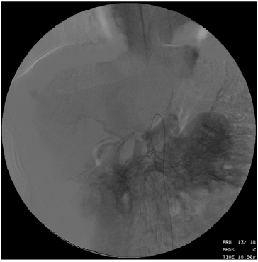
In August 2008, he suffered bleeding from the dilated veins of the esophagus and stomach. Bleeding is stopped conservatively. The diagnosis of portal hypertension syndrome, cavernous portal vein transformation. In October 2008, surgical treatment was carried out: the formation of spleno-renal anastomosis “side-byside” (Figure 1). No more bleeding was noted.

Figure 1.

Lupinepublishers-openaccess-gastroenterology-hepatology-journal

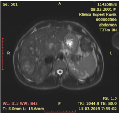
In February 2018, during a control study, multiple FNHs were identified in C1, C4, C8 with dimensions of 87x61 mm, in C7 with dimensions of 51 by 43 mm, 50 by 40 mm (Figure 2). Diagnosed with multifocal bilobar nodular liver hyperplasia after splenorenal bypass surgery for portal hypertension. Blood test for alphafetoprotein 1.66 IU / ml. During the control examination in July 2018, an increase in FNH sizes to 90 by 70 mm, an increase in blood transaminases (Table 1), alkaline phosphatase up to 218.00 IU / L (N 42 - 110), a moderate increase in bilirubin were noted. The patient complained of chronic fatigue, weakness, moderate pain in the liver. lack of appetite.

Figure 2.

Lupinepublishers-openaccess-gastroenterology-hepatology-journal

Table 1.

Lupinepublishers-openaccess-gastroenterology-hepatology-journal

After examining the child, we came to the conclusion that the observed progression of the disease can lead to the replacement of the liver parenchyma with the subsequent occurrence of organ failure. This could endanger the patient’s life, but there is no possibility of surgery due to the prevalence of the lesion. Sirolimus (Pfizer, USA) from 06.02.2018 was prescribed orally daily at a dose of 3 mg / day until a therapeutic concentration of the drug in blood serum of 6-15 ng / ml was achieved. Due to the excess of the therapeutic interval, the dose of Sirolimus was consistently reduced from 11/06/2018 to 1 mg / day, 02/05/2019 the concentration of Sirolimus was 7.8 ng / ml.

Résultats

As a result of the treatment with Sirolimus, a normalization of the level of liver enzymes, bilirubin in the blood is noted. According to radiation methods, a reduction in liver size and FNH size is up to 30%. Now the patient is feeling well. There is no pain in the liver. Playing sports. Reception of sirolimus continues to date.

Discussion

Multifocal FNH is a rare form of this disease. An even rarer situation arose in our patient, there was a progression of the disease with signs of emerging hepatic cell failure. There was a high risk of severe disease with subsequent liver transplantation. There is an interesting fact that the physiological characteristics of the patient appeared after application of a splenorenal shunt for portal hypertension. Apparently, in the growth stimulation of the nodes, the FNH played the role of impaired portal blood flow. In the available literature, we found only recommendations for liver transplantation in such patients. Teaching ability of Sirolimus to suppress vascular growth through inhibition of M-TOP, we suggested the possible effectiveness of this therapy. After an explanation with the patient and his legal representatives, we started therapy with Sirolimus at a dose of 3 mg / day, then we reduced it to 1 mg / day taking into account the concentration in the blood. The patient had no complications associated with Sirolimus therapy. After a year of treatment, we noted a clear positive trend. The patient’s condition improved, there was no pain, blood counts returned to normal (Table 1).

Conclusion

Of course, this question still requires research in other patients with multiple progressive FNH, in order to draw conclusions about the effectiveness and safety of Sirolimus. But with this patient, we got encouraging results.

Read More About Lupine Publishers Journal of Gastroenterology and Hepatology Please Click on Below Link: https://currenttrendsingastroenterology.blogspot.com/


Wednesday, 28 September 2022

Lupine Publishers| The Failure Bauhinias Damper as a Possible Cause of Chronic Autointoxication and the Development of Psoriasis

 Lupine Publishers| Journal of Gastroenterology and Hepatology


Abstract

The goal is to assess the impact of biogenetic on the condition of patients with psoriasis

Materials and Methods: The study group included 415 patients aged 19 to 65 years (34% of men and 66% of women), in whom irrigoscopy revealed the injection of contrast into the ileum, i.e., the insolvency of the baugine flap (NBZ) was diagnosed, 74 of them suffered from psoriasis. For the detection of chronic intoxication were evaluated the data of clinical manifestations, indican urine, level of middle molecules, blood serum, lipid metabolism, microbial resistance, the hydrogen breath test.

Results: NBZ is a chronic endointoxication, which regresses in collaboration with the clinical manifestations of psoriasis after biogenetic.

Conclusion: Hypothesized that NBZ is one of the causes of psoriasis, and biogenetica – link in etiopathogenetic method of treatment.

Keywords: Insolvency bauhinias dampers; Chronic endo intoxication; Psoriasis; surgery

Introduction

Recognizing the harmfulness of dysfunction of such barrier structures as cardia, pylorus, sphincter of Oddi, heart valves, venous valves of the lower extremities, etc., modern medicine ignores the failure of the ileocecal locking system (Bauhinia valve) as a possible cause of the pathology of the digestive system and associated various extraintestinal diseases [1].
But it is worth paying attention to the opinion of I.I. Grekova [2] “Despite a number of works devoted to the proximal colon, the pathology of this department is still not sufficiently explained because in these works the role of the bauhinia valve was completely ignored.” Bauhinia valve (BZ) distinguishes between the functions of the small and large intestines, isolates the small intestine from the reflux of the colonic contents, which differs sharply in chemical composition, physical condition and bacterial spectrum [3-5]. According to LG Peretz [6], in 1 ml of intestinal contents there are up to 5000 microbes, and in 1 g of the contents of the large intestine there are about 30-40 billion [6]. With the failure of the Bauhinia Damper (NBZ), billions of colon microbes are thrown into the small one [4,6], colonization of the small intestine with allochthonous (alien) microorganisms occurs, excessive bacterial growth syndrome (SIBO) or enteric dysbacteriosis develops [7,8].
Absorbing the blood products of microorganisms (indole, phenol, cresol, skatole, pyrocatechin, carbolic acid, hydrogen sulfide, mercaptan, ethane, methane, etc.) causes autointoxication phenomena that cannot be sufficiently arrested, especially for liver diseases [7,9-11].
The development of putrefactive and fermentation processes in the small intestine - The consequence of the SIBO. The lymphoid tissue in the course of the gastrointestinal tract suffers because of a violation of the barrier function of the intestinal wall [4,12], resulting in a deficiency of immunoglobulins A and M. In 70% of patients with chronic enterocolitis there is a marked decrease in the body’s immunological reactivity the body becomes less protected before microbial aggression - in patients with chronic colitis [5].
Recognizing NBZ as one of the anatomical causes of SIBR, nevertheless, the authors do not have a therapeutic effect on it [13,14], although they assign SIBOs key pathogenetic mechanisms in many diseases of the digestive tract and associated extradigestive conditions [7,10].

The interrelation between the pathology of the gastrointestinal tract (GIT) and the development of dermatosis is a well-known fact. The small intestine is the organ of the digestive canal, the most responsible for the development of dermatosis. The relationship between the intensity of skin manifestations and the activity of ileocolitis Roberts et al. [15] with psoriasis was detected in 2/3 of patients with enterocolitis [15]. Sensitization to intestinal autoflora was revealed. So sharply positive were skin tests, primarily to Escherichia coli-up to 97% [16]. Allergic dermatosis, rosacea, seborrhea, food allergy, urticaria, neurodermatitis were found in 176 (22%) of 800 patients with diseases of the alimentary canal [17]. In the etipathogenesis of dermatosis, the following enterocolitic links are distinguished [18].
a. The most responsible for the development of dermatosis is the small intestine. The immune complexes formed in the intestine penetrate the blood and settle in various tissues, including the skin with the development of immediate and delayed hypersensitivity types.
b. There is a shortage of many substances as a result of malabsorption in the small intestine, in particular vitamins, whose participation in the pathogenesis of dermatosis is large.
c. The use of sorbents, hemosorption, plasmapheresis significantly improves the effect of therapy, which confirms the role of endogenous intoxication in the development of dermatoses.
The goal is to assess the effect of Bauhinoplasty on the condition of patients suffering from psoriasis.

Materials and Methods

The study group included 415 patients aged from 19 to 65 years old (34% of men and 66% of women), in whom a reflux of contrast into the ileum, i.e. diagnosed NBZ (Figure 1). In 111 patients, reflux of the radiopaque substance to the ileum was regarded by doctors as the norm. But all surveyed to the question: “Do you consider yourself ill?” answered in the affirmative. From the onset of the first signs of the disease, 15% of those examined passed from 1 year to 5 years, 32% have from 5 to 10 years, and 53% have more than 10 years. Of the 415 patients with NBZ, 74 suffered from psoriasis. Previously, they identified: chronic gastritis - in 62.5%, chronic cholecystitis - in 37%, chronic gastroduodenitis - in 12.5%, chronic colitis - in 12.5%, chronic pancreatitis - in 6%, duodenal ulcer - 6%. In 6% of patients with a history of appendectomy.

Research methods

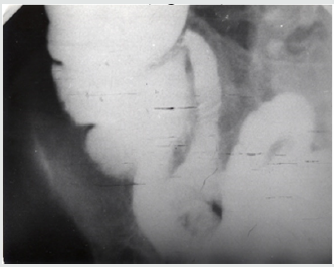
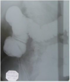
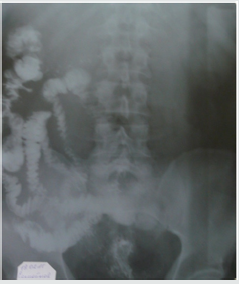
Irrigoscopy: It is important to note that the refusal to hold a tight filling of the cecum at NBZ can lead to a false-negative conclusion. A repeated X-ray examination immediately after a bowel movement is of fundamental importance, since during a bowel movement a maximum pressure is created in the bowel, which at the NBZ will be accompanied by a pronounced reflux of the radiopaque substance into the ileum (Figures 1-3).

Figure 1: Before surgery on irrigography all patients is determined by the reflux of contrast material into the ileum.

Lupinepublishers-openaccess-gastroenterology-hepatology-journal

Figure 2: Patient S, Irrigography with tight filling of the cecum was performed prior to defecation, regurgitation of contrast and the cecum into the ileum is not detected.

Lupinepublishers-openaccess-gastroenterology-hepatology-journal

Figure 3: The same patient S. Irrigography performed after defecation, define a significant reflux of barium into the small intestine.

Lupinepublishers-openaccess-gastroenterology-hepatology-journal

Chronic autointoxication was detected by such studies as urine indican (Obermeyer was tested for this purpose.), The level of the average serum molecules was measured according to N.I. Gabrielyana, lipid metabolism.
Microbial resistance was determined by the content of antibodies to peptidoglycan Staphylococcus aureus (method of Professor A.N. Mayansky). Peptidoglycan is the most common element of the cell wall of all bacteria, which makes it possible to indirectly judge the content of anti-peptidoglycan antibodies in general. The content of antibodies to peptidoglycan Staphylococcus aureus (strain 885, solubilized by ultrasound) was determined. Each sample was placed in 3 repetitions, calculating the average result. Negative controls were the wells in which the stage of treatment with the antigen (peptidoglycan) was omitted. Indicators of negative control from the results of the experiments were subtracted. A pool of 25 sera from healthy donors was used as a positive control. The results are statistically processed on the IBM / AT-286 computer using the following criteria: determination of averages, Fisher criterion, Wilcoxon-Whitney-Mann criterion.

Read More About Lupine Publishers Journal of Gastroenterology and Hepatology Please Click on Below Link: https://currenttrendsingastroenterology.blogspot.com/
Wednesday, 14 July 2021

Lupine Publishers| Utilization of Traditional Anticoagulation for Venous Thromboembolism in Patients with Liver Cirrhosis: Is It Safe?

 Lupine Publishers| Current Trends in Gastroenterology and Hepatology (CTGH)


Abstract

Patients with liver cirrhosis are known to have increased risk bleeding particularly from gastrointestinal tract. However, recent literature has shown that patients with liver cirrhosis are also at increased risk of developing deep venous thrombosis and pulmonary embolism. Therefore, it is important to consider prophylactic and therapeutic anticoagulation in cirrhotic patients. In this article, we have reviewed the available literature on the safety and efficacy of the utilization of prophylactic and therapeutic anticoagulation in cirrhotic patients.

Abbreviations: INR: International Normalized Ratio, VTE: Venous Thromboembolism, DVT: Deep Venous Thrombosis, PE: Pulmonary Embolism, PTT: Partial Thromboplastin Time, LMWH: Low-Molecular-Weight Heparin, UFH: Unfractionated Heparin, CLD: Chronic Liver Disease

Introduction

Liver plays central physiologic role in hemostasis as it synthesizes the majority of the procoagulant and anticoagulant factors. The levels of these factors are markedly affected by decrease function of liver associated with cirrhosis resulting in abnormal hemostatic mechanism. Generally, the impression in the clinical world is that liver cirrhosis is associated with decrease synthesis of procoagulant factors resulting in increased risk of bleeding. This phenomenon is known as auto-anticoagulation and is supported by elevated international normalized ratio (INR) and low platelet count usually observed in cirrhotic patients. In this regard, gastrointestinal bleeding and more specifically variceal bleed are of major concern since they contribute significantly to the mortality of patients with liver cirrhosis. At least, 30% mortality has been reported at the first episode with a 70% recurrence rate in this patient population and a 1year survival estimate ranging from 32% to 80% [1]. However, decrease function of cirrhotic liver also results in reduce level of anticoagulant factors including antithrombin III, protein S, and C which may result in increased tendency to form clots. Interestingly, recent data has also noted cases of venous thromboembolism (VTE) including both deep venous thrombosis (DVT) and pulmonary embolism (PE) in cirrhotic patients ranging between 0.5% to 6.3% [2-10]. Dabbagh et al. [4] found that even an elevated INR > 2.2 was not protective against VTE in this patient population. Gulley D et al. [10] noted that hospitalized cirrhotic patients without predisposing co-morbidities (e.g. neoplasm, congestive heart disease and chronic renal failure) had similar risks for VTE as compared to noncirrhotic patients [10]. Thus, the myth of auto-anticoagulation seems to be only partially true. Therefore, the abnormal routine blood tests (like elevated INR, Partial Thromboplastin Time [PTT], high MELD score and low platelet count) may indicate increased hemorrhage risk in this patient population but may not be necessarily completely true as these tests do not accurately reflect the activity of aforementioned anticoagulant factors in the serum. The purpose of this article is to review the epidemiological data available on the utilization of traditional VTE prophylaxis and treatment on the increased bleeding risk in patients with liver cirrhosis.

Methods

An electronic Medline search was conducted using the key terms anticoagulation, low-molecular-weight heparin (LMWH), unfractionated heparin (UFH), oral anticoagulant, deep venous thrombosis, pulmonary embolism, venous thromboembolism, liver cirrhosis, chronic liver disease, and decompensated liver disease. Studies written in the English from January 2000 to March 2018 were considered for this review article. All search results were reviewed.

Results

Intagliata N et al. [11] studied the complication rates associated with administration of thromboprophylaxis in 235 hospitalized cirrhosis patients admitted to the University of Virginia between 2007 and 2010 accounting for 355 discrete hospitalizations. They noticed only nine gastrointestinal bleeding events (2.5% of admissions) and concluded that VTE prophylaxis was not associated with increased risk of bleeding or death in their patient population [11]. Vivarelli M et al. [12] conducted a retrospective study in Italy on 229 consecutive cirrhotic patients with HCC who underwent hepatic resection. They assess the safety and effectiveness of prophylaxis for VTE in terms of thrombotic or hemorrhagic complications between those who received and those who did not receive prophylaxis with low-molecular weight heparin. They divided the patients into two groups i.e. Group A included 68.5% of patients and Group B included 31.5 % patients. Group B patients had higher Child-Pugh and MELD scores, lower platelet counts, a higher prevalence of esophageal varices and higher requirements for intraoperative transfusion of FFP. Group A received VTE prophylaxis with LMWH while Group B did not receive VTE prophylaxis. The cases of VTE were higher in group B (1.38%) as compared to the group A (0.63%) but the difference was not statistically significant (P = 0.530). On the other hand, the prevalence of hemorrhagic complications was higher in group A as compared to group B but again the difference was not statistically significant (P = 0.380). Moreover, only the presence of varices was associated with increased risk of bleeding (P=0.05) among other studied risk factors including age, CTP class and Model for End-stage Liver disease scores, platelet count and intraoperative transfusion requirements [12].

Bechmann L et al. [13] conducted a retrospective study in Germany to evaluate the safety of LMWH utilization for either prophylactic or therapeutic indications anticoagulation among 84 patients with cirrhosis. Only seven patients developed variceal bleeding at a rate comparable with the baseline rate in patients with advanced cirrhosis. The hemorrhagic complications were either secondary to bleeding esophageal varices or 2whypertensive gastropathy with no mortality. They concluded that a prophylactic use of LMWH in patients with cirrhosis appears to be safe [13].

Smith C et al. [14] also conducted a retrospective chart review of 410 chronic liver disease (CLD) patients admitted to a tertiary care academic medical center in US from August 2009 through July 2011. 225 (55%) patients received thromboprophylaxis including mechanical (154), pharmacologic (49), and combined (22). They noticed a significant decrease in overall thromboprophylaxis use and pharmacologic prophylaxis use for patients with INR >2.0 as compared to those with INR 1.4 to 2.0 (P = 0.013 and P < 0.001, respectively). 0.7% of patients developed VTE (DVT) and fifteen (3.7 %) patients had bleeding event. Out of fifteen patients who developed bleeding, nine were on mechanical prophylaxis, 1 on pharmacologic, 3 on combination, and 2 with no prophylaxis. Majority of patients experiencing a bleeding event had an INR >2.0 (P = 0.001). They concluded that use of VTE pharmacologic prophylaxis does not appear to increase bleeding risk in CLD patients with INR ≤2.0 [14].

Barclay S et al. [15] conducted another retrospective study to determine whether pharmacologic prophylaxis for VTE was associated with a decrease in the incidence of VTE or an increased incidence of bleeding in patients with CLD. The study sample consisted of a total of 1581 CLD patients hospitalized over a 3-year period and divided into two groups based on receipt of pharmacologic VTE prophylaxis. 392 (24.7%) patients received pharmacologic VTE prophylaxis. Decrease risk of VTE (0.5% vs 1.8%, p=0.05) and bleeding (2.0% vs 10.3%, p<0.001) were noted in the prophylaxis group as compared to the nonprophylaxis group [15].

In a multicenter retrospective study conducted in 5 hospitals, Reichert J et al, studied the pharmacologic VTE prophylaxis on risk of hemorrhage while hospitalized in patients with CLD and concurrent coagulopathy. They utilized the ICD-9 codes to identify subjects with CLD admitted from January 1, 2012, until December 31, 2012. 256 patients met criteria for analysis, with 80 received pharmacologic VTE prophylaxis and 176 did not receive pharmacologic VTE prophylaxis. They found that pharmacologic thromboprophylaxis was associated with increased number of hemorrhagic events among cirrhotic patients who received it as compared to those who did not (17.5% vs 7.4%, p=0.02). However, it was noted that difference in rate of overall hemorrhage was driven primarily by a difference in minor hemorrhage [16].

Conclusion

The use of anticoagulation for VTE prophylaxis and treatment in cirrhosis remains underutilized and it is still not a universal practice to provide hospitalized cirrhotic patients with such prophylaxis and treatment due to the concerns of increased bleeding risk. Also, the literature on the safety and efficacy of anticoagulation in liver cirrhosis is in initial stages and is based mainly on retrospective studies. Six studies have investigated the use of VTE prophylaxis. Two studies have shown that DVT prophylaxis does decrease the risk VTE in cirrhotic patients [12,15]. However, the results of one of the study did not reached the statistical significance which could be due to low power of the study because of the small sample size [12]. On the other hand, only one study have shown that pharmacologic thromboprophylaxis was associated with increased number of hemorrhagic events among cirrhotic patients which they attributed to increased incidence of minor hemorrhage [16]. Another study noticed higher prevalence of hemorrhagic complication in VTE prophylaxis group as compared to cirrhotic patients who did not received VTE prophylaxis, but their result did not achieve statistical significance [12]. Moreover, only the presence of varices was associated with increased risk of bleeding.

It is possible that risk of hemorrhage is underestimated in these studies because of retrospective nature of studies with possibility of underreporting of bleeding events and underutilization of thromboprophylaxis of VTE in patients at higher risk of bleeding such as those with high INR, low platelets and presence of esophageal varices. Only one study investigated the use of VTE treatment in cirrhotic patients and they found no increased bleeding risk with it [13]. However, it was a small sampled study and there was no control group utilized to compare [13]. In general, recent data suggest that utilization of VTE prophylaxis in cirrhotic patients is safe and should be considered. Underutilization of both chemical and mechanical VTE prophylaxis in hospitalized cirrhotic patients has been noted ranging to as high as 75% [8,17]. In another study, only 9% of hospitalized patients with CLD were treated with pharmacological VTE prophylaxis, while only 16% received mechanical VTE prophylaxis [2]. The major concern regarding the use of anticoagulation in this population is the gastrointestinal bleed and specifically esophageal variceal bleed. However, VTE prophylaxis appears relatively safe in cirrhotic patients and should be considered in all hospitalized cirrhotic patients with minimal varices, no evidence of clinical bleeding and with an appropriate clinical indication.

Infact, the safety of prophylactic or therapeutic anticoagulation in carefully selected patients with cirrhosis without the presence of high-risk esophageal varices appears to be comparable to general medical patients. Also, in patients with esophageal varices, primary or secondary prophylaxis with either endoscopic variceal ligation or use of non-selective beta-blockers is suggested prior to initiation of anticoagulation and after discussing about the risks and benefits of anticoagulation with the patients. Periodical screening endoscopy should be considered to assess varices and the risk of bleeding in these patients. Various classes of heparin are currently available for prophylactic or therapeutic purposes including UFH, LMWH and fondaparinux. LMWH seems to be the treatment of choice for both the prevention and treatment of VTE in cirrhotic patients. UFH is an alternative in cirrhotic patients for shorter-term use and in cases of severe renal dysfunction and/or hemodynamic instability. The possible role for the new antithrombotic drugs with direct action on factor Xa or thrombin has yet to be evaluated.

Clinical tools to evaluate the risk of VTE in cirrhotic patient population also need to be developed to target the cirrhotic patient who will benefit most from the anticoagulation. Bogari H et al. [18] conducted a retrospective cohort study to evaluate the utilization of the Padua Predictor Score (PPS) as a risk-stratification tool for the development of VTE in 163 patients with liver cirrhosis [18,19]. They categorized the patients into two groups based on whether they developed a VTE (11%) or not (89%). Patients were further risk stratified into high-risk (score ≥4) and low-risk (score <4) using the PPS. Patients in the VTE group had significantly greater mean PPS than in the non-VTE group (5.8 ± 2.0 versus 3.0 ± 2.1, respectively; p<0.001). Also, High-risk patients were more likely to have VTE (OR 12.7, 95% CI 2.8 to 57.4, p=0.001) as compared to low risk. Based on their findings, Bogari H et al. [18] concluded that PPS is an effective risk assessment tool for development of VTE in patients hospitalized with chronic liver disease. Various other factors like low serum albumin and decompensated liver cirrhosis have also shown to be associated with increased risk of VTE in patients with liver cirrhosis. In two retrospective studies conducted by Northup et al. [2], Garcia Fuster et al. [3] low serum albumin was found to be associated with increased risk of developing VTE independent from elevated INR or low platelet count [2,3]. Certain other factors like INR > 2 and low platelet count may lower the risk of developing VTE. It will be prudent to develop tools including such factors to help accurately characterizing cirrhotic patients at higher risk of developing VTE. This will help provide clinicians with more confident to start anticoagulation in cirrhotic patients at high risk of bleeding secondary to esophageal varices.

Further studies are needed to determine the utility of novel methods to monitor the anticoagulation therapy in cirrhotic patients. LMWH is conventionally monitored by anti-Xa assay, while UFH can be monitored by activated PTT (aPTT) assay in the general population. However, monitoring of LMWH anticoagulation with anti-Xa levels for dose adjustment is challenging in cirrhotic patients since the levels of anti-Xa factor have been found to be lower in cirrhotic patients compared to normal controls after administration of prophylactic or therapeutic dose of LMWH [13]. For example, Bechmann et al. [13] studied the pharmacokinetics of LMWH in patients with cirrhosis and confirmed that standard doses of enoxaparin failed to achieve target anti Xa levels recommended for prophylactic or therapeutic use against VTE [13]. However, in vitro studies evaluating the effect of LMWH on thrombin generation found that despite reduced antithrombin and anti-Xa activity levels, cirrhotic patients had an increased response to LMWH [20]. They concluded that the likely explanation of low anti-Xa levels could be a laboratory artifact while the efficacy of LMWH is preserved [20]. Thus, targeting its levels for the prophylaxis and treatment of VTE in noncirrhotic patients might underestimate the true degree of anticoagulation which could lead to incorrect dosing and subsequent increase morbidity and mortality secondary to bleeding. The role of thrombin generation in monitoring the response to LMWH in cirrhotic patients has been investigated by Lisman T et al. [21] and they found it to be a useful tool and an alternative to anti-Xa levels [21]. On the other hand, UFH is monitored with aPTT in general population. But again, aPTT levels are also known to be prolonged in patients with liver cirrhosis thus making it difficult to predict the dose accuracy of UFH in this patient population.

In the end, the treatment and prophylaxis for VTE should be utilized in patients with liver cirrhosis who are at low risk of bleeding i.e. does not have esophageal varices. Moreover, randomized prospective studies needs to be performed to confirm the efficacy of VTE prophylaxis in cirrhotic patients, to determine the safety of therapeutic anticoagulation against VTE in cirrhotic patients, to figure out accurate markers for monitoring the use anticoagulation in cirrhotic patients, and to better understand the role of coumadin and newer oral anticoagulants in VTE prophylaxis and treatment in cirrhotic patients.

Read More About Lupine Publishers Current Trends in Gastroenterology and Hepatology (CTGH) Please Click on Below Link: https://currenttrendsingastroenterology.blogspot.com/


Tuesday, 28 August 2018

Successful Endoscopic Treatment of Refractory Postoperative Duodenocutaneous Fistula using Fibrin Sealant: (CTGH)- Lupine Publishers


Iatrogenic, postoperative enteroatmospheric fistulas remain difficult to manage but common problem after gastrointestinal surgery. Surgical revision, after failure of conservative treatment is not only partially efficient but also carries a high mortality risk, mainly due to patients’ general condition and usually multiples comorbidities. Because of that alternative minimally invasive techniques are extensively studied, but unfortunately the data are still confounding [1,2]. We present a case of 38 – y.o. man with past history of nephrolithiasis, accidentally diagnosed with 30 mm gastrointestinal stromal tumour of the duodenum (Figures 1a-1c).