Showing posts with label Journal of Veterinary Sciences. Show all posts

Thursday, 31 August 2023

Lupine Publishers | The Effect of the Application of a Ruminal Activator (VITAFERT) in The First Phase of the Lactation Curve and its Influence on the Bromatological Quality of Milk

 Lupine Publishers | Journal of Dairy & Veterinary Sciences


Abstract

A total of 46 milking cows (animals in production) were selected, with a live weight of 454kg in their third lactation. Production at the beginning of the experiment was 11.3 liters/day, the animals were under the same driving conditions, the main food base was the star grass (Cynodon nlemfuensis) and a load of 2 cows/ha with a rotation period of 35 days. The animals were distributed in two groups by treatment (23 with Norgold and 23 with VITAFERT), each group was formed with animals from 10 to 100 days of lactation distributed in two treatments. Treatment 1: Norgold per cow per day. Treatment 2: 1.65kg of Norgold and 2 liters of VITAFERT (4.4ml/kg body) per cow per day. The objective of this work is 3,4kg to determine the effect of the application of the ruminal activator on the production and bromatological quality of milk. Consumption of VITAFERT significantly improved milk production in dairy cows from 10 to 100 days of lactation. The use of VITAFERT as a ruminal activator increases milk production and some dairy components. The supplement with VITAFERT is an estimated saving of imported (concentrated) foods and improved their use. It is recommended to include THE VITAFERT in the feeding of dairy cows as a way to increase milk production, favoring a partial substitution of the concentrate. Continue long-term studies on the effect of VITAFERT (ruminal activator) on dairy cow production indicators

Keywords: Dairy cows, milk quality, ruminal activator, (VITAFERT)

Introduction

The dairy industry is one of the most dynamic branches in the livestock sector, perhaps due to the increase in demand for milk in the world, stimulated among other causes by the growth of the population on the planet. Another important aspect in the dynamism of this industry is that every time, consumers demand better quality dairy products, which leads to the search for options to try to cover both the demand for milk, its quality and that of its by-products. Currently a series of research work is carried out to find ways to increase concentrations in the main dairy components in a constant way, placing a particular emphasis on fat, protein and lactose; because these components have an effect on both the taste and yield of milk and also on the performance and sensory characteristics of dairy by-products, as well as in the unsaturated fatty acid chain [1]. Tropical pastures and fodder often have particularly low digestibility of dry matter and reduced protein content, limiting microbial activity in rumen and production. These pastures can be used more efficiently when the bacterial populations of the rumen meet their energy requirements, essential protein constituents, minerals and other nutrients [2] as well as, ensuring a level of non-structural carbohydrates, necessary to optimize the synthesis of microbial protein of the ration [3-5].
In Cuba, at the Institute of Animal Science Elías & Herrera [6] reported on the production and use of a new product obtained by a simple biotech process composed of lactobacilli, yeasts, organic acids of short chains and low pH capable of controlling the development of E. coli, significantly reducing the incidence of diarrhea in animals, increasing the gain of live weight and increase the retention of energy and nitrogen. This product is called VITAFERT (MEBA), which has been used in various species with acceptable results. VITAFERT, as part of the concept of efficient activated beneficial microorganisms (MEBA), is a biological product composed of bacteria, yeasts and their metabolites, capable of producing appreciable amounts of short-chain organic acids such as lactic, acetic, propynic, succinic and pyruvic, vitamins and enzymes. It is a fermentation activator that stimulates the production of organic acids, decreases pH, increases and stabilizes the protein, increases the digestibility of dry matter and decreases the cell wall fractions of food materials that undergo its action [6,7]. The objective of this work is to determine the effect of the application of a ruminal activator (VITAFERT) on the production and bromatological quality of cow’s milk.

Materials and Methods

In a typical cow house of the Institute of Animal Science, a total of 46 milking cows (animals in production) were selected, with a living weight of 454kg in their third lactation. Production at the beginning of the experiment was 11.3 liters/day, the animals were under the same driving conditions, the main food base was the star grass (Cynodon nlemfuensis) and a load of 2 cows/ha with a rotation period of 35 days. The animals were divided into two treatments. Treatments consisted of: A.3,4 kg of Norgold per cow per day (300 grams/liter according to initial production, from the first liter). B: 1.65 kg of Norgold (50% less than A) and 2 liters of VITAFERT (4.4ml/kg pv) per cow per day, one litre was offered in each milking. Additional charges were offered during milking hours (4:30 AM and 2:30 PM). The evaluations were carried out over a 45-day period. The animals were properly identified according to treatment. The determinations made consisted of: Milk production per cow (L/cow/day); Bromatological composition of milk per cow (morning and afternoon); Body condition according to the method of 5 points, every 10 days. The animals kept an individual monitoring of the production of milk with weighing at 7, 14, 21, 28, 35, 42 days and sampling of the milk to obtain its bromatological composition, at the same time the body condition of these animals was observed. The bromatological composition was performed in the ICA ruminant laboratory, using milko-Scan 104, A/S N Foss Electric, Denmark. An analysis of processed variance was performed in the Microsoft Excel electronic tab and analyzed through the INFOSTAT statistical package. The economic analysis determined the total revenue from the production of milk per diet supplied, in the period of 45 days, after the experiment, as well as the cost of both diets for an animal in the period analyzed.

Results and Discussion

Table 1 shows the results in the two treatments, significant differences are obtained for milk production, in favor of the treatment with supply of VITAFERT compared to the treatment with Norgold those that may be related to what Elias & Herrera [6] who point out that VITAFERT is a fermentation activator that stimulates the production of organic acids, decreases the pH, increases and stabilizes the protein, increases the digestibility of the matter dries and decreases the cell wall fractions of food that are subjected to its action, Although not determined in this test, Pirela [8] noted that an amino acid deficiency, NH3 and energy reduces microbial protein synthesis, digestibility and nutrient utilization, which negatively influences consumption in this sense coincides From Wind and Palm 2015 and, therefore, on animal production perhaps in this experiment. This product (Vitafer) contains yeasts, which can contribute to improve the efficiency of food utilization, favoring production according to Robinson & Garret [9]; Rivas [10] who propose that yeast, in primipara and multipara cows, improve milk yields between 23 days and 56 days postpartum, as a result of an increase in voluntary consumption of MS and FDN aspect that coincides with what Galina [11]. Castro-Madrigal & Jimeno [12] propose that with the increase in the digestibility of MS, fibrous components (FDN, FDA, hemicelulose) and crude protein, there is greater consumption and a positive effect on milk production.

Table 1: Results in production and body condition in 100-day dairy cows.

Lupinepublishers-openaccess-Dairy-Veterinaryscience

When the bromatological composition of milk was analyzed in the two treatments, significant difference in protein, total solids and non-fatty solids was observed. While fat and lactose showed no differences. These results could indicate a better balance in diet nutritions with VITAFERT. This may have been given by the increased concentration of precursors and the effect of the diet coinciding with Stokes, Hernández & Ponce, Hernandez & Armenteros [13-15] and Elijah & Herrera [6] who demonstrate in research that there is strong correlation, between the level of degradable protein in the diet and the effectiveness in the synthesis of bacterial protein and consequently influencing the components of milk (Table 2) [16-20].

Table 2:Assessment of milk bromatology in larger components.

Lupinepublishers-openaccess-Dairy-Veterinaryscience

Cost Analysis

Sources considerations

1 litre of VITAFER - 0.11 Cuban pesos Gutierrez 2012
1kg of Norgold - 0.24 Cuban pesos Minagri 2008
Price of sale /litre of milk ? 2.50 Cuban pesos
Table 3 shows that it can be produced with cows of this production level, a saving of more than 30ctvs /cow / day in the feeding system containing VITAFER and when analyzing the system (production and food) can produce an economy of more than 10 pesos/cow/day between one system and another, motivated basically by the increase produced in milk.

Table 3: Economic Response/Treatments.

Lupinepublishers-openaccess-Dairy-Veterinaryscience

Conclusion

Consumption of VITAFERT (ruminal activator) improves milk production and some dairy components in dairy cows from 10 to 100 days of lactation. The VITAFER supplement can be a saving of imported (concentrated) foods and better use of them, if their combination is carried out in situations similar to those observed in this work. The results suggest future studies of VITAFERT in complete lactation.

Read More About Lupine Publishers Journal of Dairy & Veterinary Sciences Please Click on below Link:
https://lupine-publishers-dairy-veterinary.blogspot.com/

Saturday, 8 July 2023

Lupine Publishers | Effect of Nicarbazin on Incubation Variables of Eggs from Creole Hens of Mexico

 Lupine Publishers | Journal of Dairy & Veterinary Sciences


Introduction

Nicarbazin (NCZ) is an equimolar complex of 4,4’-dinitrocarbanilide and 2-hydroxy-4,6-dimethylpyrimidine [1]. It is an anticoccidial agent used in the poultry industry. Its main function is to prevent multiplication and proliferation of parasites of Eimeria genus in the intestinal tract, avoiding tissue damage, decreased feed consumption, reduced nutrient absorption, dehydration, blood loss, skin depigmentation and susceptibility to other pathogens in birds [2]. Nevertheless, its application has not been entirely beneficial. Ott et al. [1] reported that feeding laying hens with 100 ppm or more of NCZ caused depigmentation of the brown shell and reduced hatchability in approximately 60%. Although the mode of action of NCZ for contraceptive activity is unknown, micrographs of vitelline membranes in mallards showed severe degenerative changes and preliminary in vitro studies indicated that it may increase intracellular calcium levels and early degradation of very low-density lipoproteins that comprise egg yolk [3]. However, the effect of NCZ has not been studied in Creole hens of Mexico. Therefore, the objective of this investigation was to evaluate the effect of NCZ on incubation variables of eggs from that genotype of birds.

Materials and Methods

This study was conducted at the Poultry Experimental Farm of the Chapingo Autonomous University at Texcoco, State of Mexico, Mexico. Located at an altitude of 2,247 m [4]. A total of 1922 eggs were collected from a population of 100 Creole hens of 80 weeks of age. Two treatments were evaluated: T2 (Control), 0.00% nicarbazin, and T1, 0.05% of nicarbazin in feed. Nine hundred and sixty-one eggs from the same population were used per treatment. The collection of eggs for T1 lasted 14 days, followed by a cleaning period of 21 days (feed without nicarbazin) and subsequently collecting eggs for T2. Storage was at an average temperature of 68 °F. The fertilization of the eggs was carried out by artificial insemination. Artificial incubation with a constant temperature of 100 °F and 60% relative humidity (RH) was used until day 18, then live embryos were passed to the hatcher (100 °F and RH: 80%). At 12 and 18 days of incubation eggs were candled to identify clear eggs and eggs with dead embryos, then the embryodiagnostic was carried out. Finally, at 21 days of incubation, non-hatched eggs were separated to conclude the embryodiagnostic. Early dead embryo with no blood (ED), 1-3 days of incubation; early dead embryo with blood (EDB), 4-12 days of incubation [5]; intermediate embryonic death (IED), 13-17 days of incubation; late embryonic death (LED) 18-21 days of incubation; infertile (INFERT); contaminated (CONT) and hatched eggs (HATCH) [6-8], were the categories considered in the embryodiagnostic. The feeding program consisted of a single phase with two diets, one containing 0.05% nicarbazin and one without nicarbazin, both diets containing 16% crude protein; 2,800 kcal ME kg-1 and 4% Ca Table 1. They were provided with a program of 16 light hours. Water was supplied for free access and feed was restricted to 110 g bird-1 day-1.

Table 1: 1/Provided per kg of diet: vitamin A, 12,000 IU; vitamin D3, 1,000 IU; vitamin E, 60 IU; vitamin K, 5.0 mg; vitamin B2, 8.0 mg; vitamin B12, 0.030 mg; pantothenic acid, 15 mg; niacin, 50 mg; folic acid, 1.5 mg; choline, 300 mg; biotin, 0.150 mg; thiamine, 3.0 mg. Fe, 50.0 mg; Zn, 110 mg; Mn, 100 mg; Cu, 12.0 mg; Se, 0.3 mg; I, 1.0 mg. 2Sand was used as an inert filler. Statistical analysis Frequency data of categories from embryodiagnostic were compared by the chi-square test for independence with the PROC FREQ contingency tests with α = 0.05, SAS Institute Inc. [12].

Lupinepublishers-openaccess-Dairy-Veterinaryscience

Results

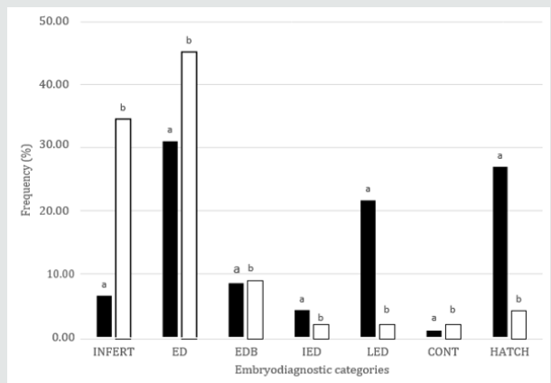
Results of the embryodiagnostic can be observed at Figure 1. The values for the seven categories in the embryodiagnostic were different (P<0.05) between treatments. A higher proportion of the eggs were classified as infertile in T1 (34.5%) comparing to T2 (6.6%). Early embryo mortality (ED + EDB = 54.0%) was also higher in T1 with respect to T2 (ED + EDB = 39.5%), however, ED category contributed with most of the dead embryos in both treatments (45.2 and 31.0% for T1 and T2, respectively). IED category just contributed with a minor proportion of the embryo mortality in T1 (2.0%) and T2 (4.3). LED show a value of 2.8 and 21.6% for T1 and T2, respectively. Contaminated eggs represented 2.5% in T1 and 1.0% in T2. As a result of the values in the previous categories of the embryodiagnostic, hatchability values of 4.2 y 26.9% were obtained for T1 and T2, respectively.

Figure 1: Embryodiagnostic categories in eggs from Creole chicken breeders fed feed without (T2) or with nicarbazin (0.05%) (T1). INFERT: Infertile, ED: Early dead embryo with no blood, EDB: Early dead embryo with blood, IED: intermediate embryonic death, LED: late embryonic death, CONT: contaminated egg, and HATCH: hatched eggs.

Lupinepublishers-openaccess-Dairy-Veterinaryscience

Discussion

Even though there are previous reports of the negative effect of nicarbazin on incubation parameters of birds [9], there is not information with respect to birds that are not raised under industrial condition environments, as the Creole chickens of Mexico. Results of the current investigation agree with those of Hughes et al. [10] and Jones et al. [11] who found reduced hatchability in eggs from birds exposed to nicarbazin. As stated by Chapman et al. [12], affection of the integrity of the vitelline membrane is the mechanism by which the coccidiostat affects the correct development of the chick embryo, so that early mortality is expected. On the other hand, given what is known about its mechanism of action [3], the first stages of development are the most negatively affected by nicarbazin. In line with this fact, our results agree with Sherwood et al. [1] who found a 3.8-fold increase in early embryo mortality when White Rock breeder hens where fed nicarbazin. So that, embryos without blood spots or blood vessels (ED category in the current study) were the most abundant in the embryodiagnostic. As previously stated, Creole chickens of Mexico are raised under certain degree of harsh environmental conditions in terms of feed, housing, and potential pathogen agents [13,14], as coccidia. So that it was considered the necessity of evaluate the effect of a coccidiostat on incubation parameters of this poultry genotype. The results obtained indicate that nicarbazin should not be used in the feed of Creole breeder hens, given its negative effect on embryo mortality, specifically on the earliest stages of development [15].

Conclusion

Nicarbazin negatively affects the incubation variables, mainly in the early embryo mortality and hatchability, of Creole hens of Mexico.

Acknowledgements

Matus-Aragón, M.A. and Zárate-Contreras. D, thank to National Council of Science and Technology (CONACyT, Mexico) for the financial support for their graduate’s studies.

Conflict of Interest

The authors declare that there is not financial or conflict of interest in this contribution.

Read More About Lupine Publishers Journal of Dairy & Veterinary Sciences Please Click on Below Link:
https://lupine-publishers-dairy-veterinary.blogspot.com/

Thursday, 1 June 2023

Lupine Publishers | Characteristic Cellular of the Vaginal Epithelium of Llamas (Lama glama) Captive in the State of Jalisco

 Lupine Publishers | Journal of Dairy & Veterinary Sciences


Abstract

With the objective of contributing data regarding the characteristic cellular forms in vaginal smear of Llamas (Lama glama) and to determine their structural forms in different physiologic states, pregnant, nurslings and you empty, they were carried out vaginal smear of 15 Llamas in captivity condition in the State of Jalisco, Mexico. In the empty Llamas of the 10 to the 30 days post weaning the superficial cells spread to go down from 41% to 7% of presence and the 30 days post weaning the intermediate cells they spread to ascend from 58% to 98% of presence. In the pregnant Llamas in the first month of gestation the naviculars cells were observed in more percentage 86% and the cells flakes in smaller presence 10%, while to the 9° month of gestation increase the presence of cells flakes of 10% to 44% and they diminished the naviculars of 86% to 55%. In the Llamas in nursing of the 10 to the 30 days of nursing the cells flakes spread to ascend from 16% to 54%, the opposite happened to the intermediate cells that spread to go down from 83% to 42% to the 30 days of nursing. The cellular differences in the vaginal epithelium of the Llamas could serve like a tool in the diagnostic of gestation and fertility.

Keywords:Camelids; vaginal cytology; gestation; nursing

Introduction

The principle of the vaginal cytology is based on identifying the type and the percentage of cells in the different stages of the cycle estral, since the hormonal changes of the vagina during the cycle are reflected in the morphology of their cells epitelials[1]. The vaginal cytology is used in most of the species to determine stages of the cycle estral in which is the female, for what is a procedure broadly used in veterinary medicine, [2], and it is used with a lot of effectiveness to determine stadiums of the cycle estral in dogs and cats [3]Stornelli[4] mentions that in the canines with estro 80% of superficial cells is observed, while in the cats with estro 88% of superficial cells is observed [5], while in the necklace Pecarí(Tayassutajacu). Mayor et al.[6] found 60% of superficial cells in estro in the vaginal smear of Alpacas (Vicugnapacos). In a work it found differences in the vaginal epithelium among empty and pregnant Alpacas observing similar cells to other species[7]. The camelids has an unique ovarian cycle, they are induced ovuladores, but their hormonal physiology differs of other ovuladores induced as the rabbit or the cat, [8,9] and they don't present estacionality for photoperiod[10]. The camelids are considered as seasonal reproducers in its natural habitat during the warmest, humid months and of more forage readiness[11]. The vagina of the Llamas measures from 15 to 25cm from the hymen to the cervix and approximately 5 diameter cm [12], these characteristics are necessary to understand its ignorance since it takes us to have a poor yield of breeding, compared with other conventional species[13].  

For such a reason the present study intends to carry out a classification at cellular level in the characteristic ways in vaginal smear of Llamas (Lama glama) captive, and to determine its structural forms in different physiologic states as a tool of diagnose reproductive. 

Materials and Methods

15 females were chosen under captivity conditions belonging to hatcheries peculiar of the state of Jalisco, Mexico, with a corporal condition, weigh and age average of 4-5, 115±8 kg and 4.5±1.5, of which 9 were pregnant, 3 empty and 3 nurslings. For the taking of the samples we use the technique described by Wave [14] where we use a hyssop of soaked sterile cotton with solution of chloride of sodium to 0.9% which 8 cm was introduced inside the vagina carrying out rotational movements against the vaginal wall with the purpose of extracting cells that come off in a natural way rotating the hyssop on a portaobjets, later on noticing with ethylic alcohol of 90°, for the tint that of Giemsa it was preferred and you proceeded that recommended by Lynch [15] where once fixed with alcohol it allows to dry off and it dives for a while in the coloring Giemsa from 15 to 30 minutes, later that washes himself with distilled water and dried off to the environment. The observation to the microscope one carries out with increase of 100X and immersion oil, the opposing cells were classified following the approach of Schutte, [16] in you scale, intermediate, naviculars and superficial.

Results

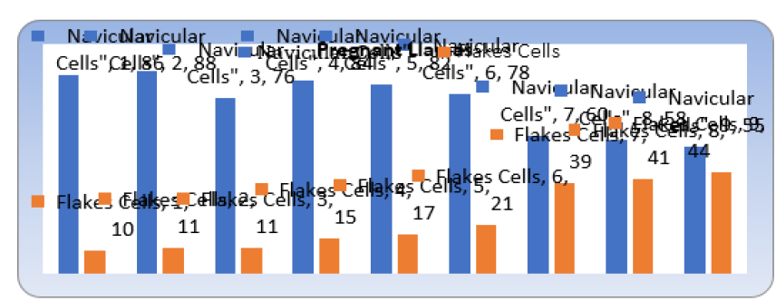
Figure 1: The following figure shows us the cellular activity from the first month of gestation up to the ninth in the 9 copies.

Lupinepublishers-openaccess-Dairy-Veterinaryscience

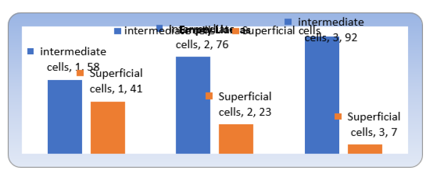
Figure 2: That shows us the cellular differences in the 3 copies of Llamas revised to the 10, 20 and 30 days search it weans.

Lupinepublishers-openaccess-Dairy-Veterinaryscience

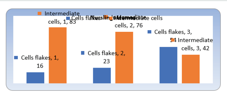
As it is appreciated in the Figures 1,2&3 previous to the first month of gestation the naviculars cells were observed in bigger percentage 86% and the flakes cells in smaller presence 10%, the opposite to the 9° month of gestation where it increase the presence of flakes cells of 10% to 44% and they diminished the naviculars of 86% to 55%. One can observe that of the 10 to the 30 days post weaning the superficial cells spread to go down from 41% to 7% of presence and the 30 days post weaning the Intermediate ones they spread to ascend from 58% to 98%, what resembles each other with the cat tames that it not indicates the existence of levels estrogenicos in the one post weaning early, [17]. As it is observed in the previous graph, of the 10 to the 30 days of nursing the cells flakes spread to ascend from 16% to 54% and the opposite happened to the intermediate cells that spread to go down from 83% to 42% to the 30 days.

Figure 3: Where show us the cellular characteristics in the 3 Llamas in nursing, from the 10 days up to the 30.

Lupinepublishers-openaccess-Dairy-Veterinaryscience

Discussion and Conclusion

In the pregnant Llamas in the first month of gestation the naviculars cells were observed in bigger percentage 86% and the flakes cells in smaller presence 10%, while to the 9° month of gestation increase the presence of flakes cells of 10% to 44% and they diminished the naviculars of 86% to 55%. In the empty Llamas of the 10 to the 30 days post weaning the superficial cells spread to go down from 41% to 7% of presence. To the 30 days post weaning in the empty Llamas the Cells intermediate spread to ascend from 58% to 98% of presence. as long as in the Llamas in nursing of the 10 to the 30 days of nursing the flakes cells spread to ascend from 16% to 54%. The opposite happened to the intermediate cells that spread to go down from 83% to 42% to the 30 days of nursing. The cellular differences in the vaginal epithelium of the Llamas could serve like a tool in the diagnostic of gestation and fertility.

Conflict of Interest

There are not conflict of interest exists.

Read More About Lupine Publishers Journal of Dairy & Veterinary Sciences Please Click on Below Link:
https://lupine-publishers-dairy-veterinary.blogspot.com/

Saturday, 11 February 2023

Lupine Publishers | Combined on-Farm Analysis of Management Procedures and Vaccination Protocols: The Way to Maximize the Health of the Herd

 Lupine Publishers | Journal of Dairy & Veterinary Sciences

Abstract

During a farm animal’s life, there are a lot of situations that carry stress and induce immunosuppression, increasing the risk of infections and diseases. At the same time, veterinarians and farmers make a big effort (technical and economical) to increase immunity, setting complete vaccination programs. Frequently, this prevention through vaccination fails because administration is carried out during immunosuppression periods. We propose an easy, accurate and understandable strategy based on a complete visit of the farm and interview with the farmer. It is purposed to analyze altogether management procedures on farm, other phenomena and vaccination protocols that occur during the ruminant’s life. After a deep analysis, we suggest changes in the farm’s routine in order to maximize health status of the ruminants, reducing stress and immunosuppression periods, increasing efficacy of vaccines and efficiency of the production system. This strategy includes the analysis of the ruminants’ life, day by day, through a spreadsheet.

Keywords: Advice; cattle; management; ruminants; stress

Introduction

Increase or maximize the health of the ruminants in a herd should be a priority for both farmers and veterinarians. Since long, the use of vaccines in order to directly increase immunity but also reduce amount of pathogens in the herd and reduction of clinical signs in infected animals has been carried out [1-3]. It is also well known that stress factors have a negative impact on the immune system and ruminants submitted to it might suffer from stress-induced immunosuppression during several days [4]. Farm animals are submitted to management, physiological status and environment that can induce stress [4,5]. Even when this management is carried out properly and welfare is excellent in the farm, ruminants can be under 30 immunosuppression for a period of time that ranges from hours to weeks [4,6,7].
This stress may, directly or indirectly, induce a decrease of vaccination efficacy, above all when administration of the vaccine is done during this period of immunosuppression. Ideally, ruminants should not be vaccinated during these immunosuppression periods. On the day by day of a farm, this golden rule is often forgotten, and vaccinations are done under stress or immunosuppression periods. Veterinarians tend to assume that vaccination protocols and management strategies they advise for are correctly done. The reality is that on farm routine brings to a bias in between what is set and what is done. The main objective of this manuscript is to provide a strategy for veterinarians, farmers and advisors in order to identify stress periods and vaccine administrations, evaluate if an increase of health and efficiency can be achieved.

Material and Methods

For this strategy a spreadsheet with several columns and multiple lines (Figure 1) is needed and an interview with the farmer/herd manager that will last 1 to 2 hours will be agreed. It is recommended to have agreed an appointment time before, it is crucial not to be in a hurry and start and finish the interview at once. It is also recommended to have this appointment visiting the farm and the animals, as if it was the first time you are in that farm.
Data collected will be all the stressing management, practices or statuses that occur in a farm from day 0 of life until completing first lactation, including dry off and the second parturition. Most common stress factors must be detailed, and the immunosuppression period attributed. It is also essential to detail all the different feeding and allocation that ruminants will have during the period under study. Finally, all the vaccination protocols will be detailed (product and day of administration -age or day after parturition-). All this data will be in columns and placed in the spreadsheet cell corresponding to the day that is done (Figure 2).

Figure 1: Template scheme used during the audit.

Lupinepublishers-openaccess-Dairy-Veterinaryscience

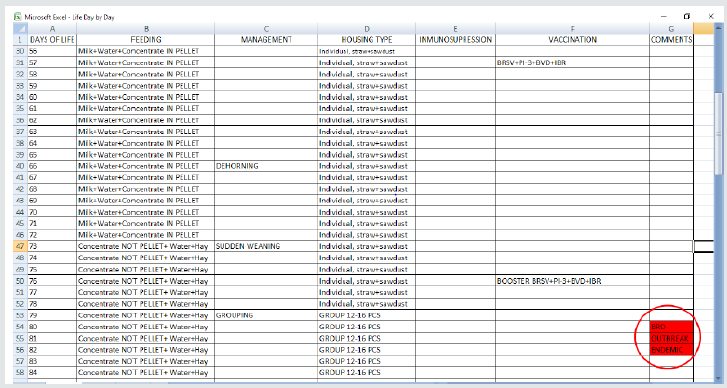
Figure 2: Detailed and completed information needed for analysis between 56 to 84 days of a dairy heifer’s life. PCS: number of calves. BRSV (Bovine Respiratory Syncytial Virus), PI-3: Bovine parainfluenza type 3 virus. BVDV (Bovine viral diarrhoea); IBR (Infectious Bovine Rhinotracheitis); BRD (Bovine Respiratory Disease).

Lupinepublishers-openaccess-Dairy-Veterinaryscience

Results and Discussion

The result of this appointment and post office work will be a spreadsheet full of different information that includes nutrition, management, immunosuppression, vaccination and disease. This way to gather information, will allow the advisor to detect, very quickly, unwanted phenomena in a farm, which reduce the health status of the animals. It will be easy to detect failures in the application of vaccinations under periods of stress or immunosuppression, different management too close together in time or too many stressing changes at once (i.e. grouping and change of feeding the same day).
Furthermore, it allows the consultant to suggest changes in the tempos of conducting management practices or suggest changes in the days of vaccination so that they do not coincide with periods of stress and immunosuppression. Thus, maximizing the potential of vaccinations, not only in their ability to generate immunity, but also just when it is desired to generate immunity. As examples, vaccination for respiratory pathogens is strongly recommended at least 2 weeks before transport or grouping and vaccination for BVDV is strongly recommended 3 to 4 weeks before insemination/ matting in order to avoid abortion or persistently infected bovines. Surprisingly, the advisor can sometimes suggest changes that will be better for health status but also for the production costs of the farmer. As an example, reduction on feeding milk replacer to calves without any impact on performance (Figures 3 & 4).

Figure 3: Critical point in the heifer rearing process in a dairy farm. Blue oval shows the stress factors in orange (late dehorning, sudden weaning and grouping) in 15 days and the immunosuppression caused, having a negative impact on the booster of respiratory disease vaccination (in red). Red oval shows the consequence: endemic respiratory disease in this group of animals. PCS: number of calves. BRSV: Bovine respiratory syncytial virus. PI-3: Bovine parainfluenza type 3 virus. BVDV: Bovine viral diarrhoea. IBR: Infectious bovine rhinotracheitis. BRD: Bovine respiratory disease.

Lupinepublishers-openaccess-Dairy-Veterinaryscience

Figure 4: Final management and vaccination schedule after analysis of situation described in Figure 3.

Lupinepublishers-openaccess-Dairy-Veterinaryscience

Blue oval shows the stress factors in orange (late dehorning, sudden weaning and grouping) in 15 days and the immunosuppression caused, having a negative impact on the booster of respiratory disease vaccination (in red). Red oval shows the consequence: endemic respiratory disease in this group of animals. PCS: number of calves. BRSV: Bovine respiratory syncytial virus. PI-3: Bovine parainfluenza type 3 virus. BVDV: Bovine viral diarrhoea. IBR: Infectious bovine rhinotracheitis. BRD: Bovine respiratory disease.
Changes on management procedures, timing on management and vaccination protocol reduced respiratory disease after grouping. Decisions taken were: Dehorning at 15 days of age, gradual weaning and vaccination protocol started a week earlier. Results were a spare of milk and a 60% decrease of BRD cases. PCS: number of calves. BRSV: Bovine respiratory syncytial virus. PI-3: Bovine parainfluenza type 3 virus. BVDV: Bovine viral diarrhoea. IBR: Infectious Bovine Rhinotracheitis. BRD: Bovine respiratory disease. ML: Milk

Conclusion

Continuous and detailed review of management procedures together with stress factors and vaccination protocols in ruminants’ farms is needed to maximize health status. This audit must be individualized. Results and changes set must be written, analyzed and described in an easy, accurate and understandable way for the farmer.

Read More About Lupine Publishers Journal of Dairy and Veterinary Sciences Please Click on Below Link:
https://lupine-publishers-dairy-veterinary.blogspot.com/


Friday, 9 December 2022

Lupine Publishers | Invitation to Use Ozone Therapy in the Veterinary Medicine, Especially in Egyptian Cattle Reproduction

 Lupine Publishers | Journal of Dairy & Veterinary Sciences


Mini Review

The aim of this article is to highlight innovative scientific research that deserves attention about ozone treatment to overcome livestock infertility problems. Reproductive efficiency is essential for sustainable dairy and meat productions and failures in reproduction lead to economic losses. There are many challenges affecting fertility in farm animals. Oxidative stress and microbial infections during reproductive cycle of cattle are important causes of decreasing fertility and is considered an important challenge affecting reproduction. These challenges affect the animal during pregnancy, during or after parturition. The risks of physical damage during the birth process or at failure to release the placenta after parturition are often leading to increasing of microbial infections in the livestock.
In the face of declining fertility in cattle, it can be appreciation and knowledge of factors that interfere with the equilibrium regulating reproduction are the first steps of solve infertility problem. The most common method of uterine treatment is either intrauterine or systemic antibiotic administration. Major challenge facing many producers is finding practical, effective ways to improve reproductive performance. This challenge requires the creation of research ideas characterized by scientific creativity and practical application. Oxidative stress (OS), which resulted from all types of stress, occurs when the balance between reactive oxygen species (ROS) and antioxidant is disrupted. Formation of ROS (in low concentration) is a continuous and normal process of cellular metabolism but in higher concentration, it affects cell membrane integrity and functions, damage DNA, lipid and protein metabolism. Most ROS are formed because of the environmental pollutants. Antioxidants (including vitamins C and E) and antioxidant cofactors (such as selenium, zinc, and copper) can dispose, scavenging, or suppressing the formation of ROS [1].
Ozone therapy (OT) is safe and nontoxic, and therefore should be widely used in the practice of veterinary and human medicine [2]. However, in Egypt, ozone therapy is still inadequately used in veterinary practice. OT with small doses is useful and stimulating, while large doses are harmful. Ozone, a highly water-soluble inorganic molecule, is a gas made of three atoms of oxygen (O) with a cyclic structure [3]. Ozone can be produced by generator which produced through flowing air at a constant flow 51/min. with 150 electrical volte. Because of ozone`s instability and short its half-life (40 min at 20 °C), ozone cannot be stored in tanks [3]. The ozonated olive oil can be stored up to 60 days at 4° till used [4].
As a powerful oxidizer and highly reactive molecule, ozone has strong antibacterial, antiviral and antifungal action [5]. Intrauterine irrigations with ozonated distilled water (400ml) with ozone concentration of 4-5 mg/l are done to provide entire contact with the site of inflammation and to exclude any damage to the mucous membrane in different forms of endometritis. On being ozonated the water via the biluminal catheter is introduced into the uterine cavity and then evacuated via the same catheter. The procedure can be repeated 3 times during one session, which is done once a day [6]. Intrauterine infusion with ozonated olive oil can be used as 50-100ml according to the size of each uterine horn. Moreover, Vaginal irrigations with ozonated saline with the volume up to 1liter and ozone concentration of 6-10 mg/L is to be done daily (8-10 procedures per course) and are to be complemented with applications with ozonated oil (1-2 times a day).
Finally, the ozone product proved to be efficient in improvement of fertility in cattle through local treatment of the postpartum uterine mucosa this is, with the advantage of no milk and meat withdrawal period due to antibiotic residues [7]. In addition, ozone has been found to be more effective in the treatment of endometritis and retained placenta in dairy cows, compared to hormonal and/or antibiotic treatment, with no negative effect on the host regarding residues [8].

Ethics

This article is an original review article based on previously published papers and does not contain new original data.

Read More About Lupine Publishers Journal of Dairy & Veterinary Sciences Please Click on Below Link:
https://lupine-publishers-dairy-veterinary.blogspot.com/

Friday, 29 July 2022

Lupine Publishers | Histochemical and Histomorphometrical Studies on the Digital Cushion of Heifers and Dairy Cows with Claw Horn Lesions

 Lupine Publishers | Journal of Dairy & Veterinary Sciences


Abstract

Background: The ruminant claw horn lesions seriously diminish histological structure and functional efficiency of the digital cushion of the dairy cows.

Objective: To investigate histological and histochemical properties of the digital cushion in the dairy cow with claw horn lesions, such as white line disease, sole damage and hemorrhage.

Results: The results showed that digital cushion loses its normal histological structure in claw horn lesions.

Conclusion: Further biochemical, physiological and feeding studies on the experimentally induced claw horn lesions are necessary to understand the nature of the events playing roles involving animal feeding in the development of the lesions, to develop new animal feeding strategies aiming to protect and maintain the normal structure of the digital cushion.”

Keywords:Protein Degradability; Goats; Intensive Feeding; Nitrogen Balance; Digestibility

Introduction

Lameness is one of the most important problems negatively affecting milk yield and animal welfare in modern dairy farming in high-performance animals. The cows with laminitis produce lower amount of milk [1-5]. Moreover, cyclic reproductive changes of these cows slow down [6] or cease because of anoestrus [7] or cystic ovarian disease [8]. Lameness is a major animal welfare problem because that it is a painful condition and causes to behavioral changes of the affected cow. A lame cow tends to display low mobility [9], to spend decreased daily feeding time and dry matter intake [10] and lying behaviour may be indicative [11]. In the dairy cow, claw horn lesions [CHL] constitute 65% of the total lesions causing to lameness [12,13]. Although possible roles of risk factors, such as claw conformation and trimming, animal welfare, management, feeding and housing have been evaluated in detail by previous researchers [14-17], pathophysiology of CHL is still unclear. Previous researchers [18,19] have suggested that CHL is arisen from traumatic lesions of the supportive tissues of the claw. Around the time of calving, increase of hoofase enzyme, oestrogen and relaxin secretions results in loosening and increased mobility of suspensor apparatus of third phalanx [19]. The bovine digital cushion locating under the third phalanx has a complex structure, which major part is constituted of fat tissue. The cushion comprises three cylindrical segments [axial, medial and abaxial segments] of adipose tissue surrounded by a thick connective tissue trabecules, those arranged in parallel to each other [20,21]. The pads of the bovine digital cushion play crucial roles in reducing the body load transferred to surface of sole via absorbing of substantial forces acting within the claw [22]. The biomechanical function of the digital cushion in distributing and attenuation of the load transferred to the base of flexor process of the distal phalanx have been well appreciated by previous researchers [21,23,24]. The aim of the present study was to determine histological and histochemical properties of the digital cushion in the heifers and primiparous, second and third parturitions, and multiparous dairy cows managed under similar feeding, housing and welfare conditions. The foot was chosen from the animals with CHL, such as white line disease, sole damage and haemorrhage, in order to define the histology and histochemical changes in the digital cushions of the animals.

Material and Methods

Animals

This experiment was approved by the Ethical Committee of Experimental Animal Production and Research Centre (2011/023) of Veterinary Faculty of Selçuk University. In the study, the 120-cadaver foot (totally 240 hooves) of Holstein heifers and cows were used as materials. The animals were culled because of moderate to severe CHL and lameness in one or more hooves. The claws of the animals with similar feeding, housing and welfare conditions were subjected to a detailed clinical examination prior to slaughter. Information on the age, number of parturitions, calving history and stage of lactation were and recorded, in addition to the clinical findings. Among the reasons given for culling were insufficient milk yield, lameness and infertility. The animals were divided into 5 groups, each consisted of 6 animals, as
group I: heifers,
group II: primiparous cows,
group III: cows with second parturition,
group IV: cows with third parturition,
group V: multiparous cows with more than three calving.
The first group animals were 20 months old averagely; other groups were between 2.6-6.2-year-old. After slaughtering, all the foot was cleaned, and lesions, such as discoloration, sole ulcer and hemorrhages were recorded, frozen at -20°C and kept until use for histological procedure.

Dissection of the Hooves and Obtaining Tissue Samples

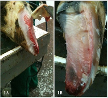
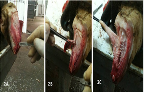
Each foot was thawed. The soft tissues of the sole and heel of each hoof were separated following the horn had been removed, in order to fully expose the axial, middle and abaxial pads of the digital cushion (Figures 1A-B & Figures 2A-C) [18]. Axial pad of the cushion was totally removed, fixed in 10% phosphate buffered (0.1M pH 7.4) formal-saline and used for histological procedures.

Figure 1: The study area, green - places of mass appearance of painted lady butterflies (Vanessa cardui L.) in 2019.

Lupinepublishers-openaccess-Dairy-Veterinaryscience

Figure 2: Separating the corium (2A), dissecting pads of the digital cushion (2B) and totally removing the pads (2C).

Lupinepublishers-openaccess-Dairy-Veterinaryscience

Histology and Histomorphometry

The tissue samples were subjected to routine histological tissue processing. Briefly, the tissue samples were washed with tap water overnight, dehydrated in ethylene series, cleared in xylene and immersed in paraffin. The sections taken at 6μm were stained as follows; hematoxylin-eosin for investigating general histology and histological changes [25], safranin-O (SO) for histochemical properties of ground substance, alcian blue at pH 2.5 (AB 2.5) for staining acid mucins and mucosubstance, alcian blue at pH 4.0 (AB 4) for demonstrating carboxylated glucose aminoglycans in the ground substance, trichrome stain for Type I collagen fibres (Coll I) and other histological detail, Verhoef’s elastic fibre (EL) stain for EL fibers [26]. The specimens were investigated under light microscope equipped with digital imaging system and digital images were recorded. The images were analyzed with digital image analysis software (BS200 PRO-2005).

Statistical Analysis

The data obtained from the digital image analyses were analyzed statistically with one-way variance analysis (ANOVA) and Tukey’s pair wise comparison tests by using SPSS software 14.01 (Release 14.01 Licence No: 9869264).

Results

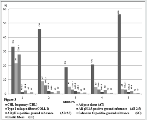
Figure 3: CHL incidence and histometrical results of the groups.

Lupinepublishers-openaccess-Dairy-Veterinaryscience

Clinical findings of the animals prior to slaughter and results of postmortem gross examination of the foot Welfare and management of the animals included into the study were far from than satisfactory. The animals were housed on the concrete floor, under poor hygiene conditions, and claw trimming was irregular. The animals were mainly fed with corn silage, alfalfa, vetch and hay. Findings of the postmortem examinations of the foot showed that the animals suffered from CHL, such as sole ulcers, hemorrhage and folding, white line disease and sole erosions in one or more hooves. The highest percentage of the claws with CHL was in the multiparous cows, in the group 5. The groups 2 and 1 the group followed it respectively. The lowest lower CHL frequencies were found in the groups 3 and 4, and the frequencies of these groups were quite similar (P>0.05, Figure 3).

Gross Macroscopic Findings of the Cushion Segments

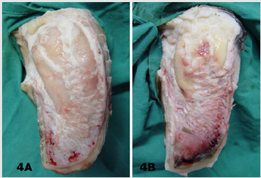
In most of the claws with CHL, segmental structure of the digital cushion was diminished. The digital cushion tissues of the heifers (Group 1) were pale and softer, whereas in the cows with the increasing the parturition number, the tissues gained yellowish colour and relatively more brittle and fragile. The cushion segments were also smaller in the animals of the groups 4 and 5, aged and multiparous cows (Figures 4A & 4B).

Figure 4: Macroscopic appearance of digital cushion tissues of a heifer from the group 1 (4A) and a cow from the group 5 (4B). The cushion pads of the group 5 animal are smaller and yellowish in colour.

Lupinepublishers-openaccess-Dairy-Veterinaryscience

Histological Findings

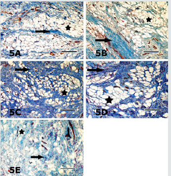
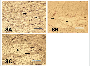
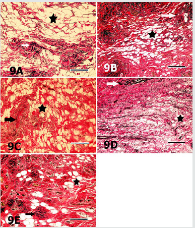
In general, the digital cushion pads of the animals were mainly formed of a loose connective tissue and unilocular adipose tissue. Adipocytes appeared as large cells with unstained cytoplasm in all of the used staining procedures (Figures 5-9). Fibrous connective tissue septa surrounded the loose connective and adipose tissues. There were striking structural differences in the histology of the digital cushion of heifers and calved cows. Loose connective tissue in the heifers, was replaced with adipose tissue in the primiparous cows, and fibrous connective tissue rich in collagen fibres in the third lactation cows. In the heifers and primiparous cows, adipocytes were seen as groups, whereas in the cows of the groups 3, 4 and 5, the cells were observed as individual cells and the cell groups consisting of small numbers of the adipocytes. Adipose tissue percentages of the groups are given in the Table 1. The highest adipose tissue percentage (21.55%) was in the heifers, group 1, and the value was significantly (P<0.001) higher than those of the other groups (Figure 3). The groups 3 and 4 displayed similar (P>0.05) adipose tissue percentages. The multiparous animals (group 5) had the lowest adipose tissue percentage (Figure 3). Type I collagenous fibres were seen as thick undulating bundles of the fibres darkly stained with aniline blue in trichrome stained sections (Figures 5A- 5E).

Figure 5: Collagen fibre bundles (arrows) and adipocytes (asterisks) are seen in the sections from lateral claw cushion of the right forefoot of the heifer from the group 1 (5A), in the medial claw cushion of the left hindfoot of the animal from the group 2 (5B), in the lateral claw cushion of the left forefoot of the animal from the group 3 (5C), medial claw cushion of the left hindfoot of the animal from the group 4 (5D) and lateral claw cushion of the left hindfoot of the animal from the group 5 (5E) are seen. In contrast to a definite decrease in the adipose tissue rate, the connective tissue gradually increased from the group 1 to the group 5. Crossmon’s trichrome. Magnification bar: 100μm.

Lupinepublishers-openaccess-Dairy-Veterinaryscience

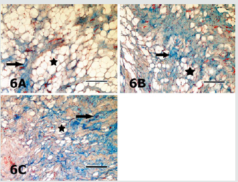
Figure 6: Sections of the digital cushion stained with AB pH 2.5 from the medial claw of the left forefoot of the animal from the group 1 (6A), the lateral claw of the right forefoot of the animal from the group 2 (6B), the lateral claw of the right hindfoot of the animal from the group 3 (6C) are seen. Staining intensity of the ground substance (arrows) in different sections is quite similar. Asterisks depict the adipocytes. AB pH 2.5 stain. Magnification bar: 100μm.

Lupinepublishers-openaccess-Dairy-Veterinaryscience

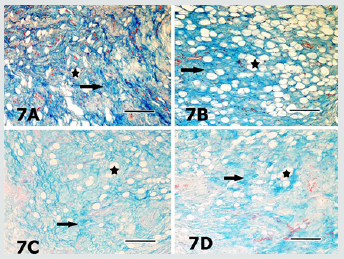
Figure 7: Sections of the digital cushion stained with AB pH 4 from the medial claw of the left hindfoot of the animal from the group 1 (7A), the medial claw of the left hindfoot of the animal from the group 4 (7B), the medial claw of the left hindfoot of the animal from the group 4 (7C) and the medial claw of the right hindfoot of the animal from the group 5 (7D) are seen. Slight differences in staining intensities of the ground substance (arrows) in different sections are seen. Asterisks depict the adipocytes. AB pH 4 stain. Magnification bar: 100μm.

Lupinepublishers-openaccess-Dairy-Veterinaryscience

Figure 8: Sections of the digital cushion stained with safranin O from the lateral claw of the left hindfoot of the animal from the group 1 (8A), the lateral claw of the left hind foot of the animal from the group 2 (8B) and the lateral claw of the right fore foot of the animal from the group 3 (8C) are seen. Staining intensities of the ground substance (arrows) in different sections are quite similar. Asterisk depicts the adipocytes. SO stain. Magnification bar: 100μm.

Lupinepublishers-openaccess-Dairy-Veterinaryscience

The heifers had significantly (P<0.001) higher type I collagen fibre percentage (27.82%) than those of the other groups those had quite similar (P>0.05) percentages (Figure 3). In AB pH 2.5 stained specimens, the AB positivity was mainly located in the ground substance around the connective tissue fibres, embedded in-between the adipocyte groups (Figures 6A-6C). The positivity was also stronger around the individual adipocytes and the groups constituted of small numbers of these cells. Percentage of the AB pH 2.5 stained ground substance was highest (2.08%) in the group 3 and group 2 followed it (1.96%), whereas the groups 1, 4 and 5 had quite similar (P>0.05) and significantly (P<0.01) lower values than those of the groups 2 and 3 (Figure 3). AB pH 4.0 mainly stained the similar ground substance regions with AB pH 2.5, around the connective tissue fibres, located around the adipocyte groups (Figures 7A-7D). Results of the histometrical investigations of the AB pH 4.0 stained specimens showed that the group 1 and group 4 had similar (P>0.05), and significantly higher (P<0.01) positivity percentages than those of the groups 2, 3 and 5 (Figure 3). In SO stained specimens, the positivity was very weak (Figures 8A-8C) in all groups. The positivity percentages of the groups were quite similar and there were no significant differences (P>0.05) between the groups (Figure 3). Elastic fibres were mainly found in-between the connective tissue trabecules surrounding adipocyte groups and tunica adventitia of the blood vessels, and seen as brownishblack, branching coarse and gently undulating fine fibres, in the sections stained with Verhoeff’s elastic fibre stain (Figures 9A- 9E). The results of digital image analyses showed that the group 2 had the highest (1.89%) elastic fibre percentage and the value was significantly (P<0.05) higher than those of the other groups. The groups 1and 3, and 4 and 5 had quite similar elastic fibre percentages (P>0.05, Figure 3).

Figure 9: Sections of the digital cushion stained with Verhoef’s elastic fibre stain from the medial claw of the right hindfoot of the animal from the group 1 (9A), the medial claw of the left hindfoot of the animal from the group 2 (9B), the lateral claw of left forefoot of the animal from the group 3 (9C), medial claw of the left hind foot of the animal from the group 4 (9D) and the lateral claw of the left hind foot of the animal from the group 5 (9E) are seen. Elastic fibres (arrows) are mainly located in connective tissue trabecules surrounding the adipocyte groups (asterisks). Verhoef’s elastic fibre stain. Magnification bar: 100μm

Lupinepublishers-openaccess-Dairy-Veterinaryscience

Discussion

In the present study, statistically significant (P<0.01) differences were found between the CHL frequencies of the groups. Particularly, high incidences of CHL in the primiparous cows (45.80%, group 2) and multiparous (56.20%) cows (group 5) were striking. The differences possibly have arisen from higher incidence of CHL in more than one or all foot of the animals in these groups. Previous researchers [27,28] have reported that the histological development and functional maturation of the digital cushion are not completed before the age of three, and also incidence of sole lesions, such as sole ulcerations and white line disease are relatively higher in the first lactation because that the cushion tissues is less elastic and rich in saturated fatty acids. In accordance with the previous researchers [27,28] Incidence of CHL was higher in primiparous cows in the present study. The roles of the additional loads and forces exposed at late pregnancy, and structural and biochemical changes in the regional tissues, in addition to trauma of both corium of sole and the cushion tissues in the pathogenesis and occurrence of CHL have been discussed in detail by previous researchers [17-19,29,30]. Moreover, collaborative effects of improper, inadequate feeding and housing practices are mentioned among significant disposing factors. In coordination of anatomic structures of the hoof [bones, joints, ligaments and bursae] leads to CHL, mainly sole ulcers and white line disease. Thus, hoof problems occur more commonly among the cows kept in cubicles or concrete yards or straw yards than in the cows kept in cowsheds. Previous researchers [18,31,32] have reported that lameness incidence increases in multiparous cows with increase of ageing and calving, especially after third lactation, due to increasing the amount of the loose connective tissue while reducing adipose tissue, decreasing body condition score [BCS], thinning of the cushion segments. In the present study, a gradually decline was found in adipose tissue percentages of the groups, with ageing and increasing parturitions. Loosening of suspensor ligament system of the hoof results in increasing in mobility of bones, and this situation disposes the sole corium and sole horn to traumatic lesions, and consequently plays significant roles in the development of CHL, such as sole ulcers, white line disease [19,33]. Loosening of suspensor system of the hoof also increases the sensitivity of the claw to the CHL depending on increase of loads applied to the foot by increase in mammary gland and foetus weights, especially during first lactation [34]. Clinical findings and histometrical results of the hooves and higher CHL incidence of the groups 2 and 5 are consistent with previous findings [18,31,32]. Moreover, higher CHL incidence of the group 5, in which significant shrinkage of the cushion tissue was observed, supports the hypothesis suggesting the decline in load absorbing capacity of the cushion increases risk of injury of sole corium. Thus, environmental factors, such as long time standing on the concrete floor, unsuitable housing conditions and in proper claw trimming are effective in embodiment of the sole trauma and haemorrhages in addition to increase of coffin bone motility in late pregnancy and parturition. Unsuitable housing and hygiene conditions in the animals of the present study were in accordance with the high CHL incidence of the animals.

The digital cushion, which is a specified connective tissue, is constituted of connective tissue cells and intercellular substance [ICS]. The ICS contains collagenous, elastic and reticular fibres embedded in an amorphous ground substance. The amount of the amorphous ground substance in young animals is higher than fibrous elements. With ageing, the amount and rate of fibrous elements increase and the rate reverses in older animals. The digital cushion, a member of supporting system of the ruminant hoof locating in subcutis, is a complex structure, composed of white and yellow adipose tissues. There are significant differences in histological structure and organization of the digital cushion among the ungulate animal species. The digital cushion of the ruminant hoof is closely resembled with the cushion of the elephant’s cushion [18]. Equine digital cushion has relatively different structural features. In the horse, the cushion is composed of small number of elastic fibres and closely packaged interlacing collagenous fibres dispersed in acidic mucous matrix, rich in hyaluronic acid. Large spaces between the cells and fibres occupied with mixoid tissue and islets of fibro cartilage tissue [35,36]. Collagen fibres, especially type I fibres are the dominant fibre type of the connective tissues. Although the collagenous fibres have limited elasticity, their tensile strength is enough to meet the high tensile forces of the foot. In the present study, axial pad of the digital cushion constituted of white, unilocular adipose tissue lobuli surrounded by connective tissue trabecules. Fibro cartilage was not observed in the specimens. In some of the groups, trabecules surrounding adipose tissue lobuli constituted of loose connective tissue and contained less amount of type I collagen fibres, the others were rich in the collagenous fibres. Group 1 contained significantly (p<0.001) higher collagen (27.82%) when compared with the other groups. A gradual decline of type I collagenous fibre percentage was typical in the remaining, older and multiparous groups (groups 3, 4 and 5). Nevertheless, they contained quite similar (P>0.05) type I collagen fibre rates. Gradual reduction of type I collagenous fibre rate with the increase of age and calving might arisen from the structural and functional changes occurred in aged animals and during the late pregnancy and post parturition. It might be predicted that shock absorbing capacity of the digital cushion considerably reduced with declining of collagen fibre rate. In accordance with this prediction, the highest (56.20%) CHL incidence was found in the group 5, which had relatively low rate of type I collagen fibres. Moreover, in accordance with the prediction above, previous researchers [19,33,37], also suggested that loosening of the suspensor system of the hoof during pregnancy and post parturition increases sensitivity of sole corium located between coffin bone and sole to trauma, and consequently playing significant roles in the pathogenesis of the CHL, such as sole ulcers and white line disease.

Although the information on the types and distribution of adipocytes in ruminant digital cushion is limited, the heifer cushion contains lesser amount of the fat cells. The digital cushion of the heifers is a whitish and gelatinous in nature containing high amounts of ground substance and lipid content gradually increases through 2-3 parturitions, and gradually decrease starting from 3 years of age [18,21,32]. Because that white fat tissue is an energy source, many metabolic and hormonal mechanisms, such as pancreatic hormones and glucocorticoids are efficient on this fat tissue. Thus, feeding and other metabolic factors are likely to efficient on both amount and composition of fat of the digital cushion. Moreover, it is well known that the fatty acid composition and amounts of the cow’s digital cushion are seriously affected by metabolic disorders, mainly by ketosis. Similarly, previous researchers [38] emphasized that the topic needs detailed evidences. In the presented study, the digital cushion of the dairy cattle comprised of islets of large unilocular adipocytes. In the digital cushion with low levels of fat tissue, individual adipocytes were frequently seen. The animals in the group 1 contained significantly (P<0.001) higher adipocyte rates (%21.55) and the group 2 it, with a significant decline. However, the group 2 contained significantly (P<0.05) higher fat tissue percentage than those of the groups 3, 4 and 5. Rastani [39] suggested that lipids are mobilized from fat tissues in order to be used in milk synthesis. Lipids in white fat depots in the digital cushion are also consumed. Considering the data obtained in this study, it is possible to assume that white adipose tissue in the ruminant digital cushion also can be used as an energy source in ketosis, which is a condition resulting in negative energy balance [NEB], and thus CHL increase post parturition period. It is well known that fat and elastic fibres of the digital cushion play significant roles in shock absorbing mechanism of the cushion via expanding sideways until their viscoelasticity is limited by stretched fibrous connective tissue septa rich in collagen fibres. Elastic fibres support expansion by flattening their undulations and support reversing to the situation before when the applied force is expired. Significant decreases in both elastic fibre and adipose tissue rates result in loss of elasticity of the digital cushion.

There is limited information on the histochemical properties of ground substance of the cow’s digital cushion, and also histometrical results evaluation of these parameters have not been documented previously. In the present study, the results of the AB pH 2.5, AB pH 4.0 and SO stains showed that ground substance of the cow’s digital cushion was rich in hyaluronic acid (hyaluronan) and other proteoglycans, although there were some differences between the groups. Because that hyaluronic acid is a macromolecule increasing the viscosity of the tissue fluid, it consequently augments resilience of the tissue by binding water molecules. Other proteoglycans stained with SO might support the digital cushion in resisting to pressure, since the proteoglycans also show high water binding affinity and tend to gelation [40]. Although the results of the present study indicated that the ground substance of cow’s digital cushion was rich in hyaluronic acid, the hoof material was obtained from the animals with varying degree of CHL lesions. The authors strongly stress that a detailed comparison should be carried out between the findings of healthy and CHL animals. The elastic fibres, which relatively thinner when compared to the collagen fibres, are able to compensate the forces applied between elasticity limits. Tissues gain elasticity and stress bearing features by their elastic fibre content. Elastic fibres are found in hoof soft tissues as in other elastic tissues. Information on distribution and localization of these fibres in the ruminant hoof soft tissues is insufficient. In a previous histological study [38], elastic fibres were found between the collagen fibre bundles in the digital cushion of African elephant. In this study, elastic fibres were more peculiarly observed in-between the collagenous fibre bundles and in blood vessel walls. Morphometrical analyses evidenced that the group 2 displayed significantly (P<0.05) higher elastic fibre percentage (1.89%) than the other groups. Although there was a declining tendency with increase of the parturition number, the other groups had relatively similar elastic fibre ratios. Besides, striking decline of the elastic fibre percentage was determined in the groups 4 and 5 in concomitant with higher CHL lesion incidence. Although further studies are needed, the results of the present study imply that elastic fibre content of the cow digital cushion is quite limited and might play only small role in the functions of the digital cushion and in the development CHL lesions.

Conclusion

Based on the results, it was concluded that CHL seriously affected both histology and functional efficiency of the digital cushion of dairy cows. Results of the present study, which show strong evidence between histological, histochemical and histometrical findings and CHL lesions are noteworthy. However, the results should be compared with those of the healthy animals in further experiments those are involving experimental models. Further studies necessary to understand the nature of the events playing roles involving animal feeding in the development of the lesions, to develop new animal feeding strategies aiming to protect and maintain the normal structure of the digital cushion, in order to maintain and augment its supportive and protective roles, to reduce incidence of laminitis.

Grant Support

This work was supported by a grant from The Scientific and Technological Research Council of Turkey with project no: TOVAG 112O332.

Read More About Lupine Publishers Journal of Dairy Please Click on Below Link:
https://lupine-publishers-dairy-veterinary.blogspot.com/

Thursday, 7 July 2022

Lupine Publishers | Gastrointestinal Parasites Found in Domesticated Animals Introduced Into the Neo-Tropics (New World Tropics)

 Lupine Publishers | Journal of Dairy & Veterinary Sciences


Abstract

This paper is the first part of a three (3) part series of reviews that serves to shed light on the parasites which inhabit the gastrointestinal tract of domestic animals imported into the New World tropics (Neo-tropics), the domesticated Neo-tropical animals and the non-domesticated Neo-tropical animals (wildlife). This first review has focused on the domesticated animals introduced into the new world tropics (Neo-tropics) which were brought by the early European settlers four to five hundred years ago. These introduced Old World domesticated animal species were cattle (Bos taurus, B. indicus), sheep (Ovis aries), goat (Capra hircus), pigs (.com scrofa), horses (Equus caballus) and chicken (Gallus domestiucus). The references used in this review were gathered and synthesized to give a clear breakdown of gastrointestinal parasites of Old World Domesticated animals. The references spanned sixty five (65) years, the earliest reference cited was in 1953 and the most recent was 2018. Most classes of gastrointestinal parasites were reviewed and tabulated by species; they were trematodes, nematodes, cestodes and coccidian. Gastrointestinal parasites which have been reported in domestic livestock species, have negatively affected the health and performances of the animals. The drugs used have been tabulated across animal species.

Keywords: Old World domesticated animals, Gastrointestinal parasites, Anthelmintic, Trematodes, cestodes, Nematodes

Introduction

In the 16th century Spanish colonist brought animals from the Old World to the New World. These original animals were cattle (Bos Taurus, B. indicus), sheep (Ovis aries), goats (Capra hircus), pigs (.com scrofa), horses (Equus caballus) and chicken (Gallus domestiucus) and they had to adapt to the new world conditions Reitz [1]. The first domestic animals came from Iberia or Canary Islands to Hispaniola. In Hispaniola, the local mammals were rodents such as hutia (Capromys pilorides), quitmi, mohug and a mule dog Reitz [1]. In Cubugua and Spanish Florida deer (possibly Mazama spp.) was present as a wild ruminant that may have suggested that the native ruminant diseases could have affected the domestic ruminants that were imported. The Old World animals at these locations were reported to have performed poorly due to diseases, high humidity and local competition for food. The soils were leached, acidic, and infertile and produced forage of low nutritive value Reitz [1].

Domesticated animals reared in the French West Indies were imported on the voyages from Europe in the 16th Century. Horses were used to penetrate the country and donkeys and mules used to carry loads over hills. In Guadeloupe, in the French west Indies, the number of Old World domestic animals in the 16th Century were 3000 cattle, 16000 horses and mules, 4000 goats and sheep Xande [2]. This paper is part of a three part series of reviews that serves to shed light on the parasites which inhabit the gastrointestinal tract of domestic animals imported into the New World tropics (Neo-tropics), the domesticated Neo-tropical animals and the nondomesticated Neo-tropical animals (wildlife). This first review has focused on the domesticated animals introduced into the new world tropics (Neo-tropics) which were brought by the early European settlers four to five hundred years ago. This review seeks to shed light on the effect of gastrointestinal parasites on Domesticated animals that were introduced to the Neo-tropics. In particular, cattle, sheep, goat, pigs, horses and chicken were reviewed. The review has addressed the presence of the parasite in each segment of the gastrointestinal tract and liver.

a) To define and explain “Parsasitology”.

b) To review the gastrointestinal parasites of the Old World Domestic Animals (Equines, Swine, Ruminants and Chickens).

c) To review the therapeutics and management of gastrointestinal parasites of the Old World domestic animals in the New World or Neo-tropics.

What is Parasitology?

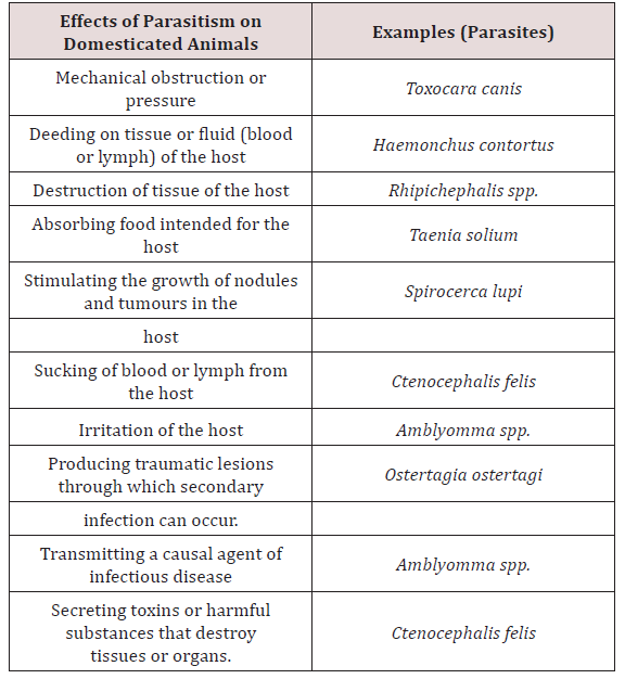
Parasitology is the study of the phenomenon of parasitism. Animal association can be broken down into (1) parasitism, (2) commensalism, (3) symbiosis and (4) mutualism. Parasitism implied a harmful association between the parasites living at the expense of the host Soulsby [3]. Krull [4] stated that a parasitic organism is one which lives on or in a host at the host’s expense. Lapage [5] also stated that a parasite was an animal which lived within another animal which was called a host and the parasite gained benefits from its host and the parasite also inflicted harm to the host. There are many species of parasites, which are relatively harmless. There are many forms which have been reported to produce pathological changes which may lead to severe illness or death of the host Soulsby [3]. Lapage [5] was in agreement with Soulsby [3] and stated that host and parasite may slowly develop mutual tolerance of each other and live together with minimal effects to each other. When this did occur the host was considered to be a carrier (Table 1).

Table 1: The effect of Parasitism on Domesticated Animals Soulsby (3); Krull (4); Lapage (5).
Lupinepublishers-openaccess-Dairy-Veterinary-Sciences

Commensalism was suggested to be where one organism benefits nutritionally from another, without at the same time harming the benefactor Soulsby [3]. In symbiosis, the original implication was that of living together between dissimilar organisms with both organisms obtaining a benefit from this relationship Soulsby [3]. Mutualism has been seen to be essentially symbiosis and the use of the term emphasises the mutual benefit derived by members of the association Soulsby [3]. An intermediate host is an organism that alternates with the definitive hosts harbours larvae or immature stages of the parasite. The definitive host is the organism that normally the sexual stages of the parasites [4,5].

Conclusions on “What is parasitology”

The writings of Lapage [5], Krull [4] and Soulsby [3] have been found to be in agreement with each other on what is a parasite. In summary, a parasite is an organism that lives in its host, is metabolically depend on the host for its survival and negatively affects the host’s health and performance either clinically or sub-clinically. The literature has also suggested that animal associations can be broken down into four relationships parasitism, commensalism, symbiosis and mutualism.

Gastrointestinal parasites of Equine

a. Stomach

The stomach hair worm Tricho strongylus axei was noted to have a direct life cycle and was widespread in many countries MSDAGVET [6], Krull [4], Dua [7]. The adults were found in the lumen of stomach gland and in the small intestines. These parasites are important contributing to the burden of mixed worm species infection. Parasites may have caused catarrhal gastritis that resulted in severe weight loss Dua [7]. The stomach hairworm irritated and eroded villi of the gut, damaged lymph vessels and blood vessels. Blood was lost into the gut causing melena, diarrhoea, anaemia, oedema and rapid loss of condition MSDAGVET [6], Krull [7]. Krull [7] assumed that the horse becomes infected through its association with ruminants.

Habronema muscae, H. microstoma and Draschia megastoma were found in the stomach of horses. Habronema muscae and H. microstma occured in the mucosal layer under the mucus while D. megastoma was found in tumours of the stomach wall MSDAGVET [6]; Krull [4]; Dua [7] (Table 1). These parasites have been observed to have indirect life cycles and were present in horses worldwide. The larvae or eggs released by the adult worm were ingested by the housefly (Musca) or the stablefly (Stomoxy) maggots which developed in manure. The maturation of the maggot to the adult fly allowed the larvae to develop into the infective third stage. Third stage infective larvae were deposited on wounds, lips and nostrils of the horse as the fly feeds. Krull [4] noted that the pre-patent period of Habronema was unknown. If licked and swallowed by host larvae matured in the stomach but if they remained in the wound deposited they didn’t complete their development and remained circumscribed to the area of the wound which caused cutaneous habronemiasis.

Another method of infection was by the ingestion of flies with feed and water Krull [4]; MSDAGVET [6]; Dua [7]. Cutaneous habronemiasis was also called summer sores and occurred on the fetlock, side if neck, mouth, medial canthus of the eye, hock and knees of horses Krull [4]. Draschia megastoma promoted the formation of tumour like growths on the stomach wall which may have ruptured and occasionally which caused blockage of the passage of food from the stomach. Larvae deposited in the wound migrated and fed which extended and prevented healing. Mild digestive disorders may have resulted from gastric habronemiasis MSDAGVET [6]; Dua [7].

b. Small intestine

The horse roundworm Parascaris equorum was observed to be common throughout the world and was highly significant in foals less than six months of age. Within the egg the second stage larvae develop on pastures within 10 days to 6 weeks MSDAGVET [6]; Krull [4]. When the eggs were swallowed the larvae hatched and penetrated the intestines and were carried by the circulatory system to the lungs and liver. Migration then occurs up the trachea where they were coughed and swallowed and mature in the small intestines Dua [7]. Clinical signs have been reported in foals less than 6 months of age. Large numbers of larvae broke into the lungs which caused haemorrhage which was also accompanied by severe enteritis Dua [7]; Krull [4].

Fouls may cough and have a fever when immature larvae were present in lungs. Enteritis resulted in alternated constipation and foul-smelling diarrhoea. Mature horses rarely showed clinical signs as previously infected horses confer good resistance MSDAGVET [6]; Krull [7]. The equine intestinal threadworm Strongyloides wersteri affected horses worldwide with a predilection site for the small intestines. The only pathogenic form is a parthenogenic female Dua [7]; MSDAVET [6]. Infection occurred through ingestion of larvae or skin penetration, it involved larvae migrating to the lung and once present in the lungs they then migrated up the trachea to be swallowed MSDAVET [6].

Dua [7] stated that mares often harbour the larval stages in their tissue and they were activated in parturition to move to mammary tissue where they infected foals via milk. The mature adults were found in the small intestine, females then layed eggs that required no fertilization to develop MSDAVET [6]. These eggs were passed in faeces and then hatched to yield first stage larvae, which developed in the manner described as typical roundworms to become infective third stage larvae. This was considered as a homogenic life cycle. A homogenic life cycle involved a host and allows for rapid increase in adult threadworm population when conditions outside the hosts were unsuitable for larval development.

However, the adult threadworm in the intestine may have also layed eggs that develop into a different kind of larvae. If environmental conditions can provide necessary levels of warmth and humidity, these larvae would undergo a series of moults on the pasture to develop into the adult worm which can live outside the host. The host could have ingested male and females of this type, mate and lay eggs that eventually yielded infective third stage larvae. The type of free ranging life cycle was termed heterogenic and requires warmth and humidity Dua [7]; MSDAVET [6].

This parasite was of major importance to foals where migrating Strongyloides larvae migrated through the lungs and caused severe haemorrhage and respiratory distress. Skin penetration may have resulted in dermatitis and adult worms may have caused erosion and sloughing of the intestinal mucosa which severe interfered with digestion MSDAGVET [6]. Krull [7] found that S. westeri produced diarrhoea and emaciation in foals and has been speculated that problematic skin ailments occurred because of cutaneous penetration of infective larvae. Anoplocephala magna, A.perfoliata are the tapeworms that were found in horses. The life cycle was indirect requiring an oribatid mite as the intermediate host.

Infection occurred when a horse ate an infected mite. The parasites were found worldwide and adults were located in the small intestines and occasionally in the large intestines and caecum. Mild infection caused no clinical signs but in large numbers they caused irritation producing haemorrhage or ulcerative enteritis. Fatal intestinal blockage could have occurred at the ileocecal junction when, A. perfoliata clustered at this point of the large intestines MSDAGVET [6]. Coccidiosis in horses was caused primarily by Eimeria leukartii which caused massive intestinal haemorrhage in foals and young horses. It occurred worldwide and infected the host via ingestion of sporulated oocyst. Foals were suspected of sudden death due to this parasite and the parasite damaged the cells of the intestinal mucosa Dua [7].

c. Large intestine

Large strongyles such as Strongylus vulgaris, S.edquinus, S. edentates and Tridontophorus spp. were observed as stout bodied round worms. Adults are located in the large intestines and caecum, the eggs developed into infective third stage larvae on pasture Dua [7]; MSDAGVET [6]. When ingested third stage larvae dropped their protective sheath in the small intestines. Strongylus vulgaris was the most important large strongyle because it is the most pathogenic. Strongylus vulgaris third stage larvae penetrated the wall of the intestinal mucosa into nearby blood vessels. The larvae wondered through arteries before reaching the anterior mesenteric artery. After moulting to immature adults S. vulgaris returned via the arteries to the large intestine and burrowed through into the lumen Dua [7]; MSDAGVET [6].

S. equinus burrowed into the submucosa to moult, subsequent to that these parasites migrated to the liver where they wonder for 6-7 weeks. They then emerged from the liver and moulted to immature adults in various abdominal organs, then returned to the large intestines. S. Edentatus larvae penetrated the intestine and travelled to the liver via the portal vein where moulting occurred. Larvae then wandered the peritoneum causing nodules. The strongyle larvae formed nodules in the gut wall which they ruptured to enter the lumen of the colon MSDAGVET [6]. S. vulgaris larvae caused roughening of arterial walls providing sites for clotting MSDAGVET [6]. These clots break off and caused infarction to various parts of the body. Weakening of the blood vessels may have caused aneurysms (verminous arteritis) which could have ruptured which lead to death. Adults fed on mucosa and capillaries causing intestinal damage, anaemia, fluid loss into the intestines and blood protein loss Dua [7]; MSDAGVET [6]. Intestinal damage caused diarrhoea, fever, oedema, anaemia, anorexia, depression, weight loss and dehydration. S. equinus and S. edantatus caused liver damage and peritonitis MSDAGVET [6].

Craterostomum spp, Oesophagodontus spp, Gyalcephalus spp and Cyathostoma spp were referred to as small stongyles MSDAVET [6]. Dua [7] stated that Triodontophorus spp. belonged to the small strongyle group of nematodes. They have a direct life cycle, were seen in horses throughout the world and the large intestine and caecum were the sites of predilection. Small stronyles caused considerable economic losses and severe disease. Damaged was caused by the adult worms in the large intestines however larvae in gut walls may also have sucked blood. These worms were observed as plug feeders and removed intestinal mucosa and blood. Clinical signs included diarrhoea, anorexia, colic and weight loss MSDAGVET [6]; Dua [7]. In heavily infected animals there was disruption in digestive and absorptive function which resulted in catarrhal enteritis in the large intestines Dua [7].

Oxyuris equi also known as equine pinworm was observed to be common in horses throughout the world and adult worms were found in the large intestine and rectum MSDAGVET [6]. Dua [7] stated that these worms were of little significance in the intestine and had direct life cycles. Adult female worms migrated out of the anus and layed eggs on the peri-anal skin. The eggs were covered with a sticky fluid and horses may have become infected by biting at larvae in sticky fluid. Eggs may also have dropped into feed and water and matured to infected larvae where they were ingested by the horse, the larvae when ingested penetrated the lining of the large intestines where they fed on mucosa. Ulceration, loss of condition and poor appearance can be seen due to larvae feeding on mucosa but intense itching of the rump was commonly seen due to the fluid with Oxyuris eggs which was attached to the rump of the animal MSDAGVET [6] (Table 2).

Table 2: The effect of Parasitism on Domesticated Animals Soulsby (3); Krull (4); Lapage (5).
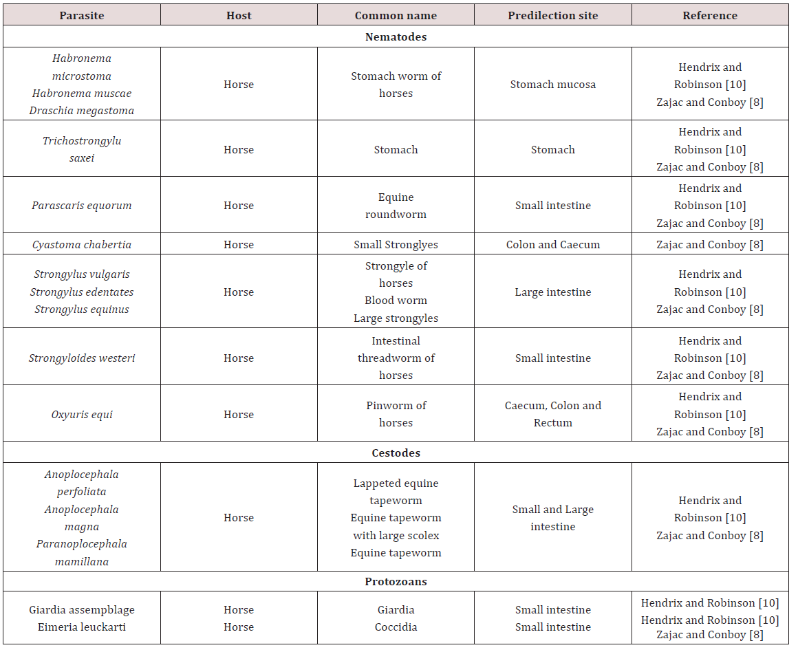
Lupinepublishers-openaccess-Dairy-Veterinary-Sciences

d. Liver

The common liver fluke Fasciola hepatica has been reported to occur in wet areas where the Lymnea snail existed. The predilection site was reported as the bile duct where adults layed eggs in the bile. The bile carried the eggs into the intestines. The eggs left the host in faeces where the immature fluke (miracidium) penetrated a snail. Multiplication occurred in the snail and the cercariae left the snail to be insisted on the blades of grasses. Definitive host was infected when they ingested encysted metacercariae on grasses. The parasites then penetrated the gut and passed over the liver where they migrated to the bile ducts. Migration of flukes through the liver damaged the tissue which caused scar tissue to develop which impaired the function of the liver in production of albumin. Flukes also ingested blood directly causing anaemia and decreased growth rate MSDAGVET [6] (Table 2).

e. Conclusions on the gastrointestinal parasites in Equines

Parasites present in the digestive system of the horse caused a wide variety of clinical signs which included diarrhoea, anemia, melena, peri-arnal irritation. Small strongyles were extremely pathogenic to horses and caused damage to digestive and circulatory system. The migration of these parasites caused damage to several organs before returning to its predilection site. These parasites can have direct or indirect life cycles. The indirect life cycles required intermediate host such as Lymea spp. (snail) or mite.

Gastrointestinal parasites of Swine

a. Oesophagus

Gongylonema ransomi was observed as the gullet worm (oesophageal worm) of pigs. This parasite was observed as causing of great economic losses in swine. It affected pigs worldwide where the worms were found embedded in the tongue of the animal. Consequently there was disposal of swine tongues which were found at abattoirs when lesions were found. The life cycle was indirect and required dung beetles (Aphodius spp.) and cockroaches (Blattella germanica) for completion of the cycle. The definitive host became infected by consumption of parasitized intermediate host. The migratory path which was taken by the parasite to reach the oesophagus in domestic animals was unknown Krull [4].

b. Stomach

Physocehphalus sexalatus was reported as the thick stomach worm of pigs Krull [4]; Dua [7]; Zajac and Conboy [8]. Krull [4] noted that these parasites have worldwide distribution but seem to be absent from the British Isles. Dua [4] is in disagreement with Krull [7] and stated that the occurrence of P. sexulatus was seen worldwide. The life cycle was indirect and the coprophagic beetle was reported as the intermediate host. When egg containing larvae were eaten by host other than the coprophagic beetle the larvae encysts in tissue Dua [7]; Krull [4]. These paratenic hosts when eaten by the pig (final host) the larvae become excysted and made their way to the stomach where they reached maturity. The prepatent period was approximately 30 days. These worms were blood sucking and in light infection no clinical signs can be seen. Heavily infected animals showed anorexia, excessive thirst and restlessness Krull [4]. Dua [7] stated that clinical signs such as those mentioned by Krull [4] were only seen when there was large numbers of worms present or if there was a reduction in body condition due to poor nutrition.

Hyostrongylus rubidus was also named the red stomach worm of pigs Krull [4]; Dua [7]. The worm had worldwide distribution and was found in the stomach. Krull [4] stated that the life cycle was direct, the eggs hatched and larvae grew and underwent a partial moult. Within 6 days the infective third stage larva was developed and hogs become infected by eating contaminated feed or rooting. In contrast Dua [7] stated that H. rubidus has an indirect life cycle with the intermediate host being a coprophagic beetle and pigs became infected by ingestion of the intermediate host. The pre-parent period is 25 days and clinical signs were seen as diarrhoea in young pigs and constipation in old sows. Anaemia, emaciation, haemorrhages were common clinical signs seen. In the stomach deep ulcerations may have perforated and caused death by peritonitis Krull [4]; Dua [7] (Table 3).

Table 3: Predilection sites and common names of gastrointestinal parasites in Pigs.
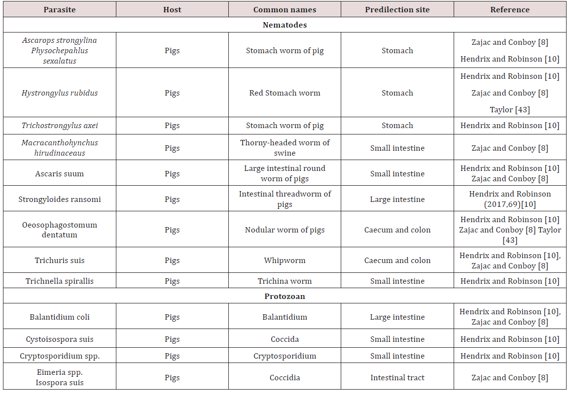
Lupinepublishers-openaccess-Dairy-Veterinary-Sciences

c. Small Intestines

Ascaris suum, the large intestinal roundworm found in pigs. A. suum can migrate to other areas of the gastrointestinal tract (bile duct, stomach, large intestine, gall bladder). The life cycle was observed to be direct and the eggs have to undergo development to become infective Krull [4]; Dua [7]. The develop larvae does not hatch from the egg but undergoes a moult, host was infected by ingestion of the egg contaminated feed, water, scavenger animal which have ingested eggs and piglets are infected by contaminated udders Krull [4].The egg hatched in small intestine of definitive host and larva must undergo extra-intestinal to the lungs and liver via blood vessels before maturing and returning to the intestines when the mature larva was coughed and then swallowed. A.suum produces serious respiratory, digestive and hepatic damage due to its larval migration Krull [4]; Dua [7].

There was a pre-patent period of 75 days and clinical signs were seen due to the migrations of the immature stages and the adult worms. Many of the livers of infected pigs must be condemned at abattoirs due to the immature larvae present Krull [4]. Respiratory signs included coughing, fever and even death. In the digestive tract they caused irritation, occlusion of bile ducts and intestines. A.suum affected the pig industry greatest by the stunted growth, wasted feed, unthriftness and condemnation of carcasses Krull [4]. Dua [7] stated that the main effect of the adult worms were reduction in grow rate of young pigs but in heavily infected animals abdominal breathing can be seen due to pulmonary oedema and consolidation caused by immature larvae.

Strongyloides ransomi, hair-like worm usually found in the small intestines of pigs worldwide. The life cycle was similar to other Strongyloides spp. in which the worms have a parasitic state and a free living state. The adult females were considered to be parasitic and were found in the wall of the small intestine. Transmission occurred commonly through colostrum to affect piglets. In light infection no clinical signs were seen but under heavy infection scouring can be seen Dua [7]. Diarrhoea seen in piglets as young as ten days and severe infections can be fatal Zajac and Conboy [8]. Coccidiosis was a serious disease of pigs causing destruction of intestinal mucosa by invasion. Species which were highly pathogenic include Eimeriade bliecki, E. neodebliecki, E. scabra, E. spinosa and Isospora suis. E. spinose.

They had been seen in pigs less than three month of age causing diarrhoea while pigs between five and fifteen days were usually affected by I. suis. It affected swine worldwide and transmission occurred via ingestion of infective oocyst. Older animals were usually resistant to clinical disease and were sources of infection to young, susceptible animals. Oocyst of Eimeria and Isospora when passed in faeces are unsporulated. Under favourable conditions oocyst sporulated and become infective. During sporulation the development of sporozoites can be seen in the sporocyst. The Eimeria species had four sporocysts each containing two sporozoites while Isospora have two sporocysts each containing four sporozoites. When infective oocyst was ingested they encysted on the small intestine and sporzoites contained within were liberated.

Asexual reproduction took place producing merozoites and the liberated merozoite affected new cells. It is the merozoites that caused destruction of intestinal cells that can lead to death of the host. Some merozoites entered host cell and transformed into gametocytes and gametocytes transform into gametes. When gametes fused a zygote was formed which develops into an oocyst. The oocyst then escaped from the host cell and was passed in faeces. Clinical signs of cocciodiosis were watery or greasy diarrhoea. In piglets the small intestines was flaccid but fibronecrotic enteritis can be seen Dua [7] (Table 3).

d. Large intestines

Macracanthorhyncus hirudinaceaus are the thorny headed worm of pigs. Adults were found in the large intestines and pigs all over the world were affected. The life cycle was indirect with various beetles serving as intermediate host. The site of attachment may have a necrotic centre surrounded by inflammation. There is no specific clinical sign but peritonitis and death may be seen if intestinal wall was perforated Dua [7]. Trichuris suis commonly referred to as the whip worm of swine. It is distributed worldwide with adult worms being found in the caecum and large intestines. The life cycle is direct and infection occurs via infection of embryonated ova. These parasites had been found in the intestinal tract of human on occasion. Most cases in animals were asymptomatic but heavy infection could have caused diarrhoea and unthriftiness Dua [7]. Commonly found in pigs and may have caused severe diarrhoea and dehydration. In severe cases bloody diarrhoea may have be seen Zajac and Conboy [8] (Table 3).

e. Conclusions of gastrointestinal parasites of swine

Parasites of the pigs which were found in the digestive tracks caused economic losses in the following ways

i. Larval migration causing of damaged organs and condemning of affected organ at the abattoir.

ii. Decrease growth rate of infected pigs.

iii. Death of animals that succumb to parasitic disease.

Strongyloides spp. has two states, a parasitic and non-parasitic state with the females being parasitic and were found in the final host. Migration of larvae throughout the body can cause digestive and respiratory signs. Adult cestodes were absent in the digestive tract of pigs.

Ruminants

a. Rumen/ Reticulum

Paramphistomes were flukes which were found in the reticulum of cattle and to a lesser extent sheep. The adults don’t cause ill effects but the immature forms which reside in the lining of the small intestine causes enteritis. The life history is similar to the liver fluke where the fresh water snail was observed as the intermediate host Belschner [9].

b. Abomasum

Haemonchus contortus was also called the large stomach worm of ruminants. While Belschner [9] stated that the barber pole worm was called Haemonchus placei .Dua [7] showed that Haemonchus placei was found in cattle while Haemonchus contortus was found in sheep and goat. Cross transmission of Haemonchus between sheep and cattle can occur with sheep being more susceptible. This worm has a worldwide distribution and adults were found in the abomasum. Sheep and goat were usually affected but may also infect ox, goat, moose, deer, antelope, bison and musk Krull [7]. In contrast Dua [7] stated that Haemonchus occurs in tropical and sub-tropical condition. The life cycle was recorded as direct; eggs are produced by females which were shed in faeces. The larva completed its development in faeces or in the ground. The larva then moulted twice to become third stage infective larva in approximately four days. Infective larvae were active and climbed unto grass to be ingested by the definitive host.

The third stage larva then became exsheathed in the stomach; entered the gastric pits and mucosa. Here it feds on blood, moulted and then returned to the lumen where the last moult occurs Krull [4]; Belschner [9]; Dua [7]. Belschner [9] stated that the pre-patent period was between twenty five (25) to twenty eight (28) days. In contrast Dua [7] found that the pre-patent period was between two to three week (14-21 days). Lesions were produced by the immature larvae and adults that attacked the mucosa of the abomasum. The worm’s mouth part possessed a lancet which was used to secure blood and elicit bleeding. Animal infected are emaciated, oedema, have rough hair coat, have pale mucus membranes and are diarrheic Krull [4]; Dua [7]. Young cattle were usually heavily infected and can cause death due to the blood loss that the parasite caused Belschner [9].

Ostertagia ostertagi was observed in the stomach is also known as the brown stomach worm primarily found in cattle Krull [4]; Dua [7]. O. circumcincta and O. trifurcate is found in sheep and goat Dua [7]. It has a worldwide distribution and adults were found in the abomasum (true stomach). These parasites were found in bison, antelope and sheep but as occasional host. These parasites occurred worldwide and have a pre-patent period of 22 days Krull [4]. Breschner [9] grouped Ostertagia ostertagi, Trichostrongylus axei and Cooperia spp. as short hair worms which was found in the fourth stomach. In young cattle it caused stunted growth and scouring. Krull [4] stated that affected animals have stomach walls which were elevated, inflamed, haemorrhagic and oedematic. Symptoms seen were emaciation, pale mucous membranes, rough hair coat and diarrhoea. Deaths were seen in severe cases and identification of the parasite cannot be done by eggs Dua [7]; Krull [4].

Krull [4] stated that Trichostrongylus axei and T. columbriformis were found in the abomasum and small intestines of sheep, goat and cattle. Dua [7] stated that Trichostongylus axei was found in the abomasum of ruminants and the stomach of horses and pigs. These parasites are extremely pathogenic and were not very host specific and caused disease to domesticated, wild and sometimes man. The life cycle was direct; eggs were discharged in faeces and moulted twice to become infective third stage larva. The infective stage larvae migrated from faeces and attach to grasses. Infection occurred when third stage larvae was ingested by definite host. The infective larvae when ingested entered the submucosa and moult before returning to the lumen. The pre-patent period may have ranged from 15-23 days Krull [4].

Clinical signs in sheep and goats were seen when the number of worms ranged from 2,000 to 10,000 per animal. Krull [4] assumed that the pathogenic effect was due to the toxins it produced but not the blood sucking. Young lambs and kids were usually affected showing acute signs of weight loss and reduced appetite. Mixed infection with Nematodirus spp. intensifies the pathogenic potential. In sheep it caused reduced gains, interference with wool production, and depression in protein digestibility and depression in calcium and phosphorus utilization. Phenothiazine was used in the treatment of twisted stomach worm and lesser stomach worm which inhabit the abomasum. Proper sanitation can control the transmission of this disease Haberman [11].

c. Small intestine

Trichostrongylus columbiformis and Trichostongylus vitrinus were found in the small intestines of ruminants worldwide. T. vitrinus was found mostly in sheep and goat and was observed as having a pre-patent period of 18-21 days. The life cycle was direct and larvae burrowed superficially in the crypts of the mucosa. Clinical signs were anorexia, persistent diarrhoea and weight loss Dua [7]. Cooperiosis was reported as a parasitic disease of ruminants caused by the parasite Cooperia. C. punctate, C. pectinate and C. onchophora parasitized cattle. C. surnabada parasitized cattle and sheep while C. curticeii affected sheep and goat. These worms don’t suck blood but caused profuse diarrhoea, anoexia and emaciation. The life cycle was direct with a pre-patent period of 12- 15 days Dua [7].

Nematodirus helvetianus affected the small intestine of cattle while N. battus affects sheep worldwide. The life cycle was reported as direct with N. battus more commonly seen in temperate regions; animals became infected by ingestion of third stage larvae. Clinical infection was usually seen from six week onward in dairy cattle but in sheep there was a sudden onset of unthriftiness, profuse diarrhoea, marked dehydration and death. Nematodirosis was common in confined lambs or weaned sheep with lesions of dehydration, catarrhal enteritis or acute inflammation of the small intestines Dua [7]. Belschner [9] agreed with Dua [7] and stated that Nematodirus spp. was the thin necked intestinal worm found most commonly in the small intestines. Heavy infection by the immature larval stages could have caused death in young cattle with a direct history similar to the barber’s pole worm. Bunostomum phlebotomum the hookworm of cattle is fairly common in coastal areas and heavy infection was usually seen in calves. The hookworm fed on blood and tissue in the intestines; it adhered to the intestinal wall and drew blood using its mouthpart. Infection could have occurred via ingestion of infective larvae or by larvae boring through the animal’s skin. The larvae migrated in the body and were carried to the lungs where they developed and were coughed up and swallowed to reach the intestine Belschner [9]; Dua [7]. Belschner [9] stated that the pre-patent period was two (2) months. Dua [7] found that the pre-patent period could have ranged from 30-56 days. Signs of infection were similar to the barber’s pole worm where there was rapid loss of condition, anaemia and bottle jaw Belschner [9]; Dua [ 7].

Neoascaris vitulorum also known as the large roundworms of cattle were commonly found in the small intestines of calves. These parasites were found worldwide affecting cattle, caribou and Indian buffalo but were more prevalent in tropical areas. The life cycle was direct and host became infected by ingesting infective eggs. The larvae then migrated to lung and liver to mature and then returned to the small intestines as adults Krull [4]; Dua [7]. Krull 1968 [4] stated that the life cycle is similar to Ascarids seen in pigs and the pre-patent period is two and a half months (75 days). Dua [7] was in disagreement with Krull [4] stated that the pre-patent period was 3-4 weeks and transmammary infection occured in pregnant cows where larvae mobilized to be passed in milk which was fed to calves. Clinical signs were anorexia, diarrhoea, colic and a lack of endurance Krull [4]; Dua [7].

Strongyloides papulosus was observed as a slender like worm measuring 3.5 – 6 mm long. These worms were found in the small intestines of ruminants worldwide. Strongyloides have unusual life cycles in which eggs produced in faeces of host can develop into infectious larvae or free living adults. Transmission occurs by ingestion or skin penetration of larvae. Infection was usually seen in calves but clinical signs were rare but large worm burden may cause diarrhoea and loss of appetite Dua [7]. Monezia benedeni was recorded as a tapeworm that occurs in cattle found in the small intestines. The microscopic mite was observed as the intermediate host and cattle became infected by ingesting mites by grazing. Heavily infected calves had intestinal obstruction with loss of body condition Belschner [9].

In ruminant coccidiosis was observed to be of great economic importance worldwide. They were found in the small intestines but can also be found in the liver. They occurred worldwide and specific species affect sheep, goats and cattle. In cattle Eimeria zuernii and E. bovis were the most pathogenic. In sheep E. crandallis, E. ovinoidalis and E. ovina was seen with E. ovinoidalis being a serious pathogen of lambs. In goats E. arloingi, E. christenseni and E. ovinoidalis were seen with the later (E. christenseni and E. ovinoidalis) being highly pathogenic to kids Dua [7]; Zajac and Conboy [8]. Transmission occurred by ingestion of infected cyst. The life cycle was similar to Eimeria species found in pigs. The virulence of these organism was linked with stressors such as poor nutrition, poor sanitation, weaning, shipping, severe weather or change in feed. Young ruminants were usually susceptible to disease and having a prepatent period of seventeen to twenty one days. Clinical signs included diarrhoea, fever, tenesmus, ill- thrift, inappetence and death Dua [7].

d. Large intestine

Oesophagustomum radiatum also known as the nodular worm was found in the large intestine Belschner [9]. O. radiatum were found in cattle and buffaloes while O. columianum was found in sheep and goats Dua [7]. The life cycle was recorded as direct and consisted of eggs being passed in faeces and larvae development occurring in optimum conditions. The infective larvae were then ingested by the host where it burrowed into the bowel wall and formed nodules on the intestines Dua [7]; Belschner [9]. The larvae left the nodule upon maturity and were present in the intestinal lumen. The pre-patent period is six weeks and clinical signs were similar to other endoparasites and entailed loss of condition, soft dropping containing mucus and haematochezia Belschner [9]; Dua [7].

Chabertia ovina was observed as the large nematode found in the colon of ruminant. It was mainly found in sheep and goat but was occasionally seen in cattle with a worldwide distribution. The life cycle was reported as being direct and in sheep soft blood flecked faeces can be seen. Death occurred with heavy infection but clinical signs were seldom seen in cattle Dua [7]. Trichris globulosa, the whipworm found in the caecum of cattle worldwide while Trichuris ovis is found in sheep and goat. The infection was common in calves with larvae and adult causing small haemorrhages and oedema in the colon. These worms have a pre-patent period of 6-12 weeks Dua [7].

e. Liver

Fasciola hepatica was reported to be found in the bile duct of sheep, goat, ox and other ruminants (Table 4). In unusual host such as man and horse the fluke was found in the lungs and under the skin. The fluke caused fasioliasis (liver fluke disease, liver rot) especially in sheep and cattle Soulsby [3]; Belschner [9]; Dua [7]. The eggs entered the duodenum with the bile and left the host in the faeces. The rate of development for hatching of the eggs was dependent upon environmental temperatures. The miricidium penetrated actively into the snail (Lymnea spp.) and developed into the sporocyst. Each sporocyst give rise to rediae which further developed into cercariae. Cercariae escaped from snails and settled on the blades of grass just below water level. The cercariae were then swallowed by the final host with plants on which they were encysted Soulsby [3]; Belschner [9]; Dua [7].

Table 4: Predilection sites and common names of gastrointestinal parasites in Ruminants.
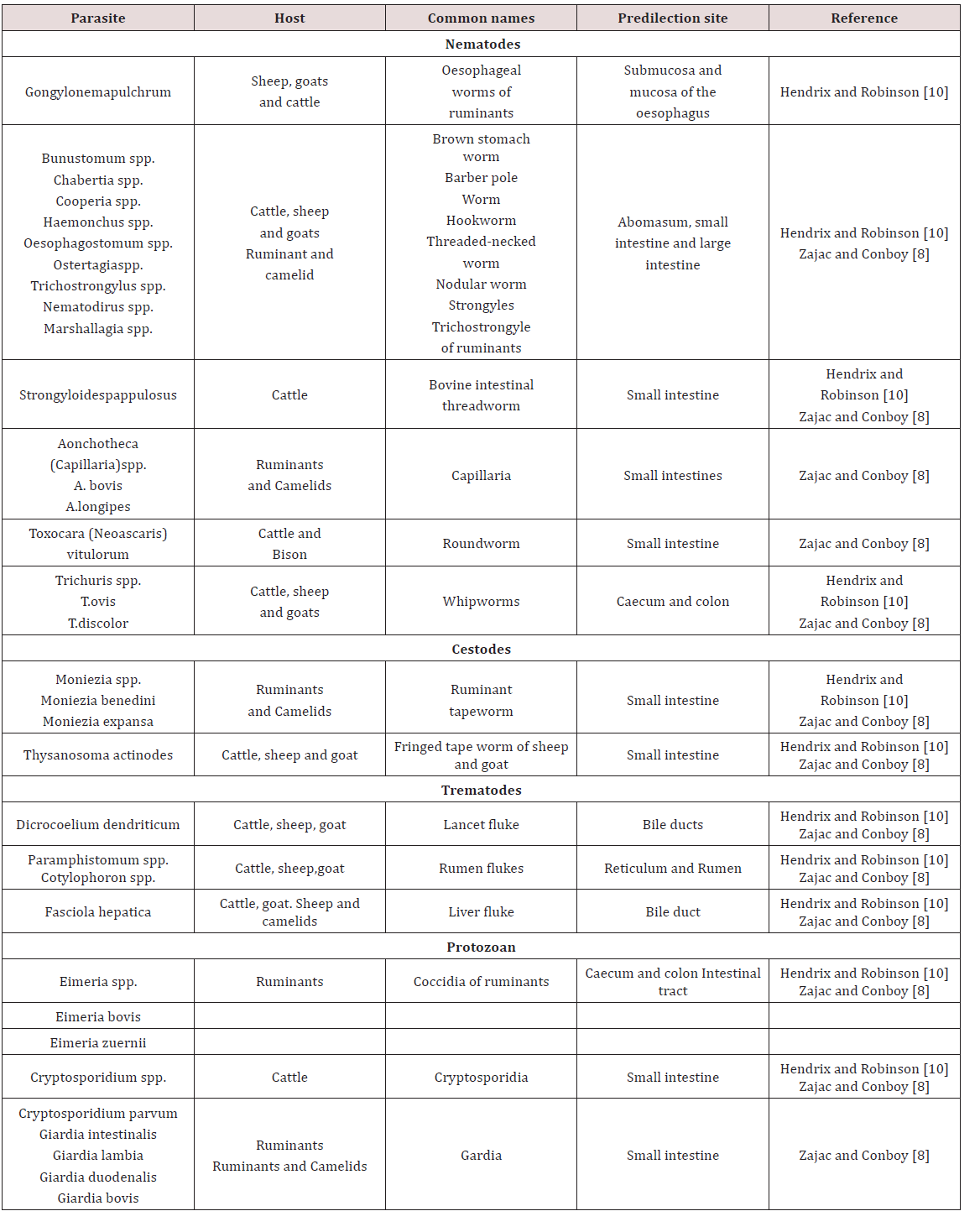
Lupinepublishers-openaccess-Dairy-Veterinary-Sciences

Following ingestion the metacercaria excysted in the duodenum. Within 24 hours of infection the majority of immature trematodes occurred in the abdominal cavity. They then penetrated the liver capsule and migrated through the liver parenchyma. The pathological lesions manifested depended upon the number of metacercariae ingested. No appreciable damage was done during penetration of intestinal wall or the peritoneal cavity Soulsby [3]. The principle lesions occurred in the liver parenchyma or bile duct. The disease entity can be divided into acute fascioliasis and chronic fascioliasis Soulsby [3]; Dua 2012 [7]. The acute form was usually seen in sheep and is essentially traumatic hepatitis produced by the migration of large numbers of immature trematodes.

Animals may die a few days after the development of clinical signs due to haemorrhage in the abdominal cavity caused by extensive destruction to liver parenchyma. A complication of this acute condition was “Black disease” caused by Clostridium oedematiens novyi. The bacterium invaded after hepatic damage by the parasite to anaerobic necrotic lesions Soulsby [3]; Belschner [9]; Dua [7]. Chronic fascioliasis was more common in sheep, cattle and other animals. The pathology was divided into hepatic fibrosis and hyperplastic cholangitis Soulsby [3]; Belschner [9]; Dua [7]. Migration of immature flukes in the liver parenchyma caused haemorrhage and necrosis. Healing and regeneration of these lesions caused collagen and fibrin to be deposited.

The hyperplastic cholangitis was caused by the presence on adult flukes in the bile ducts. At first the epithelium of the bile ducts became hyperplastic, both close to and distal to the site of fluke residence. Animals suffered from anaemia due to the blood sucking activity of the adult flukes. Oedema may have occurred due to the hypoproteinemia and was seen as “bottle jaw” Soulsby [3]; Belschner [9]; Dua [7]. Dicrocoelium dendriticum was found in the bile duct of ruminants Soulsby [3]. Two intermediate hosts were a snail and an ant. Two principal snail hosts were Zebrina detrita in Europe and Cionella lubrica in North America. The miricidium do not hatch out of the egg until the eggs have been swallowed by the intermediate host. They hatch in the gut of the snail, the sporocyst was then converted into cercariae and there was no redia stage. The cercariae emerged from the snail in damp and clump into a slime ball. The slime balls were eaten by ants of genus Formica Soulsby [3].

Metacercariae were produced in the abdominal cavity and some were found in the brain of the ant. Definitive host was infected by swallowing infected ants. Cercariae entered the liver via the bile duct, these flukes penetrated the fine branches of the bile duct. Despite heavy burdens which may have occurred pathological lesions were not seen. In advanced infection there was extensive cirrhosis and scarring of the liver surface and the bile duct is markedly distended with large number of flukes. In severe cases anaemia, oedema and emaciation could have been be seen but many cases showed no clinical signs Zajac and Conboy [8]; Hendrix and Robinson [10]. Control of the snails (Intermediate host) could be done by proper draining of water log pasture and the use of Hexachlorethane in environment Haberman [11]. Parker [12] stated that control was done by eliminating the intermediate host (snail) which will break the cycle (Table 4).

f. Conclusions on the gastrointestinal parasites of Ruminants

Haemonchus spp., Ostertagia spp. and Trichostrongylus spp. caused severe damage to the abomasum of Old World Domestic Ruminant and impeded ruminant production. Nematodirus spp. affected sheep causing devastating clinical signs whereas when these parasites are found in other ruminants caused milder clinical signs. Subcutaneous oedema (Bottle Jaw) was reported in ruminants when large numbers of gastrointestinal parasites was present which removed protein (blood, plasma protein).

Chickens

a. Oesophagus

Mukaratirwa et al. [13] found Gonglyonema inguvicola, Skrjabinocerca spp. and Capillaria contorta in the oesophagus of chickens. Mungube et al. [14] identified Capillaria contorta and Gonglyonema ingluvicola in the oesophagus of free ranging chickens in Kenya.

b. Crop

In Nairobi Kenya Maina et al. [15], Mbeere Kenya Cheg et al. [16] and Chota et al. [17] free ranging chickens Gonglyonema ingluvicola was found in the crop. Capillaria contorta and Dispharynx nasuta was found in the crops of chickens in Zimbabwe Mukaratirwa et al. [13]. Mungube et al. [14] located Capillaria contorta in free ranging chickens in Kenya.

c. Proventriculus

Tetameres americana was found in the proventriculus of free ranging chickens Maina et al. [15]; Cheg et al. [16]; Mukaratirwa et al. [13]; Mungube et al. [14]; Chota et al. [17]. Dispharynx nasuta was also found in the proventriculus in chickens in Zimbabwe Mukaratirwa et al. [13] but was found in the trachea in chickens in Kenya Mungube et al. [14]. Dispharynx spiralis was identified in Kenya Mungube et al. [14].

d. Gizzard/ Ventriculus

In Nairobi Kenya, free ranging chickens were examined. In the gizzards Acuaena hamulosa was found Maina et al. [15]. Cheilospirura spp. was found in the gizzard of chickens in Zimbabwe Mukaratirwa et al. [13]; Mungube et al. [14].

e. Small intestine

In chickens two forms of coccidiosis were seen; chronic form and acute form. The chronic form was found in the small intestines with the causative agent reported as Eimeria tenella. There was also E. nacatrix, E. maxima, E. acervulina, E. mitis, E.praecox, E. hagoni and E. brunetti which also caused intestinal coccidiosis. Destruction of intestinal cells by the parasites caused diarrhoea, bloody droppings and decreased in feed intake. Sulphur based drugs such as sulfaguanadine, sulfamethazine, sulfamerazine, sulfadiazine, sulfapyrazine and sulfaquinoxaline Haberman [11] (Table 5). Coccidia were common in domestic birds and many infections were reported to be asymptomatic but where poultry was confined Eimeria spp. were important pathogens. Sexual and asexual multiplication occurred within cells of the intestinal walls Zajac and Conboy [8]. Cryptospotidiumbaileyiand C. melaegridis were found in the gastrointestinal tract and caused diarrhoea. This parasite is zoonotic Zajac and Conboy [8].

Table 5: Predilection sites and common names of gastrointestinal parasites in Ruminants.
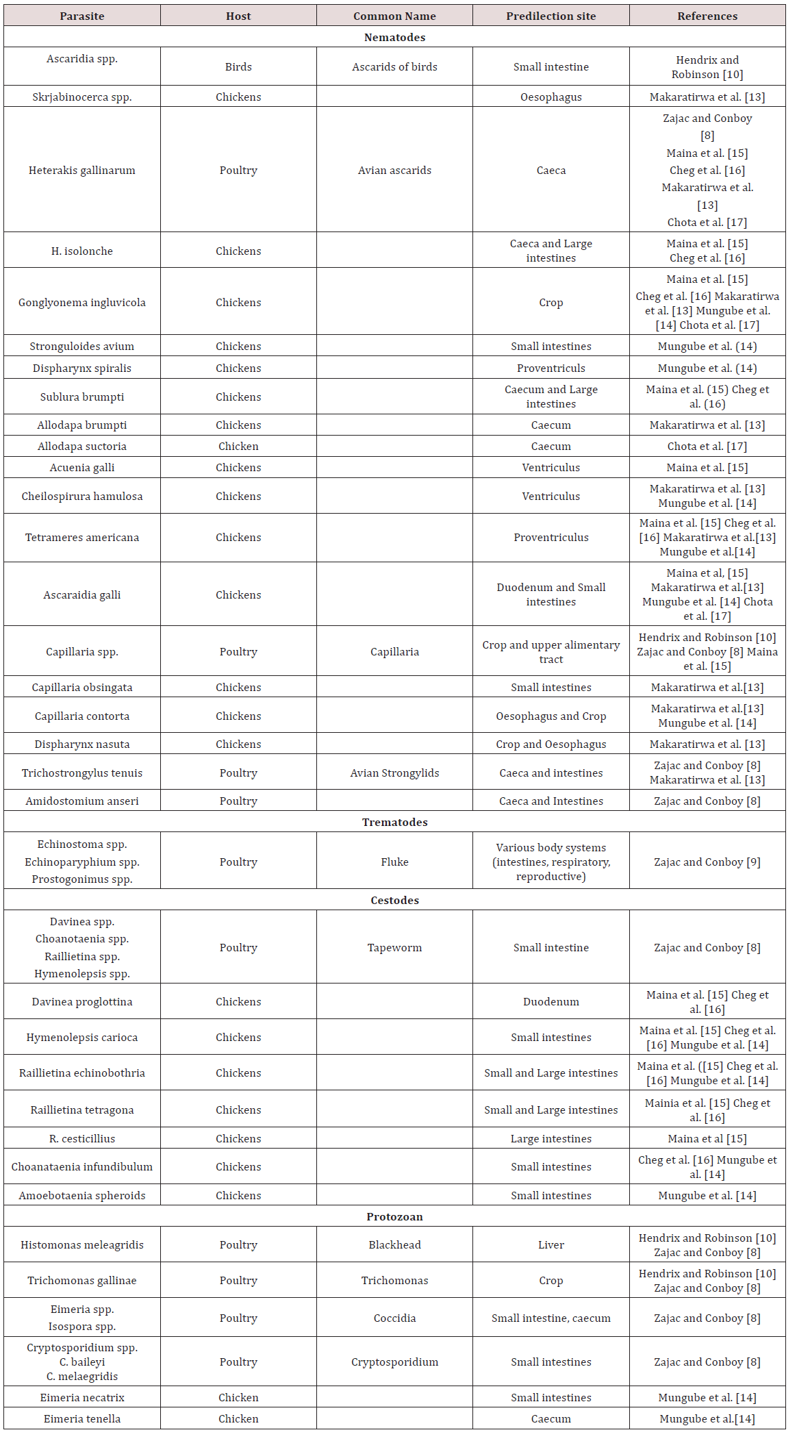
Lupinepublishers-openaccess-Dairy-Veterinary-Sciences

In the small intestines of free ranging chickens in Nairobi Kenya parasites were observed. Endoparasites found were Ascaridia galli, Raillietina echinoborthrida, Raillietina tetragona, Davinea proglottina and Hymenolepsis carioa Maina et al. [15]. Mungube et al. [14] identified cestodes; R. echinobothrida, Choanataenia infundibulum, D. proglottina and Amoebotaenia sphenoids. Nematodes identified were A. galli, Strongyloides avium, Ttrichostrongylus tenuis and coccidian Eimeria necatrix. Cheg at al. [16] found Hymenolepsis canteniana, Choanataenia infundibulum, R. echinoborthrida, Davinaea Proglottina. Mukaratirwa et al. [13] found Trichostrongylus tenuis and Capillaria contorta in the small intestines of free range chickens in Zimbabwe. Chota et al. [17] recorded A. gallinarum in the small intestines of scavenger chickens in Zambia.

found Trichostrongylus tenuis and Capillaria contorta in the small intestines of free range chickens in Zimbabwe. Chota et al. [17] recorded A. gallinarum in the small intestines of scavenger chickens in Zambia.

In the caeca and large intestines of chicken in Nairobi Kenya Heterakis gallinarum, H. isolonche, Subuluria brumpti and Raillietina cesticullius, R. echinobothrida were found Maina et al. [15]. In another county in Kenya (Mbeere) Heterakis spp., H.isolonche, H.gallinarum, Subulura brumpti, R. echinobothrida and R.tetagena was found Cheg et al [16]. Mukaratirwa et al. [13] found Allodapa brumpti and Heterakis gallinarum in the caecum of free range chicks in Zimbabwe. Mungube et al. [14] recorded Eimeria tenella in the cecum of free ranging chickens in Kenya. Chota et al. [17] identified Heterakis gallinarum in the ceaecum of chickens in Zambia (Table 6).

Table 6: Predilection sites and common names of gastrointestinal parasites in Ruminants.
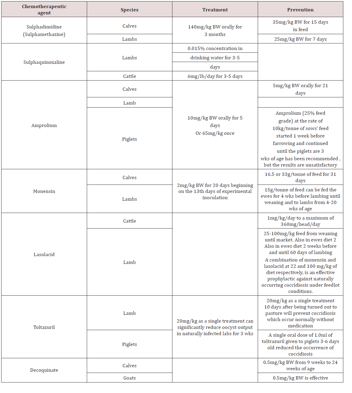
Lupinepublishers-openaccess-Dairy-Veterinary-Sciences

g. Conclusions on gastrointestinal parasites of Chickens

The majority of work on helminths in chicken was obtained from Africa, Asia and North America with limited published work done in the Caribbean. Due to the long pre-patent period of helminths they are usually found in free-ranging chickens and layers. Eimeria spp. causes major economic damage to the poultry industry due to the deleterious effects on the birds’ digestive tract.

Treatment and Control of Gastrointestinal parasites

a. Management practices and drugs strategies

In Brazil animals were reared in organic operations and traditional dairy cattle farms to compare animal performance and health. Organic farms had lower milk production per animal, lower calf mortality, less incidences of mastitis, few rates of spontaneous abortions and reducted ecto-parasitic load. However organic farms had a greater prevalence of internal parasites compared to the conventional farm Sliva et al. [18]. Farmers in Kenya perceived the use of anthelmintics in drought times had no impact on the productivity of small ruminants but states that the rainy season was the reason for increased performance. The farmers didn’t consider endoparaistes as having an effect on the animals unless clinical signs were seen Okell et al. [19].

Anindo et al. [20] used Medicated molasses urea block with fenbendazole in sheep production and recorded an increase in feed intake and decrease in faecal eggs count. Arece-Garcia et al. [21] studied ewes using two management procedures for control of parasites. One group selective use of anthelmintic was done. Only animals with clinical signs of parasites were treated whilst the alternative treatment used conventional deworming system. The results showed that there was no significant difference between the two treatments with respect to animal performance. The strategic strategy was then recommended as it reduced the amount of therapeutic drugs being used which reduces anthelmintic resistance Arece-Garcia [21]. Dried banana leaves were fed to sheep artificially infected with Haemonchus and decreased faecal egg hatchability was seen due to the tannins present in the leaves Gregorya et al. [22].

Medicated fenbendazole urea block had caused an increase in weight gain in goats as compared to the negative control group which wasn’t treated with fenbendazole Abid et al. [23]. Cassava foliage fed to goats had increased growth rates and reduced parasite infestation. This was due to the high levels of tannins in the leaves of the cassava plant Sokera and Preston [24]. Phengvichith and Preston [25] also did an experiment in supplementing goats with cassava foliage and found similar result to Sokera and Preston [24]. There was an increase in live-weight gain and reduction in faecal egg count as compared to a negative control group. In Bishoftu Town a survey was done with sheep reared in urban and peri-urban environment. The most common athelmintic used was albendazole followed by ivermectin and levamisole.

The reasons for the use of anhelmintics were general disease symptoms (45.8%) such as emaciation, rough coat, weakness. 23% of farmers treated due to digestive disturbances, 18.3% gave anthelmintics for simple deworming and finally 10% used anthelmintics for respiratory conditions Datiko et al. [26]. Mahieu and Aumont [27] investigated the use of sheep and cattle in alternating grazing systems. Lambs were grown in the alternate system (Cattle and Sheep) and the control (Sheep only) and the 70 day old lamb weight in the alterative system was higher than the control. There was also no significant difference in the faecal egg count in sheep reared under the two conditions.

b. Anthelmintic Resistance in Domestic Animals

Cheng et al. [16] investigated the efficacy of Piperazine citrate, Levamisole hydrochloride and Albendazole in chickens. Levamisole (25mg/kg) was effective against caecal worms Heterakis spp., H. isolonche and Sublura brumpti but was ineffective to tapeworms and Tertameres americana. Albendazole (20mg/kg) was effective against Heterakis spp., Sublurua brumpti, Tetrameres americana, Railletina tetrogena and R. echinobothrida. Piperazine citrate (3mg/kg) was only effective against Ascaridia galli and ineffective against nematodes and trematodes. In Bangladesh Trichostrongylus spp., Oesophagustomum spp. and Strongyloides spp. were resistant to Albendazole using the Faecal egg count reduction test (FERT). This was due to the over use of the drug by farmers. Anthelmintic resistance can be small in farm holders that use anthelmintics for clinical disease Hogue et al. [28]. In Mexico, anthelmintic resistance of parasites in Beef cattle was examined. Faecal egg reduction and inhibition of egg hatchability was used for albendazole, ivermectin and levamisole. The results showed resistance to ivermectin (32% reduction faecal egg reduction).

Albendazole and levamisole were effective (95% and 100% reduction in faecal egg reduction) Muniz-Lagunez et al. [29]. In South Africa small scale farmers were investigated for the presence of anthelmintic resistance of albendazole, levamisole and ivermectin. All anthelmitic in the study as ineffective in reduction of faecal egg count in the animals with only a couple (2) farms have susceptibility to the drugs Tsotesti et al. [30]. Algeria -Lopez [31] investigated the efficacy of ivermectin against ecto and endo parasites of cattle. The results showed that gastrointestinal parasites were resistant to ivermectin but the tick Rhicephalus were susceptible to the drug. In Ethopia the relationship of dose and efficacy of albendazole was investigated. In goats 3.8 mg/kg, 5.7 mg/kg and 7.6 mg/kg was given and all dosages was ineffective (65.5%, 81.4% and 84.1%) and showed the helminths developed resistance to the drugs Euale et al. [32]. In Trinidad the efficacy of anthelmintics were investigated in sheep. Faecal egg reduction tests were carried out and the results showed heliminth were resistant to albendazole (46-62% reduction) fenbendazole (44- 61% reduction) and levamisole (53-81%). Ivermectin was effective in decreasing the eggs count (95-97% reduction) George et al. [33].

c. Alternatives and Herbal Remedies in the control of Parasites

Lone et al. [34] investigated the use of Euphorbia helicoscopia as an anthelmintic to control Haemonchus contortus. Faecal Egg Count Reduction (FECRT), egg hatchablity and larval development inhibition was used. It reduced faecal egg count in 18 days post treatment. Adult motility showed the highest of 98% efficacy at 5mg/kg. When compared with levamisole the plant extract had low effects on egg hatchability. A collection of condensed tannins from Balanites aegyptiaca, Tamarindies indica and Celtis toka were evaluated for anthelmintic treatment. C. toka had the fastest adult worm mortality and was as high as ivermectin whilst the other two forages had no significant effect. There was no significant difference in egg hatchability between ivermectin, T. indica and B. aegytiaca. The three plant extracts (Balanites aegyptiaca, Tamarindies indica and Celtis toka) was observed to inhibit larval development by 100% which was similar to ivermectin Assefa et al. [35].

Leucaena leucocephala and Salix bablyonica was investigated as anthelmintic against Haemonchus spp., Ostertagia spp., Oesophogostomum spp., Cooperia spp., Bunustomum spp., N. battus, Chabertia spp., Strongyloides papulosus and Nematodirus spathiger. There was a reduction in the egg count for Strongyloides spp., S. papulosus, D. filarial, M. capillaris and Eimeria spp. The extract had no effect on egg reduction on Trichuris spp., Nematodirus spp. and Fasciola spp. Hernandez et al. [36]. Feitosa et al. [37] used of pumpkin seed for its anthelmintic properties in ostriches. In the study the pumpkin seeds fed at 1g/kg body weight gave a 90% reduction in faecal egg count. The control and albendazole showed no reduction in the faecal egg count. Extracts of Larrea tridentata was used as an anthelmintic against Haemonchus contortus larvae. Concentration of 12.5 to 50 mg/ml leads to low mortality of sheathed and ex-sheathed larvae. When compared to Ivermectin, Larrea tridentata had 70% efficacy with Ivermectin having 99% efficacy against larvae Garcia et al. [38].

Consensed tannins of Sesbania sesban and Desmodium intortum was investigated as an alternative anthelmintic for Haemonchus contortus in goats. It was found that levels of the 1mg/ ml tannin from D. intortum significantly inhibited larval migration but S. sesban was ineffective. Goats given D. intortum extract showed no difference in weight compared to the negative control which was attributed the incomplete removal of parasites by the tannins Debale et al. [39]. Biological control was also investigated as an alternative control for helminths in Sweden where Diddingtonia flagrans a fungus was fed to sheep which trap anthelmintic larvae. The experiment showed that there was no difference in the performance of the lambs that received treatment and the negative control group.

In the experiment the finding were attributed to the high initial level of nutrition for both groups Waller et al. [40]. Gastrointestinal parasitism in Creole goats was investigated using mixed breed grazing systems in post-weaning period (3-7 months). Four groups were grazed with different stocking rates bases on live-weight; 25% (kids 150 kg LW0.75 and cattle 450 kg LW0.75), 50% kids (Kids 300kg LW0.75 and cattle 300 kg LW0.75), 75% (kids 450 kg LW0.75 and cattle 150 kg LW0.75) and 100% kids (kids less than 600 kg LW0.75). There animals were then exposed to pasture contaminated with parasites for 3-7 months. Faecal egg count was significantly higher for groups having 100% and 75% goats.

The kids were then infected with H. contours at 11 months of age. In contrast to the faceal egg counts seen earlier faecal egg counts for the second infection (experimental) were lower for the goats which were reared at 100% and 75% kids respectively. It suggested that the animals had a degree of resistance based on their first exposure to the parasites at pasture Cei et al. [41]. Cei et al. [42] investigated the effect of growing kids in different housing environments. The results of the study showed that rearing animals gave better growth than animals in collective pens. Both groups were exposed to 10000 L3 larvae of Haemonchus contortus and the results showed that the individual reared animals performed better than the collectively reared goats. This shows with proper housing and management some of the effects of parasitism can be avoided.

d. Conclusions on the treatment and control of gastrointestinal parasites

It has been reported that a wide variety of drugs can be used to treat anthelmintics and coccidia which are present in Old World Domesticated animals. With increases in the improper use and frequency of these drugs, helminths and coccida have developed some resistance. Now farmers and scientist are searching for alternative extracts (drugs) and management technique to control gastrointestinal parasitism in these animals.

Summary of Conclusions

a) The writings of Lapage [5], Krull [4] and Soulsby [3] have been found to be in agreement with each other on what is a parasite. In summary, a parasite is an organism that lives in its host, is metabolically depend on the host for its survival and negatively affecting the host’s health and performance either clinically or sub-clinically.

b) The literature has also suggested that animal associations can be broken down into four relationships (1) parasitism, (2) commensalism, (3) symbiosis and (4) mutualism.

c) Parasites present in the digestive system of the horse caused a wide variety of clinical signs which included diarrhoea, anemia, melena, peri-arnal irritation.

d) Small strongyles were extremely pathogenic to horses and caused damage to digestive and circulatory system. The migration of these parasites caused damage to several organs before returning to its predilection site.

e) Parasites of the pigs which were found in the digestive tracks caused economic losses in the following ways; (1) larval migration causing of damaged organs and condemning of affected organ at the abattoir, (2) decrease growth rate of infected pigs (3) death of animals that succumb to parasitic disease.

f) Strongyloides spp. has two states, a parasitic and nonparasitic state with the females being parasitic and were found in the final host.

g) Adult cestodes were absent in the digestive tract of pigs.

h) Haemonchus spp., Ostertagia spp. and Trichostrongylus spp. caused severe damage to the abomasum of Old World Domestic Ruminant and impeded ruminant production.

i) Nematodirus spp. affected sheep causing devastating clinical signs whereas when these parasites are found in other ruminants caused milder clinical signs.

j) Subcutaneous oedema (Bottle Jaw) were reported in ruminants when large numbers of gastrointestinal parasites was present which removed protein (blood, plasma protein).

k) The majority of work on helminths in chicken was obtained from Africa, Asia and North America with limited published work done in the Caribbean.

l) Due to the long pre-patent period of helminths they are usually found in free-ranging chickens and layers.

m) Eimeria spp. causes major economic damage to the poultry industry due to the deleterious effects on the birds’ digestive tract.

n) It has been reported that a wide variety of drugs can be used to treat anthelmintics and coccidia which are present in Old World Domesticated animals.

o) With increases in the improper use and frequency of these drugs, helminths and coccida have developed some resistance.

p) Further research on alternative extracts (drugs) and management technique to control gastrointestinal parasitism in these animals.

Read More About Lupine Publishers Journal of Dairy & Veterinary Sciences Please Click on Below Link:
https://lupine-publishers-dairy-veterinary.blogspot.com/