Showing posts with label Journal of Oncology Medicine & Practice. Show all posts

Thursday, 22 December 2022

Lupine Publishers | Cancer: Our Body’s Global Warming Warning

 Lupine Publishers | Journal of Oncology and Medicine

Opinion

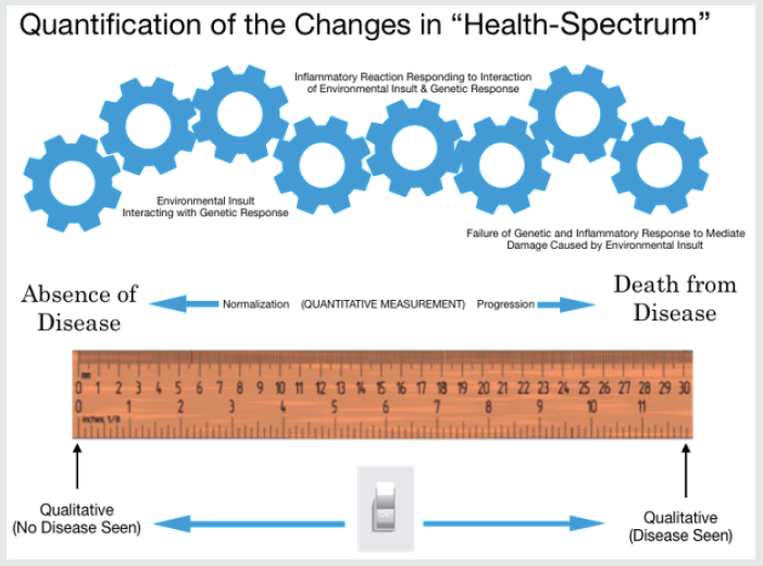
Monday 4 February 2019 was yet another World Cancer Day. We celebrated our efforts to find and eradicate cancer without admitting to ourselves that cancer isn’t a single disease, but rather a group of diseases all caused by our bodies responding to the toxic environment we are exposing ourselves to, just as our planet is responding to the toxic wastes we are dumping into it. The question is WHY do our bodies run amok. The answer lies in the same answer to why Global Warming exists. It is the untold unimaginable continued damage and destruction we cause to the world and to ourselves that accounts for the destruction of the planet and the development of cancer within us. While some individuals may have a genetic predisposition [1] for certain types of cancers, it is nonetheless this constant bombarding of ourselves with toxins, which our bodies try to react to and when overwhelmed the climate change of our bodies occur and call for the eradication of first the damaged cells and then eventually ourselves. Just as the continued bombardment of our Earth with environmental toxins is resulting in violent changes on the planet in an attempt to respond to and eradicate the cause of the toxins to the plant, so too are our bodies reacting to try to eradicate these toxins and their effect within our bodies. This process on a biological basis is reflected in how the cells of our body respond to the cellular environment as shown in Figure1. The process of developing cancer or not developing cancer is not a sudden change but rather a transitional series of events resulting from the interaction between the specific responses and expression of the genome of the cell involved and the specific environment in which the cell is immersed. This environment includes both carcinogenic and non-carcinogenic factors. As the insults occur, the cellular mechanisms to respond to those insults include a variety of responses, including cellular repair and immunologic reactions. The outcome is determined by the interaction between these two opposing sets of factors. Progression or regression is determined by these responses. Clearly no one wants to have cancer or for the planet to be destroyed. Confusion comes from not knowing what is helpful and what is hurting us. Until recently [2], we have been limited by testing (qualitative imaging, biomarkers, etc.), which at best can only provide a yes/no answer to the question of whether a person has cancer or not. Many of these tests do not even provide a yes/no answer, but rather infer there could be a problem. These qualitative approaches are unable to provide us with information warning us that these transitional changes are happening [3]; changes which we could act upon if we only knew they were occurring. Changes which when measured could be used to determine if a given treatment is working [4], harming us or having no effect. Measurable changes which can show us cancer in its early stages [5]. These transitional changes can now be measured using FMTVDM [2]. The only question is whether we use this tool to help find these transitional changes and guide our treatment regimens or whether we will continue to pretend that what we are doing is working?

Figure 1: Quantification of the “Health-Spectrum” for Cancer.

Lupinepublishers-openaccess-cancer-Oncology

Read More About Lupine Publishers Journal of Oncology and Medicine Please Click on Below Link:
https://lupine-publishers-cancer-journal.blogspot.com/

Saturday, 26 February 2022

Lupine Publishers | Targeting the Immune Checkpoint in Cancer: Is This a Viable Treatment Option for AML?

 Lupine Publishers | Journal of Oncology 


Abbrevations: AML: Acute Myeloidleukaemia; CBF: Core Binding Factor; mAb: Monoclonal Antibody; MDS: Myelodysplastic Syndrome

Editorial

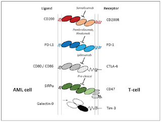
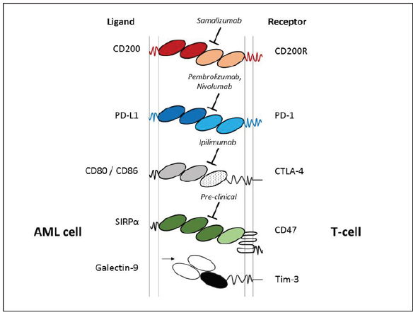
The immune suppressive mechanisms displayed by malignant cells are considered a central process in the pathogenesis of cancer. Research in this area has gained significant momentu mover the past 20 years, with several immune checkpoints identified, including; CTLA-4, CD200/CD200R, Tim-3/Galectin-9 and PD-L1/PD-1 (Figure 1). Whilst characterising the molecular basis of leukaemia for risk stratification remains at the forefront of AML research; this must now extend to understating how the seimmune checkpoint path ways fit into the equation. A good example of why this is important is to consider CD200expression level in AML, which is a negative prognostic indicator [1]. CD200 is an immunosuppressive lig and, that when engaged with its receptor CD200R, has the capacity to attenuate T-cell and NK-cell anti-tumour activity in AML. Interestingly, most cases of CBF AML express high levels of CD200, yet CBF AML performs relatively well clinically. This paradox suggests there is a complex interplay between AML molecular heterogeneity and immune surveillance. Given the recent development and FDA approval of several immune checkpoint therapies, a full understanding of these processes and integration with standard molecular risk stratification is warranted.

Figure 1: Illustrated are immune checkpoint legends expressed on AML blast cells (left) with the cognate T-cell receptors (right), including; CD200/CD200R, PD-L1/PD-1, CTLA-4, CD47 and Galectin-9/Tim-3. Currently clinical trials are exploring the therapeutic potential CD200, PD-1 and CTLA-4 with the mAb’s Samalizumab, Pembrolizumab/Nivolumab and Ipilimumab respectively. CD47 mAb therapy is at a preclinical stage.

The immune checkpoint story is becoming complex for AML, since several studies report that that these immune surveillance pathways function in tandem. For example, the Galectin-9/Tim-3 immune checkpoint has been shown to cooperate with the PDL1/ PD-1 pathway in AML, which is central in driving CD8+ T-cell exhaustion. Thus targeting both Tim-3/Galectin-9 and PD-L1/PD-1 was required to achieve significant cyto reduction and improved survival in pre-clinical models [2]. Another study illustrated that the CD200/CD200R and PD-L1/PD-1 immune checkpoints are also linked in AML. In this instance, activation of CD200R was sufficient to drive the up regulation of PD-1 on memory CD8 T-cells. Further analysis relaveled that targeting both CD200/CD200R and PD-L1/ PD-1 immune checkpoints were required to significantly restore memory CD8 T-cell function [3].

This finding indicates that these immune checkpoints may be important in driving AML relapse. Indeed, this notion is realised in a current phase-II trial (NCT02708641), which is assessing the effects of the PD-L1/PD-1 checkpoint inhibitor ‘Pembrolizumab’ as a post-remission treatment in AML. The potential use of targeting the immune checkpoint in post-remission is also recognised in findings published from a recent phase-IB/II study involving the PD-L1/PD-1 checkpoint inhibitor ‘Nivolumab’. The report shows that when used in combination with azacytidine for relapsed AML, Nivolumab showed an improvement in prognosis and increased numbers of effectors CD8+ T-cells [4]. Given that the PD-L1/ PD-1 checkpoint functions in combination with other immune checkpoints, the question now is to understand whether targeting a combination of these pathways in AML performs well clinically.

To this end, results from a current phase-II trial (NCT02530463) targeting PD-L11/PD-1 with Nivolumab in combination with the CTLA-4 inhibitor ‘Ipilimumab’ in MDS are eagerly awaited. As are the next generation of therapeutic mAb’s such as ‘Samalizumab’ (Alexion Pharmaceuticals), which is designed to target the CD200/ CD200R checkpoint. The potential for immune checkpoint therapy in AML is clearly evident, however the interplay between these pathways needs full appreciation and placed into context with molecular stratification and standard therapy (Figure 1).

Read More About Lupine Publishers Journal of Oncology Please Click on below Link:
https://lupine-publishers-cancer-journal.blogspot.com/

Thursday, 10 June 2021

Lupine Publishers | Main Components of Liquid Biopsy are Greater than it Seems?

 Lupine Publishers | Journal of Oncology and Medicine


Abbreviations: CTCs: Circulating Tumor Cells; NGS: Next Generation Sequencing; ecDNA: Circular Extra Chromosomal DNA; cfDNA: Cell Frees DNA

Editorial

Long time ago it was wrongly thought that tumor genes and cells are only existence in the exact tumor site. In spite of the fact of the hypothesis that circulating tumor cells (CTCs) are a fundamental prerequisite to metastasis ( first projected in the 1896 by Thomas Ashworth, an Australian pathologist, ) and the presence of cfDNA report in human plasma by Mandel and Metals in 1948, liquid biopsy were totally ignored till 1977. In 1977, researchers made the novel observation that cancer patients carried cell-free DNA in their peripheral blood which was Initial progress on further of liquid biopsy. Without a doubt, significant progress was not made until recent years with the advent of Next Generation Sequencing (NGS) technology, which significantly improved the sensitivity and specificity of ctDNA detection. Interestingly, research in this field of liquid biopsy has entered a “golden age” in which the huge potential of liquid biopsy main components including CTCs, cfDNA and exosomes make tumor diagnosis and treatment much clearer than before. Liquid biopsy tests are fast traction as a viable substitute to traditional diagnostic tests for cancer. It has the potential to facilitate detect cancer at earlier stages, present a less-expensive and less-invasive way to monitor patients throughout treatment, and can help doctors make better decisions about which drugs are the best fit for personal patients.

The questions are now:

a) We know all about liquid biopsy components?

b) Is there any possibility that we still are missing a main representative of tumor genetics?

c) If the answer is yes what could be that components: RNA, DNA, or cell?

Answering to these questions is not easy but it is supposed to keep this question in mind. By way of illustration, in molecular biology covalently closed circular DNAs are able to pass through double layer of eukaryotic cellular membrane. Very recently the presence of circular extra chromosomal DNA (ecDNA) has been shown by Turner KM [1] which is different in seventeen different types of cancers. In fact, ecDNA are the tricky way of oncogenes to increase their copy number. It can be imagine that the presence of ecDNA in the blood of cancer patients as a subpopulation of liquid biopsy can be possible. On the occasion of their presence in blood they will be very beneficial to cover the small amount of cell frees DNA (cfDNA). Isolation and characterization of ecDNA will be possible by a sensitive method entitled Circle-Seq. The origin of tumor more than its prognosis and diagnosis will be possible in the easiest way by using ecDNA as a liquid biopsy component. Thanks to the latest breakthroughs in gene sequencing techniques all liquid biopsy components will be discovered and determined within next decade.

Read More About Lupine Publishers Journal of Oncology Please Click on Below Link:
https://lupine-publishers-cancer-journal.blogspot.com

Thursday, 25 March 2021

Lupine Publishers | Somatic Mutations in Cancer-Free Individuals: A Liquid Biopsy Connection

 Lupine Publishers | Journal of Oncology

Abstract

Somatic mutations have been perceived as the causal event in the origin of the vast majority of cancers. Advanced massively parallel, highthroughput DNA sequencing have enabled the comprehensive characterization of somatic mutations in a large number of tumor samples for precision and personalized therapy. Understanding how these observed genetic alterations give rise to specific cancer phenotypes represents an ultimate goal of cancer genomics. However, somatic mutations are also commonly found in healthy individuals, which interfere with the effectiveness for cancer diagnostics.

Keywords: Somatic mutation; Germline; Cell-free DNA; Liquid biopsy; Next-generation sequencing

Abbreviations: NGS: Next-Generation Sequencing ; cfDNA: Cell-free DNA; MAF: Mutant Allele Frequency

Introduction

Mutations in healthy individuals are not all germline

Over the course of our lifetime, there are many millions of cell divisions in the body. By chance alone, mutations will definitely occur. Indeed, spontaneous somatic mutations constantly occur in individual cells. These background mutations arise either from replication errors or from DNA damage that is repaired incorrectly or left unrepaired, and have been detected in healthy tissues, including blood, skin, liver, colon, and small intestine [1-3]. Deepsequencing studies in normal tissues also surprisingly identified cancer-driving mutations, e.g., in blood, driver mutations can be detected in ~10%of individuals older than 65 years of age and resemble patterns seen in leukemia patients. Individuals carrying these driver mutations have an elevated future risk of blood cancers [4-6], suggesting that these are genuine precancerous clones. Further, a detailed analysis of 31,717 cancer cases and 26,136 cancer-free controls from 13 genome-wide association studies revealed that the majority, if not all, of aberrations that were observed in the cancer-associated cohort were also seen in cancer-free subjects, albeit at lower frequency [7,8].

Somatic mutations in healthy individuals are very prevalent, with an average mutation number of around 2–6 mutations/1 M bases [9,10]. The baseline somatic mutation spectrum in healthy population not only can help fill the gaps for the establishing early cancer diagnosis strategies, but also argues against the idea of using normal cells as germline control to make somatic mutation calls in sequencing tests. Moreover, the same driver mutation could exist in both tumor and normal cells yet with distinct biological effects, we should not simply define the threshold of mutation detection by removing the background mutations found in a healthy population. Taken together, we need to incorporate and carefully calibrate the background somatic mutations in healthy individuals; the fact is they are not all germline mutations.

Somatic driver mutations found in healthy population by liquid biopsy

With the dramatically decreased cost of next-generation sequencing (NGS) in recent years, it is now practical to screen a large number of individuals at ultra-deep sequencing depths to identify cancer-related mutations. Cell-free DNA (cfDNA) in the blood circulation of cancer patients (as liquid biopsy) have emerged as key biomarkers for cancer monitoring and treatment decisionmaking [11]. Both academic research groups and industry players are chasing the pan-cancer screening by a simple blood draw. However, the reliable and accurate application of cfDNA detection requires better understanding of background somatic information in healthy individuals.

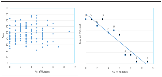
We performed ultra-deep target sequencing on 50 cancerassociated genes for plasma cfDNA from a cohort of 129 apparently healthy cancer-free subjects. To increase the confidence of the called mutations, we here defined the mutation as the variant allele frequency greater than 1% and the average depth more than 5,000 xs for demonstration. Our data revealed an age-independent mutation spectrum with average 3.12 somatic mutations per subject (Figure 1). The most frequently mutated genes are TP53 (42%), KIT (6%), KDR (5.5%), PIK3CA (5.5%), EGFR (5%) and PTEN (3.7%). These results highlighted the prevalence of some cancer-associated driver mutations in healthy individuals as background mutations. We also demonstrated the concordance between our results and a recent study for revealing the real somatic mutation in healthy population.

Figure 1: Distribution plots of somatic mutation detected in a cohort of 129 healthy subjects.
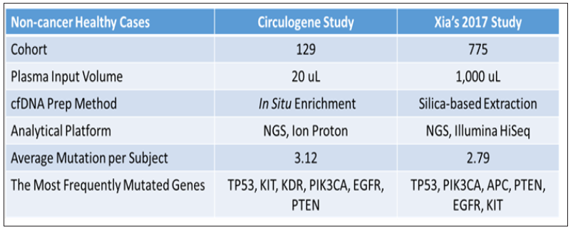
The study by Xia et al. [12] examined the background somatic mutations in white blood cells and cfDNA in healthy controls based on sequencing data from 821 non-cancer individuals with the aim of understanding the baseline profile of somatic mutations detected in cfDNA. The data comparison was summarized in Figure 2. Although there are differences in study cohort composition, sample volume, extraction methodology and analytical platform, the end results are remarkably similar, i.e., average 3 mutations per subject with an almost identical list of frequently mutated genes. Although varying mutation spectra in cancers have often been attributed to cancerspecific processes, our data suggest that at least a subset of these mutations actually reflect normal tissue-specific processes. This concept is consistent with the idea that a substantial fraction of the mutations found in cancers occur in normal stem cells [13,14].

Figure 2: Comparison of somatic mutation detection in healthy population from two studies.
Normal tissue as a germline control not justified

There is evidence for the presence of tumor-derived cfDNA in early cancers [15]. However, the real fraction of cfDNA that shed by tumor rather than the background somatic mutations is not well illustrated. For clinical application, the low level of tumor mutation as well as the heterogeneity of background mutation present in the circulation needs to be clearly addressed and differentiated to achieve accuracy. Unfortunately, this goal can’t be achieved by pushing detection limit of current advanced technology to below 0.01% mutant allele frequency (MAF). Contrarily, the higher sensitivity will guarantee higher chance to pick up background somatic mutations. Also, the clinical relevance of those lowpercentage tumor mutations is still debatable in terms of treatment decision or regimen change. Each human individual is unique. Every cancer patient is different. No two tumors are the same even resides within the same patient; to distinguish the definitive cancer-specific mutations from background signals observable in plasma is extremely daunting. Evaluation of specificity in plasma cfDNA profiles from large numbers of healthy individuals as representative controls for the cancer population seems farfetched with uncertainty, especially when standardized protocol and optimized technology are still lacking.

Unlike tissue genomic DNA, circulating cfDNA is so diluted and dynamic with a relatively short half-life, making single-point measurement not suitable for clinical application. We reason that cfDNA in circulation is truly under a continuous selection pressure to select for highly aggressive/proliferative clones, as disease progressing the low-abundant tumor clones will either evolve and dominate or vanish by the immune clean-up processes, therefore longitudinal clinical follow-up should be performed to identify the best time and target for precision therapy, meanwhile to filter out contaminating background mutations. To achieve high clinical specificity, a cfDNA-based test must be capable of distinguishing between the background signals originating from non-cancer or pre-cancerous processes and the invasive malignancy of clinical interest. It is still possible that mutational signatures in cfDNA could distinguish basic biological processes from malignant and pathological processes.

Figure 3: A representative mutational trending curve after filtering out background mutations.
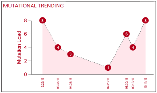
Here we propose a combined approach based on the tumor evolutional principle of “survival and domination of the fittest” in circulation that is to perform multiple time-point monitoring, filter out potential background mutations (e.g., <1% MAF), reduce sample input volume and interrogate multiple databases. A representative mutational trending curve following our approaches was shown (Figure 3). Our findings underscore the importance of an assessment of the landscape of somatic mutations in cancerfree population, and associated mutation signatures. Somatic mutations and mosaicism in healthy individuals have implications not only for early detection, diagnosis and treatment of cancer using liquid biopsy but also emerging technologies in healthcare. We recommend caution while extending the mutation conclusions to cancer patients by employing matched normal tissue as germline control. To increase sample input and push liquid biopsy sensitivity toward <1% may not serve the interest of detecting low-frequency mutant allele, but only to increase the chance of background mutation contamination. Application of artificial intelligence, machine-learning on big database to create an algorithm for highrisk population screening of cancer is a good idea for preventive medicine, yet the outcome is uncertain given the uniqueness of every patient, each tumor - one size can’t fit all.

Read More Lupine Publishers Oncology Medicine Journal Please Click on Below Link
https://lupine-publishers-cancer-journal.blogspot.com/

Tuesday, 28 August 2018

What is beyond the Nivolumab Monotherapy approval for advanced Hepatocellular Carcinoma?: (OAJOM)-Lupine Publishers


With an estimated 500,000 new cases per year, hepatocellular carcinoma (HCC) represents the third leading cause of cancer death worldwide. The incidence is rising in the west, largely due to an increasing incidence of hepatitis C virus infection [1]. The majority of HCC patients are diagnosed with disease too advanced for curative treatment. Only liver resection and liver transplantation are considered curative, with poor efficiency of other modalities such as radiofrequency ablation (RFA) and transarterial chemoembolization (TACE), although this may provide a modest prolongation in survival; however, the relapse in the majority of these patients is inevitable [2]. An array of translational research and pilot clinical trials have revealed that adoptive immunotherapy’s are safe by patients with HCC, but they lack efficacy [3]. Now, we are in the new era of immunotherapy’s such as immune checkpoint inhibitors and CAR-T strategies, which would bring benefit to the HCC patients.