Showing posts with label Journal of Pharmacology. Show all posts

Tuesday, 5 September 2023

Lupine Publishers | Assessing The Risk of Transfer of a Transdermal Hormonal Drug Through Skin-To-Skin Contact by an In Vitro Permeation Testing Method

 Lupine Publishers | Journal of Pharmacology & Clinical Research


Abstract

Objective: Transdermal hormonal drugs without protective covering may pose a risk for skin-to-skin hormone transfer through contact from the dosed to undosed person. Currently, no in vitro methodologies are available to assess the potential risk of skinto- skin drug transfer. This study aimed to develop an in vitro permeation test (IVPT) to evaluate the impact of use conditions (e.g., dosing duration and skin-to-skin contact time), on the potential risk of skin-to skin transfer of hormonal drugs.

Method: The risk of hormone transfer from the dosed skin to undosed skin was evaluated by employing human cadaver dermatomed skin mounted on vertical diffusion cells under various use conditions. Testosterone and estradiol transdermal hormonal gels were used as model hormonal products.

Results: Extent of hormone permeation through dosed skin was found directly proportional to the dosing duration of the formulation. Following dosing duration of 0.5-2 h, drug transfer from the dosed to undosed skin approached equilibrium after 24 h of skin-to-skin contact time. Following dosing duration of 8 h, drug transfer from dosed to undosed skin increased proportionally and significantly (p<0.05) after skin-to-skin contact times of 0.5-8 h.

Conclusion: A validated IVPT method may be used as a predictive tool to evaluate how dosing duration and contact time may affect the potential for skin-to-skin transfer of a hormonal drug. This IVPT tool could ultimately be utilized to develop drug products with a lower risk of skin-to-skin transfer and assess or mitigate skin-to-skin transfer risk of existing hormonal transdermal drugs.

Keywords: In Vitro Permeation Test (IVPT); Skin-to-skin transfer; Dosing duration; Topically applied drug products; Hormonal replacement therapy

Introduction

Hormonal drugs for replacement therapy are commonly administered via a transdermal route of drug administration. Hormone administration via a transdermal route has the advantage of by-passing the first-pass metabolism and minimizing the side effects that may be associated with peak plasma drug concentrations as is the case in oral administration [1]. Various topical drug products such as gels, creams, emulsions, sprays, or patches are employed for transdermal hormonal drug delivery. Although gels, creams, emulsions, or sprays are generally preferred over patches due to their lower skin irritability [2], a disadvantage of these topical products is their lack of protective covering without which residual hormonal products could pose a potential risk of drug transfer to others who may inadvertently come in direct skin-to-skin contact with the dosed skin. For instance, a small fraction of testosterone has been demonstrated to be systemically absorbed through the stratum corneum while most of the hormone remained unabsorbed on the skin surface of a patient for a longer period of time [3,4]. This unabsorbed hormone on the skin surface of a patient poses a risk of drug transfer via skin-to-skin contact to a non-patient. While transdermal testosterone and estradiol may provide substantial clinical benefit to a patient, these hormones may also lead to serious side effects to non-patients if transferred via inadvertent skin-to-skin contact such as hormonal imbalance. This risk has been described via several case reports in which children suffered pronounced virilization, early puberty, and premature epiphyseal closure of the bones due to secondary exposure to testosterone gel through skinto- skin contacts with male patients [5-9]. Additionally, children also experienced nipple swelling or breast enlargement due to transdermal estradiol exposure through contacts with female patients [10]. Skin-to-skin transfer of testosterone gel to women through male contacts has been reported to result in hirsutism, acne, coarsening of the voice, clitoral hypertrophy and male pattern alopecia [11,12]. Importantly, clothing has been shown to decrease the transfer of testosterone by preventing direct skin-to-skin contact [13,14] and washing of testosterone gel from the skin has been shown to significantly reduce the risk of skin-to-skin drug transfer [15].

The potential of drug transfer via skin contact with non-patients has been investigated in several pharmacokinetic clinical studies. In these studies, volunteers made physical contact of their dosed skin with the undosed skin of non-patients for a certain duration [4,15-17]. These studies were performed under a variety of occlusive or non-occlusive conditions [15,18]. However, these studies do not necessarily replicate the natural states of patients in which skin contact may be prolonged due to, for instance, intimate contact. Duplicating such studies with prolonged contact to a high degree of fidelity would be challenging. Moreover, conducting studies that expose non-patients to potentially toxic doses of drugs with their deleterious side effects for extended periods of time is not ethically appropriate. This is rendered even more challenging as in vivo animal studies are not considered suitable to extrapolate to human subjects because the animal skin (or fur) is structurally not similar to human skin. This leaves only in vitro approach for assessing the potential risk of drug transfer through skin contact, which to our knowledge has not yet been investigated.

The objective of the present study was to develop an in vitro permeation test (IVPT) to assess the potential of skin-to-skin drug transfer by exposing human cadaver dermatomed skin to various hormonal drug products. This study evaluated the effect of various experimental conditions, such as different formulations, formulation residence or wear time, and duration of skin-to-skin contact, on drug transfer. The formulation residence or wear time has been termed as dosing duration in this paper. Two model hormonal drug products were selected: AndroGel® (Testosterone 1.62%) and EstroGel® (Estradiol 0.06%). These two drug products were considered suitable for this study because there is a body of evidence establishing the clinically meaningful transfer of testosterone or estradiol from dosed to un-dosed subjects [3,4,19].

AndroGel® and EstroGel® are hydroalcoholic gels. Androgel® contains 1.62% of testosterone. Other excipients include carbopol 980, isopropyl myristate, sodium hydroxide, ethyl alcohol, and purified water. AndroGel® is indicated for the treatment of adult males with testosterone deficiency. One actuation of the pump delivers 1.25 g of gel which is equivalent to 20.25 mg of testosterone. Andro- Gel® is applied to the maximum surface area of the skin of the shoulders and upper arms once daily [20]. EstroGel® contains 0.06% of estradiol (17β-estradiol). Other excipients include carbomer 934P, triethanolamine, alcohol, and purified water. EstroGel® is indicated for the treatment of moderate to severe symptoms of vulvar and vaginal atrophy due to menopause in women. One actuation of the pump delivers 1.25 g of gel which is the single approved dose and is equivalent to 0.75 mg of estradiol. EstroGel® is applied over a large area (750 cm2) of arm skin, from the wrist to shoulder in a thin layer [21].

Materials and Methods

Materials

AndroGel® (Testosterone gel 1.62%), lot # D90024A, exp. date 03/2021 and EstroGel® (Estradiol gel 0.06%), lot # NHCR, exp. date 08/20, and lot # PDBT, exp. date 03/2021 were purchased from Brookville Pharmacy (Rockville, MD). Testosterone and estradiol USP reference standards were purchased from Sigma Aldrich Chemicals (St. Louis, MO). Acetonitrile and Methanol were purchased from VWR International (Radnor, PA). All other solvents and materials used in the study were of analytical grade.

Skin Samples

The human cadaver dermatomed skin of 600 ± 200 μm thickness obtained from shoulder and or thigh of donors of 40-60 years of age and free of any dermal diseases was purchased from Science Care (Phoenix, AZ USA) and stored at -80 °C.

Instrumentation

The HPLC system consisted of an Agilent-1260 series (Agilent technologies, Santa Clara, CA, USA) equipped with a diode array detector (DAD). Separation was achieved on a Phenomenex Luna C18 column (4.6 mm × 150 mm, 5 μm packing) fitted with a Phenomenex Luna C18 (2) guard column. In vitro permeation experiments were performed by using Vision Microette 18-cell system (Teledyne Hanson Research, Chatsworth, CA).

Methods
IVPT method development

An IVPT method was developed employing vertical diffusion cells to study the potential of drug transfer from a dosed skin to undosed skin through skin-to-skin contact. The diffusion cells, methodologies and study conditions utilized in the IVPT study were validated as per draft guidance for acyclovir topical cream 5% [22]. Briefly, vertical diffusion cells, consisting of a donor cell with cap (occlusive condition) or an open top (non-occlusive condition), and receiver cells of 12 mL capacity were used. The skin was thawed at room temperature and immersed in deionized water for hydration. The hydration medium was replaced with fresh medium three times every 10 minutes. The hydrated skin was dried with Kim wipes, cut into pieces of about 2.9 cm² to cover the exposure area of the receiver cell and examined visually for any surface damage with magnifying glass. The cut skin pieces were mounted onto the receiver cell orifices (1.72 cm2 diameter) with the stratum corneum side of the skin facing air and the dermal side in contact with the receiver medium. The receiver medium consisted of phosphate buffer saline of pH 7.4 for testosterone and phosphate buffer saline with 30% propylene glycol of pH 7.4 for estradiol. The receiver chambers were examined to ensure the absence of air bubbles below the dermal side of the skin. The receiver medium was maintained at 32 ± 1ºC by circulating water to mimic physiological temperature of the human skin surface and stirred at 600 ± 50 rpm. The skin integrity was tested by measuring trans-epidermal water loss (TEWL) with the help of a Vapometer. TEWL reading of 10 g/m2/h or lower ensured adequate hydration of skin.

Approximately 280 ± 20 mg of testosterone gel equivalent to 4.54 ± 0.324 mg of testosterone; or estradiol gel equivalent to 0.168 ± 0.012 mg of estradiol was applied on 1.766 cm2 of skin mounted on diffusion cell respectively. The gel was applied on the skin using a 1 mL positive displacement pipette and spread evenly over the entire skin surface with the help of a round-ended glass and then permeation experiment was started. The samples were withdrawn from the receiver medium at predetermined intervals throughout the course of permeation test for experiments A-C, described in the next subsection. The volume of receiver medium withdrawn was replaced by the fresh medium for maintaining the sink conditions. The drug permeated into the receiver medium, and drug recovered from dosed and undosed skins were quantified using validated HPLC methods.

Investigations on the effect of use conditions on skin-toskin transfer

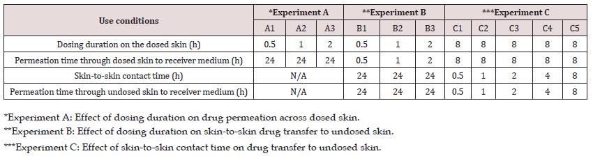
Three separate experiments (A, B, C) were carried out to study the effects of various use conditions to determine the potential for skin-to-skin drug transfer, as described in Table 1. Each experiment was replicated either four or six times based on the availability of skin.

Table 1: Experimental design to investigate the effect of dosing duration and skin-to-skin contact time on the in vitro drug transfer.
lupinepublishers-openaccess-journal-pharmacology-clinical-research-journal

In the first experiment (A), the effect of dosing duration on the rate and extent of drug permeation from the dosed skin to the receiver medium in diffusion cell was investigated. For this purpose, the dosing duration for the dosed skin was varied from 0.5 to 2 h. while permeation was continued up to 24 h. in all cases. Briefly, the formulation was applied on the skin and after the specified dosing duration, the residual formulation was carefully removed from the dosed skin with the help of cotton swabs without interrupting the permeation experiment. For this purpose, three dry and three wet (water) cotton swabs were used. All experiments were performed under non-occlusive conditions (n=6) by leaving the donor cell exposed to air. A control in vitro permeation test (n=6) under occlusive condition was also performed where the donor cell was covered by a cap after applying the formulation and no formulation was removed from the skin surface.

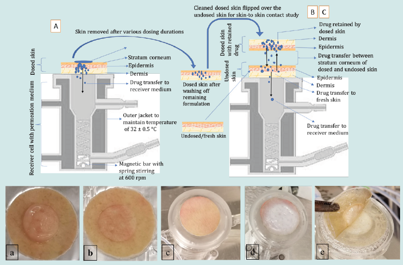
In the second experiment (B), the effect of dosing duration on drug transfer from the dosed to the undosed skin and subsequent permeation into the receiver medium was investigated. For this purpose, skin was dosed with the formulation exactly in the same fashion as in experiment A, where skin mounted on a diffusion cell was dosed for a specified dosing duration of either 0.5, 1 or 2 h and permeation allowed into receiver medium under non-occlusive condition. At the end of dosing duration, the experiment was stopped, and residual formulation was removed and cleaned from the dosed skin as described previously. Then the cleaned dosed skin was placed on an undosed skin that was already mounted on a diffusion cell containing fresh receiver medium. The dosed skins were placed on each other in such a way that the stratum corneum of both skins were in close contact with each other. A glass disc was carefully placed on the dosed skin and the cell was closed with a clamp to ensure intimate contact between the dosed and undosed skin. The permeation test was initiated to allow the skin-to-skin drug transfer for 24 h. The 24 h of skin-to-skin contact time is not likely to occur in real life. A longer contact time of 24 h of skin-toskin contact time was selected as a worst-case scenario to understand the fate of drug retained by the dosed skin and its transfer to undosed skin which, in turn, helped us to design experiment C.

In the third experiment (C), the effect of various skin-to-skin contact time on drug transfer from the dosed to the undosed skin was studied. In this experiment the dosing duration of the dosed skin was kept constant for 8 h prior to bringing it in contact with the undosed skin. The cleaned dosed skin was then flipped over the undosed skin and secured to ensure skin-to-skin contact in the same way as previously described in experiment B. The dosed and undosed skins were then removed after a specified skin-to-skin contact time of either 0.5,1, 2, 4 or 8 h and permeation also stopped at the same time. The schematic presentation of the experiments A-C is shown in (Figure 1).

Figure 1: Upper panel: schematic presentation of experiments A-C employing vertical diffusion cells. Lower panel: a) image of residual gel on the dosed skin, b) clean dosed skin after washing off residual gel, c) undosed/fresh skin mounted on the diffusion cell with fresh medium underneath, d) dosed skin flipped over the undosed skin to enable skin-to skin contact, and e) removing skin at the end of the contact time for quantifying drug transfer.
lupinepublishers-openaccess-journal-pharmacology-clinical-research-journal
Skin drug recovery

The dosed and undosed skins after removal from the diffusion cells were grounded into small pieces and then immersed in 5 mL of mobile phase. The skin in mobile phase was sonicated for 99 mins at 32 ± 0.5°C using a bath sonicator. The above skin samples were then centrifugated at 5000 rpm for 30 mins at 32 ± 0.5°C, followed by filtration. The filtrate was then analyzed by HPLC.

Analytical method

A reversed-phase HPLC method, adopted from literature, was used for the analysis of testosterone [23] and estradiol [24] contents in skin and receiver medium samples. Both methods were validated as per ICH Q2(R1) guidelines for specificity, linearity, accuracy, precision, range, limit of quantitation (LOQ), limit of detection (LOD) and system suitability [25]. Chromatographic separation of testosterone and estradiol were achieved on a Phenomenex Luna column using a methanol-water (70:30 v/v) mobile phase for testosterone and an acetonitrile-methanol-water (45:05:50 v/v) mobile phase for estradiol. Both compounds were eluted isocratically at a flow rate of 1 mL/min. Testosterone was analyzed with UV detection at 254 nm. Estradiol was analyzed with UV detection at 280 nm. The injection volume was 10 μL. The calibration range for testosterone was from 5-100 μg/mL with limit of detection (LOD) of 0.1 μg/mL with an acceptable precision (< 10%). The estradiol peak was detected by using DAD UV detector at 280 nm. The calibration range for estradiol was from 0.5-100 μg/mL with limit of detection (LOD) of 0.1 μg/mL with an acceptable precision (< 10%).

Data and Statistical Analysis

The permeation parameters such as cumulative drug permeated (Eq. 1), steady state flux (Eq. 2) and percent drug permeated (Eq. 3) were calculated per square centimeter (cm2) of the skin from the obtained AUC data using Eq. 1, 2 or 3, respectively [23,26,27]. The drug retention in the skin was normalized to a constant skin thickness of 373.33 μm for testosterone and 283.33 μm for estradiol based on the original skin sample thickness. The drug concentration in each receiver medium sample was calculated by correcting the sampling effect according to the equation described by Hayton and Chen [26].

where Q is the cumulative amount of drug permeating through the skin, A is the surface area of permeation at a given time t, Cd is the amount of drug in donor chamber and P is the permeability coefficient obtained from the slope of a plot of cumulative permeation of drug across skin samples against square root of time.

where J is the flux of drug from donor to receiver chambers in μg/cm2/h, dc/dt is the concentration gradient, D is diffusion coefficient, P is the permeability coefficient of the skin tissue to the drug, Cdonor is the amount of drug in the donor chamber, and Creceptor is amount of the drug in the receiver chamber at the end of the study.

In case of experiment B and C, the amount of drug retained by the dosed skin following various dosing duration were back calculated using Eq. 4 below. The obtained total amount retained by the dosed skin was then considered as 100% loading amount of drug on the undosed skin during skin-to-skin drug transfer studies. All data were expressed as mean ± standard deviations, with either four or six experimental replicates. Statistical significance for each experiment was determined using one-way analysis of variance (ANOVA). A p-value of less than 0.05 was considered statistically significant.

Figure 2: Effect of dosing duration (0.5, 1 and 2 h) on testosterone permeation (mean ± SD of n=6). A) cumulative amount of drug permeated as a function of permeation time, B) percent testosterone permeation after 24 h, C) Flux of testosterone.

lupinepublishers-openaccess-journal-pharmacology-clinical-research-journal

The results in Figure 2A show the effect of dosing duration 0.5, 1, or 2 h on the cumulative amount of testosterone permeated through the dosed skin into the receiver medium. At dosing duration of 0.5, 1 and 2 h, the cumulative amount of testosterone that permeated through the unit surface area of skin following 24 h of permeation was found to be 3.63 ± 1.81, 5.0 ±3.52 and 5.6 ± 2.27 μg/mL/cm2, respectively, under non-occlusive condition. Figure 2B shows the percent testosterone permeated through the dosed skin into the receiver medium was 2.3 ± 1.19, 2.7 ± 1.8 and 2.9 ± 1.17 % after 24 h of permeation when dosing duration was set at 0.5, 1 or 2 h, respectively. Figure 2C shows the steady state flux of testosterone was 2.3 ± 0.16, 2.75 ±1.30 and 3.1±1.12 μg/mL/cm2/h for dosing duration of 0.5, 1, and 2 h, respectively. These permeation results clearly indicate that increase in the dosing duration from 0.5 to 2 h exhibited an increase in testosterone permeation across the skin. However, the cumulative permeation or percent permeation was not statistically significant (p>0.05) at various dosing durations which may be attributed to close proximity of dosing durations to each other (0.5, 1 and 2 h). Notably, for the dosing durations of 0.5, 1, or 2 h studied, the retention of testosterone in the skin after allowing permeation for 24 h was found below the LOD.

Contrary to the extent of testosterone permeation, the estradiol permeation in receiver medium was found below the LOD even after 24 h of permeation for all dosing durations (0.5, 1 and 2 h). This observation may be explained by assuming very slow permeation of estradiol. Therefore, estradiol could not permeate in detectable amounts into the receiver medium during the shorter dosing duration of 0.5 to 2 h. On the other hand, in case of the control run, where the dosing duration was long enough (24 h) at occlusive condition, estradiol permeated into receiver medium in measurable quantities, as shown in Table 2. The results in Table 2 for control show the percent estradiol permeated, cumulative estradiol permeated, and estradiol flux were 34.37 ± 2.21, 2.63 ± 0.17 and 1.72 ± 0.11, respectively.

Table 2: In vitro permeation data of estradiol as control under occlusive conditions, (n=6).

lupinepublishers-openaccess-journal-pharmacology-clinical-research-journal

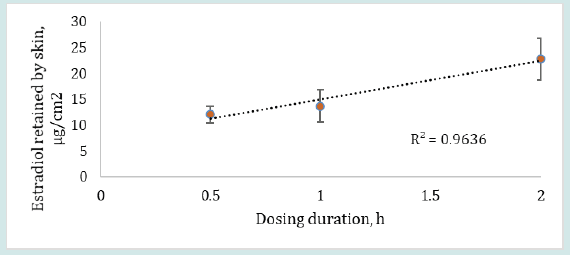
The results in Figure 3 show that the retention of estradiol by the dosed skin was found to be 12.07 ± 1.63, 13.70 ± 3.16 and 22.78 ± 4.06 μg/cm2 when dosing duration was 0.5, 1 or 2 h, respectively, after continuous permeation for 24 h. The retention of estradiol by skin clearly exhibited an increase in drug absorption when the dosing duration was increased from 0.5 to 2 h. However, the skin retention of estradiol was not statistically significant (p>0.05) for different dosing durations which may be attributed to the selected dosing durations (0.5, 1 and 2 h) being close in time to each other. The low permeation results of estradiol may be due to hormone accumulation in the subcutaneous region or due to estradiol’s relatively high affinity for the lipophilic and hydrophilic interface of the stratum corneum and epidermis from where the drug may permeate slowly into the circulation [27,28].

Figure 3: Estradiol retained by the skin (mean ± SD) after 24 h of permeation when dosing duration was varied from 0.5 – 2 h, (n=6).
lupinepublishers-openaccess-journal-pharmacology-clinical-research-journal
Effect of dosing duration on skin-to-skin drug transfer

The results in Table 3 show the effect of dosing duration on the testosterone transfer through skin-to-skin contact to undosed skin and into the receiver medium in contact with undosed skin. The testosterone recovered from the dosed skins following skin-to-skin contact of 24 h with undosed skins was 463.60 ± 60.28, 509.39 ± 22.44 and 565.99 ± 100.09 μg/cm2 for dosing duration of 0.5, 1 or 2 h, respectively. Testosterone absorbed and retained by the undosed skin following the same skin-to-skin contact time and dosing durations was 426.35 ±83.69, 449.41 ± 66.32 and 455.31 ± 42.15 μg/cm2, respectively. The cumulative amount of testosterone that finally reached into the receiver medium from the dosed through undosed skins was observed to be 5.19, 4.51 and 7.82 μg/cm2 after 0.5, 1, or 2 h of dosing duration, respectively. These results indicated that testosterone recovered from dosed skin, undosed skin or cumulative amount in the receiver medium all exhibited an increase in drug transfer with increase in dosing duration.

Table 3: Testosterone amount recovered from dosed skin, undosed skin and receiver medium after 24 h of skin-to-skin contact time (n=6).

lupinepublishers-openaccess-journal-pharmacology-clinical-research-journal

After 24 h of skin-to-skin drug transfer, most of the retained drug was assumed to be transferred from the dosed to undosed skin after this duration of skin-to-skin contact time. Surprisingly, we did not observe such results. Instead, the amount of testosterone retained by both dosed and undosed skins were approaching close to each other for all dosing durations evaluated.

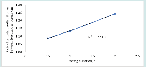
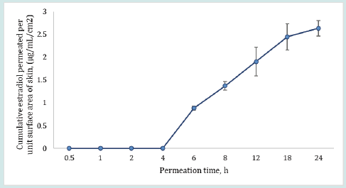
The plot in Figure 4 shows that the ratio of testosterone distribution between the dosed and undosed skin after 24 h of skin-toskin contact increased with increasing dosing duration. The testosterone distribution ratios between dosed and undosed skin were relatively close but the difference was statistically significant (p = 0.0262, regression (R2) = 0.9983). The smaller ratio of testosterone distribution after 24 h of permeation may be attributed to the drug content in dosed or undosed skin which may be slowly approaching equilibrium. On the other hand, the amount of estradiol recovered from the dosed skin or undosed skin or from the receiver medium in contact with undosed skin following the same skin-to-skin contact time after specified dosing duration were found below LOD for most of the cells (n=6). The possible reason for observing undetectable lower estradiol retention by both dosed and undosed skins could be attributed to the equilibrium distribution of the estradiol in both dosed and undosed skins which further lowered the sensitivity of the estradiol detection. Notably, the percent drug recovered from the skin wash after each dosing period (0.5 to 2 h) were 95- 98%. The cumulative estradiol permeation results for control formulation revealed that estradiol starts to appear in the receiver medium after 4 h during 24 h of permeation test study even though the estradiol formulation was kept on the skin intact for 24 h at occlusive conditions (Figure 5).

Figure 4: Ratios of testosterone distribution or transfer between the dosed and undosed skin following 0.5, 1 or 2 h of dosing duration and 24 h of skin-to-skin contact (n=4).
lupinepublishers-openaccess-journal-pharmacology-clinical-research-journal

Figure 5: Cumulative amount of drug permeated per unit surface area of the skin (μg/mL/cm2).
lupinepublishers-openaccess-journal-pharmacology-clinical-research-journal
Effect of contact time on skin-to-skin drug transfer

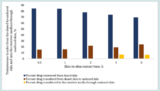
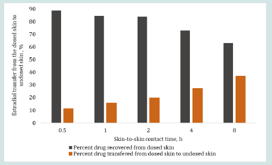
The results in Figure 6 and 7 show the effect of changing skinto- skin contact time on drug transfer from the dosed skin to the undosed skin and into the receiver medium. The dosing duration was kept constant at 8 h for all dosed skins while the skin-to-skin contact time of dosed skin with undosed skin was varied from 0.5 to 8 h. The percent testosterone recovered from the undosed skin after the specified skin-to-skin contact times, as shown in Figure 6, were 15.50, 16.03, 21.81, 19.12 and 23.86%. The testosterone recovered from the receiver mediums in contact with the undosed skin were below LOD after up to 2 h of skin-to skin contact time and 6.92 and 6.70% after 4 and 8 h of skin-to skin contact time, respectively. The overall testosterone transfer from the dosed to undosed skin showed that testosterone transfer increased linearly and significantly (p=0.000785, R2 = 0.985) with increasing skin-to-skin contact time. The testosterone content in the receiver medium after 0.5 to 2 h of skin-to skin contact was found below the LOD because of shorter permeation time. Importantly, most of the results of the testosterone drug transfer did not exceed 50% of % relative standard deviation (RSD). The variability in these experiments was at tributed to the inherent variability from differences in the donors’ race, age, sex, skin thickness and site of the skin source on the body and the pairing of two stratum corneum together. The results in Figure 7 show the effect of 0.5 to 8 h of skin-to-skin contact time on the estradiol transfer from the dosed to undosed skin and into the receiver medium following same dosing duration of 8 h. The percent estradiol recovered from the undosed skins were 11.31, 15.62, 19.58, 27.25 or 37.06%, as shown in Figure 7 for the respective contact times. In all cases, the permeated amount in the receiver medium via the undosed skins were found below LOD. Overall, the results clearly show a linear transfer of estradiol that significantly increased when the duration of skin-to-skin contact was increased (p= 0.00223, R2 of 0.9699).

Figure 6: Effect of skin-to-skin contact time on the percent testosterone transfer from the dosed skin (after 8 h of dosing) to the undosed skin and into the receiver medium in contact with undosed skin after 0.5, 1, 2, 4 and 8 h of skin-to-skin contact time. The total drug retained by the dosed skin after 8 hr of dosing duration was back calculated by employing Eq. 4.
lupinepublishers-openaccess-journal-pharmacology-clinical-research-journal

Figure 7: Effect of skin-to-skin contact time on the percent estradiol transfer from the dosed skin (after 8 h of dosing duration) to the undosed skin (including receiver medium containing undosed skin) after 0.5, 1, 2, 4 and 8 h of skin-to-skin contact time (obtained from Eq. 1). The total drug retention by the dosed skin after 8 hr of dosing duration was considered as 100% loading dose on the undosed skin for skin-to-skin contact study.
lupinepublishers-openaccess-journal-pharmacology-clinical-research-journal

Conclusions

The data provided by this work demonstrated that the potential risk of transdermal hormonal drug transfer to undosed persons through skin-to-skin contact with a dosed skin may be assessed by employing an in vitro permeation testing method. The extent of drug transfer to an undosed skin through skin-to-skin contact with a dosed skin may depend on the dosing duration of dosed skin and its duration of contact with undosed skin. An adequately developed and validated IVPT method may serve as a tool to quantify the extent of drug transfer from the dosed skin to undosed skin for product development of transdermal hormonal drugs or other transdermal drugs.

Acknowledgment

“This project was supported in part by appointing ORISEs (Behnam Dasht Bozorg and Rokon Uz Zaman) to the Research Participation Program at the U.S. Food and Drug Administration administered by the Oak Ridge Institute for Science and Education through an interagency agreement between the U.S. Department of Energy and the U.S. Food and Drug Administration.”

Disclaimer

This scientific publication reflects the views of the authors and should not be construed to represent FDA’s views or policies.

Read More About Lupine Publishers Journal of  Pharmacology & Clinical Research Please Click on the Below Link: https://lupine-publishers-pharmacology.blogspot.com/

Monday, 26 June 2023

Lupine Publishers | The Effect of all-Natural Biological Milk Silo Culture (MSC) to Protect Human Health by Improving the Safety, Quality and Quantity of the Dairy Food Products through Activation of Milk’s Natural Lactoperoxidase (LP) System

 Lupine Publishers | LOJ Pharmacology & Clinical Research


Abstract

The effect of inoculating raw milk with Milk Silo Culture (MSC), developed and manufactured by IMAC, Inc. (International Media and Cultures), on retarding the growth of spoilage and pathogenic bacteria, yeast and molds, and viruses through the activation of milk’s natural Lacto-Peroxidase System (LP), has been evaluated. The Milk silo Culture (MSC) has been used successfully for over 30 years, routinely by some dairy food manufacturers around the world. Although the validity and efficacy of MSC has been clearly proven by various leading scientific authorities, yet several of the dairy food manufacturers are skeptical and thus are not taking advantage of the marvelous all-natural biological system to improve the quality, safety, and quantity of the ready to consume dairy food products. The current worldwide Covid-19 pandemic partially or completely changed the thinking and perspective of some consumers regarding the safety of the animal-origin foods they are consuming. Consequently, they are tending towards cerealderived non-dairy products, although milk and dairy products are the best nutritionally balanced foods, with high quality protein and calcium, etc. Since the milk is of animal origin with possible microbial contamination from both animal as well as milk handlers, we felt that it is extremely important to protect the raw milk from pathogenic bacteria and viruses, etc., using all-natural and easyto- implement biological systems, so that the consumers will have utmost confidence to consume such dairy food products.

In addition, we felt that it is equally important that dairy food manufacturers should also benefit from such systems to improve the quantity of dairy food products. Thus, laboratory and commercial research experiments were conducted to study the effect of Milk Silo Culture to accomplish the aforementioned objectives. The result of the study proved that the use of Milk silo culture significantly improved the microbiological safety (significant reduction of pathogenic bacteria and viruses), quality, and quantity (by protecting the microbial degradation of milk protein-casein and fat determined by using particle size analyzer) of the dairy food products, such as widely consumed cheese and fluid milk products, etc., through the activation of milk’s all-natural Lacto- Peroxidase system. Thus, the dairy food industry can go forward safely, without any skepticism, and take advantage of using “Milk Silo culture” to improve the microbiological safety, quality, and quantity of dairy food products to protect human health and to improve consumer confidence.

Keywords: Milk Silo Culture (MSC); Lactoperoxidase (LP) System; Protection of Cheese from Pathogenic Bacteria; Bacteriophage; Extended shelf-life of Fluid Milk; Casein; RNA and DNA Viruses; Increased Cheese Yield and quality

Introduction

Raw milk starts spoiling as soon as it comes out of the cow’s udder due to extraneous microbial contamination. With all the precautions undertaken, it is literally impossible to keep the contaminants away. However, there is an inherent built-in provision in raw milk that can protect it from spoilage. This is called the Lactoperoxidase system. This is also called “LP” system, in short. The “LP” system has three components: Lactoperoxidase enzyme (present in raw milk); Thiocyanate (present in raw milk); Hydrogen Peroxide (not present in raw milk). This system will be activated only when all these three components are present in raw milk. The Lactoperoxidase enzyme acts on thiocyanate in the presence of Hydrogen Peroxide, and converts it to Thiocyanite, which is extremely inhibitory to both the spoilage and pathogenic bacteria, certain yeast and molds and some DNA and RNA viruses (in this connection the SARS-CoV-2 Corona Virus which created covid-19 pandemic is also an RNA virus). The only component which is missing in raw milk to activate the “LP” system is Hydrogen Peroxide.

The question here is how can we induce the production of minute amounts of Hydrogen Peroxide in raw milk to activate the LP system? The specific answer to this puzzle is the use of certain food grade beneficial starter lactic cultures which can produce the molecular Hydrogen Peroxide, when added to raw milk [1]. The addition of food-grade lactic cultures is legal to inoculate the raw milk (while the raw milk is in your possession) without having to declare them. However, the food-grade lactic cultures must be selected based on their intrinsic ability to produce hydrogen peroxide and bacterial-origin (natural) oxidase enzymes. These specialty cultures have been produced by IMAC (International Media and Cultures, A division of the American Dairy and food Consulting Laboratories, Inc.) designated as Milk Silo cultures (MSC) and have been used successfully for the past several years, all over the world, to protect the raw milk in the cheese plants. However, most cheese plants are still reluctant to use them because of a lack of detailed scientific information.

The most often asked question is, how long does it take to activate the LP system in raw milk using the IMAC milk silo culture? The system can be activated in a few hours (4 to 8 hours) in raw milk, in milk silos. Of course, it will be beneficial even if the raw milk is held longer in the silos with the inclusion of Milk silo culture. It has been thoroughly researched by several scientists, who came to a firm conclusion that the activated LP system followed by pasteurization will destroy both the spoilage-type bacteria as well as pathogenic bacteria, Yeast, molds and Viruses, according to Kamau et.al [2,3]. These research findings were science-based and proven through a series of experiments by leading scientists and were also published in the most dependable peer-reviewed scientific journals. I am presenting these references for better clarification to the quality control, Management, and research and development departments, as verification and validity of the statements made and published by the world’s Leading dairy researchers about LP system.

With all this published information, some of the dairy industry personnel are still unwilling to use them and requiring more experimental information to prove its total efficacy. There are two aspects of looking into Milk silo culture (MSC). The first aspect is the effect of activated LP system by MSC to significantly retard the spoilage type Psychrotrophic bacteria (Psychrotrophic - meaning cold loving or loves to grow in refrigerated raw milk) which degrades milk protein casein and milk fat in the shortest time, resulting in the reduction of dairy products yields, especially cheese. In addition, their enzymes are highly heat resistant which survives pasteurization, and start destroying the quality and functionality of cheese and other dairy products. The second aspect is to study the LP system effect on destroying the pathogenic bacteria in raw milk, which I will be addressing in the later part of this article. Earlier, the dairy scientists Lara et.al [4] reported the following regarding the improved cheese yields they have obtained using “LP system” activated raw milk. Of course, which is subsequently pasteurized to make cheese.

According to them the yields of fresh type cheese (measured on a wet or dry bases) of LP treated cow milk were significantly higher than the cheese made from control (no LP activated) milk. They have indicated that a complete transition or recovery of fat from the LP-activated milk into cheese was achieved, while only 85 % of fat was retained in the cheese made from non-LP activated milk or untreated milk. More than all, the quality and microbiological safety of the product was greatly improved due to the retardation of spoilage and harmful micro-organisms. More than 2kg (4.4 lbs.) of extra cheese can be recovered for every 100 kg (220 lbs.) of milk. These research findings were also reported in the most reputable peer- reviewed scientific journal “Milchwissenschaft”. According to other reputable scientists, Earnshaw et.al [5], the cheese made using LP-activated milk did not affect the sensory characters of the cheese and significantly improved the quality and safety of the products. According to them, the activated LP system functions as an effective natural preservation system to increase not only the safety but also quality of the cheese.

The Following Dairy Product (Cheese Etc.) Yield- Reducing and Cheese Quality Degrading Psychrophilic Spoilage Bacteria Encountered in Raw Milk, which are Inhibited by the LP System

The antibacterial activity of the LP system in milk against psychrotrophic spoilage organisms has been widely investigated by Bjorck et. al. [6,7]. Bjorck et. al. demonstrated that LP system was bactericidal (killed the bacteria) against the spoilage bacterial species belonging to pseudomonas and several coliforms such as E. coli. In addition, they have discovered that several milk spoilage type grams -negative rod- shaped bacteria were killed by at least 91%, in a relatively shortest time (4 hr). Furthermore, the activated LP system prevented the growth of the Psychrotrophic bacteria for up to 5 days in raw milk.

The Following Commonly Encountered Pathogenic Bacteria in Raw Milk and in the Subsequent Cheese Products, have been Inhibited by the LP System, According to Several Research Reports Published by the Scientific Authorities, in the Leading Peer- Reviewed Scientific Journals

a) Salmonella: Salmonella typhimurium and several other pathogenic salmonellae causes gastroenteritis in humans, primarily due to ingestion of contaminated foods, including cheese. The activated LP System exerts both bacteriostatic (stops bacterial growth) and bactericidal (kills bacteria) effects against the pathogenic (disease-causing) strains of Salmonella typhimurium in the raw milk [8].

b) Campylobacter jejuni: This is the leading cause of zoonotic infections (animals to humans and vice versa). It mostly infects children under the age of five and immunocompromised individuals. Even in small numbers (500 bacteria) in milk or cheese can cause severe illness in humans by inducing fever, abdominal pain, diarrhea, nausea, vomiting, etc. The LP system exhibits an antibacterial effect on pathogenic Campylobacter species, according to the scientists Beamer et.al. [9].

c) Bacillus cereus: These pathogenic bacteria have been isolated from several samples of raw milk. It causes Bovine Mastitis and thus enters raw milk. It causes food poisoning through the production of toxins. It causes serious clinical conditions including death involving human intestines, lungs, and brain. The good news is that according to the research work of Scientists Tenovue et.al [10], and Zajac et.al. [11], the LP System inhibits Bacillus cereus Pathogenic bacteria found in raw milk.

d) Staphylococcus aureus (Staph): This is a major Causing agent of Bovine mastitis and poses a human health problem since pathogen can enter in to milk from mastitis udders. This organism produces pasteurization resistant toxins (enterotoxins) in raw milk and thus causes severe illness due to food poisoning. Renowned scientists Kamau et.al. [12], have studied, confirmed, and published the data stating that LP System activated in raw milk is both bacteriostatic (stops multiplication) and bactericidal (kills the bacteria) to both the Staphylococcus aureus and Listeria monocytogenes. The other investigators Seifu et.al. [13], also proved LP System activated in goats raw milk inactivated Staph bacterial pathogen both being Bacteriostatic as well as bactericidal.

e) Listeria monocytogenes (Listeria): Listeria monocytogenes is a pathogen of major concern to the dairy industry as food -borne listeriosis due to consumption of milk products, specifically cheese. Listeria infection causes abortion, septicemia, and meningitis in humans. Doyle et.al. [14], confirmed and published the scientific data indicating that the risk of listerioses is amplified by the ability of listeria bacteria to grow at refrigeration temperatures and their relative heat resistance to survive pasteurization temperatures. Several leading scientists Kamau et.al. confirmed that the activated LP system in raw milk (following pasteurization) kills the Listeria bacteria by exhibiting both bactericidal and bacteriostatic effect.

f) Brucella melitensis: This organism causes human Brucellosis. It has been associated predominantly in goat’s milk and goat milks cheeses, which have been attributed to causing Brucellosis. According to scientists Ryser et.al. [15] and Seifu et.al. the activated LP System in raw goat’s milk inhibited or killed the Brucella pathogenic bacteria through exhibiting bactericidal effect.

Antifungal or Anti mold effect of Lactoperoxidase system activated in raw milk to inhibit milk associated molds and some yeasts

Yeast and molds cannot be avoided in raw milk due to extraneous contamination. Although their vegetative cells can be inactivated by pasteurization of milk, their harmful toxins (Aflatoxin) cannot be destroyed with mere thermal heat treatment. In addition, some of the mold spores may survive pasteurization. Jacob et.al. [16], have published data showing that activated Lactoperoxidase system inhibited the growth of several mold species in raw milk. Doyle and Marth [17] have demonstrated that mold-produced toxic aflatoxin can be degraded or destroyed by the Lactoperoxidase system. This is a significant finding.

Antiviral effect of the Lactoperoxidase system on RNA viruses (SARS-CoV-2 virus causing COVID-19)

The latest SARS-CoV-2 virus causing Covid-19 pandemic has brought significant awareness to the public around the world, due to its devastating effect on humanity and economy. Many people in various sections are questioning even the safety of food products and the food industry practices regarding the total assurance of not having any viral contaminants, in ready to consume foods. Wherever there is human involvement, it is hard to eliminate the presence and prevalence of pathogenic viruses. It has been proven and reported by Scientists Belding et.al. [18], and Yamaguchi et.al. [19], that Lactoperoxidase system (LP System) can kill the most resistant Polio and HIV-1 viruses. As we all know, Polio and HIV-1 viruses are RNA viruses. In addition, the notable human diseases caused by RNA viruses include the common cold, influenza, SARS, MERS, Covid 19, Dengue virus, Hepatitis-C, Hepatitis -E, West Nile Fever, Ebola, Rabies, Polio, Mumps, and Measles etc.

Since it has been proven that activated Lactoperoxidase system is viricidal to several hard to kill RNA viruses, it can be hypothesized that the raw milk’s activated LP system may also have effect on inhibiting several RNA viruses including SARS- CoV-2, which caused Covid -19 pandemic. In fact, now it is proven by Cegolon et.al. [20], in 2021 that thiocyanite of LP system even at a micromolar concentration can destroy or be viricidal to the SARS-CoV-2 virus through irreversible oxidative damage of the lipid components of the viral envelop of the nucleoproteins. Although it has been proven that the LP system inhibits the pathogenic bacteria, yeast and molds, viruses, and the spoilage organisms in raw milk, according to Reddy [21], yet some of the dairy industry personnel are requesting more concrete data and simplified approach to implement such systems in their manufacturing facilities. Thus, the following research experiments were conducted and reported in this article.

Materials and Methods

The following research experiments were conducted to study the effect of Milk Silo cultures (MSC) inoculation into raw milk, and their effect of inhibiting DNA viruses (Bacteriophages), spoilage type bacteria, reducing the breakdown of milk nutrients, and finally to improve the quantity and quality and safety of the dairy products.

a) The antiviral effect of the MSC activated Lacto-peroxidase system on DNA virus in raw milk.

b) Antibacterial effect of Milk silo culture (MSC) on retarding spoilage type bacteria in the raw milk and after pasteurization, to extend the shelf-life of grade-A fluid milk.

c) Milk protein (casein) and fat protecting effect of MSC to improve the quality and quantity of these essential nutrients, determined using Particle Size Analyzer.

d) Effect of MSC on improving the dairy product yield, safety from pathogenic bacteria, yeast and molds, and quality proven through controlled laboratories cheese yield experiments.

Experiment 1: Antiviral Effect of Lactoperoxidase System on DNA Virus (Bacteriophage)

The grade A raw milk was inoculated with DNA virus Streptococcus thermophilus bacteriophage to arrive at 120 x 106 (120 million) Phage particles/ml. The milk silo culture (MSC) supplied by IMAC (International Media and Cultures) is a frozen can comprising of food grade thermophilic and mesophilic lactic acid producing bacteria along with their growth-end products (immunomodulins). These immunomodulins include bacteriocins, antibacterial therapeutic bio-peptides, non-specific inhibitory compounds, organic acids and traces of preformed molecular hydrogen peroxide and enzymes. The thawed Milk Silo culture was inoculated into raw milk using the equivalent amount of 2 cans (260 ml.) per 100,000 lbs. of milk. The frozen Milk Silo Cultures were thawed fully and then the liquid culture was added into the milk silo. The inoculated milk was incubated at the refrigeration temperatures (comparable to milk silos) for 8 hours. Then the milk was pasteurized at 161 F. for 15 sec (commercial HTST pasteurization temperature). The bacteriophage counts were determined using plaque assay before and after pasteurization using the specific bacterial host (Streptococcus thermophilus) of the bacteriophage included in this study. The phage inoculated raw milk without the addition of milk silo culture served as control. The results are presented in the results and discussion section.

Experiment 2: The Effect of Milk Silo Culture Activated LP System on Reducing the Spoilage Type Bacteria Present in Raw Milk, and after Pasteurization to Extend the Shelf Life of Grade A Fluid Milk

Grade A raw milk obtained freshly from farm was divided into two fractions. One is inoculated with Milk Silo Culture (MSC), the other fraction without MSC was treated as control. The Milk Silo Culture inoculation was comparable to using 260 ml of culture for 100,000 lbs. of milk. Both the fractions were pasteurized at 161F for 15 seconds. Both the pasteurized samples were plated to determine the total bacterial count using SPC (standard plate count). Also, the samples were plated to determine the psychrotrophic bacterial counts as well as the coliform counts. The samples were stored in the refrigerator for an extended length of 30 days. The samples were analyzed organoleptically for flavor and taste at intervals of 15, 20,25, and 30 days, during the storage. The bacterial counts were determined also at the end of the storage at the refrigeration temperature.

Experiment 3: The Effect of Milk Silo Cultures Inoculation in Refrigerated Commercial Raw Milk on Protecting the Microbial Degradation of Milk Protein (Casein) and Milk Fat in 24 Hours determined using a Particle Size Analyzer

Normal raw milk on average will have approximately 3.5 % protein and 3.2% fat. After milking, the pooled milk is placed in a refrigerated tank at the farm for a period of few hours to a day or two. Then the milk is picked up by the trucker going from farm to farm. Such comingled milk is being transported to the cheese or any dairy product manufacturing plant. The raw milk is then pumped from truck into large, refrigerated milk silo at the processing plant. The raw milk will be in silos for at least 8-12 hours to one or two days, depending on the day of the manufacturing. All along the psychrotrophic bacteria present in raw milk will be growing, since they can grow and multiply in the refrigerated cold raw milk. Their maximum deleterious effect is towards the end of their journey. These psychrotrophic organisms produce heat resistant proteolytic (protein breaking) and lipolytic (fat breaking) enzymes, which will degrade milk proteins and fats in raw milk during storage.

Even if psychrotrophic bacteria consume 0.05% protein and 0.05% fat, it will reflect in a great loss to the cheese manufacturer due to loss of significant quantity of cheese, especially considering 95% of the cost of the cheese is attributed to milk. The end products of the growth and the enzymes elaborated by the Psychrotrophic bacteria, will significantly alter the quality of the finished dairy product. Similar thing will happen even with the pathogenic bacteria as well as Yeast and molds in the raw milk, since presence, absence and concentration of these organisms is a classical hit and miss proposition. None of the investigators earlier has studied the exact loss of the protein and fat during storage for up to 24 hours at the cheese plant or any dairy product producing facility at the minute level. Thus, we undertook a study to determine the exact loss of the protein and fat in raw milk due to growth of psychotropic bacteria, by using the particle size analyzer, which will determine the degradation of solids accurately.

The physical chemistry and mechanism of how the Particle Size analyzer work is as follows: The principle behind the Melvern particle Analyzer, to determine the degree of breakdown of milk protein and milk fat in milk is as follows: laser diffraction measures particle size distribution by measuring the angular variation in intensity of light scattered as a laser beam passes through a dispersed particulate sample. Large particles scatter light at small angles relative to the laser beam and small particles scatter light at large angles. The angular scattering intensity data is then analyzed to calculate the size of the particles responsible for the scattering pattern, using the Mie theory of light scattering. The particle size is reported as a volume equivalent sphere diameter. This research was conducted at the factory level in a plant processing over one million pounds of milk a day making Mozzarella cheese in southern Wales, U.K. The holding capacity of each milk silo is 300,000lbs.

We have elected to use 2 milk Silos and inoculated the milk in silo with 6 cans of Milk Silo Cultures. One can contain 130 grams of milk silo culture per 50,000 lbs. of milk). The other silo was not inoculated with the Milk Silo Culture served as control. Milk at zero time (prior inoculation of the milk silo culture) served as control. The raw milk after 24 hours, with and without inoculation of MSC served as positive and negative controls. All the samples were analyzed for the degradation of solids by using the particle size analyzer and for the protein and fat breakdown using standard lab tests. The cheese made with such milk was also analyzed for the microbiological as well as for the quality and quantity of the cheese. The results of this experiment are presented in the results and discussion section. The theoretical cheese yields were determined using modified Cornell Van slyke Formula, developed for the Mozzarella cheese.

Experiment 4: The Controlled Laboratory Experiments Conducted to Check the Effect of Milk Silo Culture Inoculation into Raw Milk on Improving the Safety (Freedom from Spoilage and Pathogenic Bacteria), Quantity (Yields) and Quality of the Dairy Products-Cheese and the Whey.

To study the effect of Milk Silo Culture on the retardation of casein and fat breakdown in cheese milk and thus improve the cheese yield, quality and safety of the dairy product, the following experiments were designed. Grade A raw milk (3.5% milk fat) was divided into eight, 200 ml. fractions. Four of the 200 ml. fractions were not inoculated with Milk Silo Culture and were considered as controls. The other remaining four, 200 ml. fractions were inoculated with amount comparable to inoculation of two 130 ml. cans of Milk Silo Culture /100,000 lbs. of raw milk. The treated and untreated milk were stored at the refrigeration temperature for 24, 48, 72, and 96 hours. The first set was designated as 24 hours held raw milk in Milk Silo. The milk was heated to 161 F for 15 seconds. and then cooled to 98F.

Both treated and untreated samples were inoculated with 1.0% milk grown streptococcus thermophilus bacterial culture and incubated (ripened) for 30 minutes. At the end of ripening, calf rennet was added to the ripened milk, at the comparable rate of one ounce per 1000 lbs. of milk and held for 30 minutes until it is set. The coagulum was cut, healed for 5 min., cooked to 104 F., and the whey was drained. The curd was pressed overnight, and the run-off whey was collected. The curd was weighed, and the moisture and fat were determined. Whey was also weighed, and the whey solids and fat were determined. Similar cheese making procedure was followed with all the other variables (treated and untreated samples held for 48, 72, and 96 hours). In addition, the cheese samples were plated to check for the presence of pathogenic bacteria. The cheese samples were analyzed for the flavor, body, and texture by using the expert panels. The results are presented in the results and discussion section (Tables 1-3).

Results and Discussions

Results of Experiment 1

The milk silo culture added raw milk, after pasteurization has exhibited a drop of two logs of the count. Initial phage count was 120 x 106/ml, and phage count after pasteurization was 80 x 104 /ml. Whereas the control did not exhibit any drop in the phage numbers. The phages which infect the thermophilic bacteria are more resistant to pasteurization temperature than the mesophilic lactic cultures. Thus, phages infecting mesophilic lactic cultures must be more sensitive to Lactoperoxidase activated system in raw milk. Earlier, it was also proven that SARS-CoV-2 corona virus gets inactivated with activated LP system. It has been clearly pointed out in the literature that LP system also inhibits or can destroy some other DNA viruses. However, there are no reports so far in the literature regarding the effect of Milk Silo Culture activated LP System on retarding the dairy starter culture lysing DNA Virus.

Since dairy associated bacteriophage which can destroy or lyse the mesophilic and thermophilic starter cultures are DNA viruses (with a rigid outer protein envelop), this investigation was undertaken to study the effect of LP system intermediates in raw milk followed by pasteurization, to inactivate the phage in raw milk. Raw milk is the biggest source of bacteriophage in the dairy and cheese industry. In addition to these laboratory studies, the phage protecting effect of Milk Silo Cultures were monitored in several commercial factories. So far, none of the plants which have been using the milk silo cultures routinely in raw milk, have encountered any serious phage problems.

Of course, they were also following strict sanitation (like anybody else) procedures, culture rotations, and specifically several of them use bulk starters. In addition, none of them had any pathogen problems either, whatsoever. Since 70 % of the Lactoperoxidase enzyme and natural thiocyanate present in raw milk survives pasteurization, the use of bulk starters with molecular hydrogen peroxide produced by starter cultures, further activates the LP System in the pasteurized milk, in the cheese vat, and thus retards or kills any other post pasteurization contaminated yeast and molds, and pathogenic bacteria and virus etc. Thus, combination of both inoculation of milk silo cultures into raw milk and use of bulk starters grown in phage resistant media assures good cheese yields, quality and freedom from pathogenic bacteria, some molds and yeasts, and viruses in the finished cheese and whey products.

Results of Experiment 2

The Total bacterial counts, immediately after pasteurization were 5000 /ml in the LP activated milk, in comparison to 18,000/ ml in the non-LP activated milk. The psychrotrophic bacterial counts were less than 10/ml for LP activated milk, whereas non- LP activated milk were 20/ml. Both the samples were negative for coliforms. The organoleptic tests revealed that the pasteurized non-LP activated milk did not have any fresh milk flavor on fifteenday storage in the refrigerator, and progressively exhibited slight off flavor on twentieth day, and a distinct spoiled flavor on 25th day. The psychrotrophic bacterial counts obtained on twenty-fifth day far exceeded one million bacteria /ml. Whereas the LP activated milk did not exhibit any off flavor even at 30-day storage, according to the trained panelist, both in terms of flavor and taste. The LP activated milk had less than 100 psychrotrophic bacteria (even the colonies were small) and SPC count was 17,000/ml. Thus, the LP activated milk passed the organoleptic tests even after 30- day storage, and perhaps it could have gone longer also without spoilage.

These results were highly reproducible. This experiment clearly proved that Milk Silo Culture activated pasteurized fluid milk significantly reduced the psychrotrophic bacteria and their heat resistant proteolytic and Lipolytic enzymes, judged based on organoleptic tests. Our experiments in the laboratory proved that LP activated raw milk using Milk Silo Culture, after pasteurization, can be safe to give sell by date 30 days, stored at 4C, as opposed to 21 to 24 days of the non-LP treated milk. This 6 day longer safe storage extension will be good for both the manufacturer, retail, and the consumer. Of course, 30 days sell by date can only be given on the basis that the container is not opened, and the milk is stored at proper refrigeration temperature. It has tremendous significance because the spoilage of milk associated may also have some nonbeneficial pathogenic type bacteria etc., may also harbor some of the heat resistant or post pasteurization contaminated bacteria. Milk Silo Culture addition to raw milk, prior to pasteurization assures the microbiological safety of the fluid milk.

Results of Experiment 3

This is the first of its kind of experiment conducted to determine the degree of breakdown of essential milk solids, casein, and fat, by the psychrotrophic bacteria in less than 24 hours of storage of raw milk in the refrigerated silos. The broken-down fractions of casein and fat will end up in whey and not in cheese matrix. In addition, the enzymes produced by the psychrotrophic bacteria (Proteolytic as well as lipolytic enzymes) are thermostable and resist the normal pasteurization temperature and thus continue their deleterious effect into the stored cheese and also into the subsequent whey products. Using this approach of using particle Size Analyzer and the quantitative analytical techniques we have quantified and determined that the use of IMAC’s Milk silo Cultures protected the loss of casein by 0.08% and loss of fat by 0.06% due to the growth or effect of psychrotrophs (cold loving bacteria naturally present in the raw milk) during less than or equal to 24-hour storage of raw milk in milk silos at the dairy plant (cheese plant).

Milk Silo Culture Laser Diffraction Data

Particle sizing is a unique way to study the effects of introduced variables into preexisting systems. IMAC Inc., in conjunction with Dansco Dairies Ltd. (UK) and Malvern Instruments (UK), and researchers from Oxford University, England, conducted a series of experiments to quantitatively determine the effectiveness of MILK SILO CULTURE in protecting the quality and quantity of the essential milk nutrients namely casein (milk protein) and fat. Milk contains a standard array of particles that have a definite size. Fat, protein, lactose, vitamins, and minerals can all be allocated a size range and can be measured by laser diffraction particle analysis. If degeneration of the milk components by psychrotrophic bacteria occurs, then the existing components in milk will be altered to different size particles within the milk.

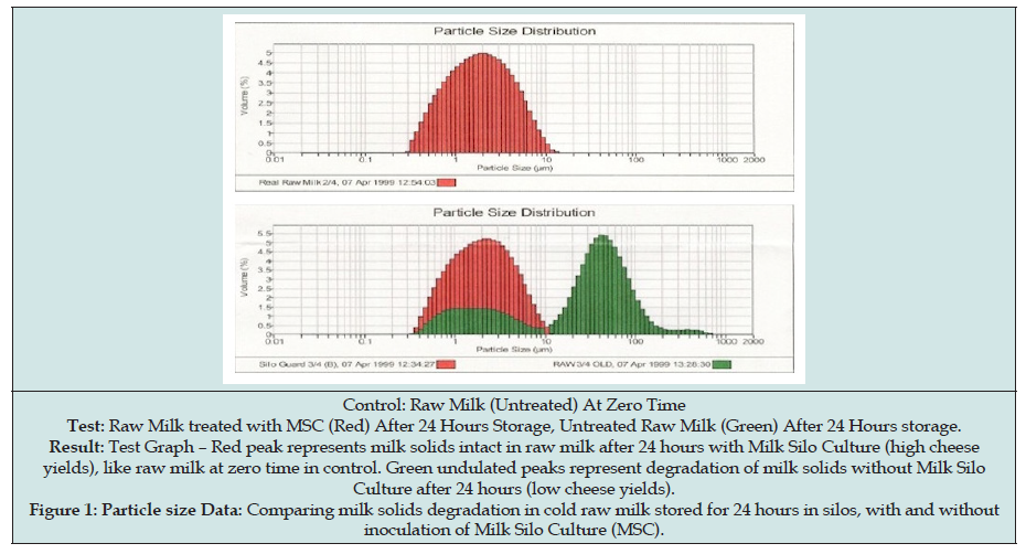
Figure 1: Particle size Data: Comparing milk solids degradation in cold raw milk stored for 24 hours in silos, with and without inoculation of Milk Silo Culture (MSC).

lupinepublishers-open-access-journal-pharmacology-clinical-research

In Figure 1, the first graph demonstrates clean, fresh, refrigerated raw milk from the silo at zero time. This sample does not contain MILK SILO CULTURE and is used as the CONTROL. The CONTROL indicates that no breakdown of milk components has occurred. The second graph in Figure 1 demonstrates the test portion of the experiment, where MILK SILO CULTURE was added in silo to the fresh raw milk in one sample, and another CONTROL sample was left untreated in the silo. Both the treated and untreated milk were held at refrigeration temperatures in the silo for 24 hours. The results are clearly evident from the graphs in Figure 1. The region in RED is the raw milk treated with MILK SILO CULTURE - it demonstrates little to no change from the aforementioned CONTROL sample - this means little to no breakdown of components within the MILK SILO CULTURE treated sample has occurred in 24 hours.

The TEST sample, which is the GREEN region on the graph, shows what happens to milk that is not treated with MILK SILO CULTURE- the particles have altered significantly, which is evident from the graphs in Figure 1. Even in the harshest of conditions, IMAC MILK SILO CULTURE protects the valuable components of milk from spoilage, resulting in improving quality, safety, and savings in terms of milk performance and losses, and even energy costs. More than all, the dairy product made with such milk has more nutritive value and is safer for consumption. It has been proven earlier that several pathogenic bacteria can be inactivated by the activated LP system in raw milk due to Milk Silo cultures. It is a win-win situation in that both dairy products manufacturers and the consumers gets benefited. Because of this, some of the Dairy product manufacturing facilities instituted the use of Milk silo cultures into raw milk in milk silos as a part of Total Quality management (TQM). Contrary to peoples thinking the maximum amount of psychrotrophic bacterial growth occurs at the end of storage of milk in the milk silos at the factory level.

Such an infinitesimal amount of casein and fat protected by the use of milk silo cultures, will save, or improve the net bottom line of the cheese plant by $1,500,000 per year, in a plant processing one million pounds of milk per day, 6 days a week. As an example, plugging the above data into theoretical yield model using the Van Slyke formula, it has been determined that cheese yield can be improved by 3.125 percent over the standard yield, (Control of mozzarella cheese yield without the use of MSC is 9.3%, whereas the yield due to use of Milk Silo cultures (MSC) is 9.6%), keeping all other factors such as moisture etc. constant. This has been confirmed further by considering the actual yield. Although there was variation between theoretical yield and the actual yield with the control milk. The variation is minimal with the milk treated with Milk Silo culture and the yields and quality are significantly superior in comparison to the control. Accordingly, you can calculate the net profit to your cheese plant depending on the volume of milk processed per day, and the number of working days in a week. None of the pathogens were detected in the dairy product (cheese).

Results of Experiment 4

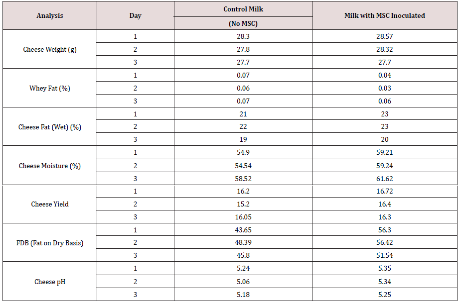
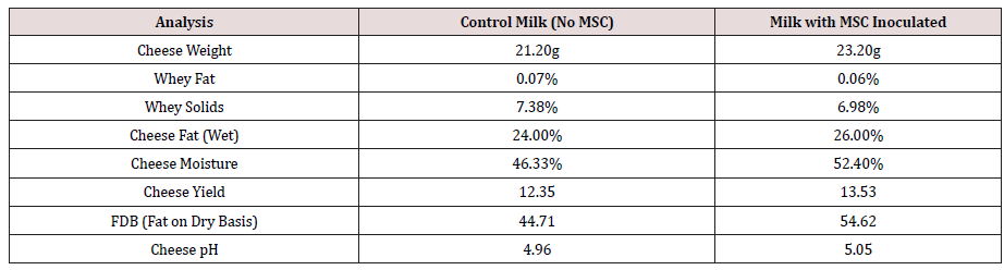
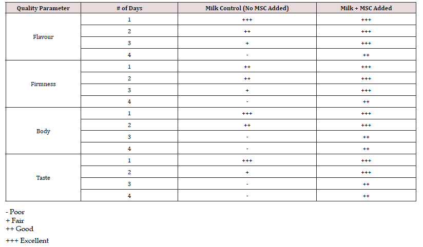
The results proved that the treated milk greatly improved the yield (Tables 1&3) and the quality of cheese in terms of texture and flavor (Table 2). On average the cheese yield improved by 0.5% compared to the untreated milk. The cheese curd or the cheese that was made on Friday on the last set of treated milk was pressed over the weekend. The results are presented in Table 3. The results clearly indicated that Milk silo culture greatly improved the yields and the quality of cheese. The treated raw milk held at 4 C for 4 days also improved the yield by 1.18 % compared to untreated milk. The quality of whey was excellent in terms of smell and taste, according to the Judges. Such whey was free from pathogenic bacteria and upon drying had excellent color (not an objectionable dark color). The safety of whey products is extremely important because of their wide usage as sweet whey, Whey protein concentrates, whey protein isolates, in the baby food formulas, and in the nutritional products and formulations especially for the athletes and the geriatric population to control or treat Sarco- Penia (muscle loss).

Table 1: The Effect of Milk Silo Culture (MSC) on Raw Milk Held at refrigeration temperature for up to 4 Days on Cheese Yield and Composition of Whey.

lupinepublishers-openaccess-journal-pharmacology-clinical-research-journal

Table 2: The Effect of Milk Silo Culture (MSC) in Milk After 4 Days storage at refrigeration temperature on cheese yields and composition of whey.

lupinepublishers-openaccess-journal-pharmacology-clinical-research-journal

Table 3: The Effect of Milk Silo Culture (MSC), (Milk Held at refrigeration temperature for up to 4 Days) on the microbiologically altered rheological properties of Cheese.

lupinepublishers-openaccess-journal-pharmacology-clinical-research-journal

Several Experiments conducted on a large scale in commercial cheese plants processing over a million pounds of milk per day also revealed comparable results. The commercial experiments with the use of Milk Silo Cultures were also conducted in various Dairy plants (cheese) located in mid-west, east coast and west coast of the U.S. and also cheese plants located in Wales, Southern and Northern Ireland, Scotland, South America, and Southeast Asia etc. Irrespective of the geographic region, the use of Milk Silo Cultures improved the quality, quantity, and safety of dairy products. None of the dairy products (cheese) produced with milk Silo Cultures had any problems with Bacteriophage, pathogenic bacteria, such as Salmonella, Listeria, Staphylococci, Enteropathogenic E. Coli, campylobacter, and Yersinia etc.

In this connection, it is worthwhile to mention that there are no Government regulations to check for the presence of pathogenic bacteria in raw milk. It is taken for granted that the legal pasteurization of 161 F for 15 secs. should eliminate all the pathogenic bacteria. It is not true anymore because it has been found that certain pathogenic strains of Listeria monocytogenes, can survive pasteurization by being embedded in the leucocytes, Reddy [21]. The effect of Global warming on microbial mutation cannot be ignored. Thus, we can no longer rely on past research results. In addition, unless the LP system is activated, the postpasteurization contaminated pathogenic bacteria or the pathogens which survived the pasteurization cannot be curtailed. Thus, it is highly recommended to use Milk Silo Cultures in raw milk, to produce superior quality dairy products with better functionality and more than all the safety of the products in terms of free from both spoilage and pathogenic bacteria, to protect consumers worldwide.

Conclusion

The laboratory experiments and commercial successful trials proved beyond doubt that the milk’s natural Lactoperoxidase system (LP System) activated through the use of Milk Silo Cultures, upon pasteurization distinctly reduced the spoilage type and pathogenic bacteria, and the breakdown of milk solids (Casein and Fat). In addition, it was proven using the particle Size Analyzer that in 24 hours’ time of retention of raw milk in silos at the dairy products manufacturing facilities, the psychrotrophic bacteria reduced casein by 0.08% and milk fat by 0.06%. Use of Milk Silo cultures prevented the degradation of both casein as well as milk fat. Milk Silo Cultures, by activating the LP System followed by Pasteurization exhibited the inhibition of DNA Bacteriophage active against most widely used starter culture Streptococcus thermophilus. It has a great significance in that the protection of the starter cultures from viral infection assures the improved quality in terms of reduction of both spoilage bacteria, as well as the pathogenic bacteria in the dairy products, such as cheese, yogurt, and other fermented products.

Experiments conducted on the prevention of spoilage of the pasteurized milk using Milk Silo Cultures proved beyond doubt that the shelf-life and the safety of the fluid milk can be significantly improved, beyond the sell by date. It has a significant effect since milk can be stored for a longer period to eliminate dumping, which can be otherwise used to feed people. Furthermore, such dumping will induce pollution problems resulting in environmental issues. The experiments conducted on improving the dairy product (cheese) yields and quality with the introduction of Milk Silo Cultures into raw milk were phenomenally successful. Not only the cheese yields increased, but also the rheological properties and more than all complete eradication of pathogenic bacteria and viruses were observed. Once again, it is extremely important because the increased amount of food will reduce the cost of production, and also improve the safety of the products and improve the confidence of the consumers. Considering the positive results of this laborious research, the dairy industry worldwide can implement the usage of the milk silo cultures into raw milk to activate the raw milk natural protective Lactoperoxidase system to improve the quality, quantity, shelf-life, and safety of the dairy products.

Acknowledgement

I am extremely thankful to Mr. V. R. Mantha, director of Quality Control and Research and Development of IMAC (International Media and Cultures), a division of the American dairy and Food consulting laboratories Inc., of Denver, Colorado, USA, for coding and compiling the data, and for helping to prepare this research article. Thanks, are also extended to the various dairy product manufacturing facilities especially cheese plants in USA, South America, British Wales, Scotland, Northern and southern Ireland, and Southeast Asia. A Special thanks to the researchers participated in the study from Oxford University, England, and Melvern Instruments personnel. Deep gratitude to Mr. Rasheed Hussain and Mr. Sridhar Reddy for their contribution to graphs and tables that are presented in the research article.

Read more about Lupine Publishers LOJ Pharmacology & Clinical Research Click on the below link:

Monday, 21 March 2022

Lupine Publishers| Impact of Blood Pressure on Presence or Absence of Chin Dimple

 Lupine Publishers| Pharmacology & Clinical Research


Abstract

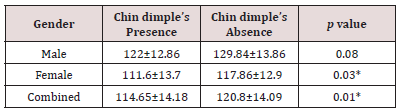
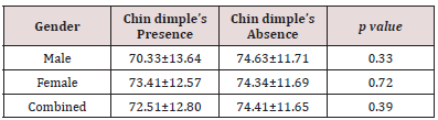
Impact of blood pressure on dimple on chin was the objective of this study. Normal pressure of blood is crucial to life. Without the power that controls our blood to stream around the circulatory structure, no oxygen passed on through our tissues and organs. High pressure of blood also causes distinct syndromes. An estimate of 189 subjects partook in this survey. Subjects were undergraduate students in Bahauddin Zakariya University, Multan, Pakistan. Blood pressure was also examined by device. It was concluded from the survey that there was impact of systolic pressure of blood but there was no impact of diastolic pressure of blood on chin dimple.

Keywords:Systolic BP; Diastolic BP; Circulatory Strain; Dimple

Introduction

Normal pressure of blood is crucial to life. Without the power that controls our blood to stream around the circulatory structure, no oxygen passed on through our tissues and organs. Blood pressure is likewise fundamental since it conveys WBCs and antibodies that causes resistance. Blood itself conveys various different properties, including its temperature. It additionally conveys one of our barriers against tissue harm, the coagulating platelets that avert blood loss. Blood moves through our body as a result of a distinction in weight. Pressure of blood is most noteworthy toward the beginning of its voyage from our heart - when it enters the aorta - and it is least toward the finish of its adventure along continuously littler parts of arteries. That weight distinction is the thing that makes blood stream around our bodies. The state of the arteries influences blood pressure and stream and narrowing of the arteries can block the supply by and large, prompting hazardous conditions including stroke and heart assault [1]. Normal pressure of blood is 120/80 mmHg. High pressure of blood causes cardiovascular disorder. Hypertension ought to be treated at 130/80 as opposed to 140/90. Pressure of blood has a day by day design. Circulatory strain is typically lower during the evening while people are in rest condition. The pulse begins to rise a couple of hours before a person wakes up. The pressure of blood keeps on ascending during the day and it is at peak in midafternoon. At that point in the late evening and night, pressure of blood starts dropping once more. Irregular pulse design, for example, hypertension during the night or promptly in the first part of the day, can imply that you have a medical issue. For unusual pulse, person can discuss with the specialist about the cause of unusual pulse or pressure of blood. Hypertension in people is a major issue today. Regularly, with increase in age, the pressure of blood also increases. Danger of hypertension starts to climb when men hit age 45, despite the fact that it can happen in more youthful men. African-Americans will in general create it more in youth and have increasingly extreme hypertension. Obesity or a family ancestry of hypertension additionally builds hazard [2].

Hypertension is particularly risky, in light of the fact that individuals can have it for a considerable length of time without knowing. Numerous elements can prompt hypertension. Obviously, diet is a vital thing for controlling blood pressure. An excessive amount of salt, too little potassium, and a lot of liquor have all been found to expand the danger of hypertension. An excessive amount of pressure and too minimal physical action both increase the risk of more hypertension. In the event that the pressure of blood is very high, there might be sure symptoms to pay special attention to them as cerebral pain, chest torment, trouble in breathing, unpredictable heartbeat. Chin dimple is a y-molded depression on the jaw. The chin dimple is due to dominant genes present in the body. The chin dimple may be an inherited trait that is passed from parents to offspring or it can be appeared after birth. The dimple at the jaw occurs where the two bones fuses. The individual having this dimple seeks the attention of people. The person having chin dimple looks beautiful and this dimple look like in several shapes. Dimple on the chin is known as fovea mentalis. Dimples are visible indentations of the skin and a prevailing attribute. Anatomically, dimples might be brought about by varieties in the structure of some body tissue for instance muscles, connective tissues, skin and subcutaneous tissue [3]. Impact of blood pressure on chin dimple was the objective of this study.

Materials and Methods

Measurement of Blood Pressure: The instrument that is used to measure blood pressure is Sphygmomanometer. To start the measurement of pressure of blood, use an appropriate size of cuff on arm. The cuff should cover the 80% area of the upper part of arm. We measured the blood pressure of each student. The students were asked to sit on a comfortable chair and then the cuff was tightly wrapped around their arm. The device was set on and it automatically told the systolic and diastolic BP of the person.

Project Design: An estimate of 189 subjects partook in this survey. Subjects were undergraduates in Bahauddin Zakariya University, Multan, Pakistan. This survey was done to study the impact of blood pressure on chin dimple.

Statistical Analysis: M-STAT was used to carry out statistical analysis.

Results

Impact of systolic and diastolic blood pressure is given in Tables 1& 2. In Table 1, results showed that females and combined (male and female) with systolic blood pressure had significant p value that was less than 0.05 but p value for male was higher than significant value but it was close to 0.05 that was significant. In Table 2, all the results were non-significant. These results showed that there was no impact of diastolic pressure of blood on chin dimple.

Table 1: Impact of systolic pressure of blood on chin dimple (Mean ± SD).

lupinepublishers-openaccess-journal-pharmacology-clinical-research-journal

p<0.05 significant results.

Table 2: Impact of diastolic pressure of blood on chin dimple (Mean ± SD).

lupinepublishers-openaccess-journal-pharmacology-clinical-research-journal

p>0.05 non-significant results.

Discussion

Recent studies have given an important advancement in research [4-11]. According to Rengin, Female Greek kids and youths running in age somewhere in the range of seven and fifteen years the nearness of cheek dimples was researched. There was anyway a noteworthy increment of dimples with age just as altogether higher quantities of uneven than symmetric expressions in all age groups. According to Carlene and Stephen, some examinations have evaluated the degree and conveyance of the blood pressure trouble around the world. The point of this examination was to evaluate the worldwide weight of illness identified with high blood pressure. Worldwide weight of infection owing to hypertension was assessed for gatherings as per age. Population affect divisions were determined with information for mean systolic blood pressure. 7·6 million unexpected losses and 92 million DALYs were ascribed to hypertension. About a large portion of this weight was in individuals with hypertension; the rest of in those with lesser degrees of hypertension.

Conclusion

It was concluded from the survey that there was impact of systolic pressure of blood on chin dimple but there was no impact of diastolic pressure of blood on chin dimple.

Read More About Lupine Publishers Journal of Pharmacology & Clinical Research Please Click on Below Link: https://lupine-publishers-pharmacology.blogspot.com/

Monday, 6 December 2021

Lupine Publishers| Understanding the TCM Role of Liver in the Treatment of Cough

 Lupine Publishers| Journal of Pharmacology & Clinical Research


Abstract

The TCM role of Liver understood with the meridian route and functions shows that the insulting sequence plays a key to understanding the relationship between Liver and Lung in the effective treatment of cough. Liver qi stagnation is the most important and common pattern in clinic that deserves attention, which can be interpreted with scientific evidence. However, there remains a long way to go for the integration of TCM with the conventional medicine.

Keywords: Cough; Liver Qi Stagnation; Liver Meridian; Yin Yang

Introduction

Pathological mucus or foreign irritants are cleared out of the airways in the lung disorders with the protective action coughing when the lung is contracted with the infections. Cough categorized as the acute, the subacute, and the chronic with the duration from less than three weeks to over eight weeks can affect the patients’ life as long as it continues for more than two weeks [1,2]. How to make acupuncture treatments effectively requires an accurate diagnosis following Zang-Fu organs, meridian theories, Yin-Yang, and acupoint indications. Although the stimulations with the acupuncture treatments on the immune system for the infection issue like cough have been verified effective to improve the symptoms resulted from the cough in the recent scientific studies [3,4], the unique technique pattern should deserve attention for the expected successful treatments. Compared to the Western medicine, symptoms in traditional Chinese medicine (TCM) are believed to be broader with the pattern identification. The pathological classifications of diseases in the Western medicine are not generally followed by TCM physicians and acupuncturists, but they strictly depend on the patterns developed with the Yin- Yang balance, Qi-Blood, and the meridian theory [5].

Glance at the Functions of the Liver Meridian

A human body is supported by the three essential and vital treasures Jing (essence), Qi, and Shen. Jing (essence) is viewed as Ying, referring to material that seems to bear high similarity to genes. The damages to Jing may cause serious issues with physiological and psychological developments. Qi is classified as Yang for functions of the body and viewed as the energy to benefit the body to fight against external evil Qi (pathogens), and transports Blood to nourish the all the systems of the body [6,7]. In addition to Jing, Qi, and Shen, spirit, the combination of body and mind, indicates whether or not Yin and Yang are at balance and serves as the foundation for diagnosis and treatment. The concept that the human body in TCM is not only seen as the interaction between humans and Heaven but as a miniature of the universe highlights the importance of Yin-Yang balance. A disease is believed and understood to be the Yin and Yang imbalance in Figure 1. The Yin- Yang balance in TCM has severed as the guideline for explaining the etiology of the diseases and the treatments for over two thousand years in the history of Chinese medicine and the key to the accurate diagnosis and successful treatment outcomes. On the basis of QiBlood, Essence, and fluids, Liver, Heart, Spleen, Lung, and Kidney are five Ying Zang organs that co-work through the meridian systems with the six Fu Yang organs Gall Bladder, Large Intestine, Stomach, Small Intestine, Sanjiao, and Bladder to connect all the body systems. The systems of the body can be affected with the activities of ascent and dispersion of Liver [8]. The Five Elements theory indicates that one of the functions of Liver is to store Blood for regulating the blood volume to nourish the constituents of the body and the Liver qi can affect the Spleen qi to ascend and the Stomach qi descend correctly and the regulation of emotions. On the other hand, whether or not Qi can flow freely within the body in order to ensure the distribution of fluids and blood is also governed by Liver [9]. Qi circulates in the traditional twelve meridians in the body to connect the tissues and organs together, collaborating with Blood to regulate the normal functions of the body and reflect pathology for diagnosis. Meridians are divided into three Hand Yin meridians, three Foot Yin meridians, three Hand Yang meridians, and three-Foot Yang meridians as shown in Table 1 [10]. The Liver meridian classified as Foot Jueyin originates at the lateral side of the big toe, goes upwards along the inner side of the leg meeting with SP12 and SP13, and then encircles the pubic region to connect with the Conception Vessel at CV2, CV3, and CV4. It ascends to Stomach and penetrates Liver to connect with Gallbladder and Lung at PC1, and finally connect with the Governing Vessel at the vertex of the head [11].

Figure 1: Characteristics of Yin and Yang.

lupinepublishers-openaccess-journal-pharmacology-clinical-research-journal

Table 1: Twelve meridians and pathways.

lupinepublishers-openaccess-journal-pharmacology-clinical-research-journal

Pathological View of Liver with Cough

It is stated that “Five Zang organs and six Fu organs make people cough, in addition to Lung.” in the 38th Chapter On cough of the volume Su Wen (Essential Questions) of the Huang-Di-Nei- Jing (Essential Questions of Yellow Emperor’s Inner Classic). This warning suggests that a TCM practitioner and an acupuncturist must consider the relationship of the other organs in treating cough based on the Five Elements theory to gain the whole picture of the etiology. Liver in the Five Elements theory refers to Wood while Lung is Metal to govern Qi in TCM. The meridian route of Liver indicates that the Liver meridian passes Lung and the insulting sequence that Liver can invade Lung explains that Liver can affect the functions of Lung with its failure in free flow of Qi to descend Lung qi, which can result in such symptoms as cough, dyspnea, and asthma. Blood regulation and dispersing stagnated Qi rely on the smooth flow of Liver qi [12,13]. Unfortunately, Liver qi stagnation, presented with the symptoms of swelling in the breast, frequent sighs, the tightness in the chest, tension in the epigastric region, the sensation of a lump in the throat called Plum Pit Qi, or sometimes stabbing pain in the hypochondriac region, is the extremely common TCM pattern in clinic and one of the pathological changes of Liver. The circulation of Qi is impeded with this change when Liver is depressed with the negative emotions like anger, worry, depression, and resentment [14,15]. Scientific studies of depression, which is understood to be the presentation of negative Shen in TCM, have shown that the neurotransmitter signaling in transforming macronutrients into molecules is dysregulated with the stagnated Liver qi. Stagnated Liver qi leads the molecules to be delivered insufficiently to the brain with the disrupted mitochondrial ATP production in neurons [16]. In addition, the failure in controlling the ratio of lymphocytes and granulocytes that can be seen as Yin and Yang by the autonomic nervous system is viewed as Liver qi stagnation [17]. On the other hand, the sympathetic nervous system controls the Natural Killer cell while the parasympathetic nervous system is assumed to be closely connected to the release of cytotoxic substances [18,19].

Discussion

The quotation “Qi is the commander of Blood and Blood acts as the mother of Qi” highlights the collaboration of Blood and Qi. Blood is seen as Yin and Qi can be classified into Yang, which can result in diseases when the imbalance of Yin-Yang occurs. Blood, produced with food qi (gu qi) by Spleen, circulates in the veins governed by Heart to nourish the organs and the systems. Blood, in addition to food qi, is also generated by the mother of Liver based on the Generating sequence Kidney, which stores prenatal Jing and produces marrow that generates to manufacture Blood. Qi circulates in the traditional twelve meridians to support the life, interacting with Blood for the Zang-Fu organs to function normally in harmony [20,21]. The studies on the relation between Qi in the twelve meridians and the oxygen metabolism highlight that one may be short of breath and experience wheezing or coughing when the normal level of blood oxygen is below, presenting the high similarity of physiological functions and pathological reactions between Qi and oxygen [22]. In other words, this suggested that oxygen, to some extent, is equivalent to Qi [23]. The Generating Sequence of the Five Elements theory shows Kidney is the mother of Liver, which suggests that Kidney’s problems can affect its child Liver. The maximum oxygen is delivered with the normal value of 45% of hematocrit [24]. Kidney produces Erythropoietin to promote the number of red blood cells to increase the capacity of the blood to carry more oxygen. In addition, the circulation of oxygen in the Kidney is closely associated with the production of Erythropoietin determined by tissue oxygen pressure [25]. In other words, stagnated Liver qi can be dispersed as long as Kidney can function normally with the delivery of the healthy Qi to Liver. In a word, the inflammation resulted from the infection can be reduced with much more oxygen delivered upwards with the blood to Lung.

Conclusion

Inductive logic thinking is the basis of the developments of TCM and acupuncture theories. TCM practitioners and acupuncturists must stick to the pattern identification developed based on Yin-Yang and the Five Elements theories that act as the two key principles in practice to the accurate diagnosis and the expected outcomes. With the collaboration of Liver and Kidney, it is suggested in this study that Qi may be viewed as oxygen in the blood and Kidney is the key to producing Qi, which makes Liver an anti-inflammatory role eventually to reduce the inflammation of the Lung. The integration of the western medicine and TCM has gained much more attention, which suggests that the trend may be the optimal choice for the public health system. However, how to link each other together needs more research and shows that there is a long way to go.

To Know More About Lupine Publishers Journal of Pharmacology and Clinical Research Please Click on Below Link: https://lupine-publishers-pharmacology.blogspot.com/