Showing posts with label Lupine Publihsers. Show all posts

Monday, 19 June 2023

Lupine Publishers | Insecticidal Activity of Plant Extracts

 Lupine Publishers | Drug Designing & Intellectual Properties International Journal


Abstract

Insecticides are a group of compounds with heterogenous toxicity, whose intended purpose is to kills insects. Synthetic insecticides are known for their very harmful environmental and health impacts. This therefore generates a need for a safer solution. Botanicals are a special group of insecticides with a natural origin, obtained primarily from plant parts. There is a continued interest to screen plants for their insecticidal activities in light of the great destruction done to crops that affect the livelihood of the populace worldwide, especially in countries, whose GDP depends primarily on agriculture. Guyana, a country on the mainland of South America is no exception. Agricultural produce is now threaten by emerging new species of insect pests, such as in the rice sector. Thus, there is a need to screen Guyana’s richly biodiversified flora for its insecticidal activities and to promote research in biological control of crop pests and boost the agro industry.

Keywords: Insectides; environmental; Guyana; biodiversified flora

Introduction

Background

Insects, though useful in many ways to mankind, have had negative effects in the field of agriculture. They have attacked plant parts such as stored grains, seeds, flowers, leaves, stems, fruits etc and have had a significant effect on the agroeconomy of many countries [1-3] . One way to control insects deleterious effects on crops is to utilize chemical pest control, which employs potent chemical pesticides to curb, reduce or eliminate pests and thus sustain crop production throughout the world. However, most synthetic insecticides used to date are deleterious to human health and the environment [4]. Thus, an alternative strategy is necessary. That alternative strategy is the use of plant extracts and phytochemicals as natural antifeedant, insect deterrents and repellent [4]. Plant extracts are safe alternatives that are of low cost, convenient to use and environmentally friendly. Plant products have been successfully exploited as insecticides, insect repellents and insect antifeedants [1-3]. In addition, natural products insectides have been isolated from plants and serve the basis for structure mimicry synthesis. Figure 1 show a list of structure of some isolated natural products from crude plant extracts. Figure 2 shows a notorious insecticidal natural product, Azadirachtin Many countries whose GDP (Gross Domestic Product) depends on agriculture, have in recent times being affected by insects. Guyana, a country on the mainland of South America is of no exception. Agricultural produce is currently threaten by emerging new species of insect pests. One notable example is in the field of rice cultivation. Recent research in Guyana has shown that rice production is threatened by paddy bugs, leaf miners, water weevils and caterpillars [6]. Natural insecticides from Guyana flora, should be a good choice, apart from the synthetic analog imported. Thus, there is a need to screen Guyana’s richly biodiversified flora for its insecticidal activities and to promote research in natural pesticides control. There is not much report of plant being screened for their insectidal activities here, even though research is currently being done. So, this area of research needs intensification in Guyana. In addition, the crude plant extracts can be subjected to chromatographic separation, which will lead to the isolation of known and unknown natural products which will continue to provide the platform for novel insectidal discoveries and structure activity relationship.

Figure 1: The structure of some selected isolated insecticides.

Figure 2: Azadirachtin, a notorious insecticidal isolate from Azadirachta indica.

Brief survey of some selected natural insectides

Internationally, there are many reports of plant extracts being used for their insecticidal properties. Several will be briefly reviewed here. For example, Morinda citrifolia leaf extract was tested for larvicidal activity against three medically important mosquito vectors such as malarial vector Anopheles stephensi, dengue vector Aedes aegypti, and filarial vector Culex quinquefasciatus Insecticidal activity of essential oil [7], extracted from Morinda lucida was tested on pulse beetle Callosobruchus maculatus, which is a pest that causes serious damage to several pulses. The insecticidal activity was compared with two pesticides, Phostoxin and Primo-ban-20. Results clearly indicate that M. lucida essential oil can be used as an effective alternative for pulse beetle C. maculatus control, and it could be tested against other pulse beetles affecting Asia and Africa and throughout the world, thereby reducing use of synthetic pesticides [8].

The insecticidal and antifeedant activity of the ethanolic extracts from Allium rotundum L has been noted against the larvae of the L.decemlineata [9]. It was found that the ethanolic extracts from the aerial part possessed moderate level (40.0%) of insecticidal properties against the larvae of the L. decemlineata and low against the imago (6.7-13.3%0) [9]. The petroleum ether, ethanol and aqueous extracts from the leaves of Artemisia herbalba, Asso, Euclayptus camaldulensis Dehnh and Rosmarinus officinalis L were investigated for their insectidal activity against 3 to 4 days old Myzus persicae individuals at 1,2.5, 5 and 10% and observations were made after 24 hours. Etheric extracts of all plants were effective and induced mortalities (100%, 53% and 60% respectively) at the highest concentration. However, the ethanolic and aqueous extracts did not show any significant insecticidal effect [10] Sixteen aromatic plant extracts from three species belonging of the Asteraceae family, were tested for their insectidal activity against adults and larvae of confused flour beetle Tribolium confusum du Val (Coleoptera Tenebrionidae).

The methanolic and ethyl acetate extracts of Mantisalca duriaei Briq. Cavill. and petroleum ether, chloroformic and methanolic extracts of Raconteur acute DC, significantly induced larval growth. Antifeedant properties were detected in methanolic extracts of M. duriaei and R. acaule, petroleum ether and chloroformic extract of R. acaule and ethyl acetate extract of M. duriaei. For all extracts, mortality was higher for larvae than adults. Values of 83% and 77% were obtained using petroleum ether and methanolic extracts of R. acaule. These results suggest that M. duriaei and R. acaule may be used in grain storage against insect pests [11]. Leaf oil of Psidium guajava L obtained from Soxhlet extraction was tested for insecticidal effect and phytochemical screening against khapra beetle, Trogoderma granarium Everts (Coleoptera: Dermestidae. The application of the oil at the rate of 0.5ml gave significantly (p < 0.05) higher percentage mortality than the control [12].

A study was conducted to determine the insecticidal activity of essential oils from oregano, Origanum onites L. (Lamiales: Lamiaceae), savory, Satureja thymbra L. (Lamiales: Lamiaceae), and myrtle, Myrtus communis L. (Rosales: Myrtaceae) against three stored-product insects such as the flour moth Ephestia kuehniella Zeller (Lepidoptera: Pyralidae), the Indian meal moth Plodia interpunctella Hübner (Lepidoptera: Pyralidae) and the bean weevil Acanthoscelides obtectus Say (Coleoptera: Bruchidae). A. obtectus was the most tolerant species against the essential oils. However, the insecticidal activity of the myrtle oil was more pronounced than other oils tested against A. obtectus adults. The essential oils of oregano and savory were highly effective against P. interpunctella and E. kuehniella, with 100% mortality obtained after 24hrs [13].

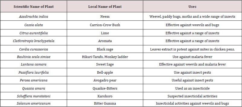
Table 1: Some selected plants from the Guyanese flora with folklore insecticidal activities.

lupinepublishers-openaccess-drug-designing-journal

Six different indigenous plants were screened for antifeedant and insecticidal activities against fourth instar larvae of Epilachna beetle, Henosepilachna vigintioctopunctata, a severe pest on brinjal. Amongst the plants screened, Achyranthes aspera showed higher activity against the selected pest. Ethyl acetate extracts of A. aspera showed higher antifeedant index and insecticidal activity against fourth instar larvae of H. vigintioctopunctata. Preliminary phytochemical analysis revealed that the presence of alkaloid and quinines in the ethyl acetate extracts indicating higher percentage of activities [14]. The insecticidal activities of four local plants extracts Rhazya stricta Decne, Lantana camara L., Ruta chalepensis L. and Heliotropium bacciferum Forssk against subterranean termites Psammotermes hybostoma (Desneux) were reported. Of the four extracts, the hexane extract of R. stricta was more pronounced, having an acute (24hr) and chronic (48hr) LC50s of 194.8 and 147.4ppm, compared to 221.7 and 149.9; 288.9, 185.6 and 391.3 and 244.5ppm for L. camara, R. chalepenesis and H. bacciferum respectively [15]. There are other reports of plant extracts used for their insecticidal activities [16-22]. There are few reports of plants from the Guyana flora with insecticidal activities, even though research has been pursued in that direction. Table 1 shows a list of ten plants with selected insecticidal activity from the richly biodiversified Guyana flora.

Conclusion

Insects are indeed a threat to mankind, especially in the field of agriculture. Many countries, whose economy depends on the agro-industry are threaten with new emerging destructive insects. Guyana is of no exception. Whist synthetic insecticides can do the job in many cases, there use is deleterious to the environment etc. Insecticides derived from plant extracts is far more environmentally friendly and far less expensive to make compare to synthetic ones. From an economical point of view, it would be best to use the aqueous extract of selected plants with insecticidal activities. There are many plants with folklore insecticidal activities in Guyana and their extracts: organic and aqueous need insectidal investigation. Also, the isolation of natural products from the crude plant extracts can lead to the discovery of new isolates and it would form the basis for continued novel drug discovery.

Read more about Lupine Publishers Journal of Drug Designing and Intellectual Properties Click on the below link: https://lupine-publishers-drug-designing.blogspot.com/


Wednesday, 14 June 2023

Lupine Publishers | Sustainability, gives Impetus to Innovation and Research

 Lupine Publishers | Latest Trends in Textile and Fashion Designing


Abstract

An increasing population of the Globe in general and that of India and China in particular brings before us a number of challenges of providing the people enough food, shelter, clothing, sanitation & health facilities, infrastructure, etc. To meet these challenges, unbridled manufacturing activities disregarding environment and society has been taking place almost all over the world which has resulted in alarming level of increase in Green House gases and depletion of valuable nonrenewable resources and water bodies. Under this backdrop business practices based on sustainability are becoming inevitable and textile sector is no exception to it. India’s aspiring young population, like consumers from developed countries, further demands quality apparels with multifunctional performance properties and thus Technical Textiles consumption is growing rapidly. The present paper explains how innovation and sustainability driven research can meet the challenges of coming decades. In this paper a few examples of the research work and their findings are summarized and their potential in meeting such challenges is presented.

Introduction

Indian Textile Industry being Mother industry, its growth is weaved with the future of over 55 million people connected directly or indirectly to this sector. Many a times Business as usual manufacturing activities are carried out with unbridled freedom, and total disregard to optimization and use of raw materials/ processes and energy sources. This is leading to air & water pollution; Green House Gases Emission and rapid depletion of fossil fuels and water resources. It has been reported that Globally we have ploughed in extra 30% of earth’s resources depriving our future generations. Hence, it is natural that Sustainability issues take center stage. It is inevitable that we adapt to Sustainable value model.

Environment has to be protected and people have to be respected and looking after the profitability of any business enterprise. Planet, People and Profit are these three pillars on which business success is to be measured. While following sustainability agenda, the steps to be taken include more and more use of renewable raw materials and energy; process intensification to conserve energy, water, and use of utilities, etc. increase the use of products which are biodegradable, etc. Thus, SUSTAINABILITY is never to be considered as financial burden; in fact, it is a key driver for Innovation based research as well as Technological Developments which can give an enterprise competitive advantage.

When we look at the respect to environment and people over the profitability, it is not a philosophical or wishful thinking. Top management needs to have commitment to sustainability; intern it, it is a matter of commitment to ethically doing the business. Indeed, when ethics becomes core component of business, only then no one would compromise environment and the society for the paltry sum of profits. Sustainability requires multipronged approach. It necessitates exploration of newer ways of processing, newer raw materials, technologies which require minimum energy/water inputs and has lowest level of emissions and effluents. Hence, naturally the regular processes and technologies need to be relooked in to the sustainability lens and explored giving impetus to research and developments. Fortunately, awareness about use of products made by eco-friendly processing technologies is increasing and such products based on green technology are being preferred even paying slight premium. Positive development in India is the introduction of new company law, making it compulsory for the companies to spend 2% profit of theirs for Corporate Social Responsibility projects, which again creates mental frame among the corporates about social commitment and corporate philanthropy.

India is a Young Nation as 65 % of its population is below the age of 35 years. This aspiring young Population with increasing standard of living and surplus disposable income, are quite aware about Consumer rights and environment, and are Brand conscious and quality conscious. They need improved quality of life and Standard Medical care. And to meet these demands of enlightened and economically affording section of customer base, we have to switch from traditional Textile manufacturing to the diversification towards manufacture of Technical Textiles. Coming decade is thus going to be decisive in terms of adapting to various products based

on Technical Textiles of different segments.

Many of the reported technologies such as Digital Printing; Transfer Printing; Ultrasonic /Laser Energy, Supercritical Carbon dioxide dyeing of Polyester; Plasma application etc. can be further extrapolated and up-scaled to see their application potential on large scale level. Also, a number of efforts in Process intensification giving rise to optimum use of utilities, with improved performance of the product and quality , various Fibre Modification processes and Nano -Technological applications need to be thoroughly investigated to extract the un-tapped value in these technologies which can propel us toward the fulfillment of our sustainability agenda.

Personally, I have been privileged to guide the research in the field of fibre science, technical textiles and textile processing for close 40 years with over 200 students for Master and Ph D degrees. Due to the paucity of the space and time, it will be prudent to describe here the outlines of a few pieces of our research work which are deeply rooted in sustainability, process intensification and technical textile applications. The examples taken here are not exhaustive and they are just representative samples.

Ecofriendly and Economical Finishing agents for Cotton and Cellulosic fibres

Cotton and cellulosic fibres enjoy important position in clothing. In fact, today Cotton garments have become revenue earners and are the materials for those who can afford to have them. However, fibre lacks wrinkle resistance and it is important that these required properties are imparted to cotton. Traditional way is to modify this fibre by resin finishing, which makes use of formaldehyde-based resin. This itself gives rise to the issue of release of formaldehyde during wearing. Hence low formaldehyde and formaldehyde free finishing agents are in demand. Polycaroxylic acid such as 1,1,2,2, butanae tetra carboxylic acid(BTCA) is presented as an alternative to this; however, it is cost prohibitive and hence used only in a limited extent. We have worked on similar lines making use of polycarboxylic acids such as polymaleic acid, citric acid and their combination and we have found interesting results with respect to crease recovery properties. The use of Polyethylene emulsion further helps in reducing the stiffness caused and thus fabric does not suffer on that account too. In addition, when this formulation is mixed with Chitosan, (We had prepared the naturally occurring biopolymer Chitosan from shrimp shells obtained from the fish market), it gets further enhanced with regard to imparting antibacterial properties. Padding through the solution was done with Wet pick up of 95% and then drying on pin frames at 80°C for 2 min. This was followed by curing at 160°C for 2 min .The results indicate very good degree of CRA and wrinkle resistance properties as well as acceptable softness level, and antibacterial properties [1].

Dyeing of finished Garments as per the Market needs

The limited aim was to address the need of the Garment dyers to quickly deliver the goods as per the purchase order, which is fluid. Since the demand for typical fashion colors keep changing fast, it is important to avoid the unnecessary large inventory of the garments in specific color combinations. The wise things to do is to have the garments finished in un-dyed form which can be dyed in a short time as per the orders placed and made available promptly. Technologically this is a bit difficult thing to do as after finishing the color uptake of the garments is reduced to as low as 10% of the unfinished garments.

We undertook this work with an aim to convert the finished un-dyed garment into dyeable form as and when needed as per color requirements. To that effect we used N- containing Additives in finishing formulation for garment dyeing. Nitrogen containing additives such as Triethanol amine can act in acidic conditions of dyeing enabling protonation of the fabric due to cationisation effect and the exhaustion of the reactive dye takes place very easily which can then be fixed well. The results were quite promising, and additives used in finishing were found to give the dyeability as good as or near about equivalent to that when finish was absent [2].

Simultaneous ACID Dyeing and Finishing of Cotton

It is well known that Acid dyes are cheaper than that of direct dyes; however, they do not have any substantivity for cotton and hence one is compelled to restrict the use of acid dyes only to proteinic fibres. In this piece of work, we attempted to establish linkage between acid dyes and cotton using a bridging chemical such as multifunctional resin or polycarboxylic acids. The resin as well as polycarboxylic acid while reacting with hydroxyl groups in cellulosic chains on one end, can be made to react with amino groups of acid dyes at the other end ,in one single operation of pad, dry and cure technique. A lot of trials to optimize conditions were taken and it was concluded that acid dyes which contain free primary or secondary amino groups can participate in this reaction with cross-linking agents, which intern can also react with the cellulosic fibre and impart wrinkle resistance properties as well as dyeability. The padding bath contained cross-linking agent, catalyst, acid dye and after padding the fabric with 100%expression, it was dried and cured at 150oC for 3 mins. Detail studies showed that the dye forms a covalent bond with the cross-linking agent which inturn is covalently bonded with the fabric. Hence the fastness properties exhibited by these dyeing were exceptionally good. The Advantages of this process include capacity to make dyeing operation continuous; acid dye application on Cotton and cellulosic fibres; high performance properties; simultaneous resin finishing happening and thus saving on energy, chemicals and water. Shortening of the process will give rise to extra production and thus this process is Eco-friendlier and more sustainable one [3,4].

Development of Hygienic, Fragrant and Mosquito repellent Cotton

Fragrance and essential oil have specific effects on individual’s feelings and emotions such as relaxation, exhilaration, sensuality, happiness and wellbeing through odour via stimulation of brain. Essential oils also have antibacterial, insect repellent, and mosquito repellent properties. However, many of these oils are volatile and hence lose their property very rapidly. However, the storage life of a volatile compound can be increased markedly by micro-encapsulation technique. Microencapsulation of fragrance compounds maintain fresh aroma on textiles, so that material retains its freshness for longer duration. Capsules rupture by friction during wearing giving necessary fragrance and under normal conditions remain intact. Fragrance oil has been encapsulated in gum Arabic and gelatin. The microcapsules containing these oils (Cederwood , Lavender, Lemon grass oils) with aroma are applied on cotton fabric from resin as well as binder bath.

The evaluation of the final finished fabric involved estimation of intensity of aroma, washing durability of the aroma, Antibacterial activity of these oils in microcapsules and efficiency of Mosquito repellency of microcapsule treated fabric. All these anticipated properties were found to be imparted on to the cotton fabric using this one shot natural herbal recipe. In other words, finished fabric in addition to being wrinkle resistant, also showed, pleasant aroma, antibacterial property, as well as mosquito repellency. The feasible end uses of such products include ribbons, handkerchiefs, curtains and furnishing fabrics [5].

Conclusion

These are just a few examples of actual research having carried out by my group of students, it shows that there lies a great potential to exploit commercially the findings of these pieces of research work and convert them into acceptable novel technologies/products which can satisfy the parameters of sustainability and requirement of technical textiles demanded by the modern consumers. Indeed, they can provide the products of high-quality performance, the products which can be delivered in shorter dwell time, the products having optimized use of utilities and thus low Carbon foot prints. The novel products and technologies developed can offer a very good potential of industrial exploitation.

Read more about Lupine Publishers Journal of Textile and Fashion Designing Click on the below link

https://fashion-technology-lupine-publishers.blogspot.com/


Tuesday, 13 June 2023

Lupine Publishers | Unique Anatomy of the Spleen of One Humped Camel (Camelus Dromedarius): A Review

 Lupine Publishers | Open Access Journal of Biomedical Engineering and Biosciences


Abstract

Over the past years, defensive immunity has been recognized as having an important role as a front-line mechanism and as an integral part of the adaptive immune response. Immunity in One Humped Camel (Camelus dromedarius) exposed too many diseases are spleen-dependent. In this review, we discuss the general aspects of the gross anatomy of the spleen (both internal and external), based on location, position, relation, blood supply and innervations of the organ. The micro-architecture of the camel anatomy was discussed using eosin and hematoxylene stain and the cells visible in the aspect of normal tissue histology. The aim of the write-up is to examine the anatomy of the different sections in the spleen of the one-humped camel (Camelus dromedaries).

Keywords: Defensive Immunity; Camelus Dromedarius; Spleen; Gross Anatomy; Histology

Introduction

Camelids originated in North America, where they are now extinct, and did not reach the Old World until about two million years ago [1]. Camels belong to the suborder Tylopoda of the Artiodactyls (even-toed ungulates, along with pigs). Within their current native range of North Africa and central and western Asia the one-humped camel or dromedary (Camelus dromedarius) is a highly revered species, used for food, fibre, and racing [2]. The species reportedly has no known natural predators throughout this range. Australia is the only country in which camels have established a feral population, and is now the only location where the species lives in natural populations [3]. Spleen, located between the stomach, left kidney and diaphragm, the spleen is the largest lymphoid organ in the body, performing functions for the blood similar to those performed by the lymphnodes for the lymph [4]. It is a soft organ, conforming to the contours of the organs and structures surrounding it. At the hilus on the visceral surface, the splenic artery brings blood into the spleen, the splenic vein takes blood from the spleen to the hepatic portal system, and lymphatic drain lymph from the spleen.[5]. In some domestic species such as the horse and dog, the spleen functions as a reservoir from which blood can be mobilized when needed and in these species, smooth muscle is a prominent feature of the capsule and traberculae of the spleen [6].

Structure

The spleen represents the largest reticulo-endothelial accumulation in the body [6]. It has a thin fibrous capsule, to which the peritoneum adheres intimately. The fibrous tissue of the capsule extends into the spleen to form a series of traberculae between which lies the splenic pulp [5]. The spleen has an extensive blood supply consisting of traberculae arteries, central arteries, penicillar arteries, sinusoids, red pulp veins, and traberculae veins [7]. It is surrounded by a capsule, has traberculae, and is divided into red and white pulp. The spleen is very vascular and has red and white pulp [3].

Embryologically

The spleen develops in the dorsal mesentery of the stomach (dorsal mesogastrium). The spleen arises from cells of the mesentery, which migrate into the plane between the layers of the mesentery [8]. The mesentery covering the spleen becomes the visceral peritoneum of the spleen. The mesentery between the spleen and the gut tube becomes the gastrosplenic ligament. The mesentery between the spleen and the dorsal body wall becomes the splenorenal ligament (most of which subsequently fuses to become parietal peritoneum) [9].

Grossly

The spleen is about the size of the cupped hand. If forms the left lateral extremity of the lesser sac [10]. The spleen is a peritoneal organ in the upper left quadrant that is related to the left 9th, 10th, and II the ribs. Clinically, fracture of these ribs may injured or lacerate the spleen. The Spleen is located in the upper left abdominal quadrant. Unlike the lymph node, the spleen is inserted in the blood stream. The spleen clears the blood of aged blood cells and foreign material. It is the site of an immune response to bloodborne antigens, especially in children. In adults, the spleen is not essential to life.

In general, white pulp is the site of antibody synthesis and lymphocyte production. Red pulp is chiefly concerned with the removal and destruction of worn out erythrocytes. In as much as the spleen lies above the costal margin, a normal-sized spleen is not palpable. Enlarged spleen may be palpated below the left costal margin. The splenic artery and vein reach the hilus of the spleen by traversing the splenorenal ligament. Passing from it are the gastrosplenic ligament to the greater curvature of stomach (carrying the short gastric and left gastroepiploic vessels) and the lienorenal ligament to the posterior abdominal wall (carrying the splenic vessels and tail of the pancreas) [5]. The spleen is a peritoneal organ in the upper left quadrant that is related to the left 9th, 10th, and II the ribs. Fracture of these ribs may lacerate the spleen [5]. In as much as the spleen lies above the costal margin, a normal-sized spleen is not palpable. The enlarged spleen may be palpated below the left costal margin. The splenic artery and vein reach the hilus of the spleen by traversing the spleno-renal ligament.

Relations:

a) Ventrally: The left diaphragm, separating it from the pleura, left lung and the 9th, 10th and 11th ribs.

b) Cranially: The stomach.

c) Dorsally: The splenic flexure of the colon.

d) Medially: The left kidney.

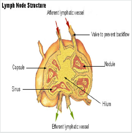
The tail of the pancreas abuts against the hilum of the spleen through which vessels and nerves enter and leave this organ Figure 1.

Figure 1: Lymph Node Structure.

lupinepublishers-openaccess-journal-environmental-soil-sciences


Histologically

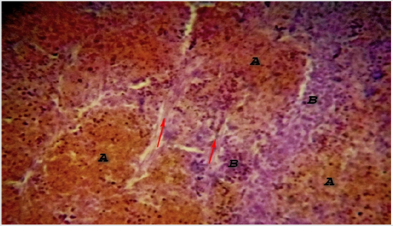
The structure resembles the lymph node except in some anatomical aspect, which includes; only efferent lymphatic drain the spleen. The sinusoids are blood vascular not lymphatic. As a result, all cells entering the spleen do so via the blood. Scattered primary nodules (germinal centers) are found throughout the spleen. These contain abundant lymphocytes and make up the “white pulp” (also known as Malpighian corpuscles). Plasma cells and macrophages are found here as well. Surrounding the white pulp nodules are venous sinuses containing abundant erythrocytes - the “red pulp”. These sinuses are lined with macrophages (RE cells) which are highly phagocytic. Between the sinuses are cords of cells - lymphocytes, monocytes and macrophages - referred to as the Cords of Billroth. The immune function of the spleen is especially important in infancy and early childhood. At these times, the rest of the lymphoid system is somewhat underdeveloped Figure 2.

Figure 2: Photomicrograph of Camel Spleen showing clearly differentiated zones of white pulp (A) and Red pulp (B) cells with inter parenchymal traberculae collagen connective tissue fibers (Red arrow) H&E x200.

lupinepublishers-openaccess-journal-environmental-soil-sciences

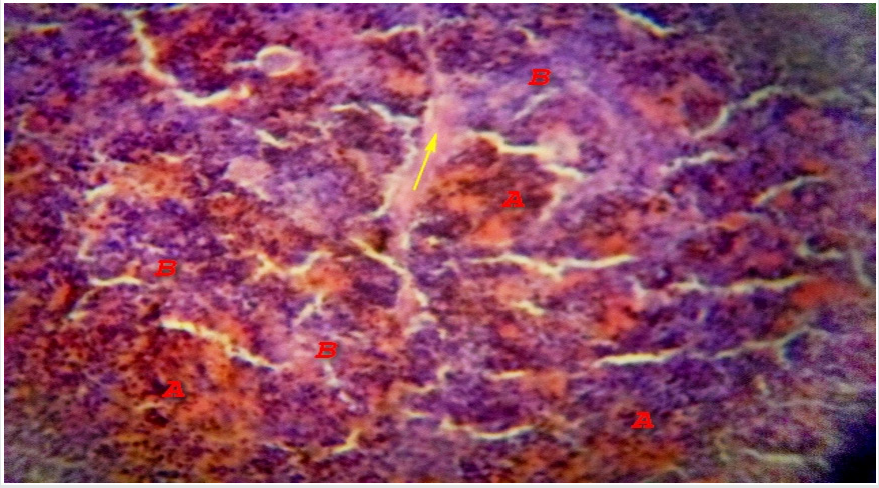
Microscopically, the spleen appears to consist of discrete 0.5-1 mm white nodules, called the white pulp, embedded in a red matrix called the red pulp. Microscopically, as shown here, the white pulp WP consists of lymphoid aggregations and the red pulp RP, making up the bulk of the organ, is a highly vascular tissue [10]. The spleen has a thin fibroelastic outer capsule C from which short traberculae T extend into the parenchyma Figure 3. The capsule is thickened at the hilum and is continuous with supporting tissues that sheath the larger blood vessels entering and leaving the organ. In dogs and horses the spleen is also a reservoir of blood and these supporting tissues contain smooth muscle to pump blood out, but in humans only a few smooth muscle cells persist Figure 4. The splenic artery divides into several major branches, which enter the hilum and branch to form numerous arterioles. [11].

Figure 3: Photomicrograph of Camel Spleen showing clearly differentiated zones of white pulp (A) and Red pulp (B) cells with communication of paranchymal traberculae connective tissue fibers connective tissue (Red arrow) and thick connective tissue fibers capsule (Yellow arrow) H&E x200.

lupinepublishers-openaccess-journal-environmental-soil-sciences

Figure 4: Photomicrograph of Camel Spleen showing clearly zones of white pulp (A) and Red pulp (B) cells with developed inter-paranchymal traberculae collagen connective tissue fibers (Yellow arrow) H&E x200

lupinepublishers-openaccess-journal-environmental-soil-sciences

An overview of the splenic ultra-structure and circulation is shown in Figure 1 above; blood enters the spleen in the splenic artery which branches repeatedly within the parenchyma [5]. The larger arteries are surrounded by a fibrocollagenous sheath that disappears in the smaller branches. These central arteries A, are so named because they have a cylindrical cuff of lymphoid tissue around them, the periarteriolar lymphoid sheath PALS, consisting mainly of TH cells. The central artery gives off a number of short branches at right angles, which are called penicilliary arteries PALS and these terminate in two to three sheathed capillaries with only one is shown for each penicilliary artery [7]. These unique vessels are small blind-ending capillaries with no endothelial lining but surrounded instead by an aggregate of macrophages. Thus the blood arriving in a sheathed capillary must traverse this wall of macrophages before entering the red pulp RP. The sheathed capillaries therefore form the first part of the filtering mechanism of the spleen [6]. The splenic white pulp is of two types, T cell and B cell, together making up 5-20% of the total mass of the spleen. The functions of these areas appear to be similar to those of the paracortex and superficial cortex of lymph nodes respectively. The non-filtering areas of red pulp parenchyma as probably be considered part of the splenic lymphoid tissue mass also, but its immunological function remains to be elucidated [12].

White Pulp

White pulp consists of lymphoid tissue that unsheathes the central arteries (periarterial sheath) along with the associated nodules and germinal centers. The periarterial sheath is populated mainly by T lymphocytes [9]. The peripheral white pulp and germinal centers are populated mainly by B lymphocytes. In the white pulp, the T cell areas surround the central arteries, forming the periarteriolar lymphoid sheath. In humans, this lymphoid tissue is less well organized than in other animals, but the term PALS persists [12].

Red Pulp

Red pulp consists of splenic cords of Billroth and venous sinusoids [8]. Defective red blood cells resulting from aging or disease (as in sickle cell anemia, hereditary spherocytosis, or thalassemia syndromes) are delayed in their passage from Billroth cords into the venous sinusoids and phagocytosed by macrophages lining the cords. The splenic parenchyma is permeated by an interconnected network of sinuses S that drain in turn into larger sinuses, tributaries of the splenic vein and finally the hepatic portal vein [3]. The sinuses are lined by endothelial cells resting upon a basement membrane with numerous narrow slits. The reticulin fibers of the sinusoidal basement membrane are arranged in a circular fashion and are continuous with the reticulin meshwork of the parenchyma [1].

Blood Supply

The splenic artery is one of the three main branches of the coeliac axis. The splenic vein is joined by the superior mesenteric to form the portal vein [6]. Blood cells entering the parenchyma from the sheathed capillaries squeeze through the walls of the sinuses to drain out of the organ via the splenic vein, an arrangement known as the open circulation [4]. Note that the splenic vessels also provide the principal blood supply of the pancreas. [6].

Read more about Lupine Publishers Journal of Biomedical Engineering and Biosciences Click on the below link : https://biomedical-sciences-lupine-publishers.blogspot.com/


Wednesday, 7 June 2023

Lupine Publishers | Maurice Roy’s Protest

 Lupine Publishers | Journal of Modern Approaches in Dentistry and Oral Health Care


Opinion

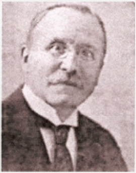
Maurice Roy was born on 24 December 1866. At 14, he entered a dental surgery as an apprentice technician. In 1884, he enrolled at the Dental School of Paris where he was awarded his diploma in 1886. He became a tenured teacher in 1900. He passed his baccalaureate. He then started medical studies. In order to meet his own needs, he worked as a dentist. He was exempted from military service because of his myopia; he was able to devote himself to writing his thesis he sustained in 1894. When the Dreyfus Affair broke out, at the cries of Zola in J’Accuse! published in 1898, he signed the petition for the rehabilitation of the captain. Having contributed towards the creation of FDI World Dental Federation in 1901, he became a member of its executive body in 1904, then its vice-president in 1911. He was re-elected to the post in 1936. In 1902 he founded the French Dentists Cooperative Society (Cooperative Society of Dentists of France), which supplied equipments to the practitioners of the country. In 1914 he created the relief committee for the wounded to the jaws and the face (Relief Committee of the wounded of the jaws and the face). He dedicated himself: he established surgical and prosthetic rehabilitation protocols that were used by other centers. In 1915 he created the Fraternal aid for French and Belgian war victims dentists (Fraternal help to French dentists and Belgian victims of war). In 1923 he took the lead of The Odontology magazine. Roy made it a key player in dentistry of that time. Every themes were subjectively studied. In 1928 he left the public welfare [1-3].

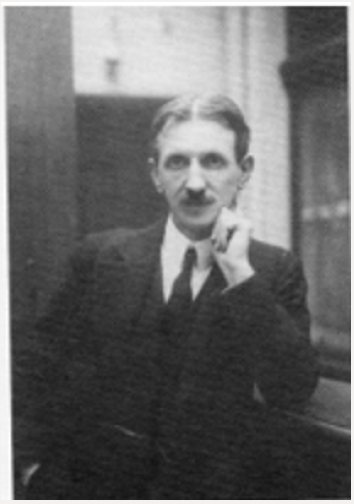
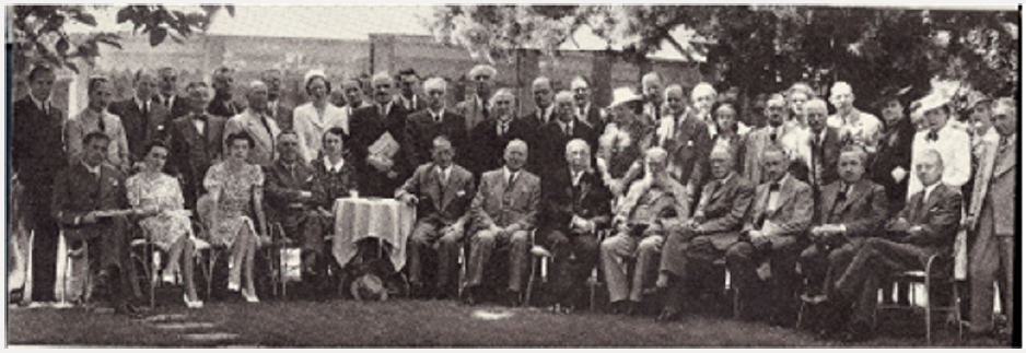
On 28 July 1933, Roy made the executive body pass a motion during FDI World Dental Congress in Edinburgh after Georges Villain, his president and friend, gave a thundering speech to denounce Nazi crimes and to help Jewish German dentists, that stated that: “The executive body of FDI, that duly gathered in session in Edinburgh on 28 July 1933 only to consider the protection of rights obtained by dentists from all over the world thanks to the diplomas granted by the competent authorities of their respective countries [4], declared that no matter of race, of religion or of politics under no circumstances must limit the liberty and the practising of our duly qualified colleagues. Likewise, no restriction which would lead them to breaches of their moral and professional obligations must be imposed on them.” Having made everything to help their colleagues although unsuccessfully the committee passed this motion unanimously excluding Germany who chose to withdraw from FDI. During the 1936 congress of Vienna, Roy received the Miller Prize for his work: the highest distinction that was awarded by the 33-member countries of the executive body including Germany. He received the Legion of Honor this same year. As they were shouted down during the 1938 congress, the Germans were absent from the 1939 congress of Zürich [5]. While the country was occupied, Roy hid aviators and Resistance fighters in his house. While he was the head teacher of the Dental School of Paris, he prohibited the wearing of the yellow star there, this could have caused him an arrestation from the Pétainist font. During a congress in Paris, he left the inaugural session just as the Germans appeared. That provoked a tremendous outcry. While the demands of the Germans were getting insistent, he stopped practicing his dental surgery in 1942, but kept his positions at the Dental School of Paris, at The Odontology magazine and at the Cooperative until his death [6,7]. His convictions caused him insulting articles in the Occupation newspapers: I’m everywhere. Because he was suspected by the Gestapo, he received only routine visits. Maurice Roy died on 5 January 1947 (Figures 1-6).

Figure 1: Maurice Roy (Roy Family, 2011).

Lupinepublishers-openaccess-dentistry-oralhealth

Figure 2: Georges Villain (1881-1938), ©BIUM, 2008

Lupinepublishers-openaccess-dentistry-oralhealth

Figure 3: Dental School of Paris - 57, rue Rochechouart (public domain).

Lupinepublishers-openaccess-dentistry-oralhealth

Figure 4: Congress of the Fédération Dentaire Internationale in Zurich, in 1939 (Ennis, 1967). Maurice Roy is in the 1st rank, 2nd on the right. There is no German dentist in this meeting.

Lupinepublishers-openaccess-dentistry-oralhealth

Figure 5: Reichszahnärzteführer Dr. Ernst Stück (1893 – 1974) (public domain).

Lupinepublishers-openaccess-dentistry-oralhealth

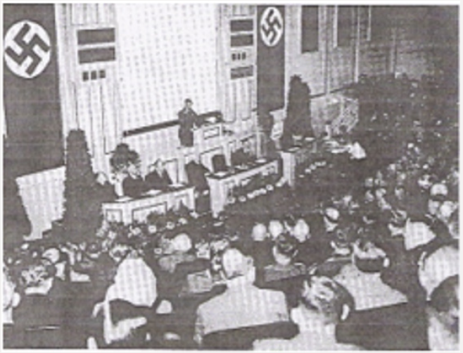
Figure 6: VIIth congress of the German dentists directed by Ernst Stück in 1935, in Berlin (public domain).

Lupinepublishers-openaccess-dentistry-oralhealth


Friday, 29 July 2022

Lupine Publishers | Histochemical and Histomorphometrical Studies on the Digital Cushion of Heifers and Dairy Cows with Claw Horn Lesions

 Lupine Publishers | Journal of Dairy & Veterinary Sciences


Abstract

Background: The ruminant claw horn lesions seriously diminish histological structure and functional efficiency of the digital cushion of the dairy cows.

Objective: To investigate histological and histochemical properties of the digital cushion in the dairy cow with claw horn lesions, such as white line disease, sole damage and hemorrhage.

Results: The results showed that digital cushion loses its normal histological structure in claw horn lesions.

Conclusion: Further biochemical, physiological and feeding studies on the experimentally induced claw horn lesions are necessary to understand the nature of the events playing roles involving animal feeding in the development of the lesions, to develop new animal feeding strategies aiming to protect and maintain the normal structure of the digital cushion.”

Keywords:Protein Degradability; Goats; Intensive Feeding; Nitrogen Balance; Digestibility

Introduction

Lameness is one of the most important problems negatively affecting milk yield and animal welfare in modern dairy farming in high-performance animals. The cows with laminitis produce lower amount of milk [1-5]. Moreover, cyclic reproductive changes of these cows slow down [6] or cease because of anoestrus [7] or cystic ovarian disease [8]. Lameness is a major animal welfare problem because that it is a painful condition and causes to behavioral changes of the affected cow. A lame cow tends to display low mobility [9], to spend decreased daily feeding time and dry matter intake [10] and lying behaviour may be indicative [11]. In the dairy cow, claw horn lesions [CHL] constitute 65% of the total lesions causing to lameness [12,13]. Although possible roles of risk factors, such as claw conformation and trimming, animal welfare, management, feeding and housing have been evaluated in detail by previous researchers [14-17], pathophysiology of CHL is still unclear. Previous researchers [18,19] have suggested that CHL is arisen from traumatic lesions of the supportive tissues of the claw. Around the time of calving, increase of hoofase enzyme, oestrogen and relaxin secretions results in loosening and increased mobility of suspensor apparatus of third phalanx [19]. The bovine digital cushion locating under the third phalanx has a complex structure, which major part is constituted of fat tissue. The cushion comprises three cylindrical segments [axial, medial and abaxial segments] of adipose tissue surrounded by a thick connective tissue trabecules, those arranged in parallel to each other [20,21]. The pads of the bovine digital cushion play crucial roles in reducing the body load transferred to surface of sole via absorbing of substantial forces acting within the claw [22]. The biomechanical function of the digital cushion in distributing and attenuation of the load transferred to the base of flexor process of the distal phalanx have been well appreciated by previous researchers [21,23,24]. The aim of the present study was to determine histological and histochemical properties of the digital cushion in the heifers and primiparous, second and third parturitions, and multiparous dairy cows managed under similar feeding, housing and welfare conditions. The foot was chosen from the animals with CHL, such as white line disease, sole damage and haemorrhage, in order to define the histology and histochemical changes in the digital cushions of the animals.

Material and Methods

Animals

This experiment was approved by the Ethical Committee of Experimental Animal Production and Research Centre (2011/023) of Veterinary Faculty of Selçuk University. In the study, the 120-cadaver foot (totally 240 hooves) of Holstein heifers and cows were used as materials. The animals were culled because of moderate to severe CHL and lameness in one or more hooves. The claws of the animals with similar feeding, housing and welfare conditions were subjected to a detailed clinical examination prior to slaughter. Information on the age, number of parturitions, calving history and stage of lactation were and recorded, in addition to the clinical findings. Among the reasons given for culling were insufficient milk yield, lameness and infertility. The animals were divided into 5 groups, each consisted of 6 animals, as
group I: heifers,
group II: primiparous cows,
group III: cows with second parturition,
group IV: cows with third parturition,
group V: multiparous cows with more than three calving.
The first group animals were 20 months old averagely; other groups were between 2.6-6.2-year-old. After slaughtering, all the foot was cleaned, and lesions, such as discoloration, sole ulcer and hemorrhages were recorded, frozen at -20°C and kept until use for histological procedure.

Dissection of the Hooves and Obtaining Tissue Samples

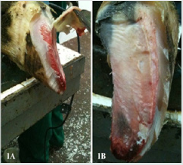
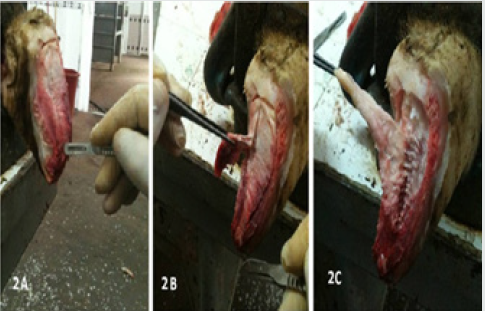
Each foot was thawed. The soft tissues of the sole and heel of each hoof were separated following the horn had been removed, in order to fully expose the axial, middle and abaxial pads of the digital cushion (Figures 1A-B & Figures 2A-C) [18]. Axial pad of the cushion was totally removed, fixed in 10% phosphate buffered (0.1M pH 7.4) formal-saline and used for histological procedures.

Figure 1: The study area, green - places of mass appearance of painted lady butterflies (Vanessa cardui L.) in 2019.
Lupinepublishers-openaccess-Dairy-Veterinaryscience

Figure 2: Separating the corium (2A), dissecting pads of the digital cushion (2B) and totally removing the pads (2C).
Lupinepublishers-openaccess-Dairy-Veterinaryscience

Histology and Histomorphometry

The tissue samples were subjected to routine histological tissue processing. Briefly, the tissue samples were washed with tap water overnight, dehydrated in ethylene series, cleared in xylene and immersed in paraffin. The sections taken at 6μm were stained as follows; hematoxylin-eosin for investigating general histology and histological changes [25], safranin-O (SO) for histochemical properties of ground substance, alcian blue at pH 2.5 (AB 2.5) for staining acid mucins and mucosubstance, alcian blue at pH 4.0 (AB 4) for demonstrating carboxylated glucose aminoglycans in the ground substance, trichrome stain for Type I collagen fibres (Coll I) and other histological detail, Verhoef’s elastic fibre (EL) stain for EL fibers [26]. The specimens were investigated under light microscope equipped with digital imaging system and digital images were recorded. The images were analyzed with digital image analysis software (BS200 PRO-2005).

Statistical Analysis

The data obtained from the digital image analyses were analyzed statistically with one-way variance analysis (ANOVA) and Tukey’s pair wise comparison tests by using SPSS software 14.01 (Release 14.01 Licence No: 9869264).

Results

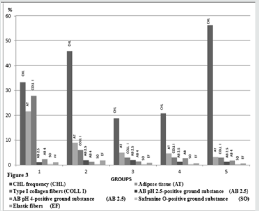
Figure 3: CHL incidence and histometrical results of the groups.
Lupinepublishers-openaccess-Dairy-Veterinaryscience

Clinical findings of the animals prior to slaughter and results of postmortem gross examination of the foot Welfare and management of the animals included into the study were far from than satisfactory. The animals were housed on the concrete floor, under poor hygiene conditions, and claw trimming was irregular. The animals were mainly fed with corn silage, alfalfa, vetch and hay. Findings of the postmortem examinations of the foot showed that the animals suffered from CHL, such as sole ulcers, hemorrhage and folding, white line disease and sole erosions in one or more hooves. The highest percentage of the claws with CHL was in the multiparous cows, in the group 5. The groups 2 and 1 the group followed it respectively. The lowest lower CHL frequencies were found in the groups 3 and 4, and the frequencies of these groups were quite similar (P>0.05, Figure 3).

Gross Macroscopic Findings of the Cushion Segments

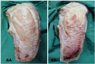
In most of the claws with CHL, segmental structure of the digital cushion was diminished. The digital cushion tissues of the heifers (Group 1) were pale and softer, whereas in the cows with the increasing the parturition number, the tissues gained yellowish colour and relatively more brittle and fragile. The cushion segments were also smaller in the animals of the groups 4 and 5, aged and multiparous cows (Figures 4A & 4B).

Figure 4: Macroscopic appearance of digital cushion tissues of a heifer from the group 1 (4A) and a cow from the group 5 (4B). The cushion pads of the group 5 animal are smaller and yellowish in colour.
Lupinepublishers-openaccess-Dairy-Veterinaryscience

Histological Findings

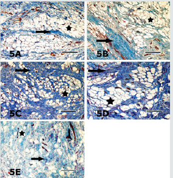
In general, the digital cushion pads of the animals were mainly formed of a loose connective tissue and unilocular adipose tissue. Adipocytes appeared as large cells with unstained cytoplasm in all of the used staining procedures (Figures 5-9). Fibrous connective tissue septa surrounded the loose connective and adipose tissues. There were striking structural differences in the histology of the digital cushion of heifers and calved cows. Loose connective tissue in the heifers, was replaced with adipose tissue in the primiparous cows, and fibrous connective tissue rich in collagen fibres in the third lactation cows. In the heifers and primiparous cows, adipocytes were seen as groups, whereas in the cows of the groups 3, 4 and 5, the cells were observed as individual cells and the cell groups consisting of small numbers of the adipocytes. Adipose tissue percentages of the groups are given in the Table 1. The highest adipose tissue percentage (21.55%) was in the heifers, group 1, and the value was significantly (P<0.001) higher than those of the other groups (Figure 3). The groups 3 and 4 displayed similar (P>0.05) adipose tissue percentages. The multiparous animals (group 5) had the lowest adipose tissue percentage (Figure 3). Type I collagenous fibres were seen as thick undulating bundles of the fibres darkly stained with aniline blue in trichrome stained sections (Figures 5A- 5E).

Figure 5: Collagen fibre bundles (arrows) and adipocytes (asterisks) are seen in the sections from lateral claw cushion of the right forefoot of the heifer from the group 1 (5A), in the medial claw cushion of the left hindfoot of the animal from the group 2 (5B), in the lateral claw cushion of the left forefoot of the animal from the group 3 (5C), medial claw cushion of the left hindfoot of the animal from the group 4 (5D) and lateral claw cushion of the left hindfoot of the animal from the group 5 (5E) are seen. In contrast to a definite decrease in the adipose tissue rate, the connective tissue gradually increased from the group 1 to the group 5. Crossmon’s trichrome. Magnification bar: 100μm.
Lupinepublishers-openaccess-Dairy-Veterinaryscience

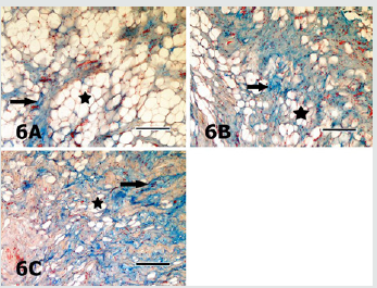
Figure 6: Sections of the digital cushion stained with AB pH 2.5 from the medial claw of the left forefoot of the animal from the group 1 (6A), the lateral claw of the right forefoot of the animal from the group 2 (6B), the lateral claw of the right hindfoot of the animal from the group 3 (6C) are seen. Staining intensity of the ground substance (arrows) in different sections is quite similar. Asterisks depict the adipocytes. AB pH 2.5 stain. Magnification bar: 100μm.
Lupinepublishers-openaccess-Dairy-Veterinaryscience

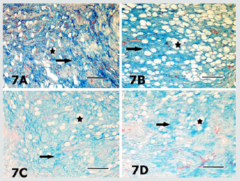
Figure 7: Sections of the digital cushion stained with AB pH 4 from the medial claw of the left hindfoot of the animal from the group 1 (7A), the medial claw of the left hindfoot of the animal from the group 4 (7B), the medial claw of the left hindfoot of the animal from the group 4 (7C) and the medial claw of the right hindfoot of the animal from the group 5 (7D) are seen. Slight differences in staining intensities of the ground substance (arrows) in different sections are seen. Asterisks depict the adipocytes. AB pH 4 stain. Magnification bar: 100μm.
Lupinepublishers-openaccess-Dairy-Veterinaryscience

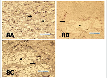
Figure 8: Sections of the digital cushion stained with safranin O from the lateral claw of the left hindfoot of the animal from the group 1 (8A), the lateral claw of the left hind foot of the animal from the group 2 (8B) and the lateral claw of the right fore foot of the animal from the group 3 (8C) are seen. Staining intensities of the ground substance (arrows) in different sections are quite similar. Asterisk depicts the adipocytes. SO stain. Magnification bar: 100μm.
Lupinepublishers-openaccess-Dairy-Veterinaryscience

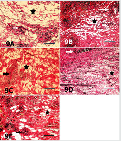
The heifers had significantly (P<0.001) higher type I collagen fibre percentage (27.82%) than those of the other groups those had quite similar (P>0.05) percentages (Figure 3). In AB pH 2.5 stained specimens, the AB positivity was mainly located in the ground substance around the connective tissue fibres, embedded in-between the adipocyte groups (Figures 6A-6C). The positivity was also stronger around the individual adipocytes and the groups constituted of small numbers of these cells. Percentage of the AB pH 2.5 stained ground substance was highest (2.08%) in the group 3 and group 2 followed it (1.96%), whereas the groups 1, 4 and 5 had quite similar (P>0.05) and significantly (P<0.01) lower values than those of the groups 2 and 3 (Figure 3). AB pH 4.0 mainly stained the similar ground substance regions with AB pH 2.5, around the connective tissue fibres, located around the adipocyte groups (Figures 7A-7D). Results of the histometrical investigations of the AB pH 4.0 stained specimens showed that the group 1 and group 4 had similar (P>0.05), and significantly higher (P<0.01) positivity percentages than those of the groups 2, 3 and 5 (Figure 3). In SO stained specimens, the positivity was very weak (Figures 8A-8C) in all groups. The positivity percentages of the groups were quite similar and there were no significant differences (P>0.05) between the groups (Figure 3). Elastic fibres were mainly found in-between the connective tissue trabecules surrounding adipocyte groups and tunica adventitia of the blood vessels, and seen as brownishblack, branching coarse and gently undulating fine fibres, in the sections stained with Verhoeff’s elastic fibre stain (Figures 9A- 9E). The results of digital image analyses showed that the group 2 had the highest (1.89%) elastic fibre percentage and the value was significantly (P<0.05) higher than those of the other groups. The groups 1and 3, and 4 and 5 had quite similar elastic fibre percentages (P>0.05, Figure 3).

Figure 9: Sections of the digital cushion stained with Verhoef’s elastic fibre stain from the medial claw of the right hindfoot of the animal from the group 1 (9A), the medial claw of the left hindfoot of the animal from the group 2 (9B), the lateral claw of left forefoot of the animal from the group 3 (9C), medial claw of the left hind foot of the animal from the group 4 (9D) and the lateral claw of the left hind foot of the animal from the group 5 (9E) are seen. Elastic fibres (arrows) are mainly located in connective tissue trabecules surrounding the adipocyte groups (asterisks). Verhoef’s elastic fibre stain. Magnification bar: 100μm
Lupinepublishers-openaccess-Dairy-Veterinaryscience

Discussion

In the present study, statistically significant (P<0.01) differences were found between the CHL frequencies of the groups. Particularly, high incidences of CHL in the primiparous cows (45.80%, group 2) and multiparous (56.20%) cows (group 5) were striking. The differences possibly have arisen from higher incidence of CHL in more than one or all foot of the animals in these groups. Previous researchers [27,28] have reported that the histological development and functional maturation of the digital cushion are not completed before the age of three, and also incidence of sole lesions, such as sole ulcerations and white line disease are relatively higher in the first lactation because that the cushion tissues is less elastic and rich in saturated fatty acids. In accordance with the previous researchers [27,28] Incidence of CHL was higher in primiparous cows in the present study. The roles of the additional loads and forces exposed at late pregnancy, and structural and biochemical changes in the regional tissues, in addition to trauma of both corium of sole and the cushion tissues in the pathogenesis and occurrence of CHL have been discussed in detail by previous researchers [17-19,29,30]. Moreover, collaborative effects of improper, inadequate feeding and housing practices are mentioned among significant disposing factors. In coordination of anatomic structures of the hoof [bones, joints, ligaments and bursae] leads to CHL, mainly sole ulcers and white line disease. Thus, hoof problems occur more commonly among the cows kept in cubicles or concrete yards or straw yards than in the cows kept in cowsheds. Previous researchers [18,31,32] have reported that lameness incidence increases in multiparous cows with increase of ageing and calving, especially after third lactation, due to increasing the amount of the loose connective tissue while reducing adipose tissue, decreasing body condition score [BCS], thinning of the cushion segments. In the present study, a gradually decline was found in adipose tissue percentages of the groups, with ageing and increasing parturitions. Loosening of suspensor ligament system of the hoof results in increasing in mobility of bones, and this situation disposes the sole corium and sole horn to traumatic lesions, and consequently plays significant roles in the development of CHL, such as sole ulcers, white line disease [19,33]. Loosening of suspensor system of the hoof also increases the sensitivity of the claw to the CHL depending on increase of loads applied to the foot by increase in mammary gland and foetus weights, especially during first lactation [34]. Clinical findings and histometrical results of the hooves and higher CHL incidence of the groups 2 and 5 are consistent with previous findings [18,31,32]. Moreover, higher CHL incidence of the group 5, in which significant shrinkage of the cushion tissue was observed, supports the hypothesis suggesting the decline in load absorbing capacity of the cushion increases risk of injury of sole corium. Thus, environmental factors, such as long time standing on the concrete floor, unsuitable housing conditions and in proper claw trimming are effective in embodiment of the sole trauma and haemorrhages in addition to increase of coffin bone motility in late pregnancy and parturition. Unsuitable housing and hygiene conditions in the animals of the present study were in accordance with the high CHL incidence of the animals.

The digital cushion, which is a specified connective tissue, is constituted of connective tissue cells and intercellular substance [ICS]. The ICS contains collagenous, elastic and reticular fibres embedded in an amorphous ground substance. The amount of the amorphous ground substance in young animals is higher than fibrous elements. With ageing, the amount and rate of fibrous elements increase and the rate reverses in older animals. The digital cushion, a member of supporting system of the ruminant hoof locating in subcutis, is a complex structure, composed of white and yellow adipose tissues. There are significant differences in histological structure and organization of the digital cushion among the ungulate animal species. The digital cushion of the ruminant hoof is closely resembled with the cushion of the elephant’s cushion [18]. Equine digital cushion has relatively different structural features. In the horse, the cushion is composed of small number of elastic fibres and closely packaged interlacing collagenous fibres dispersed in acidic mucous matrix, rich in hyaluronic acid. Large spaces between the cells and fibres occupied with mixoid tissue and islets of fibro cartilage tissue [35,36]. Collagen fibres, especially type I fibres are the dominant fibre type of the connective tissues. Although the collagenous fibres have limited elasticity, their tensile strength is enough to meet the high tensile forces of the foot. In the present study, axial pad of the digital cushion constituted of white, unilocular adipose tissue lobuli surrounded by connective tissue trabecules. Fibro cartilage was not observed in the specimens. In some of the groups, trabecules surrounding adipose tissue lobuli constituted of loose connective tissue and contained less amount of type I collagen fibres, the others were rich in the collagenous fibres. Group 1 contained significantly (p<0.001) higher collagen (27.82%) when compared with the other groups. A gradual decline of type I collagenous fibre percentage was typical in the remaining, older and multiparous groups (groups 3, 4 and 5). Nevertheless, they contained quite similar (P>0.05) type I collagen fibre rates. Gradual reduction of type I collagenous fibre rate with the increase of age and calving might arisen from the structural and functional changes occurred in aged animals and during the late pregnancy and post parturition. It might be predicted that shock absorbing capacity of the digital cushion considerably reduced with declining of collagen fibre rate. In accordance with this prediction, the highest (56.20%) CHL incidence was found in the group 5, which had relatively low rate of type I collagen fibres. Moreover, in accordance with the prediction above, previous researchers [19,33,37], also suggested that loosening of the suspensor system of the hoof during pregnancy and post parturition increases sensitivity of sole corium located between coffin bone and sole to trauma, and consequently playing significant roles in the pathogenesis of the CHL, such as sole ulcers and white line disease.

Although the information on the types and distribution of adipocytes in ruminant digital cushion is limited, the heifer cushion contains lesser amount of the fat cells. The digital cushion of the heifers is a whitish and gelatinous in nature containing high amounts of ground substance and lipid content gradually increases through 2-3 parturitions, and gradually decrease starting from 3 years of age [18,21,32]. Because that white fat tissue is an energy source, many metabolic and hormonal mechanisms, such as pancreatic hormones and glucocorticoids are efficient on this fat tissue. Thus, feeding and other metabolic factors are likely to efficient on both amount and composition of fat of the digital cushion. Moreover, it is well known that the fatty acid composition and amounts of the cow’s digital cushion are seriously affected by metabolic disorders, mainly by ketosis. Similarly, previous researchers [38] emphasized that the topic needs detailed evidences. In the presented study, the digital cushion of the dairy cattle comprised of islets of large unilocular adipocytes. In the digital cushion with low levels of fat tissue, individual adipocytes were frequently seen. The animals in the group 1 contained significantly (P<0.001) higher adipocyte rates (%21.55) and the group 2 it, with a significant decline. However, the group 2 contained significantly (P<0.05) higher fat tissue percentage than those of the groups 3, 4 and 5. Rastani [39] suggested that lipids are mobilized from fat tissues in order to be used in milk synthesis. Lipids in white fat depots in the digital cushion are also consumed. Considering the data obtained in this study, it is possible to assume that white adipose tissue in the ruminant digital cushion also can be used as an energy source in ketosis, which is a condition resulting in negative energy balance [NEB], and thus CHL increase post parturition period. It is well known that fat and elastic fibres of the digital cushion play significant roles in shock absorbing mechanism of the cushion via expanding sideways until their viscoelasticity is limited by stretched fibrous connective tissue septa rich in collagen fibres. Elastic fibres support expansion by flattening their undulations and support reversing to the situation before when the applied force is expired. Significant decreases in both elastic fibre and adipose tissue rates result in loss of elasticity of the digital cushion.

There is limited information on the histochemical properties of ground substance of the cow’s digital cushion, and also histometrical results evaluation of these parameters have not been documented previously. In the present study, the results of the AB pH 2.5, AB pH 4.0 and SO stains showed that ground substance of the cow’s digital cushion was rich in hyaluronic acid (hyaluronan) and other proteoglycans, although there were some differences between the groups. Because that hyaluronic acid is a macromolecule increasing the viscosity of the tissue fluid, it consequently augments resilience of the tissue by binding water molecules. Other proteoglycans stained with SO might support the digital cushion in resisting to pressure, since the proteoglycans also show high water binding affinity and tend to gelation [40]. Although the results of the present study indicated that the ground substance of cow’s digital cushion was rich in hyaluronic acid, the hoof material was obtained from the animals with varying degree of CHL lesions. The authors strongly stress that a detailed comparison should be carried out between the findings of healthy and CHL animals. The elastic fibres, which relatively thinner when compared to the collagen fibres, are able to compensate the forces applied between elasticity limits. Tissues gain elasticity and stress bearing features by their elastic fibre content. Elastic fibres are found in hoof soft tissues as in other elastic tissues. Information on distribution and localization of these fibres in the ruminant hoof soft tissues is insufficient. In a previous histological study [38], elastic fibres were found between the collagen fibre bundles in the digital cushion of African elephant. In this study, elastic fibres were more peculiarly observed in-between the collagenous fibre bundles and in blood vessel walls. Morphometrical analyses evidenced that the group 2 displayed significantly (P<0.05) higher elastic fibre percentage (1.89%) than the other groups. Although there was a declining tendency with increase of the parturition number, the other groups had relatively similar elastic fibre ratios. Besides, striking decline of the elastic fibre percentage was determined in the groups 4 and 5 in concomitant with higher CHL lesion incidence. Although further studies are needed, the results of the present study imply that elastic fibre content of the cow digital cushion is quite limited and might play only small role in the functions of the digital cushion and in the development CHL lesions.

Conclusion

Based on the results, it was concluded that CHL seriously affected both histology and functional efficiency of the digital cushion of dairy cows. Results of the present study, which show strong evidence between histological, histochemical and histometrical findings and CHL lesions are noteworthy. However, the results should be compared with those of the healthy animals in further experiments those are involving experimental models. Further studies necessary to understand the nature of the events playing roles involving animal feeding in the development of the lesions, to develop new animal feeding strategies aiming to protect and maintain the normal structure of the digital cushion, in order to maintain and augment its supportive and protective roles, to reduce incidence of laminitis.

Grant Support

This work was supported by a grant from The Scientific and Technological Research Council of Turkey with project no: TOVAG 112O332.

Read More About Lupine Publishers Journal of Dairy Please Click on Below Link:
https://lupine-publishers-dairy-veterinary.blogspot.com/