Showing posts with label PAPN. Show all posts

Friday, 15 September 2023

Lupine Publishers | Congenital Diaphragmatic Hernia: Case Report

 Lupine Publishers | Journal of Pediatrics and Neonatology


Abstract

Congenital diaphragmatic hernia is a malformation resulting from incomplete fusion of the pleuroperitoneal membrane, it occurs in around 1/5000 live newborns, it produces in the vast majority of cases severe symptoms of respiratory failure and pulmonary arterial hypertension in this age group. Treatment is based on maintaining the best respiratory conditions in the child through intensive neonatal therapy management and, once this objective is achieved, surgical correction of the anatomical defect should proceed. Unfortunately, despite the progress in respiratory management of the seriously ill newborn, mortality from this disease is reported above 75%. We present a case in which the left diaphragmatic hernia associated with pulmonary hypertension led to a fatal outcome in a newborn, despite timely and adequate interventions.

Keywords: Congenital Diaphragmatic Hernia; Pulmonary Hypertension; Respiratory Failure of The Newborn; Surgical Correction

Introduction

Congenital diaphragmatic hernia (CDH) is the most common developmental abnormality of the diaphragm and is one of the most common malformations in the newborn, occurring with a frequency of 1 in 2000 pregnancies and 1 in 2,200 to 5,000 live births. Its presentation is greater in males and is generally located on the left side of the diaphragm, at the site of the Boch dalek foramen, although it is occasionally right (Morgagni foramen) or, with less incidence, it can be bilateral or central [1,2]. The etiology is generally unknown, and most cases are likely to be multifactorial; It has also been associated with chromosomal abnormalities from 20% to 53% and an autosomal recessive form has been described. Despite intense research and aggressive therapeutic intervention in infants with CHD, morbidity and mortality continue to be elevated between 40% and 70% because the hypoplasia and persistent pulmonary hypertension that occur in these patients complicate efforts to maintain a gas exchange with conventional ventilation [3,4].

Case Presentation

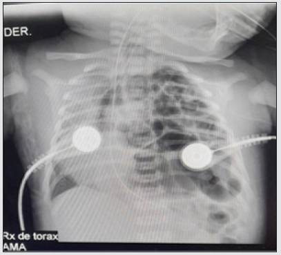
From a nearby municipality, a 37-week-term male newborn was referred to us by Ballard, obtained on 04/16/21 by vaginal delivery without immediate complications. It is the product of the second pregnancy, the son of a 20-year-old mother with a controlled pregnancy twice without ultrasound control. He was born with an APGAR score of 8/10 at 1 minute and at 5 minutes, but at 14 hours of age he presented an episode of apnea with desaturations and generalized cyanosis, requiring positive pressure ventilation and fluid resuscitation with little improvement, due to the persistence of distress. respiratory system, a chest X-ray was performed where intestinal loops were observed in the left lung field, diagnosing diaphragmatic hernia, which is why they were referred to our institution. He was admitted to the neonatal intensive care unit in a fair general condition, cyanotic, hypotonic, desaturated and with a distant audible moan, requiring an orotracheal intubation protocol and invasive mechanical ventilation. The diagnosis was corroborated with a chest X-ray (Figure 1). Orogastric tube was placed to decompress abdominal content, water support and inotropics to maintain adequate mean arterial pressure. He was evaluated by pediatric surgery, who requested presurgical examinations, in which he found prolonged clotting times. Given the instability and the alteration in the coagulogram, surgery was postponed, management with vitamin K was started, and a pediatric hematologist was consulted, who indicated a transfusion of fresh plasma.

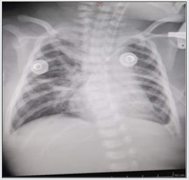
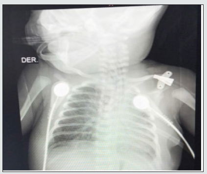
Once his hemodynamic part had stabilized and the clotting times normalized, the patient was taken to surgery on 04/24/21 where a left diaphragmatic defect of more or less 5 cm in diameter was observed and inside the thoracic cavity the presence of small intestine, descending colon, stomach and spleen, for which a reduction of the contents to the abdominal cavity was performed, hernial defect was corrected, diaphragmatic integrity was verified and a chest tube was left, ending the procedure without immediate complications. At 12 hours after surgery, a control X-ray was performed, showing a cardiac silhouette in good position and left lung expansion, and diffuse infiltrates predominantly in the middle lobe were seen in the right lung (Figure 2). An echocardiogram was performed that reported severe pulmonary hypertension with PSAP of 80 mmHg, requiring a phosphodiesterase-5 inhibitor (sildenafil), triple inotropic support and minimal manipulation protocol. Subsequently, a new chest X-ray was performed (Figure 3). Since the newborn presented clinical deterioration, increased work of breathing, generalized mucocutaneous paleness, peri-buccal and distal cyanosis; bradycardia, marked hypotension and desaturation despite established management and high ventilatory parameters. The patient presented cardiorespiratory arrest for which he began advanced cardiopulmonary resuscitation maneuvers for more than 20 minutes without obtaining a response, for which he was declared dead.

Figure 1: Chest X-ray before the surgical procedure.

lupinepublishers-openaccess-journal-pediatrics-neonatology

Figure 2: Chest x-ray after surgical procedure.

lupinepublishers-openaccess-journal-pediatrics-neonatology

Figure 3: Enlarged cardiac silhouette, with enlargement of the atrial cavities and veiling of the left pulmonary trunk, findings suggestive of pulmonary hypertension.

lupinepublishers-openaccess-journal-pediatrics-neonatology

Discussion

The first description of the anatomy and pathophysiology of congenital diaphragmatic hernia was made by Hunter and Mc Cauley in the year 1754; this was defined as the protrusion of the abdominal organs into the thorax, or the descent of the thoracic organs into the abdomen as a result of incomplete closure of the pleuroperitoneal canals. Its incidence is estimated at 1 in 2000 to 5000 births. The defect is more frequent on the left side for 80% and the rest for the right; bilateral ones are extremely rare. The incidence of associated malformations is 10–50% and mortality is high, reaching up to 70% of cases [5,6]. There are reports of high morbidity in patients who survive the initial management of congenital diaphragmatic hernia. Long-term use of mechanical ventilation is associated with bronchopulmonary dysplasia in a higher proportion of children with diaphragmatic hernia. Other types of problems are esophageal ectasia with gastroesophageal reflux in cases of very large defects; recurrence of the hernia or intestinal obstruction are serious complications, although not frequently reported. In a group of patients in which the extracorporeal oxygenation membrane was used, a higher incidence of neurocognitive problems is reported at school [7,8]. Even though there are innovative methods in intrauterine treatment and immediate postnatal therapy, the real solution to this problem will be, in the future, through the knowledge and prevention of the embryological cause and the creation of techniques that induce lung growth [9,10]. In conclusion, this case report brings us closer to knowing this pathology and thus being able to make an early diagnosis and timely management, taking into account its high morbidity and mortality and associated complications.

Read More About Lupine Publishers Journal of Pediatrics and Neonatology Please Click on Below Link:
https://lupinepublishers-pedaitrics.blogspot.com/

Friday, 28 July 2023

Lupine Publishers | Resistance of Neisseria gonorrhoeae on oral and mucosa : A Review Article

 Lupine Publishers | Journal of Pediatrics and Neonatology


Abstract

Introduction: Gonorrhea is one of the four most common sexually transmitted diseases worldwide. Globally, the highest incidence of gonorrhea cases is in the African and Western Pacific regions (including China, Indonesia and Australia). Gonorrhea is a sexually transmitted disease caused by the bacteria Neisseria gonorrhoeae. These bacteria are gram-negative bacteria in the form of diplococci located intracellularly from PMN cells. Infection from Neisseria gonorrhoeae can be transmitted by sexual intercourse or vertical transmission at the time of delivery. In children, infection can occur as a result of sexual abuse by an individual infected with Gonorrhea or it can also occur from touching items contaminated with N. gonorrhoeae bacteria.

Discussion: Gonococcal infection occurs by invading the mucosa, so that gonococcal infection can occur in many places. Gonococcal infection can occur in male and female external genitalia, anorectal, pharynx and eyes. There can be a local infection in the genital area (most common), infection in the abdominal cavity (peritonitis), infection around the liver (perihepatitis), meningitis, endocarditis, dermatitis, arthritis and can spread to the bloodstream and cause a Disseminated Gonococcal Infection (DGI) which can spread systemically. The manifestations of gonococcal infection in the female external genitalia can be asymptomatic but may also include periurethral edema and urethritis. There may be a purulent discharge from the cervix but it does not represent vaginitis, dysuria or painful urination, dyspareunia or pain during sexual intercourse and lower abdominal pain. There can also be a vulvovaginitis, bartolin abscess, and in gonococcal infections that are not treated properly, a complication can occur in the form of pelvic inflammatory disease (PID), characterized by lower abdominal pain, increased body discharge from the vagina and urethra, dysuria and intermenstruals. bleeding accompanied by signs of peritonitis, endocervicitis, endosalfingitis and endometritis, causing a deep pelvic or lumbar pain. Currently, the resistance rate of Neisseria gonorrhoeae to antibiotics is increasing rapidly. This condition is not good for prognosis.

Conclusion: The administration of dual therapy with this drug regimen is expected to increase the cure rate so that it can prevent more serious complications and reduce the possibility of resistance to cephalosporins.

Keywords: Gonorrhoea; Resistance; Oral Management

Introduction

Gonorrhea is an infectious disease of the mucosa caused by gram-negative cocci bacteria Neisseria gonorrhoeae which can be transmitted through sexual or perinatal contact. This infection occurs co-infection with Chlamydia trachomatis [1,2]. Gonorrhea can occur in men or women, generally at productive age, but infection by bacteria in women is asymptomatic. This infection can also occur in newborns due to vertical media from the mother during labor. Most gonorrhea manifests as infection of the genital tract, but it can also cause pharyngitis, proctitis and conjunctivitis in certain groups. Clinical findings of conjunctivitis were found in infants with a history of perinatal infection[1-3]. Gonococcal infection is the most common cause of urethritis in men, who experience complaints of pus from the genitals. In women, gonococcal infection causes cervicitis, but this condition manifests asymptomatically so that patients present after a pelvic inflammatory disease (PID), infertility, ectopic pregnancy, or a chronic pelvic inflammation. Diagnosis of a gonococcal infection is done by taking pus from the genitalia, rectum, oropharynx or eye secretions in conjunctivitis gonorrhea and doing a gram examination, where an image of gramnegative diplococcal bacteria can be found in PMN cells, which is confirmed by culture on gonococcal selective media such as Martin. -Lewis medium and Thayer-Martin medium [1,4].

The management of gonorrhea is done by administering antibiotics with a recommended treatment regimen using Ceftriaxone 250 mg IM in a single dose, plus offering Azithromycin 1 gram orally in a single dose. In patients with cephalosporin allergy, an alternative therapeutic regimen can be used using a single dose of Gentamycin 240 mg IM and Azithromycin 1 g orally as a single dose [1,2,4,5]. Currently, the resistance rate of Neisseria gonorrhoeae to antibiotics is increasing rapidly. The high rate of Gonorrhea resistance to the penicillin, tetracycline and quinolone classes has made this drug class no longer accepted for use as a Gonorrhea therapy in most countries in the world. Even in some countries, it has been found that Gonorrhea resistance to cephalosporin therapy creates the 3rd that is given orally[6,7].

Discussion

Gonorrhea is one of the four most common sexually transmitted diseases worldwide. In 2015, the World Health Organization (WHO) estimated that around 357 million new infections from one of the four infectious diseases occurred each year [8]. In an epidemiological study based on data collected from 2005 to 2012, it was found that the global incidence of gonorrhea in women was 0.8%, while in men, it was found that the global incidence of gonorrhea was 0.6% of the worldwide population in 2012, so that if converted In total, there were 26,819,000 cases of gonorrhea worldwide in men and women aged 15-49 years, where there were 19 cases per 1000 women and 24 cases per 1000 men based on World Bank data in 2012 [9,10]. Globally, the highest incidence of gonorrhea cases is in the African and Western Pacific regions (including China, Indonesia and Australia). There are several potential causes for the increased incidence of Gonorrhea globally. One possible cause of this is the increasing prevalence of HIV infection in developing countries. Improvements in diagnostic capabilities and case reporting systems in several countries can also contribute to increasing the incidence of Gonorrhea globally [3].

Gonorrhea is a sexually transmitted disease caused by the bacteria Neisseria gonorrhoeae. These bacteria are gram-negative bacteria in the form of diplococci located intracellularly from PMN cells. Infection from Neisseria gonorrhoeae can be transmitted by sexual intercourse or vertical transmission at the time of delivery. These bacteria mainly attack the columnar epithelium of the host, so that all mucous membranes can be infected by these bacteria[1,2]. There are many factors that influence N. gonorrhoeae bacteria on their virulence and pathogenicity. Fili help the attachment of bacteria to the mucosal surface thereby contributing to the resistance that occurs by preventing ingestion and destruction of bacteria by neutrophils. Opacity-associated (Opa) protein increases the adhesion between gonococcal bacteria and phagocytes, thereby increasing the ability of bacteria to invade host cells and can cause a decreased immune response [4].

Clinical Manifestation

Manifestations of gonococcal infection in male external genitalia can be urethritis, which is the most common manifestation of Gonorrhea in male external genitalia, balanoposthitis, balanitis, prostatitis, epididymitis, vesiculitis or cystitis, often characterized by a purulent and thick body body, sometimes accompanied by complaints of pain when urinating or dysuria. The manifestations of gonococcal infection in the female external genitalia can be asymptomatic but may also include periurethral edema and urethritis. There may be a purulent discharge from the cervix but it does not represent vaginitis, dysuria or painful urination, dyspareunia or pain during sexual intercourse and lower abdominal pain. In gonococcal infection that is not treated properly, a complication can occur in the form of pelvic inflammatory disease (PID) [1,5]. Anorectal manifestations include proctitis accompanied by pain and a purulent fever. In some cases complaints of burning or pain during defecation, tenesmus, and blood in the stool may also be found. Gonorrhea diagnosis is done by taking anamnesis and physical examination that leads to a gonococcal infection and finding an intracellular gram-negative diplococcal image from the results of gram staining of the secretions or body fluids of a patient suspected of a gonococcal infection and the growth of N. gonorrhoeae from culture results. The differential diagnosis of a gonorrhea urethritis includes urethritis caused by genital herpes, urethritis due to C. trachomatis, urethritis due to Ureaplasma urealyticum, urethritis due to Trichomonas vaginalis, bacterial vaginosis, Reiter’s syndrome [11].

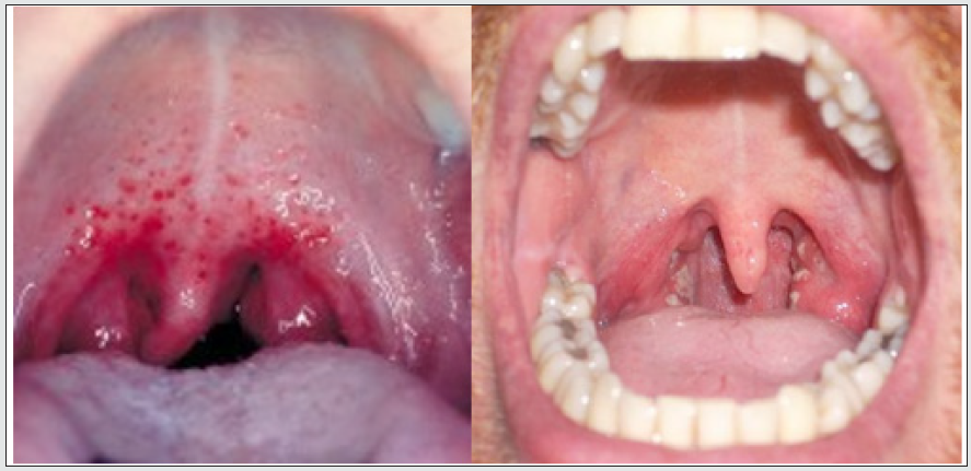
The manifestations of gonococcal infection in the male external genitalia can be in the form of urethritis which is the most common manifestation of Gonorrhea in male external genitalia, balanoposthitis, balanitis, prostatitis, epididymitis, vesiculitis or cystitis. This condition is often characterized by the presence of a body discharge that varies from clear and clear to purulent and thick, sometimes accompanied by complaints of pain during urination or dysuria. In some cases, edema may also be found in the external urethral meatus, prepuce or penis. Gonorrhea manifestations of the pharynx occur due to sexual exposure to oral-genital contact. It can also occur as a result of an inoculation after holding an infected limb or object and then putting the hand in the mouth. In this condition, there is erythema of the pharynx, mild sore throat, and often an infection of the genitalia is also found [1,2]. There is a hyperemic pharynx and swelling of the tonsils and there is a purulent white discharge on the wall of the pharynx. It is important to dig for the coitus suspectus with orogenital sexual intercourse and to do a throat swab to check with gram stain or for culture (Figure 1).

Figure 1: Gonorrhea of the pharynx (Pharyngitis Gonorrhea).

lupinepublishers-openaccess-journal-pediatrics-neonatology

Gonorrhea manifestations in the eye may include conjunctivitis, palpebral edema, chemosis and profuse and purulent discharge. In some cases, this gonococcal infection can even cause a corneal ulcer to perforate which can lead to permanent blindness. In neonates, ophthalmia neonatorum can occur, which is a bilateral conjunctivitis condition characterized by eye pain, conjunctival hyperemia, and the presence of purulent secretions[1]. Diagnosis of gonorrhea is carried out by taking anamnesis and physical examination that leads to a gonococcal infection and finding an intracellular gramnegative diplococcal image from the gram stain of the secretions or body fluids of a patient suspected of a gonococcal infection. However, because the sensitivity is low, a negative gram test result is not sufficient to rule out a diagnosis of gonorrhea, so it is necessary to have another gram-test or a culture examination done. Swab culture examination of the infected area is currently. The standard for diagnosis of Gonorrhea, which is very useful especially in cases with unclear clinical picture, when there is a condition of failure of therapy or when it is difficult to extract the history of the disease. However, empiric therapy can be done first considering the culture results take 24-48 hours [2,4].

Gonorrhea Resistance

Antibiotic resistance, especially in the case of Gonorrhea, is a threat in reducing the consequences of sexually transmitted diseases worldwide. Based on data from WHO, the incidence of gonorrhea infection that is resistant to antibiotics is increasing every year. This is of particular concern to public health observers, especially for the prevention of infertility in women [8]. Gonorrhea is currently still one of the four largest sexually transmitted diseases worldwide that can be cured. However, in recent years, Gonorrhea has become increasingly resistant to antibiotics that were previously sensitive, which has led to fears that Gonorrhea will not cure. Gonorrhea with multiple drug resistance (MDR) was reported by more than 36 countries in the world in 2015 [9-10]. Untreated gonorrhea can lead to more severe complications, such as female infertility, complications in pregnancy and blindness in newborns who are infected during pregnancy. the delivery process [12]. Resistance of Neisseria gonorrhoeae to penicillin and tetracyclines was first discovered in Asia in 1970. The high rate of resistance to quinolones (Ciprofloxacin) emerged since mid-2000. Recent data shows that there is an increase in resistance and failure of Neisseria gonorrhoeae treatment against third generation cephalosporins given orally. Several serotypes of N. gonorrhoeae that have been associated with failed cephalosporin therapy have also been shown to have resistance to other antibiotics and have been classified as multi-drug resistant gonococci [13].

Therapeutic Management

Initially, the therapeutic regimen given in cases of gonorrhea can be done by providing single therapy using cephalosporins. However, due to the resistance of these bacteria to oral cephalosporins as monotherapy, dual therapy using ceftriaxone and azithromycin is recommended as the standard therapy for gonorrhea cases worldwide, especially in the United States. Dual therapy with ceftriaxone and azithromycin should be given simultaneously on the same day and under direct supervision. Since most N. gonorrhoeae infections are co-infected with C. trachomatis, the principle of dual therapy is also considered quite effective because basically azithromycin is a class of antibiotics given to C. trachomatis infections [14]. In recent years, gonorrhea has become increasingly resistant to previously sensitive antibiotics, which has led to fears of incurable gonorrhea. Gonorrhea with multiple drug resistance (MDR) was reported by more than 36 countries in the world in 2015. Untreated gonorrhea can lead to more severe complications, such as female infertility, complications in pregnancy and blindness in newborns who are infected during pregnancy. childbirth process. Due to the resistance of these bacteria to oral cephalosporins as monotherapy, dual therapy using ceftriaxone 250 mg IM single dose and azithromycin 1 g PO single dose is recommended as the standard therapy in cases of gonorrhea worldwide. The administration of dual therapy with this drug regimen is expected to increase the cure rate so that it can prevent more serious complications and reduce the possibility of resistance to cephalosporins [15,16].

Read More About Lupine Publishers Journal of Pediatrics and Neonatology Please Click on Below Link:
https://lupinepublishers-pedaitrics.blogspot.com/

Thursday, 15 June 2023

Lupine Publishers | Prevalence and Determinants of Low Birth Weight in Burundi: An Investigation from The National Demographic and Health Survey

 Lupine Publishers | Journal of Pediatrics and Neonatology


Abstract

Background: Birth weight is an important determinant of neonatal and child health outcome. For instance, evidence has shown that low birth weight (LBW) has a negative impact on the baby’s growth, cognitive development, and on neuro-motor development and immune function. Underweight babies are prone to increased risk of infections and stunting. LBW remains prevalent worldwide and is more pronounced in low- and middle-income countries. Several factors including mothers’ socio-economic characteristics, maternal health behaviors and maternal and pregnancy health conditions determine birth weight outcome. This study sought to determine the prevalence and investigate determinants of LBW among Burundian women of reproductive age.

Methods: This study used data extracted from the 2016-2017 National Demographic and Health Survey (DHS) conducted on 7047 women who reported a live birth history in the five years preceding the survey and whose birth weight was recorded at childbirth. The study used linear regression to explore socio-economic, maternal, and pregnancy related factors that determine birth weight and further employed a logistic model to unpack factors with higher likelihood of LBW.
Results: Of 7,047 babies born between 2012 and 2017, 660 (10%) were underweight. Findings suggested that birth weight decreases with older women’s age, multiple pregnancies (twin or triplet), and female babies. Conversely, birth weight increases with a diabetes condition, wealthier quintiles, and higher party orders. Results from the linear regression were supported by those implemented in the discrete model. In fact, higher parity orders and wealthier women were more likely to deliver normal weight babies. High blood pressure, smoking, multiple pregnancies, and female child’s sex were negative predictors of normal birth weight. For instance, twin babies were twice more likely to be underweight compared to single pregnancies.

Conclusion: This study unpacked high prevalence of LBW in Burundi and further highlighted areas of improvement to deliver on global neonatal and child health targets.

Results: From this work could be used to implement targeted interventions to reduce poverty, tackle chronic conditions in pregnancy, and reduce tobacco use among pregnant women as the above predicted LBW. Other interventions include modern contraception through health educational programs.

Keywords: Low birth weight; Determinants; Burundi

Introduction

Normal birth weight, defined as live-born neonates weighing from 2500 g to 4000 g at birth, is an important determinant of better neonatal and child health. Evidence has established the impact of low birth weight (LBW) on increased neonatal deaths and child stunting [1,2]. Furthermore, low-birth babies experience perinatal growth failure, reduced cognitive and neuro-motor functioning, and poor school performance [3, 4]. There exists a widening negative correlation between birth weight and children’s better health with extreme low-birth-weight babies experiencing major health conditions [4, 5]. Most importantly, underweight babies who survive tend to have impaired immune function and increased risk of disease; they are likely to remain undernourished, with reduced muscle strength, cognitive abilities, and intelligence quotient (IQ) throughout their lives [6]. Despite efforts to improve pregnancy experience and combat LBW, many countries still report high numbers of underweight babies until today. In 2012, the World Health Assembly (WHA) resolution set the goal to achieve 30% reduction in the number of LBW newborns by 2025 [7]. This target was further reemphasized by the Sustainable Development Goals (SDGs) agenda by setting an aim to “end all forms of malnutrition” including among pregnant women to “reduce neonatal mortality to at least as low as 12 per 1,000 live births and under-5 mortality to at least as low as 25 per 1,000 live births” [8]. Global efforts did not yield expected results as some countries continue to experience high rates of LBW.

Of about 21 million low birth babies – representing nearly 15% of all live-births worldwide – 19 million of too small babies are from Africa and Asia (excluding Japan). Altogether, more developed regions namely Northern America, Europe, Japan and Australia, and New Zealand account for less than 10% of low births [5]. According to available data, the whole sub-Saharan Africa excluding Rwanda suffers from LBW. Countries with higher LBW rates (15 to 20% of all live births) include Angola, Benin, Burundi, Botswana, Cote d’Ivoire, Guinea Equatorial, Madagascar, Namibia, Togo, and Western Sahara. Guinea Bissau exceeds 25% of LBW rate and is among the highest worldwide [5]. Despite Burundi having declined LBW from 17.4% [CI: 11.9–22.9] in the year 2000 to 15.1% [10.9– 19.4] in 2015; the country remains above the global average and is further top-ranked LBW rates [5, 9]. To better tackle LBW and deliver on maternal and children global targets, countries need to invest into evidence-based interventions that have been found to undermine childbearing and fraught with child growth among pregnant women.

An amounting evidence has established a causal link between a woman’s characteristics, maternal and pregnancy health conditions, and maternal health behaviors with birth weight outcome. For instance, WHO developed a framework that explains causal pathways for an increased risk of LBW. The framework comprises distal and proximal or immediate factors leading to small babies. Among distal determinants of LBW include

a) maternal characteristics such as extreme age, multiple parity, poor birth spacing, and wealth index.
b) maternal health conditions namely chronic diseases
which have been found to increase maternal risk (i.e. high blood pressure and diabetes).
c) maternal malnutrition characterized by anemia and extreme maternal weight.
d) and other risk behavioral factors such as increased alcohol and tobacco consumption [5]. During the course of the pregnancy, LBW can result from a premature birth (a birth occurring before 37 weeks of pregnancy) and/or the growth faltering in the mother’s womb [5]. Other researchers of whom Alfred Kwesi Manyeh in Ghana [10] and Getaneh Baye Mulu in Ethiopia [11] found similar evidence. They both established the effect of the mother’s age, wealth, parity, gestational hypertension, maternal height, antenatal care (ANC), mother’s education attainment and the child’s sex on birth weight [10, 11]. Furthermore, a study conducted on 10 developing countries incriminated the place of the woman’s age, ANC, literacy level, body max index, and wealth on babies weight at birth [12]. Despite the topic being of national focus today, little has been done to explore factors leading to LBW in Burundi. The aim of this study was to determine the prevalence and investigate determinants of LBW in Burundi. Results of this study inform the design of maternal and neonatal policies with an aim to deliver on global and national targets by 2030.

Methods

Source of data

This study is a secondary data analysis using the Burundi Demographic and Health Surveys (DHS) 2017 datasets. To better understand predictors of birth weight, the study used women’s individual recorde dataset. This study used a sample of 7,047 women who reported a birth history during five years prior to the survey and whose information on birth weight was included in the dataset.

Outcome and explanatory variables

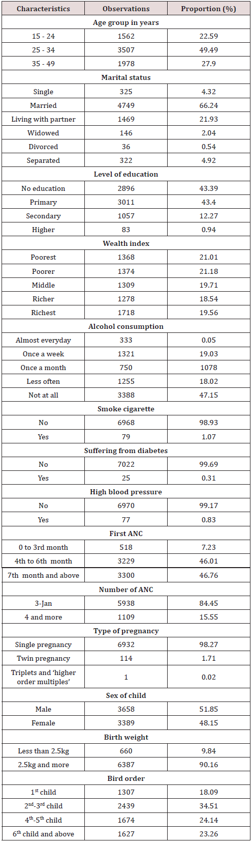
This study used “birth weight” as the dependent variable which was considered as continuous first to allow a linear regression analysis and again as dichotomous to enable the discrete model. We based on WHO guidelines to define cut-offs of the dichotomous “birth weight” outcome [13]. A dummy variable was generated taking value 0 for babies weighing less than 2,500 g at birth and value 1, otherwise. Selection of independent variables was informed by the literature search and by the understating of local context. We included individual woman’s characteristics, behaviors and underlying health conditions as well as factors related to pregnancy health. With an aim to better grasp the effect of coefficients on birth weight, all explanatory variables were categorized as summarized in Table 1.

Table 1: Socio-economic and demographic characteristics.
lupinepublishers-openaccess-journal-pediatrics-neonatology

Key: X+SD: Mean + Standard deviation.

Data management and models specification

In the first instance, owing to DHS study design which used multiple sampling stages, the dataset was survey set before analysis. In the second stage, we constructed the linear and logistic models as specified below:

In the above linear estimation, Yi is the outcome variable (i.e. birth weight). The model includes an intercept and a random error term. Independent variables are represented by a vector of covariates Xi and B1 captures the magnitude of change in birth weight corresponding to a unit change in explanatory variables. Significance of linear coefficients was ascertained based on p-value at α = 0.05.

Equation 2 is the logistic specification model. In this model, the dependent variable is the log odds that a woman i delivers a normal weight baby (i.e. a baby weighing at least 2,500 g) relative to those giving birth to underweight babies (i.e. babies with less than 2,500 g). 0 β captures fixed effects and 1 β detects random effects on the probabilities of giving birth to normal weight babies. The vector of covariates Xi includes independent variables described in Table 1. For the logistic model, significance of explanatory factors was determined based on a corresponding 95% confidence interval that does not contain value 1.

Results

Results of this study are sectioned into three main subheadings. In the first instance, we describe socio-demographic characteristics of study participants. In the second time, we summarize findings from the linear model of socio-economic and maternal health factors on birth weight. In the final stage, we present results of the logistic model on the probability of low versus normal birth weight.

Socio-economic characteristics

Table 1 summarizes socio-economic characteristics of participants. Of 7047 women of reproductive age, half were aged between 25 and 34 years old. Overall, majority of women did not achieve university education. Further, the prevalence of tobacco and alcohol consumption as well as the prevalence of health conditions with evidence to complicate pregnancy or childbirth was considerably low. Results also showed that mothers delayed in attending ANC as only about 7% attended their first ANC in the first trimester and only about 16% achieved recommended four or more ANC visits during the course of the pregnancy. More than half of surveyed women were married, and majority had a parity of two to three. Surveyed women were evenly scattered across the wealth quintiles. Low birth weight babies represented the vast minority (less than 10%) and male babies constituted a slightly higher proportion.

Determinants of birth weight

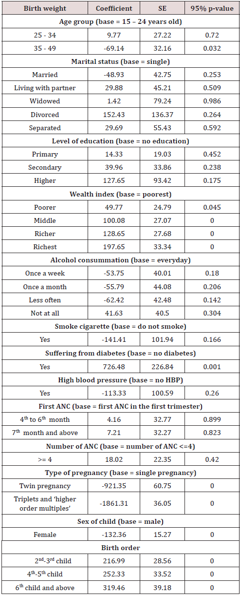
As can be viewed in Table 2 below, birth weight significantly decreases with older women’s age and increases with a diabetes condition. Also, there was evidence of a sharp increase in birth weight with wealthier quintiles. For instance, women who belong to the richer and richest quantiles gave birth to babies with almost 130 grams and 200 grams more; respectively. Moreover, there was a significant increase of birth weight with party. Mothers who had four to five and six and more parity gave birth to babies with 253grams and 320 grams more respectively. Most importantly, diabetic women gave birth to babies with 750 grams more compared to women without diabetes condition. The effect of a woman’s age on birth weight is linearly negative with a 70 g decrease for women above the age of 35 years. In contrary to the above, multiple pregnancies and female babies are associated with low birth weights. The decrease in birth weight is more evident for triplet pregnancies; reaching nearly 1800 grams lower compared to single pregnancies. The decrease in birth weight halves from triplet to twin pregnancies (1800 versus 900 grams). In the same perspective, female births tended to yield lower birth weights up to 130 grams less compared to male babies.

Table 2: The effect of socio-economic determinants on birth weight.
lupinepublishers-openaccess-journal-pediatrics-neonatology

Determinants of low birth weight

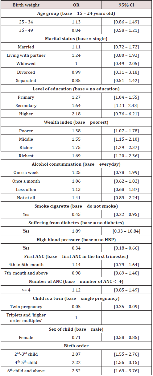
On the one hand, women’s education attainment, parity, and wealth index were predictors of higher likelihood of normal birth weight. With reference to poorest women, the likelihood that a woman gives birth to a normal weight baby increases with wealth quintiles to nearly double for women belonging to richer quantiles. Similarly, higher education level predicted higher likelihood of normal birth weight. For instance, women who attained tertiary education were about twice more likely to deliver normal weight babies compared to their counterparts who did not attend schooling. Furthermore, compare to women with one parity, the likelihood that a woman gives birth to a normal weight baby increases with high parity to become two times more and nearly three times for women with four to five and six and more parity respectively. On the other hand, high blood pressure, multiple pregnancies, smoking cigarette, and bearing a female child were negative predictors of normal birth weight. In other words, chances of low birth weight among women who reported smoking cigarette and those with high blood pressure were more than twice likely compared to women without the above conditions. Furthermore, the likelihood that women give birth to underweight babies was nearly double among women bearing female children or multiple pregnancies. Results of the logistic model are summarized in Table 3.

Table 3: The effect of socio-economic determinants on low birth weight.
lupinepublishers-openaccess-journal-pediatrics-neonatology

Discussion

This study used secondary data from the national Demographic and Health Survey 2017 to determine LBW prevalence and explore socioeconomic and demographic factors that predict LBW among Burundian women. LBW in Burundi was found to be close to other low- and middle-income countries namely Ethiopia and Iran. In Burundi, LBW is nearly 10% against 10% and 9.4% in Ethiopia [14] and Iran [15]; respectively. Conversely, LBW was found to be much higher in other countries such as India with one fifth of live births weighing less than 2,5 kilograms [16]. In our study, significant determinants of birth weights included wealth, parity, chronic health condition, pregnancy type (single versus multiple), woman’s age, and child’s sex. On the one hand, birth weight consistently increases with higher wealth quintiles, higher parity levels, and having diabetes. Compared to poorest women, birth weight increases of 50 grams for women belonging to poorer families. This increase doubles (100 grams) for women from middle class and again doubles for richest women who gave birth to babies weighing 200 grams more. Similarly, second and third order newborns weighed 220 grams more compared to first order babies. The increase in birth weight was consistent with higher birth orders and reached more than 300 grams for babies born to multiparous women from sixth pregnancy going forward. Diabetes was associated with an increase of more than 700 grams. On the other hand, multiple pregnancies, older age, and female babies were significant predictors of decreased birth weight. The most appealing evidence concerns multiple pregnancies which cause birth weight to decrease of about 1900 grams for triplet babies and about 920 grams for twin babies. Women older than 35 years gave birth to babies lower of 70 grams weigh and female newborns weighed 130 grams less compared to their male counterparts. Results from the linear model corroborate with those implemented using the logistic regression. Wealth index and higher parity determined the likelihood of giving normal weight babies while smoking, multiple pregnancies, child’s sex, and a woman’s chronic condition were significant predictors of the likelihood of LBW. Higher birth orders were associated with more than twice likelihood of bearing a normweight baby compared to first pregnancies.

In the same perspective, wealthier mothers and those highly educated were nearly two times more likely to give birth to normal weight babies. However, similar to the linear model results, twin pregnancies and female babies were nearly twice more likely to be underweight. Additionally, there was a double chance of giving birth to underweight babies among women who smoke cigarettes during the course of pregnancy were and those who had high blood pressure. Determinants of birth weight in Burundi corroborate with evidence from other settings. In Sri Lanka for instance, wealthier women and higher educated women were more likely to give birth to normal weight babies [17]. In this study, other significant determinants of birth weight were mother’s age at childbirth, newborn’s sex, and parity level. Contrary to the context of Burundi where antenatal care did not predict birth weight, Indian women who completed at least four recommended ANC visits were more likely to deliver normal weight babies [17]. Similar evidence has also been found in India [18]. Furthermore, similar to our findings, a wealth evidence has established the correlation between women with chronic health conditions such as diabetes with macrosomia [19, 20]. Multiple pregnancies were significant predictors of LBW in Burundi and in other similar settings. The example is the study by Taywade et. Al [21] in India where women bearing multiple pregnancies were 21 more likely to give birth to underweight babies [21]. Additionally, male sex was found to be a protective predictor of LBW in India and Ghana [18, 22]. Smoking was also a negative predictor as women who reported having smoked cigarette during pregnancy were at high risk of LBW in Ethiopia [23] and in India [21].

Conclusion

This study which used a nationally representative sample of Burundian women of reproductive age yielded evidence on LBW prevalence and factors affecting it. The prevalence of LBW was considerable when compared to many countries around the world (10%). Among important predictors affecting birth weight included a women’s age, wealth, education, parity, chronic health condition, smoking, pregnancy type (single versus multiple) and child’s sex. Markedly, LBW was highly associated with multiple pregnancies, female babies, the history of smoking over the course of the pregnancy, and high blood pressure. Despite considerable efforts put in place by the government to improve maternal and child health, LBW remains a public health concern. This study suggests the need for the implementation of targeted innovative interventions to tackle identified LBW risk factors among women of reproductive age. For instance, socio-economic interventions targeting poor household may help to improve economic status of women with an aim to reduce LBW prevalence among poorer women. Our study used secondary data analysis which has limit in establishing causal inference between predictors and the study outcome. This shortcoming could be addressed by cohort studies of pregnant women which have the possibility to unpack factors determining LBW with a much stronger evidence than cross-sectional surveys. Therefore, this study provides important evidence and a threshold for further research works in the field.

Read More About Lupine Publishers Journal of Pediatrics and Neonatology Please Click on Below Link:
https://lupinepublishers-pedaitrics.blogspot.com/

Saturday, 8 April 2023

Lupine Publishers | Aquatic Exercise Intervention Is Effective for Spasticity Inhibition in Children with Cerebral Palsy: A Clinical Controlled Study

 Lupine Publishers | Journal of Pediatrics and Neonatology


Abstract

Introduction: Spasticity has been implicated as a major hindrance to motor development and overall functional performance in children with cerebral palsy (CP). Recent evidence have suggested aquatic therapy as an alternative means for inhibiting spasticity in children with CP. However, these previous studies have provided contrasting results, thereby creating some dearth of evidence in the use of aquatic therapy to manage spasticity in children with CP.

Objective: To investigate the effectiveness of aquatic exercise intervention in inhibiting spasticity in children with CP.

Materials and methods: Thirty children aged 1-12 years participated in this study. They were randomised into 2 groups (experimental and control). The experimental groups received manual passive stretching on affected muscles of both upper and lower extremities followed by weight bearing exercises in water (temperature 28-32ºC) while the control group received same exercises on land for 10 weeks. Degree of spasticity on treated group of muscles was assessed using Modified Ashworth Scale (MAS). Mann-Whitney-U test was used to compare the change in degree of spasticity between both groups. The level of significance was set at p<0.05.

Results: The experimental group showed significant reduction in spasticity of all the tested muscle groups while the control group showed no significant improvement in spasticity of wrist flexors and knee flexors. No significant difference was observed for change in spasticity between both groups.

Conclusion: Aquatic exercise intervention is effective for inhibiting muscle spasticity in children with cerebral palsy.

Keywords: Aquatic Exercise; Intervention; Spasticity; Cerebral Palsy.

Introduction

Spasticity is a widespread problem among children with cerebral palsy (CP) as it affects function and can lead to musculoskeletal complications [1]. It occurs as a result of pathologically increase in muscle tone and hyperactive reflexes mediated by a loss of upper motor neuron inhibitory control [2]. Spasticity is considered an important neural contributor to muscle hypertonia in children with cerebral palsy [3].It adversely affects muscles and joints of the extremities, causing abnormal movements, and it is especially harmful in growing children[4]. Reduction of spasticity and improvement of motor control is a prerequisite for functional performance in children with cerebral palsy [5,6]. With reduction in spasticity, improvement in gross motor function and independent performance of ADLs are expedited which in turn reduces the burden cerebral palsy poses on the children and their caregivers [5,6,7] .The management of spasticity in children with CP is often complex and poses a great challenge to healthcare professionals. Effective treatment requires a multidisciplinary approach involving pediatricians, physiotherapists, occupational therapists, orthotists and surgeons. Previous studies have suggested various treatment approaches and modalities to manage spasticity associated with spastic CP. These include the use of oral neuropharmacological agents, injectable materials such as botulinum – a toxin or surgical treatment, use of orthotic devices, massage techniques, strengthening exercises to the antagonist musculature, use of electrical stimulation and the application of cryotherapy or ice therapy [8,9]. Aquatic intervention is one of the most popular supplementary treatments for children with neuromotor impairments, particularly CP [10]. Several studies have reported aquatic intervention as a veritable tool in the rehabilitation of children with CP [11-13]. The intervention may provide safe and beneficial alternative low-impact exercise for children with disabilities [14]. This study was aimed at investigating the effectiveness of aquatic exercise intervention in inhibiting spasticity in children with spastic cerebral palsy.

Methods

Thirty children undergoing rehabilitation were recruited for this study from a center for developmental challenges in Lagos, Nigeria. Their age range was from 1 to 12 years and were diagnosed with the spastic form of cerebral palsy. Those with other neurodevelopmental conditions were excluded from the study.

Ethical Approval and Informed Consent

Ethical approval for this study was obtained from the Health Research and Ethics Committee of the Lagos University Teaching Hospital, Idi-Araba, Lagos, Nigeria (Ref. No: ADM/DCST/HREC/ APP/1525). Parents of the participating children gave their informed consent.

Procedure

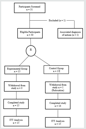
A detailed history and thorough physical examination was initially carried out on each participant. Those who met the criteria for the study were then randomly assigned into experimental and control groups. The flow chart of the study is presented in Figure 1.

Assessment Protocol

Functional Level Assessment: This was assessed using the Gross Motor Function Classification System- Expanded and Revised according to standard [15].

Degree of Spasticity Assessment: This was done using the Modified Ashworth Scale (MAS). Both upper limbs (shoulder adductors, elbow flexors and wrist flexors) and lower limbs (hip adductors, knee flexors and ankle plantar flexors) were assessed for spasticity bilaterally (left and right) according to the pattern presented by the participants. Each participant was placed in a relaxed supine position and the starting position of testing each muscle group was a maximal placement of the joint in a position of their primary function and then maximally moved to a lenghtened position opposite to their primary movement. Participants were then scored based on the amount of resistance to passive movement as described in MAS. To accommodate the score of “1+” modification for numeric analysis, grade ‘‘1+’’ was recorded as 1.5[16].

Figure 1: Flow chart of the study. Key: R: Randomization. ITT: Intention –to-treat.

lupinepublishers-openaccess-journal-pediatrics-neonatology

Intervention

The participants in both experimental and control groups had a total of 20 treatment sessions consecutively for 10 weeks at the rate of two sessions per week. It was ensured that participants in both groups were not on any form of anti-spastic medication throughout the study.

Those in the experimental group received exercise training in water of temperature ranging between 28-32 ºC for 10 weeks with the exercised parts fully immersed in water. The exercise protocol consisted of 3 categories of exercises:

Exercise 1 (Manual passive stretching): Each joint involving the spastic group of muscles was passively stretched and held in this in fully lengthened position of the muscle groups for up to 60 seconds follow by relaxation of 30 seconds. This procedure was repeated five times for each part

Exercise 2: Weight bearing one or both upper extremities in sitting with or without support for 60 seconds and repeated 5 times.

Exercise 3: Weight bearing on both lower extremities in standing with or without support for 60 seconds and repeated 5 times.

Control group

Participants in the control group received same exercise intervention as those in the experimental group except that the intervention was carried out on land.

Post-intervention Assessments

Participants in both groups were re-assessed after 4 weeks, after 8 weeks and after 10 weeks of intervention for changes in spasticity using the Modified Ashworth Scale (MAS). All assessments were carried out by blinded assessors.

Data Analysis

Data was analyzed using the Statistical Package for Social Sciences version 21.0 version. Participants in both groups were compared for age using the Independent t-test. Friedman test was used to compare post intervention changes in degree of spasticity within each group across the duration of treatment while Mann- Whitney U test was used to compare the change in spasticity between both groups. All statistical tests were performed at the 0.05 level of significance (p<0.05).

Results

Physical Characteristics

The mean age of all participants was 5.20+2.43 years and no significant difference in age was found between both groups of participants at baseline Table 1,2. Most of the participants were in level IV of the GMFCS. Quadriplegic spastic type of CP predominated among all the participants as shown in Figure 2.

Figure 1: Distribution of Type of Spastic Cerebral Palsy among Participants.

lupinepublishers-openaccess-journal-pediatrics-neonatology

Table 1: Comparison of Age and Mobility Level between both Groups at Baseline.

lupinepublishers-openaccess-journal-pediatrics-neonatology

Key: X+SD: Mean + Standard deviation.

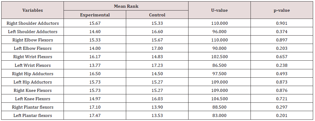
Table 2: Mean Rank Comparison of Spasticity between both Groups at Baseline.

lupinepublishers-openaccess-journal-pediatrics-neonatology

Aquatic Intervention and Spasticity

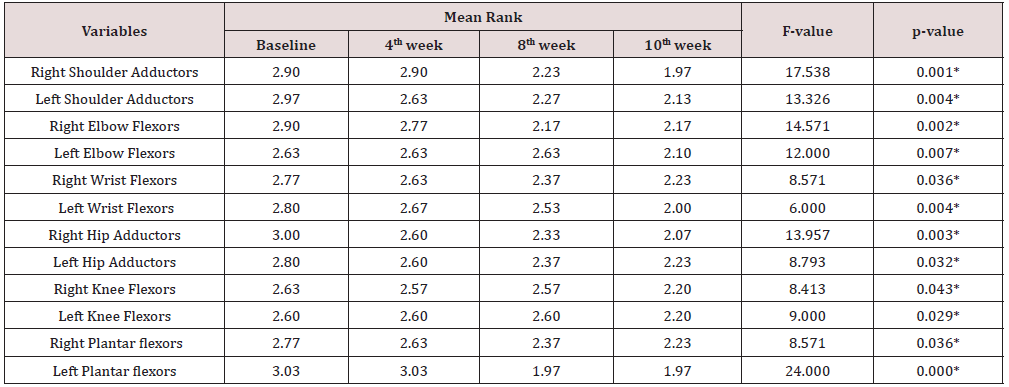
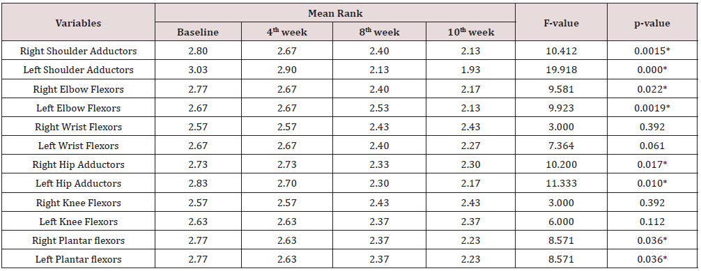
Significant change in spasticity was observed on all tested group of muscles of participants in the experimental group following aquatic exercise intervention Table 3.

Land-based Exercises and Spasticity

Significant change in spasticity was also observed on tested group of muscles among participants in the control group following land-based exercise intervention, except for right and left wrist flexors; and right and left knee flexors as shown in Table 4.

Table 3: Comparison of Mean Rank Changes in Spasticity in Experimental group of Participants across the duration of Intervention.

lupinepublishers-openaccess-journal-pediatrics-neonatology

*Significant at p<0.05

Table 4: Comparison of Mean Rank Changes in Spasticity in Control group of Participants across the duration of Intervention.

lupinepublishers-openaccess-journal-pediatrics-neonatology

*Significant at p<0.05.

Comparison of Spasticity Change between Experimental and Control Groups

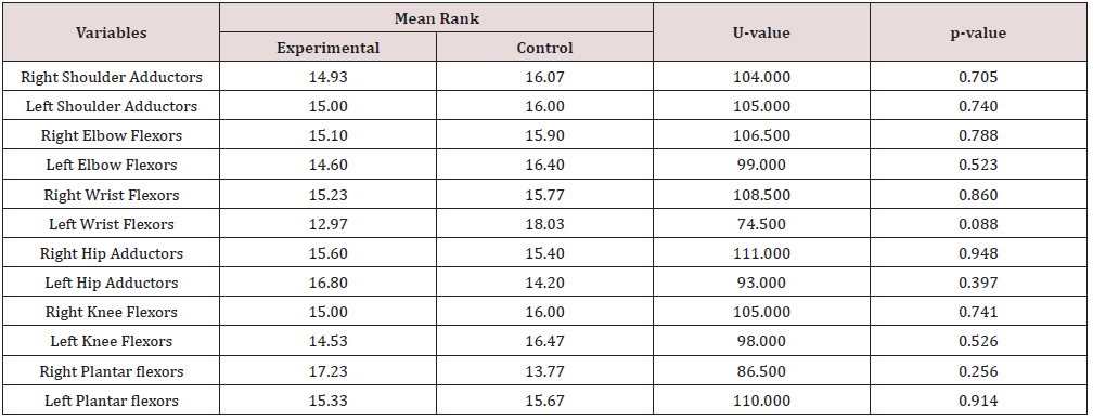
Despite the gross improvement in spasticity of participants in experimental group compared to the control group which showed no improvement for few groups of muscles, no significant difference was found in the spasticity change between both groups as represented in Table 5.

Table 5: Mean Rank Comparison of Spasticity between both Groups after 10 weeks of Intervention.

lupinepublishers-openaccess-journal-pediatrics-neonatology

Discussion

Quadriplegic type of spastic CP was more predominant in this study. This is consistent with reports from earlier studies in Nigeria [17-19]. However, reports from studies in other parts of the world showed that there were more diplegic type of spastic CP [11,20][21- 23]. Geographical differences may be considered for this variation.

In this present study, statistically significant improvement was observed in both intervention groups for spasticity of shoulder adductors, elbow flexors, hip adductors and ankle plantar flexors. This finding shows that therapeutic exercises such as manual passive stretching and weight bearing exercises which are used by physiotherapists in the management of spasticity in children with cerebral palsy are effective as documented in previous studies [21,24,25,26]. The report of statistically significant improvement in spasticity of hip adductors and knee flexors following 10 weeks of aquatic intervention in this study has been supported by a study by Chrysagis et al in 2009 where there was further significant improvement in the passive range of motion of the hip and knee joints [11]. However, the aquatic training adopted for their own study was different from the one used in this present study. This improvement in spasticity is attributable to the therapeutic benefit of buoyancy property of water which can help to relax spastic muscles by providing antigravity positioning. This study revealed that landbased exercise intervention improved the spasticity of shoulder adductors, elbow flexors, hip adductors and ankle plantar-flexors as observed among participants in the control group. This finding was corroborated by a systematic review on the effectiveness of passive stretching on spasticity in children with cerebral palsy [24]. They found out that 4 of the 6 reviewed studies reported significant reduction in spasticity including that of shoulder adductors, elbow flexors, hip adductors and ankle plantar flexors following passive stretching while the remaining 2 studies did not consider spasticity in their outcome variables. This improvement in spasticity may be attributed to fatigability of stretch receptors as a result of stretch on the muscle spindles which induces inhibition on the gamma-motor neurons and allowed more lengthening of muscle fibres thereby counteracting the effect of spasticity.

There was no significant improvement observed for spasticity of the wrist flexors and knee flexors in this present study after 10 weeks of land-based exercises. These findings are contrary to the findings of Akinbo et al. (2007) and Akinbo et al. (2007) who in their separate studies found out that manual passive stretching alone significantly improved spasticity of wrist flexors, passive range of motion of the wrist as well as hand function in children with spastic cerebral palsy with and without the use of cryotherapy. They however both concluded that a combination of passive stretching, and cryotherapy produced better improvement in the spasticity of wrist flexors in children with spastic cerebral palsy [25,26]. The reason for lack of significant improvement in spasticity of wrist flexors and knee flexors following 10 weeks of land-based exercises in this present study could not be ascertained but there may be needed to go further beyond 10 weeks to get a significant improvement. Comparison between the experimental and control groups revealed no significant difference in the change in spasticity of all tested muscle groups. This implies that either of the two forms of intervention can be adopted in the management of spasticity in children with cerebral palsy. However, considering the result of this study which had earlier shown that aquatic intervention significantly improved the spasticity of all the tested muscle groups including those of wrist flexors and knee flexors which were not significantly improved by land-based exercises after 10 weeks, the beneficial pendulum might as well be tilted towards the aquatic exercise intervention when there is need for clinical decision making in the management of a patient presenting with spasticity of all the tested muscle groups in this study.

Conclusion

Aquatic exercise intervention is effective in inhibiting spasticity in children with Cerebral Palsy and should be an intervention of choice when considering affectation of all muscles of both upper and lower extremities.

Read More About Lupine Publishers Journal of Pediatrics and Neonatology Please Click on Below Link:
https://lupinepublishers-pedaitrics.blogspot.com/
Friday, 3 March 2023

Lupine Publishers | Late Intrahospital Bath Is Safety in Newborns

 Lupine Publishers | Journal of Pediatrics and Neonatology


Abstract

Introduction: Preterm infants in many neonatal intensive care units (NICU) are routinely bathed despite the lack of evidence to support frequent bathing as a way to reduce the risk of infection. In fact, research has shown that routine bathing causes behavioral and psychological instability in premature infants

Materials and methods: Prospective, randomized clinical trial. Neonates older than 34 weeks of gestation were included, treated in the Neonatal Intermediate Care Unit at the University Hospital in the period from October 1, 2018 to February 28, 2019.

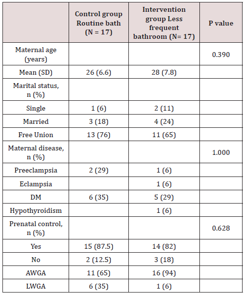
Results: A total of 34 patients were included in the study, which were assigned by lot to two groups, 17 patients corresponded to the control group (daily bath) and 17 patients to the intervention group (bath every 3 days). There was no significant difference between the two groups regarding gestational age, however, a value of p = 0.034 was found regarding trophism.

Conclusion: There were no differences in the presence of saprophytic flora or pathogenic flora that colonize the skin of the neck region between both groups. The main saprophytic microorganism found was coagulase negative Staphylococci; and the main pathogens detected in the cultives of both groups were Staphylococci Aureus and E. Coli.

Keywords: Bath; Colonization; Cultivation; Germ; Newborn; Pathogenic Flora; Saprophytic Flora

Introduction

Globally 3.1 million children die each year in the neonatal period (the first 28 days of life). Half of these deaths occur within the first 24 hours of birth, 75% occur in the early neonatal period (0 to 6 days) due to preterm births, severe infections, and perinatal asphyxia. Essential care can contribute to reducing the incidence of neonatal morbidity and mortality [1]. Recommendations for care in the first week of life include improvement in thermal care practices such as drying, skin-to-skin contact, immediate breastfeeding, and delayed bathing [2]. Although some aspects of care are understood, others have not been adequately investigated or explained, the infant bath is an example. Preterm infants in many neonatal intensive care units (NICU) are routinely bathed despite the lack of evidence to support frequent bathing as a way to reduce the risk of infection. In fact, research has shown that routine bathing causes behavioral and psychological instability in premature infants [3]. The skin functions as a protective barrier against pathogens and toxins, provides thermal regulation, and maintains fluid balance [4]. The skin of the term newborn has a barrier function similar to that of the adult, whereas the skin of the premature newborn provides a poor barrier [3]. Among the clinical implications are greater susceptibility to microbial attack, greater risk of maceration due to moisture retention and greater response to irritants, in addition to the fact that soaps and cleaners can alter the epidermal barrier and the acid mantle causing dry skin and more susceptible to damage [5].

The greater colonization of newborns occurs at birth, the new evidence suggests that factors in labor and the environment at birth such as the route of birth, the frequency of vaginal examination, the use of antibiotics, influence the maternal microbiome and the newborn [6,7]. Newborn skin contains a variety of microorganisms and is determined to some extent by the type of birth, vaginal or cesarean. The skin of newborns obtained vaginally contains bacterial “communities” that resemble the maternal vaginal flora, whereas the skin of newborns by caesarean section reflects the flora of the skin [3]. Vaginal birth remains the most common method of birth and is achieved in 68% of women in the United States. The vaginal microbiome is a mutualistic environment of organisms that depend on environmental conditions and host factors that promote homeostasis in the vagina and inhibit the growth of bacteria that have been associated with preterm labor and chorioamnionitis. When born vaginally, the fetus is exposed to a large number of maternal bacteria from the vagina and the perianal area. Infants delivered via caesarean section, regardless of exposure to prophylactic antibiotics, harbor a less diverse microbiome that resembles the skin, mouth, and bacteria on surrounding maternal surfaces [8]. Infants born via cesarean section tend to harbor a large portion of antibiotic resistant genes compared to those born vaginally [9]. Sponge bathing and bathing in premature newborns can negatively impact some physiological parameters such as heart rate, oxygenation, and present behavioral signals that indicate stress. Due to concerns about stress in premature babies during bathing, studies evaluated the impact of bathing with a mild cleanser every 2 days versus every 4 days on colonization of skin and pathogenic bacteria. Concern for thermoregulation of newborns has traditionally driven bathing practices, as effective self-regulation of body temperature can be a major challenge for newborns [10]. Temperature regulation is of vital importance for nurses caring for newborns. Although there are many benefits of bathing, such as maintaining the integrity of the skin, reducing the risk of infection and elevation of temperature, promoting mobility and maintaining joint function, the act itself has become a ritual and is based on tradition. rather than empirical evidence [11]. The times for bathing are flexible, depending on the characteristics and stability of the newborn and the wishes of your family. However, the best time for the bath to occur is unknown. The newborn’s bath should not last more than 5 minutes [5]. There are 3 types of bath available for infants. Sponge bathing is the act of bathing with a cloth or sponge without being immersed in a bath of water. The bathtub bath is the act of bathing the baby immersed in a bathtub with water. Finally, the wrapped bath is the immersion of the infant, who is first wrapped in a towel, in water, washing 1 limb at a time while maintaining a flexed and contained position throughout the bath. The objective of this study was to correlate the bacterial colonization of the neck region with the presence of sepsis, signs of systemic inflammatory response, or local infections, in neonates with an indication of daily bath versus bath every 3 days. The hypothesis raised was that the second bath of the newborn after 3 days of hospital stay does not increase the incidence of skin or systemic infections.

Materials and Methods

Prospective, randomized clinical trial. Neonates older than 34 weeks of gestation were included, treated in the Neonatal Intermediate Care Unit at the University Hospital in the period from October 1, 2018 to February 28, 2019. Within the selection criteria, the inclusion criteria were: Neonates older than 34 weeks of gestation, admitted to the Neonatal Intermediate Care Unit, euthermic and that the first bath was performed in the first 24 hours of hospital stay. The exclusion criteria were: Newborns admitted to hospital with an infectious history during their pregnancy, patients with the presence of an infectious process at the time of admission and / or administration of antibiotics. The elimination criteria were: Neonates who are discharged within 3 days of hospital stay, who do not perform any of the cultives and / or who present an incomplete clinical record. Informed consent was obtained, which was signed by both parents and two witnesses. The neonates were divided into 2 groups by means of a lottery, in both groups a basal culture was taken in the neck region at the time of admission, then the first bath was performed, and a second culture was taken after 30 minutes. The control group bathed every 24 hours and the intervention group every 3 days, using the sponge bath technique. In all patients, a third culture was collected at 3 days, before bathing and a fourth culture 30 minutes after bathing. The skin cultives were processed in the Microbiology Laboratory of the “Dr. José Eleuterio González”. To obtain the skin cultives, a Stuart culture medium was used, the sample was obtained from the neck region, cleaning 10 times, in 2 directions, 5 horizontal and 5 vertical, in 2cm [2]. The type of skin flora was identified and the number of Colony Forming Unit (CFU) per square centimeter was obtained for each microorganism. It will be determined after 48hr of incubation at 35°C. The sample was processed in the Microbiology Laboratory of the “Dr. José Eleuterio González” within 30 minutes, after obtaining each sample[12].

The normal flora was defined as coagulase-negative staphylococcus and diphtheroids [3], the pathogenic flora as any microorganism other than those mentioned [13]. When a pathogen was identified, an antibiogram was performed, the treating physician was informed, and the result was documented in the clinical record of each patient. The bathing process of the newborns included in the study consists of a sponge bath, a new Grisi® neutral soap was used for each bath and tap water. The bath was carried out in the morning shift, by nursing personnel. After completing the bath, the infants were dressed in diapers and a clean sheet. Descriptive statistics, simple frequencies and percentages were used for the analysis of the studied variables. They were used for data collection in the Microsoft Excel 2013 program and for statistical analysis the SPSS version 21.0 program (IBM Corporation ©). For the comparison between groups, inferential statistics were used by means of the student’s T distribution test for parametric variables and chi square and Pearson’s for non-parametric variables, considering a significant p <0.05. The calculation of the sample size was performed using a formula for equivalence of 2 means with a confidence of 95% and a power of 80% bilateral, with a mean of 18.43 CFU * 105 and standard deviation of 17.25 with an interval to demonstrate the equivalence of +/- 10 required a total of 30 participants per group.

Results

A total of 34 patients were included in the study, which were assigned by lot to two groups, 17 patients corresponded to the control group (daily bath) and 17 patients to the intervention group (bath every 3 days).

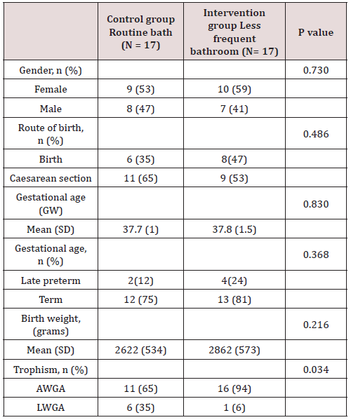
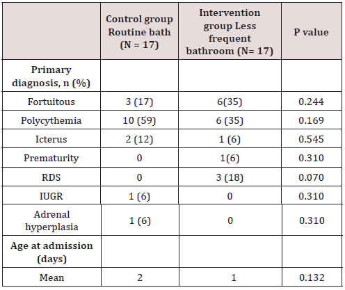
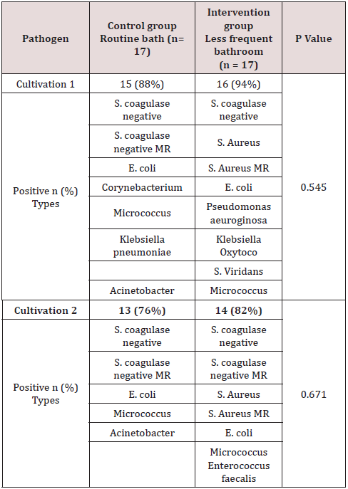
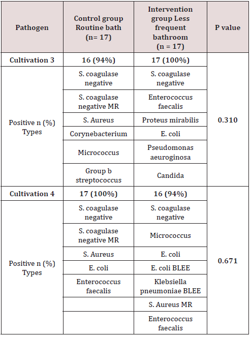
Of the total of 34 patients included, 56% (n=19) corresponded to the female gender and 44% (n=15) to the male gender. Likewise, 41% (n=14) of births in the general population were deliveries and 59% were caesarean sections (n=20). In the control group (group 1) a gestational age was recorded in weeks of 37.7 with a SD ± 1 and, in the intervention group (group 2), a gestational age of 37.8 ± 1.5. 6 late preterm patients were included (between 34-36.6 SDG), 2 corresponding to the first group and 4 to the second, the rest were term patients, no patient was older than 42 weeks of gestation (post-term). Regarding the weight at birth, an average weight of 2622 grams (± 534) was reported in the control group and 2862 grams (± 573) in the intervention group. Of the entire universe of patients, 79% (n=27) had an adequate weight for gestational age (AWGA), 7 patients reported a low weight (LWGA), the majority belonging to the control group. There was no significant difference between the two groups regarding gestational age, however, a value of p = 0.034 was found regarding trophism. Table 1 shows the demographic characteristics of each group. Among the most common admission diagnoses, we find polycythemia 47% of the general population (n=16), accidental delivery 26% (n=9), jaundice 9% (n=3), respiratory distress syndrome 9% (n=3), other diagnoses were prematurity, intrauterine growth retardation, and adrenal hyperplasia. A p = 0.070 was found in the diagnosis of RDS since all patients were from the intervention group (Table 2). Among the maternal characteristics we find an average maternal age of 27 years, 70% (n=24) in a free union marital status. The maternal diseases that occurred during pregnancy were pre-eclampsia and eclampsia. The existing maternal diseases were diabetes mellitus and hypothyroidism. According to the guidelines established by the Official Mexican Standard NOM-007-SSA2-2016, for the care of women during pregnancy, childbirth and the puerperium, and of the newborn, in relation to control, we found that 85% (n=29) had adequate prenatal care in both groups. Baseline cultives were obtained before and after the first bath, and 3 days after admission in each patient. 68 cultives from each group were obtained throughout the study (n=17 for both groups). The results of the cultives of each group are presented in tables 3,4 and 5. Despite the presence of pathogens in the skin cultives, none of the patients developed any signs or symptoms of infection during participation in this study.

Table 1: Demographic characteristics of newborns.

lupinepublishers-openaccess-journal-pediatrics-neonatology

Table 2: Admission diagnosis.

lupinepublishers-openaccess-journal-pediatrics-neonatology

Table 3: Maternal demographic characteristics.

lupinepublishers-openaccess-journal-pediatrics-neonatology

Table 4: Cultivation results.

lupinepublishers-openaccess-journal-pediatrics-neonatology

Table 5: Cultivation results.

lupinepublishers-openaccess-journal-pediatrics-neonatology

Discussion

Controversy exists about when to bathe hospitalized newborns to reduce the risk of colonization of harmful flora and whether this predisposes to local and systemic infections; without affecting physiological and behavioral stress, since an increase in stress has been described, especially in premature babies [12]. In this study, late preterm and term neonates were studied, however, a limitation of this study is that the changes in behavior of the baby were not objectively measured, as to confirm this theory. It is recommended to seek strategies to reduce this stress, one of them is a change in the frequency of bathing in patients hospitalized in the Neonatal Intensive Care Unit. It has been seen that the decrease in the frequency of bathing has shown beneficial effects in the patient admitted to the NICU since it preserves the protective properties of the skin, mainly in premature infants [1]. The newborn’s skin becomes colonized with microorganisms within 24 hours of birth. The first areas to be colonized are armpit, navel, genitals, groin, and abdomen. The prevalent microorganisms are coagulase-negative staphylococcus and diphtheroid [3]. Staphylococcus epidermidis, one of the 13 species of coagulase-negative staphylococci, is the most common vaginal organism and rapidly colonizes the surface of the skin, it is the organism that predominates on the skin of most newborns [10]. In preterm newborns, many invasive infections are caused by this bacteria [13]. Other common microorganisms are Staphylococcus saprophyticus (perineum), Staphylococcus capitis (sebaceous areas), corynebacterium (intertriginous areas), propionibacterium (follicles, sebaceous glands), acinetobacter (moist, intertriginous areas, perineum [14], malas. Lactobacillus spp dominates the vaginal area [6].

Consistent with what has been reported in the literature, the main saprophytic microorganism found in this study was coagulasenegative staphylococcus. The microbiome of neonates requiring admission to the NICU differs from healthy term neonates. The microbiome of the infant in the NICU is less diverse and less in the number of anaerobes, which contribute to an increase in the rates of infection and inflammatory processes in this population [15]. Pammi et al. in 2017 reported that the most abundant phyla in premature skin were Firmicutes and Proteobacteria, and the predominant genus was Staphylococcus. The skin microbiome of preterm infants has increased the abundance of Firmicutes and decreased that of Proteobacteria compared to full-term infants. They found a greater abundance in Staphylococcus, Corynebacterium and Prevotella species and a decrease in the abundance of Brevundimonas, Flavobacterium and Sphingobacterium species at the genus level in premature newborns [16]. In 2018, Young et al. included a total of 61 infants, 41 term newborns and 20 preterm infants, 35 of the term newborns went to joint accommodation with the mother, the rest, including preterm newborns, entered the hospital. NICU area. The dominant bacterial phylum on the skin of neonates was Proteobacteria and Firmicutes. They found that the skin microbiota of preterm infants differs from term newborns, with higher enrichment of Staphylococcus and taxa that are typically associated with the fecal microbiota, such as Escherichi [13]. The results in this study demonstrated no statistical significance in the noxious flora between both assigned groups (daily bath vs bath every third day). According to what is reported in the literature, pathogens such as Staphylococcus Aureus and E. Coli were detected in most of the cultives in both groups, before and after bathing; E. Coli BLEE and MRSA were found in the 4th culture (after bathing) in the intervention group, however, no newborn from any of the assigned groups developed an infection as a result of their participation in this study.

Conclusion

There were no differences in the presence of saprophytic flora or pathogenic flora that colonize the skin of the neck region between both groups. The main saprophytic microorganism found was coagulase negative Staphylococci; and the main pathogens detected in the cultives of both groups were Staphylococci Aureus and E. Coli. There were no manifestations of skin or systemic infectious processes during the study. The findings in this study suggest that late bathing every 3 days in the NICU is safe. This allows the nursing staff to redesign their routines and allocate this time to other priority activities. However, a study with a larger sample size is required to reduce the risk of presenting a type II statistical error.

Read More About Lupine Publishers Journal of Pediatrics and Neonatology Please Click on Below Link:
https://lupinepublishers-pedaitrics.blogspot.com/
Tuesday, 3 January 2023

Lupine Publishers | Viral Infections in Infants with Neonatal Hepatitis

 Lupine Publishers | Journal of Pediatrics & Neonatology


Abstract

Introduction: Neonatal cholestasis is a common manifestation of various pathologies. These pathologies include direct hepatocellular injury, defects in hepatocyte bile formation, or mechanical obstruction of bile flow. Histologically, most of the biliary tract diseases, infections, genetic and metabolic diseases can display parenchymal inflammation leading to the diagnosis of Neonatal Hepatitis (NH). Hence, NH implies a pattern of neonatal liver disease rather than a specific cause of liver injury. Among non-obstructive causes, various viral infections are associated with NH. The unmasking of the etiology of NH in further patients’ management is extremely important. Also, the prevalence of viral associated NH varies among different studies. Therefore, we planned to investigate the frequency of common viral agents in infants diagnosed with Idiopathic NH (INH).

Objective: A total of 138 consecutive infants with conjugated hyperbilirubinemia undergoing liver biopsy were collected as well as thirty-one umbilical cord tissues belonging to normal infants as controls. Patients with known etiology of NH were excluded from the study, and 74 cases diagnosed with INH were enrolled. Nucleic acid from the liver tissues of patients was extracted and real-time PCR for the detection of EBV, CMV, HSV1, HSV2, and HHV6 was done.

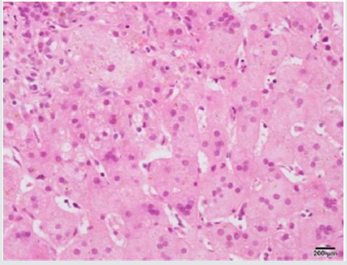
Results: The mean age of the patients at the time of liver biopsy was 70 days (minimum: 22, maximum: 98 days). There were 36 males and 38 females. CMV DNA was detected in six patients and HHV6 DNA was detected in one patient, while all control tissues were negative for the viral DNAs (p<0.05). None of the other tested DNA viruses could be detected in liver tissues. In addition, none of the liver samples were positive for CMV inclusions in hepatocytes or biliary epithelium under the light microscope.

Conclusion: In conclusion, we found CMV DNA in six liver biopsies without any histological evidence of CMV infection. Using more sensitive methods such as PCR is warranted to determine viral DNAs in the liver tissues and establish their role in the development of NH. Further studies with larger populations should be conducted to provide more information regarding the role of viruses in the development of NH and compare their frequency in different populations.

Keywords: Neonatal Hepatitis; Cytomegalovirus; Cholestasis; PCR

Introduction

Neonatal conjugated hyperbilirubinemia is a common manifestation of mechanical obstruction of bile flow, direct hepatocellular injury and also defects in hepatocyte bile formation [1]. The distinction between biliary tract obstruction and hepatocellular injury is critical for clinical management of Neonatal cholestasis. Thus, the primary goal in patients’ management is to differentiate obstructive and nonobstructive causes of hyperbilirubinemia [2]. Once the obstructive causes are excluded, the next step would be evaluating the nonobstructive etiologies of hepatocellular injury. Due to the immaturity of the biliary system and hepatocytes, different injuries result in similar clinical presentations with a significant overlap of laboratory and pathological findings [3]. Indeed, most of the biliary tract diseases, infections, genetic and metabolic diseases can display parenchymal inflammation leading to the diagnosis of Neonatal Hepatitis (NH) by pathologists. Therefore, NH means a pattern of neonatal liver disease instead of a specific cause of liver injury. Advances in the understanding of neonatal liver diseases as well as the implication of new testing modalities allow for the unmasking of the etiology in most cases. The most common etiologies include infectious and metabolic diseases [4]. Metabolic causes can be suggested based on histological and clinical clues and can be confirmed using biochemical, molecular and genetic tests. However, despite all investigations, no etiology has been found in approximately 30% of infants with conjugated hyperbilirubinemia and this group of patients is addressed as Idiopathic Neonatal Hepatitis (INH) [3].

Many viruses, including Enterovirus, Adenovirus, hepatitis A‐E viruses, Coxsackievirus, Cytomegalovirus (CMV), Herpes simplex virus, Epstein Barr virus, Influenza virus and Human Herpesvirus 6 are associated with NH [5,6]. The virus may be transmitted from mother to fetus or acquired after birth [7]. While most of the infants with congenital infection show no evidence of disease at birth, approximately 5% of infants have neurological, respiratory, hematopoietic and gastrointestinal involvement by virus leading to long-term sequels [5]. The prevalence of infection leading to NH varies in different age groups and increases with age, also depending on socioeconomic status and the viral detection method. Congenital CMV infection is observed in about 0.2 to 2.4% of all live births [5] with seroprevalence of 60-90% in Central Europe [8, 9] and the USA [8, 10, 11]. In a study by Funato [12] CMV DNA was detected in 58.1% of infants with NH [12]. Another study on Korean children showed evidence of CMV infection in 31% of the kids with acute non-A, B, C viral hepatitis [13]. Continuous states or latent infection is characteristic of infection by most of these viruses [6]. Therefore, positive serological tests are not sufficient to confirm an association of viral infection with hepatitis. Additionally, the overlapping histological and clinical features in infectious associated NH and INH result in difficulty in distinguishing etiologies in most cases. The etiology of NH in further patients’ management is extremely important and the prevalence of viral associated NH varies among different studies. CMV isolation in liver tissues in patients with INH has been rarely reported, and little information exists in the literature in this regard [14-18]. Therefore, we planned to investigate the frequency of common viral agents, including CMV, EBV, HHV6, HSV1 and 2, in infants diagnosed with INH using Polymerase Chain Reaction (PCR).

Methods

The study was conducted in the pathology department of Children Medical Center Hospital, affiliated to Tehran University of Medical Sciences. From April 2009 to July 2016, a total of 138 consecutive infants with conjugated hyperbilirubinemia undergoing liver biopsy were collected. Thirty-one umbilical cord tissues belonging to normal infants with no illness in their sixmonth follow up were included in the study as controls. Neonatal cholestasis was defined as jaundice within the first 4 months of age with conjugated bilirubin of more than 20% of the total serum bilirubin. The Biliary atresia was diagnosed according to Fischler et al criteria [18,19]. Patients with biliary tract obstruction, family history of progressive familial intrahepatic cholestasis, alpha one antitrypsin deficiency, known metabolic causes such as tyrosinemia, sepsis and those with insufficient tissues were excluded from the study, leading to a final number of 74 cases diagnosed with INH. No other defined etiologies were found in the formation of cholestasis in these patients, all were full-term infants with normal birth weights with unremarkable perinatal histories. The diagnosis of INH was based on clinical findings, laboratory evaluations, biochemical data, imaging tests and liver biopsy. Age, gender and clinical manifestations were extracted from the archive. Serum aminotransferases (AST and ALT), Gamma Glutamyl Transpeptidase (GGT), total and direct bilirubin levels, serum-concentration of alpha one antitrypsin, sweat chloride test results, HIDA scan of the gallbladder and biliary system, abdominal ultrasound, TORCH study, intraoperative cholangiography, associated anomalies, serum amino acid chromatography and any other factor with possible etiologic importance were retrieved from medical records when available. Ongoing CMV infection was defined by the detection of serum CMV IgM or evidence of the presence of CMV in the urine [8-11, 20]. Hematoxylin and eosin, PAS, trichrome and reticulinstained slides were reviewed for all 74 cases with special attention to the presence of viral cytopathic effect and proper paraffin blocks were selected for nucleic acid extraction and PCR study.

Nucleic Acid Purification

Using disposable blades, two 5μm sections of paraffin blocks were made and transferred to DNAse/RNAse free tubes followed by deparaffinization. Nucleic acids were extracted using Roche High Pure PCR Template Preparation Kit (Roche Diagnostics, Indianapolis, IN) as instructed by the manufacturer. Briefly, deparaffinized tissue was incubated with tissue lysis buffer and proteinase K at 55◦C until tissue particles disappeared completely. Then binding buffer and isopropanol were added. The DNA was eluted into a final volume of 200 μL, quantified photometrically using nanodrop, Thermoscientific 1000, USA and stored in −70◦C. Each sample was subject to Real-time PCR for the detection of EBV, CMV, HSV1 and 2 and HHV6 (AmpliSens PCR kits, Bratislava, Slovak republic) on rotor gene 6000 (Corbett Research, Mortlake, Australia). Negative, positive and internal controls were run in each performed PCR assays. The study was approved by the Research Ethics Committee of Tehran University of medical sciences and was in agreement with the Helsinki Declaration. Statistical analysis was performed using SPSS version 16.0.1 (SPSS Inc., Chicago, IL, USA). To report the frequencies and differences between groups, descriptive statistics and Student’s t-test were employed respectively in which P-values of less than 0.05 were considered as significant.

Results

A total of 138 consecutive infants with conjugated hyperbilirubinemia undergoing liver biopsy with a final diagnosis of NH were studied. Cases with biliary tract obstruction, family history of progressive familial intrahepatic cholestasis, alpha one antitrypsin deficiency, known metabolic causes such as tyrosinemia, sepsis and those with insufficient tissues were excluded from the stud, leading to a final number of 74 cases. The mean age of the patients at the time of liver biopsy was 70 days (minimum: 22, maximum: 98 days). There were 36 males and 38 females. The clinical data was gathered with the most common clinical presentation being jaundice and the onset of jaundice was on 24.2 ±17 days of life. The mean birth weight was 2.4 ±0.7 kg. IgM and IgG serology for CMV were available for 52 patients and 30 patients out of these 52 patients also had blood CMV PCR results in their charts. No data regarding CMV serology or blood CMV PCR was found for the rest of patients. CMV IgM and IgG were positive in 8 and 11 patients (out of 52 patients), respectively and PCR for blood CMV DNA was positive in 6 patients (out of 30 patients). Using PCR, CMV DNA was detected in six and HHV6 DNA in one patient while all control tissues were negative for the viral DNAs (p<0.05). None of the other tested DNA viruses could be detected in liver tissues. Among patients with positive CMV serology, three infants showed CMV DNA in the liver. These three patients also were positive for blood CMV DNA. For one case with positive liver CMV DNA no data was available regarding serology or blood CMV PCR. The other two cases with positive liver CMV DNA showed only positive viral serology (both high IgM and IgG), but negative blood CMV DNA PCR results.

None of the liver samples were positive for cytomegalovirus inclusions in hepatocytes or biliary epithelium (Figure 1). The mean level of serum alanine aminotransferase (ALT) levels was 187 IU/L (12-572 IU/L), while serum alanine aminotransferase (AST) counterpart was 308 IU/L (29-1592 IU/L). The mean level of AST and ALT, GGT, total and direct bilirubin levels and serumconcentration of alpha one antitrypsin did not differ between cases with positive and negative DNAs (p>0.05). Table 1 shows the clinical and laboratory characteristics of patients enrolled in the study.

Figure 1: Liver biopsy shows neonatal hepatitis as giant cell transformation of hepatocytes in a patient with positive CMV serology and PCR (H&E).

lupinepublishers-openaccess-journal-pediatrics-neonatology

Table 1: Shows the clinical and laboratory characteristics of patients.

lupinepublishers-openaccess-journal-pediatrics-neonatology

Discussion

INH is an important cause of neonatal cholestasis. Classically, infants with INH reveal normal biliary trees and elevated levels of conjugated bilirubin. The clinical picture is not unique and often there is an overlap with other causes of neonatal cholestasis. As the treatment protocols for each category of diseases differ, extensive radiological, histological, biochemical, microbiological and even molecular workup is an essential part of the diagnostic algorithm. Histologically, hepatic giant cells, inflammation and lobular cholestasis are evident while there are no clues to obstructive changes of biliary atresia, the paucity of intrahepatic bile ducts, viral inclusion or abnormally stored materials inside the hepatocytes. However, in the first few months of life, we cannot rely on histology or serology alone to rule out storage disease or infectious processes as the false-negative rate is high. As the infectious process is one of the leading causes of NH which can be missed on routine histological or serological studies, we retrospectively did PCR on liver tissues of infants diagnosed with INH to understand the frequency of few common viral infections as possible etiology and their clinical and histological correlates [1]. We found CMV DNA by real-time PCR system in the liver tissues of six patients with NH, which was not observed in the control group. CMV infection is one of the main causes of NH among other viruses [6, 9, 12, 21]. Direct evidence of the role of viruses in the disease process is difficult to be established; however, the detection of the virus from the liver tissue is regarded as evidence of the role of CMV in NH [6,12 9,21]. We identified IgM anti-CMV antibodies and markedly elevated titers of IgG anti- CMV antibodies in three out of six cases with positive liver CMV PCR and so CMV was the most likely cause of neonatal hepatitis in these infants. Primary CMV infection can be detected by the presence of serum anti-CMV IgM antibody or the seroconversion of the IgG antibody. However, in infants, the antibody production is limited due to the immaturity of the immune system resulting in negative results for anti-CMV IgM antibodies. In addition, the passive acquisition of antibodies from the mother may complicate the serodiagnosis of CMV infection. We reviewed all the biopsies again with special attention to the viral cytopathic effect in these six infants, but no viral cytopathic effect was observed. The absence of CMV inclusion may be related to its rarity in the biopsies of immunocompetent patients [6].

5 infants in this study were positive in terms of anti-CMV IgM but negative with respect to CMV DNA. This may be due to false-positive results, the passage of antibodies from mothers, or low sensitivity of our PCR assay in the presence of low levels of CMV DNA. Indeed, serology has a low specificity for the diagnosis of CMV hepatitis and cannot differentiate between active and latent infections. Few reports have been published describing the frequency of the association of CMV infection with NH. The prevalence of the finding of CMV in our stud is lower than in Funato et al. and Shibata et al, both with the rate of infection of 23–67% of infants [6, 12]. Here, we should mention that by different PCR techniques, the rate of viral DNA isolation varies. Different study populations are other important factors. It also implies the negative test results for other DNA viruses in the present study. HHV-6 DNA was detected in one 60-day girl with mildly increased levels of ALT and AST. Although this virus shows replication mainly in lymphocytes, in the Pischke study it has also been amplified from liver tissue especially in patients with graft hepatitis [8]. Our study has some limitations. First, although we found CMV DNA in six patients with hepatitis, its responsibility for the development of hepatitis cannot be confirmed by our method. By the way, direct detection of CMV DNA in liver tissue can support its direct role in hepatitis development. Second, we cannot conclude that the negative PCR result is because of the absence of viruses as the amount of viral DNA may be lower than the test threshold. Third, blood CMV PCR and CMV serology results were not present in all patients so we could not determine the sensitivity of the tests. In summary, we found CMV DNA in six liver biopsies without any histological evidence of CMV infection. Using more sensitive methods such as PCR is warranted to find out CMV or other viral DNAs in the liver tissues in the absence of viral cytopathic effects and establish their role in the development of NH. Antiviral therapy may help NH recovery and reduce potential complications of infections. Further studies with larger populations should be carried out to provide more information regarding the role of viruses in the development of NH and compare their frequency in different populations.

Read More About Lupine Publishers Journal of Pediatrics & Neonatology Please Click on Below Link:
https://lupinepublishers-pedaitrics.blogspot.com/